Báo cáo y học: "Computed tomography assessment of exogenous surfactant-induced lung reaeration in patients with acute lung injur" pps
Bạn đang xem bản rút gọn của tài liệu. Xem và tải ngay bản đầy đủ của tài liệu tại đây (1.11 MB, 10 trang )
RESEARC H Open Access
Computed tomography assessment of exogenous
surfactant-induced lung reaeration in patients
with acute lung injury
Qin Lu
1*
, Mao Zhang
2
, Cassio Girardi
3
, Belaïd Bouhemad
1
, Jozef Kesecioglu
4
, Jean-Jacques Rouby
1
Abstract
Introduction: Previous randomized trials failed to demonstrate a decrease in mortality of patients with acute lung
injury treated by exogenous surfactant. The aim of this prospective randomized study was to evaluate the effects
of exogenous porcine-derived surfactant on pulmonary reaeration and lung tissue in patients with acute lung
injury and acute respiratory distress syndrome (ALI/ARDS).
Methods: Twenty patients with ALI/ARDS were studied (10 treated by surfactant and 10 controls) in whom a spiral
thoracic computed tomography scan was acquired before (baseline), 39 hours and 7 days after the first surfactant
administration. In the surfactant group, 3 doses of porcin e-derived lung surfactant (200 mg/kg/dose) were instilled
in both lungs at 0, 12 and 36 hours. Each instillation was followed by recruitment maneuvers. Gas and tissue
volumes were measured separately in poorly/nonaerated and normally aerated lung areas before and seven days
after the first surfactant administration. Surfactant-induced lung reaeration was defined as an increase in gas
volume in poorly/non-aerated lung areas between day seven and baseline compared to the control group.
Results: At day seven, surfactant induced a significant in crease in volume of gas in poorly/non-aerated lung areas
(320 ± 125 ml versus 135 ± 161 ml in controls, P = 0.01) and a significant increase in volume of tissue in normally
aerated lung areas (189 ± 179 ml versus -15 ± 105 ml in controls, P < 0.01). PaO
2
/FiO
2
ratio was not different
between the surfactant treated group and control group after surfactant replacement.
Conclusions: Intratracheal surfactant replacement induces a significant and prolonged lung reaeration. It also
induces a significant increase in lung tissue in normally aerated lung areas, whose mechanisms remain to be
elucidated.
Trial registration: NCT00742482.
Introduction
Acute respiratory distress syndrome (ARDS) or acute
lung injury (ALI) is characterized by hypoxemia, high
permeability type pulmonary edema, decreased lung
compliance and loss of aeration. Inactivation or defi-
ciency of surfactant is directly involved in ARDS patho-
physiology [1]. Pre-clinical experiments show that
mechanical ventilation itself can also have a deleterious
impact on endogenous surfactant [2,3].
Currently, intratracheal replacement of surfactant is
recognized as the standard therapy for premature neo-
nates and children with acute respiratory failure [4,5]. In
patients with ARDS/ALI, despite the efficacy of surfac-
tant on arterial oxygenation and lung compliance [6],
randomized trials have failed to demonstrate a decrease
in mortality [7,8]. Inadequate doses of surfactant and
short treatment duration may account for the lack of
beneficial effect on mortality rate [9,10]. Administrati on
of natural surfactant rather than synthetic surfactant
increases the treatme nt ef fic acy and decreases mortality
rates in neonates [11]. A recent randomized multicenter
trial, however, failed to demonstrate any improvement
in mortality following the bolus administration of
* Correspondence:
1
Multidisciplinary Intensive Care Unit, Department of Anesthesiology and
Critical Care Medicine, Assistance Publique-Hôpitaux de Paris, La Pitié-
Salpêtrière Hospital, UPMC Univ Paris 06, 47-83 boulevard de l’hôpital, 75013
Paris, France
Lu et al. Critical Care 2010, 14:R135
/>© 2010 Lu et al.; licensee BioMe d Ce ntral Ltd. This is an open acces s ar ticle d istributed under the terms of the Creative Commons
Attribution License ( nses/by/2.0), which pe rmits unrestricted use, distribution, and reproduction in
any medium, provided the original work is properly cited.
exogenous natural porcin e surfactant in patients with
early ALI/ARDS [12]. Moreover, oxygenation was not
improved by surfactant replacement in this trial. In
ARDS/ALI, loss of lung aeration does not have a uni-
form distribution. In the supine position, aeration loss
largely predominates in the lower lobes as a result of
external compression by the abdomen and heart [13,14].
The deficiency of surfactant also contributes to the loss
of lung aeration. As a result, in a vast majority of
patients fulfilling the ALI/ARDS criteria, upper lobes
remain entirely or partly nor mally aerated [15]. During
mechanical ventilation with positive end-expiratory pres-
sure (PEEP), alveolar recruitment and lung overinflation
occur simultaneously in different parts of the lung
[16,17]. If natural surfactant administered by intratra-
cheal route reaches the distal lung, it should logically
rea erat e nonaerated lung regions, induce a more homo-
genous regional distr ibutio n of tidal volume and PEEP,
and consequently result in a reduction of mechanical
ventilation-induced lung injury.
Computed tomography (CT) is the reference method
for measuring alveolar recruitmen t [18] because it pro-
vides the possibility of performing a regional analysis
taking into account normally and poorly or nonaerated
lung regions separately. Alveol ar recruitment can be
defined as the volume of gas penetrating into poorly
and nonaerated lung areas following various therapies
such as PEEP, recruitment maneuver or surfactant
administration. Based on this CT method, we undertook
a prospe ctive randomized study aimed at evaluatin g the
effect of porcine-derived lung surfactant administered by
the intratracheal route on lung reaeration in patients
with ARDS/ALI.
Materials and methods
Study design
The present study is a part of an international, multicen-
ter, randomized, controlled, open , parallel group study
conducted between January 2003 and May 2004 [12].
Twenty mechanically ventilated critically ill patients
admitted to t he multidisciplinary ICU of La Pitié-Salpê-
trière Hospital, University Pierre et Marie Curie, Paris,
France, for ALI/ARDS were included in the study and
randomized either to the surfactant group (three doses of
surfactant in addition to usual care, n = 10) or to the
control group (usual care alone, n = 10). Inclusion was
restricted to the first 60 hours from the start of mechani-
cal ventilation. Exclusion criteria were: age 18 years or
less, acute bronchial asthma attack or suspected pulmon-
ary thrombo-emboli sm, daily medication for chronic
obstructive pulmonary disease at time of admission, need
for mechanical ventilation for more than 48 hours con-
tinuously within one month prior to the current ventila-
tion period, pneumonectomy or lobectomy, untreated
pne umothorax, tracheosto my, surgical procedures under
general anesthesia performed within six hours, mean
arterial blood pressure below 50 mmHg despite adequate
fluid administration and/or need for vasoactive drugs,
partial pressure of arterial oxygen (PaO
2
)below
75 mmHg with a fraction of inspired oxygen (FiO
2
) of 1.0
not responding to adjustment of PEEP, head injury, life
expectancy less than three months due to primary disease
and treatment with any investigational drug within the
previous four weeks. The institutional review board of La
Pitié-Salpêtrière approved the study protocol. Two
informed consents were obtaine d from each patient or
their next of kin: one for inclusion in the international,
multicenter, randomized, controlled study conducted
between January 2003 and May 2004 [12] and another
for the present study.
Surfactant administration
A freeze-dried natural surfactant isolated from pig lungs
(HL-10, Leo Pharmaceutical Products, Ballerup, Den-
mark; Halas Pharma GmbH, Oldenburg, Germany)
composed of approximately 90 to 95% phospholipids, 1
to 2% surfactant hydrophobic proteins (surfactant pro-
teins SP-B and SP-C) and other lipids was administered
to the patients. The product was delivered as a solution
containing 50 mg/ml of HL-10 (100 ml vials containing
3 g of HL-10 dispersed in 60 ml warm 37 to 40°C sal-
ine). Baseline was defined as the time after randomiza-
tion preceding the first large bolus of surfactant. Up to
three doses of HL-10, totalling a maximum cumulative
amount of 600 mg/kg (200 mg/kg/dose) were instilled at
0 hour, 12 and 36 hours thereafter. Before each large
bolus, patients were sedated and paralyzed. HL-10 was
then placed in two 300 ml syringes, with half of the
total dose in each. The mechanical ventilator was set on
volume control mode with a tidal volume of 6 ml/kg
predicted body weight (PBW), FiO
2
of 1.0 and PEEP left
unchanged. The patient was turned to one side, the
endotracheal tube was cla mped at expiratory hold, the
mechanical ventilator was disconnected from the
patient, and the HL-10 injected into the endotracheal
tube as fast as possible. The patient was reconnected to
the ventilator, the tube was unclamped and the tidal
volume was temporarily increased to 12 ml/kg PBW
with PEEP reduced to 5 cmH
2
O to optimize the pul-
monary distribution of HL-10. After five breaths, PEEP
was set 5 cmH
2
O above pre-HL-10 administration
values for 30 minutes, to avoid transient hypoxemia.
After all the HL-10 had disappeared from the tube, the
patient was turned back to the supine position and the
tidal volume was put back to 6 ml/kg PBW. Aft er a
steady state was obtained, the patient was turned to the
opposite side and the administ ration process was
repeated to the other lung.
Lu et al. Critical Care 2010, 14:R135
/>Page 2 of 10
Computed tomography measurement of lung reaeration
Each patient was transported to the Department of
Radiology by two physicians (QL, MZ, CG, BB). Spiral
CT sections were acquired from the apex to the dia-
phragm using a spiral Tomoscan SR 7000 (Philips, Eind-
hoven, The Netherlands) at PEEP 10 cmH
2
Oat
baseline, 39 hours (H39) or within 3 hours after the
third bolus of HL-10 for surfactant group and day 7.
During the acquisition, airway pressure was monitored
to ensure that PEEP 10 cmH
2
O was actually applied.
Contiguous axial 5 mm thick sections were recon-
structed from the volumetric data using standard filter
in order to avoid an artifactual increase in the hyperin-
flated compartment [19].
Computed tomography measurement of lung, gas and
tissue volumes
CT data were analyzed using a specifically designed soft-
ware (Lungview, Institut National des Télécommunica-
tions, France) including a color-coding system [20]. The
following lung compartm ents were identified: hyperin-
flated, made up voxels with CT numbers between -1000
and -900 HU; normally aerate d made up voxels with CT
numbers between -900 and -500 HU; poorly aerated made
up voxels with CT numbers between -500 and -100 HU;
nonaerated made up voxels with CT numbers between
-100 and +100 HU. Using the color-coding system of
Lungview, each nonaerated voxel was colored in red, each
poorly aerated voxel in light gray, each normally aerated
voxel in dark gray and each hyperinflated voxel i n white.
The overall volume of gas present in both lungs at PEEP
10 cmH
2
O was defined as end-expiratory lung volume.
Volumes of gas and tissue and hyperinflated lung volume
of the whole lung were measured as described in the addi-
tional file at baseline, H39 and day 7 [see Additional file 1].
Computed tomography measurement of surfactant-induced
lung reaeration
Surfactant-induced lung reaeration was computed on all
CT sections according to a method proposed by Mal-
bouisson and colleagues for measuring PEEP-induced
alveolar recruitment [18]. Such a method is based on the
concept of measuring reaeration not only in nonaerated
but also i n poorly aerated lung regions on the whole
lung. Accordingly, surfactant-induced reaeration was
defined as t he increase in t he volume of gas entering
nonaerated and poorly aerated lung regions after three
doses of surfactant administration (day 7) compared with
baseline. In the control group, lung reaeration was com-
puted as the increase in gas volume within poorly and
nonaerated lung regions be tween day 7 and baseline. The
detail regional CT analysis is described in Figure 1.
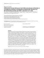
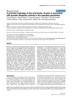
Figure 1 Representa tive CT sections of upper and l ower lobes obtained at baseline and day 7 in a patient with acute respiratory
distress syndrome. Computed tomography (CT) sections at baseline and day 7 are at the same lung region as attested by the anatomical
landmarks present on the rough images at baseline and day 7 (aortic arch and vascular divisions for upper lobe CT sections and vascular
divisions for the lower lobe CT sections). As previously described [18], poorly and nonaerated lung areas of right and left upper and lower lobes
are manually delineated (dashed line) at baseline (before HL-10 administration) with the aid of the software Lungview) that identifies poorly and
nonaerated lung areas in light gray and red, respectively. Delineation performed at baseline is manually ‘transposed’ to the CT section
corresponding to the same anatomical level obtained at day 7. Surfactant-induced lung reaeration is defined as the increase in gas volume
within the delineated zone between day 7 and baseline. The same process is repeated on each CT section in order to assess overall surfactant-
induced lung reaeration.
Lu et al. Critical Care 2010, 14:R135
/>Page 3 of 10
Computed tomography assessment of lung distribution of
surfactant
In both surfactant and control patients, right upper and
middle lobes, right lower lobe, left upper lobe and left
lower lobe were analyzed se parately at baseline and H39.
By referring to anatomical landmarks such as pulmonary
vessels, fissures, and segmental bronchi, the different pul-
monary lobes were identified on each CT section
obtained at baseline and H39 and manually delineated
using the roller ball of the computer. As the CT scan at
H39 in the surfactant group was performed within three
hours following the third bolus of HL-10, the increase of
volume of tissue at H39 provided an estimated volume of
the third bolus of HL-10. Therefore, the increase in
volume of tissue at H39 was compared with the volume
of HL-10 intratracheally administrated. The distribution
of surfactant between upper and lower lobes was com-
puted as the increase in lung tissue in each lobe.
Statistical analysis
The normal distribution of data was verified by a Ko lmo-
gorov-Smirnov test. Patients’ characteristics and regional
changes in volumes of gas and tissue between day 7 and
baseline were compared with a chi-squared t est or an
unpaired bilateral student test. Gas and tissue volumes at
baseline and their changes between H39 and baselin e
within the lobes were compared by Friedman repeated
measures analysis of variance on ranks followed by a
Tukey test. Correlations between instilled volume of HL-
10 and increase o f tissue volume were made by linear
regressio n. Cardiorespiratory and CT variables measured
at different days were compared between the two groups
using a two-way analysis of variance for a repeated factor
and a grouping factor. The statistical analysis was per-
formed with Sigmastat 3.1 (Systat Software Inc., Point
Richmond, CA, USA). Data were expressed as mean ±
standard deviation or median and interquartile range (25
to 75%) according to the data distribut ion. The statistical
significance level was fixed at 0.05.
Results
Patients
Among the 20 patients, one in the surfactant group died
at day 4 from severe hypoxemia. Of patients with ALI/
ARDS, 30% were related to extrapulmonary sepsis. The
overall mortality rate was 30%. As shown in Table 1, the
clinical characteristics and cardiorespiratory parameters
at baseline were not different between the control and
surfactant patients.
Cardiorespiratory changes in control and surfactant
groups
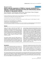
As shown in Figure 2, PaO
2
/FiO
2
ratio increased signifi-
cantly from baseline to H39 and day 7 in b oth groups
Table 1 Baseline clinical characteristics of the patients
Variables Control Surfactant P value
(n = 10) (n = 10)
Male/female 9/1 8/2 NS
Age (years) 59 ± 16 62 ± 12 NS
SAPS II 40 ± 10 41 ± 10 NS
LISS 2.3 ± 0.4 2.6 ± 0.5 NS
Septic shock (%) 80% 70% NS
Survival (%) 70% 70% NS
Cause of ALI/ARDS
Bronchopneumonia 46
Aspiration pneumonia 11NS
Lung contusion 20
Sepsis 33
PaCO
2
(mmHg) 38.4 ± 8.2 37.9 ± 7 NS
PaO
2
/FiO
2
(mmHg) 200 ± 63 201 ± 64 NS
TV/kg (ml) 5.7 ± 0.8 6 ± 0.9 NS
RR (breaths/min) 23 ± 4 20 ± 6 NS
Ppeak (cmH
2
O) 32 ± 5 32 ± 6 NS
Pplat (cmH
2
O) 23 ± 4 23 ± 5 NS
PEEP (cmH
2
O) 9.7 ± 0.9 9.4 ± 1 NS
Crs (ml.cmH
2
O
-1
) 38 ± 12 41 ± 23 NS
HR (beats/min) 110 ± 23 88 ± 24 NS
MAP (mmHg) 86 ± 15 87 ± 20 NS
ALI, acute lung injury; ARDS, acute respiratory distress syndrome; Crs,
compliance of respiratory system; FiO
2
, fraction of inspired oxygen; HR, heart
rate; LISS, lung injury severity score; MAP, mean arterial pressure; NS, not
significant; PaCO
2
, partial pressure of arterial carbon dioxide; PaO
2
, partial
pressure of arterial oxygen; Ppeak, peak airway pressure; PEEP, positive end-
expiratory pressure; Pplat, plateau airway pressure; RR, respiratory rate; SAPS II,
simplified acute physiology score II; Surfactant, porcine-derived lung
surfactant; TV, tidal volume.
Data are expressed as mean ± standard deviation.
Figure 2 PaO
2
/FiO
2
ratio at baseline, 39 hours after baseline
(H39) and day 7 in control (open circles) and surfactant groups
(closed circles) of patients with acute lung injury/acute
respiratory distress syndrome. FiO
2
, fraction of inspired oxygen;
PaO
2
, partial pressure of arterial oxygen.
Lu et al. Critical Care 2010, 14:R135
/>Page 4 of 10
and in sim ilar proportions. All o ther cardiorespiratory
parameters remained unchanged between baseline and
day 7 in both groups.
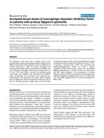
Distribution of HL-10 in the lungs
The mean volume of HL-10 instilled into the lungs per
instillation was 240 ± 30 ml. In the surfactant group,
between H39 (immediately after the third administration
of HL-10) and baseline, CT tissue volume increased by
311 ± 200 ml. The increase in tissue volume correlated
linearly with the instilled volume (R = 0.81, P = 0.008, Y
=-987+5.4X).AsshowninFigure3,atbaseline,CT
gas volume was significantly less in lower lobes than in
upper lobes whereas tissue volume was significantly
greater in the right upper lobe than in left lower lobe.
At H39, gas volume remained unchanged whereas tissue
volume significantly increased in similar proportion in
the upper and lower lobes.
In the control group, gas volume was not different
between baseline and H39. Tissue volume of right lower
lobe decreased significantly at H39 compared with the
value of baseline (Table 2).
Assessment of lung reaeration after HL-10 replacement
At baseline and PEEP 10 cmH
2
O, total lung volume, gas
volumeandtissuevolumewerenotdifferentbetween
control and surfactant groups. As shown in Figure 4,
total gas volume did not change significantly between
Figure 3 Volumes of gas and tissue at baseli ne before HL-10 instillation (upper part of the figure) and changes in volume of gas and
tissue between H39 (within three hours following the third bolus of HL-10) and baseline (lower part of the figure). Results shown in
right upper and middle lobes (RUL), left upper lobe (LUL), right lower lobe (RLL) and left lower lobe (LLL) in patients with acute lung injury/
acute respiratory distress syndrome instilled with 200 mg/kg of HL-10. Comparisons were performed by Friedman repeated measures analysis of
variance on ranks followed by a Tukey test. P values above the horizontal brackets indicate significant difference between RUL, LUL, RLL and LLL
using Friedman repeated measures analysis of variance.* P < 0.05 versus RUL,
§
P < 0.05 versus LUL.
Lu et al. Critical Care 2010, 14:R135
/>Page 5 of 10
baseline, H39 and day 7 in control and surfactant
groups. In contrast, HL-10 induced a significant increase
in tissue volume at H39 that persisted at day 7 (int erac-
tion P <0.001).Theincreaseintissuevolumebetween
day 7 and baseline correlated linearly with the instilled
volume of HL-10 (R = 0.72, P = 0.03, Y = -1594 +
7.6X). Hyperinflated lung volume was not different
between both groups at baseline, H39 and day 7.
As shown in Figure 5, in poorly or nonaerated lung
regions, gas volume significantly increased at day 7 com-
pared with baseline in both cont rol and surfactant
groups. The increase in gas volume at day 7 was signifi-
cantly greater in the surfactant group than in the con-
trol group (320 ± 125 ml versus 135 ± 161 ml, P = 0.01,
Figure 5a). In the control patients, tissue volume of
poorly or nonaerated lung regions significantly
decreased (Figure 5b, P = 0.04) between day 7 and base-
line whereas it remained unchanged in surfactant group.
In normally aerated lung regions, gas volume did not
change between day 7 and baseline in both groups
(Figure 5c). However, HL-10 induced a significant
increas e in tissue volume at day 7 (189 ± 179 m l versus
-15 ± 105 ml, P = 0.007, Figure 5d).
Discussion
The present study demonstrates that intratracheal
administration of porcine-derived surfactant to patients
with ALI/ARDS induces a significant l ung reaeration of
poorly or nonaerated lung regions. This beneficial effect,
however, is associated with a significant increase in lung
tissue in normally aerated lung areas at day 7 whose
mechanisms remain to be elucidated.
Distribution of surfactant within the lung is likely to
be an important factor that determine s the efficacy of
surfactant therapy. Delivery technique and lung mor-
phology influence surfactant distribution. In a previous
randomized clinical trial [7], the unsuccessful surfactant
treatment was related to the techn ique of aerosoli zation
that provided less than 10% distal lung deposition [21].
Intratracheal instillation by a catheter positioned just
above the carina has been shown to be much more
effective in animals and patients with ARDS [6,22]. In
patients with ARDS/ALI, the loss of lung aeration does
not have a uniform distribution and, in the supine posi-
tion, dependent and caudal lung regions are virtually
nonaerated as a result of external compression by the
abdomen and heart [13,14]. The distribution of
Table 2 Volumes of gas and tissue at baseline and H39 in
the control group of patients
Baseline H39 P value
Volume of gas (ml)
Right upper and middle lobe 864 ± 440 934 ± 411 NS
Left upper lobe 752 ± 321 722 ± 295 NS
Right lower lobe 178 ± 206 241 ± 244 NS
Left lower lobe 142 (47-277) 111(23-296) NS
Volume of tissue (ml)
Right upper and middle lobe 317 ± 115 313 ± 105 NS
Left upper lobe 281 ± 69 272 ± 71 NS
Right lower lobe 321 ± 106 275 ± 87 0.02
Left lower lobe 299 ± 89 267 ± 49 NS
Data are expressed as mean ± standard deviation or median and 25 to 75%
interquartile range. H39, CT scan performed 39 hours after baseline. NS, not
significant.
Figure 4 Computerized tomography assessment of total gas and tissue volumes at baseline, 39 hours after baseline (H39) and day 7,
in control (open circles) and surfactant groups of patients (closed circles).
Lu et al. Critical Care 2010, 14:R135
/>Page 6 of 10
exogenous surfactant in aerated and nonaerated parts of
the distal lung has never been assessed and it is
unknown whether instilled surfactant does penetrate
into nonaerated lower lobes. In the present study, the
CT scan at H39 in the surf actant group was performed
within three hours following the third administration of
HL-10. Base d on the fact that the tissue volume did not
change at H39 compared with its baseline value in the
control group, we can assume that the increase in lung
tissue between baseline and H39 in the surfactant group
is representative of instilled e xogenous surfactant. The
present data show that the overall volume of instilled
HL-10 was homogeneously distributed between upper
and lower lobes and between normally and poorly or
nonaerated lung regions (Figure 3). This result demon-
strates that the procedure of instillation (successive
bolus in right and left lateral positions followed by
consecutive recruitment maneuvers) resulted in unifo rm
bilateral surfactant distribution. A predominant distribu-
tion of HL-10 in normally aerated lung regions can be
ruled out.
Although several randomized trials have failed to
demonstrated beneficial effects of e xogenous surfactant
in adults patients with ARDS in terms of mortality and
ventilator-free days [7,8,23], the effect of surfactant ther-
apy on lung aeration had never been evaluated. In the
present study, using CT regional analysis of normally
and poorly or non-aerated lung regions, a significant
higher lung reaeration was evidenced at day 7 in
patients treated by surfactant replacement as compared
with control patients (Figure 5a). This finding provides
evidence that tracheal instillation of HL-10 induces a
substantial and prolo nged reaeration of poorly or nona-
erated lung regions and more specifically of nonaerated
Figure 5 Individual and mean changes in volume of gas and tissue in poorly/nonaerated lung regions (upper part of the figure) and
normally aerated lung regions (lower part of the figure). Volume changes were measured on computed tomography scans acquired at
baseline and seven days in patients who received either usual care (control, open circles) or usual care plus intratracheal porcine-derived
surfactant (HL-10, closed circles). In the surfactant group, each patient is identified by a specific symbol.
Lu et al. Critical Care 2010, 14:R135
/>Page 7 of 10
lower lobes. This encour aging result supports the ratio-
nale for exogenous surfact ant replacement as indication
for lung reaeration in adult patients with ALI/ARDS.
HL-10-induced lung reaeration was, however, asso-
ciated with a long lasting increase in lung tissue in pre-
viously normally aerated lung areas. Its mechanism
remains unknown and several hypotheses can be dis-
cussed. A delayed alveolar clearance of the la rge doses
of HL-10 administered to aerated lung regions, where
endogenous surfactant is already present, is a possible
mechanism that could explain the sustained increase in
lung tissue. In newborn infants, the surfactant half life is
around 35 hours [24]. In patients with ARDS treated by
recombinant surfactant, co mponents of exoge nous sur-
factant were retrieved in bronchoalveolar lavage (BAL)
two days after initi al administration, but were no longer
detectable five days later [6]. The dose of surfactant
used in the present study was orders of magnitude
beyond what was commonly used in neonates, older
children and adults. The high volume of phospholipids
administered may have prolonged the turn-over time,
explaining the persistent increase in lung tissue. Another
hypothesis explaining the increase of lun g tissue could
be an inflammatory reaction resulting from the interac-
tion of HL-10 with active endogenous surfactant present
in aerated lung regions [25]. As illustrated in the present
study, normally aerated lung region s in ARDS/A LI are
characterized by an excess of lung tissue [15] and an
increased vascular permeability [26], two abnormalities
increasing the vulnerability of lung parenchyma to exter-
nal aggressions. In these regions, saline diluted HL-10
could induce depletion of endogenous surfactant [27],
increased release of TNF and IL-6 in response to overin-
flation [28] and a resulting increase in lung micovascular
permeability. The consecutive influx of albumin into the
alveolar space could inactivate further endogenous sur-
factant [29], and aggravate lung injury. In addition, 720
ml of saline (4 ml/kg/bolus) containing HL-10 were
instilled in both lungs over 36 hours. By itself, such an
amount of liquid could induce lung injury in experimen-
tal normal lungs. Lastly, breakdown products of the
phospholipids in surfactant, specifically lysophosphati-
dylcholine, can provoke inflammation. In this study,
BAL after surfactant replacement was not performed.
Further study is required to explore the co rrelation
between the presence of inflammatory mediators, com-
ponents of exogenous surfactant, protein and cells in
BAL, and the CT increase in lung tissue in normally
aerated lung areas.
Exogneous surfactant has strong immunomodulatory
properties [30-32]. In patients with ARDS, exogenous
surfactant therapy decreases IL-6 concen trations in the
plasma and BAL of patients with ARDS, suggesting
either a direct anti-inflammatory effect or a reduction of
ventilator -induced lung stretch [6]. However, in the pre-
sent study, despite surfactant-induced recruitment of
poorly or nonaerated lung regions, CT lung hyperinfla-
tion was similar in both groups. Unexpectedly, HL-10-
induced reaeration was not associated with a significant
improvement in arterial oxygenation. Very likel y, HL-10
instillation in normally aerated lung regions worsened
regional ventilation/perfusion ratios through an increase
in lung tissue. In other words, benefit in terms of aera-
tion of poorly or nonaerated regions of the lung was
likelytobecounteractedbyanegativeimpactofHL-10
on aeration of previously normally aerated lung.
Conclusions
Although the rationale for exogenous surfactant replace-
ment in patients with ARDS/ALI is strong with some
phase II studies showing positive responses [33,34], all
clinical phase III studies failed to demonstrate a benefi-
cial effect in terms of mortality and duration of ventila-
tion [7,8,12]. Our study demonstrates that non-selective
tracheal administration of porcine-derived surfactant
reaerates poorly or nonaerated lung regions, but induces
a prolonged increase in lung tissue in regions remaining
normally aerated; therefore, gas exchange is not
improved. Further studies are needed to examine
whether a more selective instillation of exogenous sur-
factant in poorly or nonaerated lung regions would be
beneficial in te rms of improvement of oxygen ation,
reduction of mortality and ventilator-free days.
Key messages
• Intratracheal surfactant replacement reaerates
pooly and nonaera ted lung regions in patients with
ALI/ARDS.
• Intratracheal surfactant replacement induces a pro-
longed increase in lung tissue in normally aerated
lung regions.
Additional material
Additional file 1: Computed tomography measurement of lung
volumes of gas and tissue. The detail method of computed
tomography measurement of volumes of gas and tissue is described.
Abbreviations
ARDS: acute respiratory distress syndrome; ALI: acute lung injury; BAL:
bronchoalveolar lavage; CT: computed tomography; FiO2: fraction of inspired
oxygen; IL: interleukin; PaO2: partial pressure of arterial oxygen; PBW:
predicted body weight; PEEP: positive end-expiratory pressure; TNF: tumor
necrosis factor.
Acknowledgements
Porcine-derived surfactant was provided by LEO Pharma (HL-10, Leo
Pharmaceutical Products, Ballerup, Denmark). Other support was provided
from institutional and/or departmental source.
Lu et al. Critical Care 2010, 14:R135
/>Page 8 of 10
Author details
1
Multidisciplinary Intensive Care Unit, Department of Anesthesiology and
Critical Care Medicine, Assistance Publique-Hôpitaux de Paris, La Pitié-
Salpêtrière Hospital, UPMC Univ Paris 06, 47-83 boulevard de l’hôpital, 75013
Paris, France.
2
Department of Emergency Medicine, Second Affiliated
Hospital, Zhejiang University, School of Medicine, 88 Jiefang Road, 310009
Hangzhou, China.
3
Department of Anesthesiology, Federal University of Sao
Paulo, Escola Paulista de Medicina, Rua Napoleão de Barros, 715 - 5° andar,
Vila Clementino CEP 04024002 São Paulo, Brazil.
4
Department of Intensive
Care Medicine, University Medical Center Utrecht, Heidelberglaan, 100, 3584
CX Utrecht, The Netherlands.
Authors’ contributions
QL carried out the study and drafted the manuscript. MZ, CG, and BB
participated in the study and study analysis. JK participated in the
interpretation of the results and gave the advices for improving the
manuscript. JJR initiated the study, participated in the design of the protocol
and helped to draft the manuscript. All authors read and approved the final
manuscript.
Competing interests
The authors declare that they have no competing interests.
Received: 12 February 2010 Revised: 27 April 2010
Accepted: 15 July 2010 Published: 15 July 2010
References
1. Hallman M, Spragg R, Harrell JH, Moser KM, Gluck L: Evidence of lung
surfactant abnormality in respiratory failure. Study of bronchoalveolar
lavage phospholipids, surface activity, phospholipase activity, and
plasma myoinositol. J Clin Invest 1982, 70:673-683.
2. Nakamura T, Malloy J, McCaig L, Yao LJ, Joseph M, Lewis J, Veldhuizen R:
Mechanical ventilation of isolated septic rat lungs: effects on surfactant
and inflammatory cytokines. J Appl Physiol 2001, 91:811-820.
3. Malloy JL, Veldhuizen RA, Lewis JF: Effects of ventilation on the surfactant
system in sepsis-induced lung injury. J Appl Physiol 2000, 88:401-408.
4. Suresh GK, Soll RF: Overview of surfactant replacement trials. J Perinatol
2005, 25(Suppl 2):S40-44.
5. Willson DF, Thomas NJ, Markovitz BP, Bauman LA, DiCarlo JV, Pon S,
Jacobs BR, Jefferson LS, Conaway MR, Egan EA: Effect of exogenous
surfactant (calfactant) in pediatric acute lung injury: a randomized
controlled trial. JAMA 2005, 293:470-476.
6. Spragg RG, Lewis JF, Wurst W, Hafner D, Baughman RP, Wewers MD,
Marsh JJ: Treatment of acute respiratory distress syndrome with
recombinant surfactant protein C surfactant. Am J Respir Crit Care Med
2003, 167:1562-1566.
7. Anzueto A, Baughman RP, Guntupalli KK, Weg JG, Wiedemann HP,
Raventos AA, Lemaire F, Long W, Zaccardelli DS, Pattishall EN: Aerosolized
surfactant in adults with sepsis-induced acute respiratory distress
syndrome. Exosurf Acute Respiratory Distress Syndrome Sepsis Study
Group. N Engl J Med 1996, 334:1417-1421.
8. Spragg RG, Lewis JF, Walmrath HD, Johannigman J, Bellingan G, Laterre PF,
Witte MC, Richards GA, Rippin G, Rathgeb F, Hafner D, Taut FJ, Seeger W:
Effect of recombinant surfactant protein C-based surfactant on the
acute respiratory distress syndrome. N Engl J Med 2004, 351:884-892.
9. Schmidt R, Markart P, Ruppert C, Wygrecka M, Kuchenbuch T, Walmrath D,
Seeger W, Guenther A: Time-dependent changes in pulmonary surfactant
function and composition in acute respiratory distress syndrome due to
pneumonia or aspiration. Respir Res 2007, 8:55.
10. Markart P, Ruppert C, Wygrecka M, Colaris T, Dahal B, Walmrath D,
Harbach H, Wilhelm J, Seeger W, Schmidt R, Guenther A: Patients with
ARDS show improvement but not normalisation of alveolar surface
activity with surfactant treatment: putative role of neutral lipids. Thorax
2007, 62:588-594.
11. Ainsworth SB, Beresford MW, Milligan DW, Shaw NJ, Matthews JN,
Fenton AC, Ward Platt MP: Pumactant and poractant alfa for treatment of
respiratory distress syndrome in neonates born at 25-29 weeks’
gestation: a randomised trial. Lancet 2000, 355:1387-1392.
12. Kesecioglu J, Beale R, Stewart TE, Findlay GP, Rouby JJ, Holzapfel L, Bruins P,
Steenken EJ, Jeppesen OK, Lachmann B: Exogenous natural surfactant for
treatment of acute lung injury and the acute respiratory distress
syndrome. Am J Respir Crit Care Med 2009, 180:989-994.
13. Rouby JJ, Constantin JM, Roberto De AGC, Zhang M, Lu Q: Mechanical
ventilation in patients with acute respiratory distress syndrome.
Anesthesiology 2004, 101
:228-234.
14. Malbouisson LM, Busch CJ, Puybasset L, Lu Q, Cluzel P, Rouby JJ: Role of
the heart in the loss of aeration characterizing lower lobes in acute
respiratory distress syndrome. CT Scan ARDS Study Group. Am J Respir
Crit Care Med 2000, 161:2005-2012.
15. Puybasset L, Cluzel P, Gusman P, Grenier P, Preteux F, Rouby JJ: Regional
distribution of gas and tissue in acute respiratory distress syndrome. I.
Consequences for lung morphology. CT Scan ARDS Study Group.
Intensive Care Med 2000, 26:857-869.
16. Nieszkowska A, Lu Q, Vieira S, Elman M, Fetita C, Rouby JJ: Incidence and
regional distribution of lung overinflation during mechanical ventilation
with positive end-expiratory pressure. Crit Care Med 2004, 32:1496-1503.
17. Vieira SR, Puybasset L, Richecoeur J, Lu Q, Cluzel P, Gusman PB, Coriat P,
Rouby JJ: A lung computed tomographic assessment of positive end-
expiratory pressure-induced lung overdistension. Am J Respir Crit Care
Med 1998, 158:1571-1577.
18. Malbouisson LM, Muller JC, Constantin JM, Lu Q, Puybasset L, Rouby JJ:
Computed tomography assessment of positive end-expiratory pressure-
induced alveolar recruitment in patients with acute respiratory distress
syndrome. Am J Respir Crit Care Med 2001, 163:1444-1450.
19. Reske AW, Busse H, Amato MB, Jaekel M, Kahn T, Schwarzkopf P,
Schreiter D, Gottschaldt U, Seiwerts M: Image reconstruction affects
computer tomographic assessment of lung hyperinflation. Intensive Care
Med 2008, 34:2044-2053.
20. Malbouisson LM, Preteux F, Puybasset L, Grenier P, Coriat P, Rouby JJ:
Validation of a software designed for computed tomographic (CT)
measurement of lung water. Intensive Care Med 2001, 27:602-608.
21. Lewis JF, McCaig L: Aerosolized versus instilled exogenous surfactant in a
nonuniform pattern of lung injury. Am Rev Respir Dis 1993, 148:1187-1193.
22. Henry MD, Rebello CM, Ikegami M, Jobe AH, Langenback EG, Davis JM:
Ultrasonic nebulized in comparison with instilled surfactant treatment of
preterm lambs. Am J Respir Crit Care Med 1996, 154:366-375.
23. Davidson WJ, Dorscheid D, Spragg R, Schulzer M, Mak E, Ayas NT:
Exogenous pulmonary surfactant for the treatment of adult patients
with acute respiratory distress syndrome: results of a meta-analysis. Crit
Care 2006, 10:R41.
24. Torresin M, Zimmermann LJ, Cogo PE, Cavicchioli P, Badon T, Giordano G,
Zacchello F, Sauer PJ, Carnielli VP: Exogenous surfactant kinetics in infant
respiratory distress syndrome: A novel method with stable isotopes. Am
J Respir Crit Care Med 2000, 161:1584-1589.
25. Grotberg JB, Halpern D, Jensen OE: Interaction of exogenous and
endogenous surfactant: spreading-rate effects. J Appl Physiol 1995,
78:750-756.
26. Sandiford P, Province MA, Schuster DP: Distribution of regional density
and vascular permeability in the adult respiratory distress syndrome. Am
J Respir Crit Care Med 1995, 151:737-742.
27. Hafner D, Germann PG:
A rat model of acute respiratory distress
syndrome (ARDS) Part 2, influence of lavage volume, lavage repetition,
and therapeutic treatment with rSP-C surfactant. J Pharmacol Toxicol
Methods 1999, 41:97-106.
28. Stamme C, Brasch F, von Bethmann A, Uhlig S: Effect of surfactant on
ventilation-induced mediator release in isolated perfused mouse lungs.
Pulm Pharmacol Ther 2002, 15:455-461.
29. Strohmaier W, Trupka A, Pfeiler C, Thurnher M, Khakpour Z, Gippner-
Steppert C, Jochum M, Redl H: Bilateral lavage with diluted surfactant
improves lung function after unilateral lung contusion in pigs. Crit Care
Med 2005, 33:2286-2293.
30. van Putte BP, Cobelens PM, van der Kaaij N, Lachmann B, Kavelaars A,
Heijnen CJ, Kesecioglu J: Exogenous surfactant attenuation of ischemia-
reperfusion injury in the lung through alteration of inflammatory and
apoptotic factors. J Thorac Cardiovasc Surg 2009, 137:824-828.
31. Wemhoner A, Rudiger M, Gortner L: Inflammatory cytokine mRNA in
monocytes is modified by a recombinant (SP-C)-based surfactant and
porcine surfactant. Methods Find Exp Clin Pharmacol 2009, 31:317-323.
32. Erpenbeck VJ, Fischer I, Wiese K, Schaumann F, Schmiedl A, Nassenstein C,
Krug N, Hohlfeld JM: Therapeutic surfactants modulate the viability of
Lu et al. Critical Care 2010, 14:R135
/>Page 9 of 10
eosinophils and induce inflammatory mediator release. Int Arch Allergy
Immunol 2009, 149:333-342.
33. Kesecioglu J, Schultz MJ, Maas JJ, De Wilde RB, Steenken EI, Lachmann B:
Treatment of acute lung injury and ARDS with surfactant is safe
[abstract]. Am J Respir Crit Care Med 2004, 169:A349.
34. Gregory TJ, Steinberg KP, Spragg R, Gadek JE, Hyers TM, Longmore WJ,
Moxley MA, Cai GZ, Hite RD, Smith RM, Hudson LD, Crim C, Newton P,
Mitchell BR, Gold AJ: Bovine surfactant therapy for patients with acute
respiratory distress syndrome. Am J Respir Crit Care Med 1997,
155:1309-1315.
doi:10.1186/cc9186
Cite this article as: Lu et al.: Comput ed tomography asses sment of
exogenous surfactant-induced lung reaeration in patients with acute
lung injury. Critical Care 2010 14:R135.
Submit your next manuscript to BioMed Central
and take full advantage of:
• Convenient online submission
• Thorough peer review
• No space constraints or color figure charges
• Immediate publication on acceptance
• Inclusion in PubMed, CAS, Scopus and Google Scholar
• Research which is freely available for redistribution
Submit your manuscript at
www.biomedcentral.com/submit
Lu et al. Critical Care 2010, 14:R135
/>Page 10 of 10