Báo cáo y học: "Human protein C concentrate in the treatment of purpura fulminans: a retrospective analysis of safety and outcome in 94 pediatric patient" pps
Bạn đang xem bản rút gọn của tài liệu. Xem và tải ngay bản đầy đủ của tài liệu tại đây (395.49 KB, 7 trang )
RESEARC H Open Access
Human protein C concentrate in the treatment of
purpura fulminans: a retrospective analysis of
safety and outcome in 94 pediatric patients
Alex Veldman
1*
, Doris Fischer
2
, Flora Y Wong
1
, Wolfhart Kreuz
2
, Michael Sasse
3
, Bruno Eberspächer
4
,
Ulrich Mansmann
5
, Rudolf Schosser
4
Abstract
Introduction: Purpura fulminans (PF) is a devastating complication of uncontrolled systemic inflammation,
associated with high incidence of amputations, skin grafts and death. In this study, we aimed to clarify the clinical
profile of pediatric patients with PF who improved with protein C (PC) treatment, explore treatment effects and
safety, and to refine the prognostic significance of protein C plasma levels.
Methods: In Germany, patients receiving protein C concentrate (Ceprotin®, Baxter AG, Vienna, Austria) are
registered. The database was used to locate all pediatric patients with PF treated with PC from 2002 to 2005 for
this national, retrospective, multi-centered study.
Results: Complete datasets were acquired in 94 patients, treated in 46 centers with human, non-activated protein
C concentrate for purpura fulminans. PC was given for 2 days (median, range 1-24 days) with a median daily dose
of 100 IU/kg. Plasma protein C levels increased from a median of 27% to a median of 71% under treatment. 22.3%
of patients died, 77.7% survived to discharge. Skin grafts were required in 9.6%, amputations in 5.3%. PF recovered
or improved in 79.8%, remained unchanged in 13.8% and deteriorated in 6.4%. Four adverse events occurred in
3 patients, none classified as severe. Non-survivors had lower protein C plasma levels (P < 0.05) and higher
prevalence of coagulopathy at admission (P < 0.01). Time between admission and start of PC substitution was
longer in patients who died compared to survivors (P = 0.03).
Conclusions: This retrospective dataset shows that, compared to historic controls, only few pediatric patients with
PF under PC substitution needed dermatoplasty and/or amputations. Apart from epistaxis, no bleeding was
observed. Although the data comes from a retrospective study, the evidence we present suggests that PC had a
beneficial impact on the need for dermatoplasty and amputations, pointing to the potential value of carrying out a
prospective randomised controlled trial.
Introduction
Dermal and systemic thrombosis of the microcirculation,
referred to as purpura fulminans (PF), is a devastating
complication of widespread endothelial destruction due
to uncontrolled systemic inflammation, associat ed with a
high incidence of multiple organ failure, need for ampu-
tations, skin grafts and death. PF most frequently occurs
in the pediatric age group, with a peak incidence in
infants (1 to 3 years of age) and adolescents (16 to
18 years of age). Although most frequently seen in the
context of severe septic shock, in particular in patients
with meningococcemia, PF also occurs in the rare sce-
nario of homozygous or double heterozygous protein C
(PC) deficiency [1]. Indeed, the clinical manifestation of a
severe PC deficiency in the form of PF has resulted in the
description of PF as the clinical symptom of an acute PC
pathway failure [2]. Consequently, many intensivists have
used PC substitutio n in patients with PF , mostly with
promising results [3-6]. The biological rationale for
this use was the anti-coagulant, anti-inflammatory,
* Correspondence:
1
Monash Newborn, Monash Medical Centre; The Ritchie Centre, Monash
Institute for Medical Research and Department of Pediatrics, Monash
University, 246 Clayton RD, Clayton 3168, Melbourne, Australia
Full list of author information is available at the end of the article
Veldman et al. Critical Care 2010, 14:R156
/>© 2010 Veldman et al.; licensee Bio Med Central Ltd. This is an open access article distributed under the terms of the Creative
Commons At tribution License ( which permits unrestricted use, distribution, and
reproduction in any medium, provided the original work is properly cited.
pro-fibrinolytic, anti-apoptotic and barrier enhancing
action of PC [7-10].
This retrospectiv e multi-centered study analyzes clini-
cal features, safety and outcome in 94 pediatric and
adolescent patients with PF who receiv ed a human non-
activated PC concentrate as rescue therapy in Germany
from 2002 to 2005. In this study, we aimed to clarify the
clinical profile of pediatric patients with PF who
improved with PC treatment, explore treatment effects
and safety, and to refine the prognostic significance of
PC plasma levels. Finally, this study will help to establish
hypotheses and endpoints for future prospective studies
on human PC in patients with PF.
Materials and methods
The ethics committee of the J.W. Goe the University,
Frankfurt, Germany, approved the study protocol for
this retrospective, mult i-center study. Patients who
received a human plasma-derived, virus-inactivated PC
concentrate (Ceprotin
®,
BaxterAG,Vienna,Austria)in
Germanywereregisteredbythemanufacturerduetoa
post-marketing commitment reque sted by the European
Medicines Agency. This Ge rman database was used to
locate all pediatric patients treated with PC concentrate
from 2002 to 2005. The flow of the patients throughout
the study is displayed in Figure 1. If a patient who
received PC concentrate was identified, the principal
investigator contacted the treating physician with an
invitation to participate in the study. If the treating
physician agreed to participate, the hospital was visited
by a medical monitor (physician) for standardized
(form-based clinical reporting) data collection. For ana-
lysis, patients were stratified into three outcome groups
by survival to hospital discharge and complications
(negative outcome = death and/or amputation; inter-
mediate outcome = survival with skin grafts/dermato-
plas ty; positive outcome = survival without amputations
or skin grafts). As data were collected on anonymous
forms, the e thics committee waived the need for
informed consent by the patient or relatives.
Statistics
Descriptive statistics were used for categorical (tables,
rates, 95% confidence intervals (CI)) and continuous
(quartiles, minimum, maximum, mean, standard devia-
tion) variables. Differences in categorical variables
between groups were tested by the chi-square test.
Differences in continuous variables between groups
were tested by the non-parametric Mann-Whitney U
test. Comparisons of relevant parameters were per-
formed between survivors and non-survivors. The rele-
vance of PC plasma levels at admission on the
probability to survive the d isease was assessed by a
logistic regression and an odds ratio was calculated to
quantify the influence of the level of PC plasma activity
at admission on survival to discharge.
Results
Demographics
Of the 102 patients located, 94 entered the final analysis
(Figure 1): 52 (55.3%) were male and 42 (44.7%) female;
8 (8.9%) were newborn (< 28 days old), 36 (38.3%) were
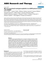
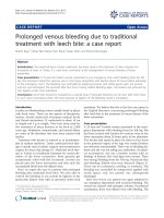
Figure 1 Flow of patients through the study and exclusions of patients. The diagnosis of purpura fulminans (PF) was regarded as definite
in the presence of livid to partly necrotic lesions of irregular shape and with sharp, clearly defined borders with either rapid progression or
already ubiquitous appearance. The diagnosis of PF was regarded as probable in the presence of livid to partly necrotic lesions of irregular shape
and with sharp, clearly defined borders. The diagnosis of PF was regarded as unclear in the presence of just livid to partly necrotic lesions
without any of the other criteria. The diagnosis of PF was not confirmed in any patient with lesions not fulfilling the above defined criteria. PC,
protein C.
Veldman et al. Critical Care 2010, 14:R156
/>Page 2 of 7
infants (28 days to 2 years), 29 (30.9%) were children
(2 to 12 years) and 21 (22.3) were adolescents (12 to
18 years).
Patients were treated in 46 different centers, with
36 centers treating 2 or less patients and 10 centers
treating 3 or more patients. None of the centers partici-
pated in or recruited pediatric patients for treatment
studies with activated PC at the time of this study.
Origin of PF
PF was the result of acquired sepsis rather than conge-
nital PC deficiency in all patients. Neisseria meningitides
was isolated in 75 (79.8%) patients, in 52 of those whose
sero-groups were specified.
Survival and outcome of PF
Twenty-one (22.3%) patients died at a mean duration of
two days, and 73 (77.7%) patients survi ved to discharge.
There was no significant difference in age or gender
between survivors and patients who did not survive.
Skin grafts were required in nine (9.6%) and amputa-
tions in five (5.3%) patients.
PF reco vered or improved in 75 (79.8%) patients,
remained unchanged in 13 (13.8%) patients and de terio-
rated in 6 (6.4%) patients.
Shock
At admission, no difference in mean arterial pressure
was detected between survivors and non-survivors (med-
ian 61.5 vs. 70 mmHg, P = 0.168). However, already at
admission, survivors presented with lower heart rates
(median 150 v s. 185 bpm, P = 0.0132) higher Glasgow
coma scale scores (mean 12.5 vs. 9.9, P = 0.027) and
less negative base excess compared with non-survivors
(median -4.95 vs. -11.85 mmol/l, P = 0.021).
A total of 63 pa tient s received inotropic support for a
median duration of two days.
Protein C treatment
Non-survivors had significantly lower PC plasma act ivity
at admission than survivors (median 10 vs. 30%, P =
0.011). A higher level of PC plasma activity by 1% at
admission improved the odds to survive significantly
(P = 0.0285) by a factor of 1.06 (95% CI for odds ratio =
1.01 to 1.12). PC was given for a median of 33 hours
(range 1 to 645 hours) with a median daily dose of 100
IU/kg (range 28 to 375 IU/kg). PC was administered as
a bolus every fou r to six hours in 78 patients, and as an
initial bolus followed by continuous infusion in the
remaining 16 patients. There was no significant differ-
ence in survival between the bolus group (21.8% died)
and the bolus plus infusion group (25.0% died;
P = 0.961). Plasma PC levels increased from a median of
27% (range 1 to 75%) prior to PC treatment to a median
of 71% under treatment (range 14 to 184%, Table 1).
Once under PC substitution, there was no significant
difference in plasma PC levels between survivors and
non-survivors (P = 0.605).
The time interval between admission and the start of
PC substitution was significantly longer (median 8.6 vs.
4hours,P = 0.03) in patients who died compared with
those who survived, and also longer in patients who had
amputations and/or died compared with those who fully
recovered (median 9.25 vs. 4 hours, P = 0.016).
Inflammatory response
C-reactive protein (CRP) levels were significantly lower
at admission in non-survivors compared with survivors
(Median: 5.9 vs. 11.6 mg/dl, P = 0.002). White blood
cell counts were also lower on admission and during
treatment in the non-survivors, but this trend did not
reach statistical significance (Table 1).
Coagulopathy
Non-survivors showed a significantly higher prevalence
than survivors of coagulopathy with prolonged activ ated
partial thromboplastin time (aPTT) at admission (median:
108 vs. 52 seconds, P < 0.0001). A significantly higher rate
of coagulopathy was still documented with PC treatment
in the non-survivors (Table 1). The platelet count did not
differ significantly at admission between survivors and
non-survivors; however, during treatment, survivors
showed significa ntly higher platelet counts than patients
who died (median: 103 vs. 61 G/l, P = 0.0047).
Fresh frozen plasma
Of the 94 patie nts, 71 received fresh frozen plasma
(FFP): 45% receive d one FFP transfusion, 30% two and
25% received three or more FFP transfusions. The total
amount of FFP given was 33.3 ml/kg in patients being
transfused, on day one a median of 20 ml/kg, on day
two a median of 22 ml/kg and on day three a median of
13 ml/kg. There was no difference in survival between
those patients transfused compared with those who did
not receive FFP (P = 0.345). However, there was a trend
towards a better survival in those patients receiving high
volume (≥ 25 ml/kg/d) FFP on day one compared with
those who received less or no FFP (P = 0.069).
Length of stay and mechanical ventilation
Length of stay on the ICU was a median of eight days
(1 to 95 days) in the whole group, a median of two days
for non-survivors and a median of nine days for survi-
vors (P < 0.0001). The duration of mechanical ventila-
tion was a median of 6.5 d ays for those who survived.
Survivors were discharged out of hospital after a median
of 18 days, and non-survivors had a median hospital
stay of 2 days.
Veldman et al. Critical Care 2010, 14:R156
/>Page 3 of 7
Table 1 Clinical and laboratory parameters of all patients, survivors, non-survivors before and during treatment with
protein C concentrate
All Survived
(n = 73)
Died
(n = 21)
P
Male/female
(n)
52/42 42/31 10/11 n.s.
Age
(years)
2.46 2.93 1.69 n.s.
PC treatment
PC total dose
(IU/kg)
258.8 (127.6-410.9) 277.8 (132.4-444.4) 153.8 (126.0-375.0) n.s.
PC daily dose
(IU/kg/d)
100 (73.4-136.6) 100
(78.74-133.3)
81.08 (71.09-153.8) n.s.
PC therapy duration (d) 2
(1-4)
3
(2-4)
2
(1-4)
n.s.
Bolus/
Bolus + cont. inf.
78/16 61/12 17/4 n.s.
Haematological paramenters prior to PC treatment
WBC
(pl)
10.4
(5.0-17.02)
11.3
(5.3-18.7)
7.21
(4.6-10.75)
n.s (0.062)
CRP
(mg/dl)
10.48
(5.65-16.47)
11.6
(7.81-17.31)
5.9
(1.99-8.92)
0.0018
Platelets
(G/l)
110
(66-183)
116
(74-182)
78
(52-178)
n.s.
PT
(%)
41
(32-54)
44
(33-56)
31
(22-36)
< 0.001
aPTT
(sec.)
59
(43-91)
52
(39-71)
108
(81-160)
< 0.001
Fibrinogen
(mg/dl)
270
(174-440)
347
(217-503)
129
(82-202)
< 0.001
D-Dimers
(mg/l)
2.38
(0.93-8.99)
2.13
(0.89-8.62)
6.40
(1.08-12.00)
n.s.
AT
(%)
76
(57-87)
80
(60-88)
70
(46-80)
n.s.
PC
(%)
27
(14-39)
30
(18-41)
10
(10-18)
< 0.05
Haematological parameters during PC treatment
WBC
(pl)
21.25
(12.75-27.28)
23.65
(13.65-28.75)
16.35
(7.7-20.90)
< 0.05
CRP
(mg/dl)
14.70 (7.0-21.8) 15.91
(7.1-23.64)
9.36
(6.90-15.88)
n.s.
Platelets
(G/l)
96
(57-130)
103
(65.5-136.5)
61
(30-80.75)
< 0.01
PT
(%)
69
(48.5-87)
77.8
(55-91)
45.5
(37.75-55.75)
< 0.01
aPTT
(sec.)
43
(33-52.75)
41
(33-47)
61
(47.5-88.4)
< 0.01
Fibrinogen
(mg/dl)
558.5
(342-747.2)
600.5
(418-766)
214.5
(184-299.2)
< 0.01
D-Dimers
(mg/l)
1.95
(0.8-6.16)
1.6
(0.68-5.96)
2.93
(1.14-6.4)
n.s.
AT
(%)
87
(68.5-102.2)
87
(68-101.5)
78
(70-102)
n.s.
PC
(%)
71
(53.5-108.4)
79
(54.7-106.8)
68.5
(33.75-108.5)
n.s.
Data on patient characteristics, outcome and laboratory findings. Shown as median and inter-quartile range (range between first and third quartile). Note that
not all laboratory parameters could be obtained in all patients at each time-point.
aPTT, activated partial thromboplastin time; AT, antithro mbin; CRP, C reactive protein; n.s., not significant; PC, protein C; PT, prothrombin time; WBC, white blood
cell count.
Veldman et al. Critical Care 2010, 14:R156
/>Page 4 of 7
Adverse events and hemorrhage
Four adverse events were reported in three patients.
None was classified as severe by the treating physician
(two events of hemorrhage from nose and/or throat,
one event of pleural effusion, one event of transient
increase in body temperature). The first patient with an
adverse event developed bleeding from the nose six
hours after receiving 66 IU/kg PC. The bleeding stopped
spontaneously after one hour and the patient received
the next scheduled dose two hours after the onset of the
event without further complication s. The treating physi-
cian classified the severity as moderate and causative
relation to PC treatment as unknown. The second
patient was a severely coagulopathic child, who devel-
oped hemorrhage from the throat and nose immediately
after a difficult endo-tracheal intubation approximately
13 hours after receiving 83 IU/kg PC. The patient
received the next dose as scheduled four hours after the
event without further complications. The same patient
developed a pleural effusion (serious with later blood
staining) more that 24 hours after the PC therapy was
ceased. The treating physician classified the severity of
both events as moderate, a causative relation to PC
treatment as unlikely. A third patient showed a transient
(10 minutes) and minor increase in body temperature
(from 39.0°C to 39.5°C) shortly after receiving 100 IU/kg
PC. No treatment was required; the treating physician
classified the severity as mild.
Discussion
Here we report what is to date the largest series of
pediatric patients with PF, as a consequence of acute PC
pathway failure in sepsis, treated with PC concentrate.
In the majority of patients, the underlying infection was
identified as meningococcemia. PC substitution was well
tolerated, safe and the need for amputations and skin
grafts with 5.3% and 9.6%, respectively, both markedly
lower than previously reported in children with PF.
PF, if associated with septic shock, is a devastating dis-
ease carrying a high mortalit y and significant morbidity.
Although improvement in health care delivery for these
very sick patients has dramatically improved survival in
recent years [11,12], permanent disability as a r esult of
amputation of limbs or digits, extensive scarring, or neu-
rological injury remains problematic. Gurgey and collea-
gues reported a series of 16 children; nine (69%) of the
13 children aged 4 years or younger and one of the
older children (age range 9 - 12 years) (33%) required
amputation [13]. A case series by Wheeler and collea-
gues on 21 patients reported amputations in nine (43%)
patients [14]. Recent dat a from Rotterdam reports
amputations in 8%, skin grafts in 16% and skin scar ring
in 48% of survivors of m eningococcal disease with PF,
combined with orthopedic sequelae in 14% [15]. These
patients did not receive PC. In five patients treated in a
burn center including therapy with activated PC, an
impressive 100% survival was achieved; however, ampu-
tations were still needed in two of t he five children
(40%) [16].
We and others have previously described replacement
therapy with human, non-activated PC in patients with
meningococcemia in case reports and smaller case series
[3,4,6,17-19]. Other studies have explored the use of
activated PC in patients with PF. Vincent and colleagues
published a post-hoc analysis of recent studies using
activated PC in adult and pediatric patients with severe
sepsis, presenting with PF, meningitis or meningococcal
disease [5]. The authors identified 119 pediatric patients
suitable for the analysis, 87 of them with PF. In that
group, which had a comparable incidence of coagulopa-
thy and low PC plasma levels, but a slightly more severe
thrombocytopenia compared with the group reported
here (85; 42 to 122 vs. 110; 66 to 183 G/l median; first
to third quart ile), serious bleeding events were noticed
in two patients during the infusion and in six patients
over a 28-day period. Fourteen-day mortality was 9.4%,
which is probably lower than the 22.3% in-hospital mor-
tality in the group reported here. It is difficult to com-
pare the illness severity of these two groups, because
Vincent and colleagues did not report the rates for
amputation or skin grafts. Another study, investigating
the use of activated PC in childre n with severe sepsis,
which was terminated prematurely due to lack of effi-
cacy, showed an increased rate of hemorrhage in the
activated PC compared with the p lacebo group, espe-
cially in children younger than 60 days [20].
In contrast, significant bleeding complications were
not seen in our large group of pediatric patients and
have also not been associated with the use of human,
non-activated PC concentrate in adult studies so far.
As published previously, this study confirmed low
plasma PC levels to be associated with negative outcome
[21]. In fact, even a difference as small as 1% in PC
plasma activity at admission changed the odds of survi-
val significantly. For a number of medical conditions,
minimizing the amount of time from patient presenta-
tion to initiation of treatment represents an important
consideration in the improvement of trea tment out-
comes. Analysis of a large, hospital-level database sug-
gested that e arlier trea tment with activated PC is
associated with a lower in-hospital mortality in patients
(n = 1179) with severe sepsis [22]. Another retrospective
study, analyzing adults with severe sepsis and APC treat-
ment found an increas e in mortality from 33% if treated
on the day of diagnosis to 40% if treatment was delayed
by one day and to 52% if treatment was delayed by two
days or more [23]. In our analysis, the interval between
admission to ICU and the commencement of PC
Veldman et al. Critical Care 2010, 14:R156
/>Page 5 of 7
replacement therapy was significantly longer in patients
whodiedcomparedwiththosewhosurvived,which
may point towards the benefits of early therapy in this
rapidly progressive condition.
During PC therapy, coagulatory and inflammatory
markers may be important prognostic markers. The pre-
valence of ongoing coagulopathy, as reflected in a signif-
icantly more abnormal aPTT and prothrombin time,
and in significantly lower fibrinogen levels at admission
and during therapy, was higher in non-survivors com-
pared with survivors. During therapy, but not at admis-
sion, platelets were found to be significantly lower in
patientswhodidnotsurvive,alsopointingtowards
ongoing coagulopathy as a negative prognostic marker.
Interestingly, although non-survivors showed lower PC
plasma levels and higher incidence of coagulopathy, the
inflammatory response in terms of CRP levels and leu-
kocytosis was less marked in the non-survivors, which
could i mply a degree of immuno-paralysis in those
patients.
Being a retrospe ctive multi-center analysis, the limita-
tions of this study are obvious: the lack of a control
group and a prospective design makes it impossible to
comment of the effects of PC on survival. The patients
were treated in many different centers with a conse-
quently large potential for intra-observer variabil ity. On
the other hand, the fact that even with so many different
centers and protocols involved, the safety profile was
still favorable in this unselected high-risk population is
very encouraging.
Conclusions
This study shows encouragingly low rates of amputa-
tions and skin grafts in a large group of pediatric
patients with PF, combined with improvement or reso-
lution of PF in most of the patients across all pediatric
age groups, with no significant adverse side effects. Our
study supports the biological rationale of human non-
activated PC concentrate as a treatment for severe
acquired PC deficiency, presenting with PF. Future stu-
dies investigating the effect of human, non-activated PC
concentrate should focus on the potential benefit of
early PC therapy in PF, and amputations and skin grafts
as important outcome measures in this condition,
which, despite recent advances in mortality cont rol, still
carries a high risk of disabling long-term morbidity.
Key messages
• Low plasma levels of PC are negatively correlated
with survival in patients with PF in the context of
meningococcemia.
• In this gro up of pediatric patients, substitution
with human, non-acti vated PC concentrate resulted
in an improvement of PF in the majority of patients,
without causing significant bleeding.
• The need for amputations and skin grafts was low
compared with historical controls, but there was no
obvious effect on mortality.
• Future studies investigating the effect of human,
non-activated PC concentrate should focus on early
PC therapy in PF, with amputations and skin grafts
as important outcome parameters.
Abbreviations
aPTT: activated partial thromboplastin time; CI: confidence interval; CRP: C
reactive protein; FFP: fresh frozen plasma; PC: protein C; PF: purpura
fulminans.
Acknowledgements
We would like to thank Dr. K.H. Jünemann for his valuable efforts in data
collection. Baxter Deutschland GmbH, Heidelber g, Germany, financially
supported this study.
The protein C study group: Dr. med. J. Urban, Kinderkrankenhaus
Josefinum, Augsburg; Dr. med. S. Spieler, Kreiskrankenhaus, Bad Hersfeld; Dr.
med. V. Varnholt, Charite/Virchow Krankenhaus, Berlin; Fr. Dr. med. U. Brosch,
Vivantes Klinikum, Berlin; Dr. med. K. Bunke, Helios Kliniken Berlin Buch,
Berlin; Dr. med. H. Schwalm, Klinikum Bremen-Mitte, Bremen; Dr. med. M.
Rachold, ZKH Bremen -Nord, Bremen; Dr. med. Schlicht, Allgemeines
Krankenhaus, Celle; Dr. med. W. Schäfer, Allgemeines Krankenhaus Hagen,
Hagen; Fr. Dr. med. S. Griethe, St. Salvator Krankenhaus, Halberstadt; Dr. med.
M. Thobaben, Universitätskrankenhaus Eppendorf, Hamburg; Dr. med. U.
Thiede, Klinikum Nord/HH-Heidberg, Hamburg; Dr. med. N. Geier, Klinikum
Heilbronn GmbH, Heilbronn; Dr. med. Hans G. Limbach, Universitätsklinikum
des Saarlandes, Homburg/Saar; Fr. Dr. med. E. Bungert, Westpfalz Klinikum,
Kaiserslautern; Dr. med. D. Faas, Städtisches Klinikum Karlsruhe gGmbH,
Karlsruhe; Dr. med. H. Schröder, Universitätsklinikum Kiel, Kiel, Dr. med. C.
Andree, Dr. med. P. Heister, Klinikum Krefeld, Krefeld; Dr. med. T. Werner, Dr.
med. M. Streitberg, Klinikum des Landkreises Lörrach, Lörrach; Dr. med. M.
Kohl, Universitätsklinikum Schleswig- Holstein, Lübeck; Dr. med. H. Frenzke,
Märkische Kliniken GmbH, Lüdenscheid; PD Dr. med. J. Sonntag, Städtisches
Klinikum, Lüneburg; Dr. med. V. Aumann, Universitätsklinikum, Magdeburg;
Dr. med. S. Bastuck, SHG- Kliniken Merzig, Merzig; Dr. med. K. Kurnik, Dr.
med. C. Bidlingmaier, Dr. von Haunersches Kinderspital, München; Dr. J.A.
Harding, Klinikum Dritter Orden Nymphenburg, München; Prof. Dr. med. U.
Nowak-Göttl, Universitätsklinikum, Münster; Dr. med. B. Kinder, Fr. Dr. med. K.
Manzke, Dietrich- Bonhoeffer- Klinikum, Neubrandenburg; Fr. Dr. med. C.
Bergheim, Dr. med. E. Jung, Kinderklinik Kohlhof, Neunkirchen; Dr. med. F.
Küchel, Kinderklinik Neustadt am Rübenberge, Neustadt a. R.; Dr. med. B.
Voigt, Klinikum Offenbach, Offenbach, Dr. med. M. Viemann, Elisabeth
Kinderkrankenhaus, Oldenburg; Prof. D. Radke, Ernst von Bergmann Klinikum,
Potsdam; Dr. med. B. Zimmermann, Dr. med. C. Hein, Universitätsklinikum
Rostock, Rostock; Dr. L. Hempel, Thüringen- Kliniken “Georgius Agricola”,
Saalfeld; Dr. med. R. Geib-König, Klinikum Winterberg, Saarbrücken; Dr. med.
S. Röll, Dr. med. H. Orth, St. Elisabeth Klinik, Saarlouis; Dipl. Med. B. Schenk,
Helios Klinikum, Schwerin; Dr. med. Z. Uyanik, Städtisches Klinikum, Solingen;
Dr. med. R. Berg, Olgahospital, Stuttgart; Dr. med. T. Trips, Klinikum
Traunstein, Traunstein; Dr. med. C. Block, Mutterhaus der Borromäerinnen,
Trier; Dr. med. M. Kumpf, Dr. A. Bosk, Universitätsklinikum Tübingen,
Tübingen; Dr. H. Vielhaber Klinikum Weiden, Weiden in der Oberpfalz; Dr.
med. M. Heldmann, Helios Klinikum Barmen, Wuppertal.
Author details
1
Monash Newborn, Monash Medical Centre; The Ritchie Centre, Monash
Institute for Medical Research and Department of Pediatrics, Monash
University, 246 Clayton RD, Clayton 3168, Melbourne, Australia.
2
Department
of Pediatrics, J.W. Goethe University Hospital, Theodor Stern Kai 7, 60590
Frankfurt/Main, Germany.
3
Department of Pediatric Cardiology and Pediatric
Intensive Care, University Childrens Hospital Hannover, Carl Neuberg Str. 1,
30625 Hannover, Germany.
4
Baxter BioScience, EdisonStr. 4, 85716
Veldman et al. Critical Care 2010, 14:R156
/>Page 6 of 7
Unterschleißheim, Germany.
5
Department of Medical Informatics, Biometry,
and Epidemiology, L. Maximilian University, Marchioninistr. 15, 81377 Munich,
Germany.
Authors’ contributions
AV was involved in study design, data analysis and interpretation, and
writing of the manuscript. DF, MS and FW were involved in data analysis
and interpretation, and writing of the manuscript. WK was involved in study
design and data analysis. BE was involved in study design and data
collection. UM was involved in data management, statistical analysis and
data interpretation. RS was involved in study design, data analysis, and
writing of the manuscript. All authors read and approved the final
manuscript.
Competing interests
AV was a member of a Baxter advisory board and received as such an
honorarium. At the time the study was performed, RS and BE were
employees of Baxter Deutschland GmbH, Heidelberg, Germany. All authors
had full and unrestricted access to the dataset. Baxter had no influence on
the data selection, interpretation or publication.
Received: 21 April 2010 Revised: 15 July 2010
Accepted: 19 August 2010 Published: 19 August 2010
References
1. Dreyfus M, Magny JF, Bridey F, Schwarz HP, Planche C, Dehan M,
Tchernia G: Treatment of homozygous protein C deficiency and neonatal
purpura fulminans with a purified protein C concentrate. N Engl J Med
1991, 325:1565-1568.
2. Baker PM, Keeling DM, Murphy M: Plasma exchange as a source of
protein C for acute-onset protein C pathway failure. Br J Haematol 2003,
120:167-168.
3. White B, Livingstone W, Murphy C, Hodgson A, Rafferty M, Smith OP: An
open-label study of the role of adjuvant hemostatic support with
protein C replacement therapy in purpura fulminans-associated
meningococcemia. Blood 2000, 96:3719-3724.
4. Ettingshausen CE, Veldmann A, Beeg T, Schneider W, Jager G, Kreuz W:
Replacement therapy with protein C concentrate in infants and
adolescents with meningococcal sepsis and purpura fulminans. Semin
Thromb Hemost 1999, 25:537-541.
5. Vincent JL, Nadel S, Kutsogiannis DJ, Gibney RT, Yan SB, Wyss VL, Bailey JE,
Mitchell CL, Sarwat S, Shinall SM, Janes JM: Drotrecogin alfa (activated) in
patients with severe sepsis presenting with purpura fulminans,
meningitis, or meningococcal disease: a retrospective analysis of
patients enrolled in recent clinical studies. Crit Care 2005, 9:R331-343.
6. de Kleijn ED, de Groot R, Hack CE, Mulder PG, Engl W, Moritz B, Joosten KF,
Hazelzet JA: Activation of protein C following infusion of protein C
concentrate in children with severe meningococcal sepsis and purpura
fulminans: a randomized, double-blinded, placebo-controlled, dose-
finding study. Crit Care Med 2003, 31:1839-1847.
7. Kisiel W, Canfield WM, Ericsson LH, Davie EW: Anticoagulant properties of
bovine plasma protein C following activation by thrombin. Biochemistry
1977, 16:5824-5831.
8. Joyce DE, Gelbert L, Ciaccia A, DeHoff B, Grinnell BW: Gene expression
profile of antithrombotic protein c defines new mechanisms modulating
inflammation and apoptosis. J Biol Chem 2001, 276:11199-11203.
9. Cheng T, Liu D, Griffin JH, Fernandez JA, Castellino F, Rosen ED,
Fukudome K, Zlokovic BV: Activated protein C blocks p53-mediated
apoptosis in ischemic human brain endothelium and is neuroprotective.
Nat Med 2003, 9:338-342.
10. Riewald M, Petrovan RJ, Donner A, Mueller BM, Ruf W: Activation of
endothelial cell protease activated receptor 1 by the protein C pathway.
Science 2002, 296:1880-1882.
11. Booy R, Habibi P, Nadel S, de Munter C, Britto J, Morrison A, Levin M:
Reduction in case fatality rate from meningococcal disease associated
with improved healthcare delivery. Arch Dis Child 2001, 85:386-390.
12. Maat M, Buysse CM, Emonts M, Spanjaard L, Joosten KF, de Groot R,
Hazelzet JA: Improved survival of children with sepsis and purpura:
effects of age, gender, and era. Crit Care 2007, 11:R112.
13. Gurgey A, Aytac S, Kanra G, Secmeer G, Ceyhan M, Altay C: Outcome in
children with purpura fulminans: report on 16 patients. Am J Hematol
2005, 80:20-25.
14. Wheeler JS, Anderson BJ, De Chalain TM: Surgical interventions in children
with meningococcal purpura fulminans–
a review of 117 procedures in
21 children. J Pediatr Surg 2003, 38:597-603.
15. Buysse CM, Oranje AP, Zuidema E, Hazelzet JA, Hop WC, Diepstraten AF,
Joosten KF: Long-term skin scarring and orthopaedic sequelae in
survivors of meningococcal septic shock. Arch Dis Child 2009, 94:381-386.
16. Hassan Z, Mullins RF, Friedman BC, Shaver JR, Alam B, Mian MA: Purpura
fulminans: a case series managed at a regional burn center. J Burn Care
Res 2008, 29:411-415.
17. Fischer D, Schloesser RL, Nold-Petry CA, Nold MF, Veldman A: Protein C
concentrate in preterm neonates with sepsis. Acta Paediatr 2009,
98:1526-1529.
18. Kreuz W, Veldman A, Escuriola-Ettingshausen C, Schneider W, Beeg T:
Protein-C concentrate for meningococcal purpura fulminans. Lancet
1998, 351:986-987, author reply 988.
19. Hodgson A, Ryan T, Moriarty J, Mellotte G, Murphy C, Smith OP: Plasma
exchange as a source of protein C for acute onset protein C pathway
failure. Br J Haematol 2002, 116:905-908.
20. Nadel S, Goldstein B, Williams MD, Dalton H, Peters M, Macias WL, Abd-
Allah SA, Levy H, Angle R, Wang D, Sundin DP, Giroir B, REsearching severe
Sepsis and Organ dysfunction in children: Drotrecogin alfa (activated) in
children with severe sepsis: a multicentre phase III randomised
controlled trial a gLobal perspective (RESOLVE) study group. Lancet 2007,
369:836-843.
21. Venkataseshan S, Dutta S, Ahluwalia J, Narang A: Low plasma protein C
values predict mortality in low birth weight neonates with septicemia.
Pediatr Infect Dis J 2007, 26:684-688.
22. Ernst FR, Johnston JA, Pulgar S, He J, Ball DE, Young JK, Cooper LM: Timing
of drotrecogin alfa (activated) initiation in treatment of severe sepsis: a
database cohort study of hospital mortality, length of stay, and costs.
Curr Med Res Opin 2007, 23:235-244.
23. Wheeler A, Steingrub J, Schmidt GA, Sanchez P, Jacobi J, Linde-Zwirble W,
Bates B, Qualy RL, Woodward B, Zeckel M: A retrospective observational
study of drotrecogin alfa (activated) in adults with severe sepsis:
comparison with a controlled clinical trial. Crit Care Med 2008, 36:14-23.
doi:10.1186/cc9226
Cite this article as: Veldman et al.: Human protein C concentrate in the
treatment of purpura fulminans: a retrospective analysis of safety and
outcome in 94 pediatric patients. Critical Care 2010 14:R156.
Submit your next manuscript to BioMed Central
and take full advantage of:
• Convenient online submission
• Thorough peer review
• No space constraints or color figure charges
• Immediate publication on acceptance
• Inclusion in PubMed, CAS, Scopus and Google Scholar
• Research which is freely available for redistribution
Submit your manuscript at
www.biomedcentral.com/submit
Veldman et al. Critical Care 2010, 14:R156
/>Page 7 of 7