Báo cáo y học: "Inhibition of monocyte chemoattractant protein-1 prevents diaphragmatic inflammation and maintains contractile function during endotoxemi" pot
Bạn đang xem bản rút gọn của tài liệu. Xem và tải ngay bản đầy đủ của tài liệu tại đây (573.52 KB, 11 trang )
RESEARC H Open Access
Inhibition of monocyte chemoattractant protein-1
prevents diaphragmatic inflammation and
maintains contractile function during
endotoxemia
Katherine Labbe
1
, Gawiyou Danialou
1
, Dusanka Gvozdic
1
, Alexandre Demoule
1,2
, Maziar Divangahi
1
,
John H Boyd
1,3
, Basil J Petrof
1,3*
Abstract
Introduction: Respiratory muscle weakness is common in sepsis patients. Proinflammatory mediators produced
during sepsis have been implicated in diaphragmatic contractile dysf unction, but the role of chemokines has not
been explored. This study addressed the role of monocyte chemoattractant protein-1 (MCP-1, also known as CCL2),
in the pathogenesis of diaphragmatic inflammation and weakness during endotoxemia.
Methods: Mice were treated as follows (n = 6 per group): (a) saline, (b) endotoxin (25 μg/g IP), (c) endotoxin +
anti-MCP-1 antibody, and (d) end otoxin + isotype control antibody. Muscles were also exposed to recombinant
MCP-1 in vivo and in vitro. Measurements were made of diaphragmatic force generation, leukocyte infiltration, and
proinflammatory mediator (MCP-1, IL-1a, IL-1b, IL-6, NF-B) expression/activity.
Results: In vivo, endotoxin-treated mice showed a large decrease in diaphragmatic force, together with
upregulation of MCP-1 and other cytokines, but without an increase in intramuscular leukocytes. Antibody
neutralization of MCP-1 prevented the endotoxin-induced force loss and reduced expression of MCP-1, IL-1a, IL-1b,
and IL-6 in the diaphragm. MCP-1 treatment of nonseptic muscles also led to contractile weakness, and MCP-1
stimulated its own transcription independent of NF-B activation in vitro.
Conclusions: These results suggest that MCP-1 plays an important role in the pathogenesis of diaphragmatic
weakness during sepsis by both direct and indirect mechanisms. We speculate that its immunomodulatory
properties and ability to modify skeletal muscle function make MCP-1 a potential therapeutic target in c ritically ill
patients with sepsis and associated respiratory muscle weakness.
Introduction
Sepsis is a major risk facto r for the development of criti-
cal illness myopathy [1], a nd impaired skeletal muscle
function has been directly linked to systemic infections in
humans [2]. The diaphragm is the primary muscle of
respiration, and acute respiratory failure occurs in a large
proportion of patients with severe sepsis [3]. Major losses
of diaphragmatic force-gene rating capacity have been
documented in several different sepsis models [4-7].
Substantial data link this decreased diaphragmatic func-
tion to the associated systemic inflammatory response
syndrome (SIRS) and to the local expression of proin-
flammatory mediators (for example, reactive oxygen spe-
cies, nitric oxide, cytokines) within skeletal mu scle fibers
(see reference [8] for recent review). Interestingly, evi-
dence also indicates that the diaphragm is particularly
prone to exaggerated proinflammatory gene upregulation
and impaired force production during different forms of
enhanced systemic inflammation [7,9,10].
Monocyte chemoattractant protein (MCP)-1, also
known as CCL2, is a prototypical member of the CC
subfamily of chemokines [11]. H igh serum levels of
* Correspondence:
1
Meakins-Christie Laboratories, McGill University, 3626 Saint Urbain, Montreal,
Quebec, Canada H2X 2P2
Full list of author information is available at the end of the article
Labbe et al. Critical Care 2010, 14:R187
/>© 2010 Petrof et al.; licensee BioMe d Central Ltd. This is an open access article distributed under the terms of the Creative Commons
Attribution License ( which permits unrestricted use, distribution, and reproduction in
any medium, provided the original work is properly cited.
MCP-1 have been demonstrated in animal models of
sepsis or SIRS [12-15], as well as in sepsis patients [16].
In a recent study profiling a large number of cyt okines
in the plasma of patients with severe sepsis, MCP-1
levels showed the best correlation with organ dysfunc-
tion and mortality [17]. MCP-1 is primarily a chemoat-
tractant for monocytes, memory T lymphocytes, and
natural killer cells, with some recent studies also point-
ing to a potential role in attracting neutrophils [11,18].
However, it is important to recognize that the actions of
MCP-1 extend well beyond leukocyte chemoattraction.
In particular, MCP-1 has important effects on the bal-
ance between pro- and anti-inflammatory cytokines
[13,19,20]. In addition, MCP-1 exposure can lead to
increased insulin resistance in skeletal myocytes [21]
and also affects muscle repair mechanisms [22,23], sug-
gesting a potential to significantly modify muscle func-
tion in critically ill patients.
In the present study, our principal objective was to
determine whether MCP-1 is involved in the pathogen-
esis of diaphragmatic dysfunction associated with SIRS
induced by endotoxin administration. Our principal
aims were as follows: (a) to evaluate whether endotoxin
administration leads to increased MCP-1 expression in
skeletal muscles; (b) to assess whether an increased
exposure to MCP-1 has direct effects on skeletal muscle
function; and (c) to de termine whether MCP-1 neutrali-
zation is able to modulate proinflammatory mediator
expression and contractile function in the diaphragm
during acute endotoxemic sepsis.
Materials and methods
Animal experiments
Exp eri ments were performed in 8- to 10-week-old male
C57BL/6 mice (Charles River Laboratories, Saint-
Constant, QC, Canada). All procedures were approved
by the institutional animal care and ethics committee, in
accordance with the guidelines issued by the Canadian
CouncilonAnimalCare.Themicewereanesthetized
with a mixture of ketamine (130 μg/g) and xylazine (20
μg/g) prior to sacrifice.
Sepsis model
Mice were injected intraper itoneally with either Escheri-
chia coli endotoxin (LPS, serotype 055:B5) (25 μg/g) or
an equivalent volume of saline. Mice were sacrificed at
12 hours (unless specifically stated otherwise) after
administering LPS, and the muscles (diaphragm, exten-
sor digitorum longus (EDL), tibialis anterior) and other
tissues (lungs, liver, blood) were removed for the various
biochemical, histologic, and physiological analyses
described later in detail. For all MCP-1 neutralization
studies, the mice were pretreated with intraperitoneal
injection of an anti-MCP-1 neut ralizing ant ibody (1 μg/
g) (BD Biosciences, San Diego, CA) at 12 and 24 hours
before LPS administration; this antibody and dose have
previously been shown to be effective in mice with sep-
tic peritonitis [14]. Control animals received the same
dose of an irrelevant isotypic control immunoglobulin,
administered in the same manner.
Local administration of MCP-1
To test the effects of exogenous MCP-1 on skeletal
muscle contractility, recombinant murine MCP-1 (100
pg in 10 μl of saline) (R&D systems, Minneapolis, MN)
was directly injected into the EDL muscle of the hin-
dlimb. The contralateral EDL was injected with an iden-
tical volume of saline at the same time to serve as a
within-animal control group, thereby eliminating any
potential differences related to systemic absorption of
the injected MCP-1. Both EDL muscles were surgically
exposed to ensure an accurately placed injection, and
after wound closure with sutures, the animals emerged
from anesthesia and resumed normal behavior. Mice
were sacrificed at 12 hours after administering MCP-1,
and both EDL muscles were removed.
Cell culture experiments
To evaluate the direct effects of MCP-1 on cytokine
expression by diaphragmatic muscle cells, primary dia-
phragmatic muscle cell cultures were established [9] by
using single living muscle fibers to isolate myoblast pre-
cursors (satellite cells). In brief, excised diaphragm mus-
cle strips were subjected to collagenase digestion and
trituration to liberate individual fibers. The individual
fibers were transferred into Matrigel-coated (Becton
Dickinson, Franklin Lakes, NJ) plates. Diaphragmatic
myoblasts were expanded in growth medium (20% fetal
bovine serum, 10% horse serum, 1% chick embryo
extract in DMEM) until attaining approximately 75%
confluence. The cultures were then placed in differentia-
tion medium (2% fetal bovine ser um, 10% horse serum,
0.5% chick embryo extract in DMEM) to induce myo-
blast fusion into differentiated myotubes. All experi-
ments were perform ed on day 5 of maintenance in
differentiation medium. Diaphragmatic myotubes were
washed with DMEM before stimulation with recombi-
nant murine MCP-1 (100 ng/ml).
To determine the effects of MCP-1 on NF-B activity
in muscle cells, myoblasts were simultaneously trans-
fected with a NF-B-driven firefly luciferase reporter
plasmid (pNF-B; Clontech, Mountain View, CA) and a
constitutively active thymidine kinase promoter-driven
Renilla luciferase plasmid (pRL-TK; Promega, Madison,
WI), as previously described [24]. In this system, the
constitutively active Renilla luciferase serves as an inter-
nal control to adjust for any differences in transfection
efficiency. For these studies, we used the C2C12 skeletal
muscle cell line (ATCC, Manass as, VA) rather than pri-
mary skeletal muscle cells, as the latter are known to be
Labbe et al. Critical Care 2010, 14:R187
/>Page 2 of 11
resistant t o standard transfe ction techniques [25].
C2C12 myoblasts (5 × 10
5
) were seeded onto 60-mm
plates and transfected the following day at approxi-
mately 50% confluence, by using Lipofectamine 2000
(Invitrogen, Carlsbad, CA). On day 5 in differentiation
medium, the cells were stimulated with murine MCP-1
(100 ng/ml) (R&D Systems, Minneapolis, MN), and the
activity levels of both forms of luciferase (firefly and
Renilla) were quantified by using the Dual-Luciferase
Reporter Assay System (Promega). Light emission was
measured in an L
max
384 luminometer (Molecular
Devices, Downingtown, PA), and the results are
expressed as the ratio of firefly (reflecting NF-B activ-
ity) to Renilla luciferase activities in relative light units.
Analyses of protein and mRNA expression
A commercial ELISA kit for murine MCP-1 (R&D Sys-
tems, Minneapolis, MN) was used to measure serum
and tissue MCP-1 protein levels i n duplicate, according
to the manufacturer’s instructions. Serum was collected
by cardiac puncture, and total protein was extracted
from the diaphragm, tibialis anterior, liver, and lung.
Frozen tissue samples were homogenized in lysis buffer
(1% Triton X-100, 50 mM HEPES (pH 8.0), 150 mM
NaCl, 10% glycerol, 2 mM EDTA, 1.5 mM MgCl
2
,10
μg/ml apro tinin, 10 μg/ml leupeptin, 1 mM phenyl-
methylsulphonyl fluoride, 1 mM sodium orthovanadate).
Homogenates were centrifuged 10 minutes at 10,000
rpm, and the supernatant protein content measured
with Bradford assay (BioRad Laboratories, Hercules,
CA).
To measure mRNA expression levels of MCP-1 and
its receptor CCR2, IL-1a,IL-1b,andIL-6,totalRNA
from tissue or cell cultures was extracted by using Tri-
zol reagent (Invitrogen) according to the manufacturer’s
protocol.
32
P-labeled, anti-sense RNA probes were
synthesized from commercially available Multi-Probe-
Template sets (BD Biosciences, San Diego, CA). Ribop-
robes were hybridized overnight at 56°C with 10 μgof
sample RNA, according to the manufacturer’sinstruc-
tions. Protected RNA fragments were separated by using
a 5% polyacrylamide gel and analyzed with autoradiogra-
phy. For each RNA probe, all experimental groups were
run on a single gel to allow quantitative comparisons.
The bands representing mRNA content were quantified
by using an image-analysis system (FluorChem 8000;
Alpha Innotech, San Leandro, CA), and the signals nor-
malized to the L32 housekeeping gene as a loadi ng
control.
Analyses of leukocyte infiltration
To quantify macrophages and neutrophils, skeletal mus-
cle cryosections (5 μm t hick) were r eacted with mono-
clonal antibodies directed against either macrophage F4/
80 (1:75 dilution) (Abcam, Cambridge, MA) or neutro-
phil Ly-6G (1:50 dilution) (BD Biosciences). Nonspecific
binding sites were blocked by incubating sections for 1
hour with PBS containing 3% BSA and 5% goat serum,
followed by goat anti-mouse IgG Fab fragment (1:20
dilution) (Jackson Laboratories, West Grove, PA) for 30
minutes. Biotinylated rabbit anti-rat IgG secondary anti-
body (1:100 dilution) (Vector Laboratories, Burlingame,
CA) was added and revealed by using the Vectastain
streptavidin-HRP system (Vector Laboratories) wit h
DAB substrate (Sigma-Aldrich Canada, Oakville, ON,
Canada). To quantify inflammatory cell infiltration, the
central and adjacent 20 × fields of the tissue were
photographed by using a digital camera, and a stereol-
ogy software package (Image-Pro Plus; Media Cyber-
netics, Silver Spring, MD) was used to overlay a
275-point grid onto each image (six photographs per
muscle). Inflammatory cells were quantified by using a
standard point-counting method, in which an abnormal
point was defined as falling either on an inflammatory
cell or on a myofiber invaded by such cells. The percen-
tage area of inflammation was then calculated by divid-
ing the number of abnormal points by the total number
of points falling on the muscle tissue section [26]. The
muscle images were selected in random order, with the
operator blinded to the identity of the experimental
groups.
As an additional index of neutrophil activity within
tissues, myeloperoxid ase (MPO) activity was determined
[27]. In brief, frozen tissues were homogenized in 1 ml
ice-cold 50 mM potassium phosphate buffer at pH 6.0.
Homogenates were centrifuged at 12,000 g for 15 min-
utes at 4 degrees Celsius, and the supernatant was dis-
carded. Pellets were resuspended, homogenized,
centrifuged, and the pellets were resuspended in buffer.
Assays were performed in duplicate on supernatant
added to buffer containing 0.167 mg/ml o-dianisidine
and 0.0005% H
2
O
2
. Enzymatic activity was determined
spectrophotometrically by measuring the change in
absorbance at 460 n m over a 3-minute period. Values
are expressed as units per gram of tissue, with each unit
representing the change in optical density per minute.
Muscle contractile function
The diaphragm or EDL muscle was surgically excised
for in vitro contractility measurements, as previously
described [7,28]. Muscles from the different experimen-
tal groups were selected in random order, with the indi-
vidual performing the contractility measurements being
blinded to their identity. After removal from the animal,
muscles were tr ansferred into K rebs solution (118 mM
NaCl, 4.7 mM KCl, 2.5 mM CaCl
2
,1.2mM MgSO
4
,1
mM KH
2
PO
4
,25mM NaHCO
3
,and11mM glucose)
chilled to 4°Celsius and perfused with 95% O
2
/5% CO
2
Labbe et al. Critical Care 2010, 14:R187
/>Page 3 of 11
(pH 7.4). The muscles were then mounted in a jacketed
tissue-bath chamber filled with continuously perfused
Krebs solution warmed to 25°Cel sius. After a 15-minute
thermoequilibration period, muscle length was gradually
adjusted to optimal length (L
o
, the length at which max-
imal twitch force is obtai ned). The force-frequency rela-
tion was determ ined by sequential supramaximal
stimulation for 1 second at 5, 10, 20, 30, 50, 100, 120,
and 150 Hz, with 2 minutes between each stimulation
train. At the end of the experiment, L
o
was directly
measured with a microcaliper and the muscle blotted
dry and weighed. Specific force (force/cross-sectional
area) was calculated, assuming a muscle density of 1.056
g/cc and expressed in N/cm
2
.
Statistical analysis
All data are presented as mean values ± SD (n =6per
group). Group mean differences were determined with
Student’s t test, or with one-way or two-way ANOVA
with post hoc application of the Tukey test to adjust for
multiple comparisons. A statistics soft ware pac kage was
used for all analyses (SigmaStat V2.0; Jandel Scientific,
San Rafael, CA). Statistical significance was defined as
P < 0.05.
Results
Effects of sepsis on MCP-1 expression and inflammatory
cells in the diaphragm
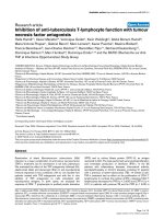
To evaluate mRNA expression levels of MCP-1, dia-
phragms from saline and LPS groups of mice were
analyzed with RNase protection assay, as shown in
Figure 1a. MCP-1 mRNA was not detected in co ntrol
diaphragms, but was greatly increased in the diaphragms
of septic animals (Figure 1b). Conversely, expression
levels of CCR2, the only known receptor for MCP-1,
were downregulated in the diaphragm after LPS admin-
istration (Figure 1c). The upregulation of MCP-1 mRNA
transcript levels was associated with a similar increase in
MCP-1 protein content within the septic diaphragm, as
shown in Figure 2a. MCP-1 protein levels were also
found to be significantly elevated in the serum (Figure
2b), as well as i n the lung, liver, and the tibialis anterior
2
3
MCP
-
1
Saline LPS
)b()a(
Saline
LPS
(a.u.)
2
3
0
1
2
MCP
1
L32
N/D
MCP-1 transcript
1
2
0
*
2
(c)
1
2
t
rary Units
6
h
r
s
2
4
h
r
s
*
*
(c)
0
*
Arbi
t
†
†
†
SL SL
Figure 1 Transcript levels of MCP-1 and its receptor in the septic diaphrag m. (a) Representative RNase protection assay showing MCP-1
mRNA in the diaphragm. (b) Quantification of MCP-1 mRNA levels in the diaphragm, normalized to the L32 housekeeeping gene. *P < 0.05 for
saline versus LPS groups; N/D, not detectable. (c) Quantification of mRNA levels of the MCP-1 receptor, CCR2 (open bars, tibialis anterior muscle;
solid bars, diaphragm; S, saline control group; L, LPS group). *P < 0.05 for tibialis versus diaphragm under the same conditions; +P < 0.05 for
saline versus LPS groups in the same muscle.
Labbe et al. Critical Care 2010, 14:R187
/>Page 4 of 11
muscle (Figure 2c) of LPS-group animals. Interestingly,
MCP-1 protein levels were two- to threefold higher in
the diaphragm than in the hindlimb muscle (tibialis
anterior) under septic conditions.
To determine whether the augmented levels of MCP-1
detected in the septic diaphragm were associated with
increased leukocyte infiltration into the muscle, immu-
nohi stochemical analysis was performed with antibodies
directed against markers for macrophages and neutro-
phils. As shown in Figure 3, no measurable differences
between control and septic diaphragms were found in
the numbers of either leukocyte population. This was
further confirmed for the neutrophil populat ion by the
lack of change in diaphragmatic MPO activity, wherea s
MPO activity was greatly increased in the lungs of septic
animals (Figure 3f).
Effects of MCP-1 on skeletal muscle proinflammatory
markers in vivo and in vitro
The ability of MCP- 1 to modulate proinflammatory
cytokine gene expression in the diaphragm during sepsis
in vivo was investigated by pretreating animals with
anti-MCP-1 neutralizing antibody. As indicated in Fig-
ure 4, transcript levels for IL-1a,IL-1b,andIL-6,as
well as for MCP-1 itself, were all significantly lower in
the diaphragms of mice that were pretreated with the
MCP-1 neutralizing antibody before LPS administration.
Therefore, systemic blockade of endogenous MCP-1
150
200
0.30
0.35
0.40
0.25
0.30
p
rotein)
*
m
l)
150
200
Saline
LPS
)b()a(
0.35
0.40
0.30
Saline
LPS
*
ng/ml
0
50
100
0.00
0.05
0.10
0.15
0.20
0.25
0.00
0.05
0.10
0.15
0.20
Dia
p
hra
g
m
MCP-1 (pg/ug
p
N/D
MCP-1 (pg/
m
0
50
100
Serum
0
0.05
0.10
0.15
0.20
0.25
09
1.0
1.1
1.2
pg
*
Saline
LPS
r
otein)
(c)
09
1.0
1.1
1.2
0.0
0.1
0.2
0.3
0.8
0
.
9
0
0.1
0.2
0.3
*
*
MCP-1 (pg/ug p
r
0.8
0
.
9
0
Lung
Liver
Tibialis
anterior
Figure 2 MCP-1 protein in the diaphragm and other organs during sepsis. MCP-1 protein content determined with ELISA in (a) diaphragm,
(b) serum, and (c) organs and hindlimb muscle (tibialis anterior). *P < 0.05 for saline versus LPS groups. N/D, not detectable.
Labbe et al. Critical Care 2010, 14:R187
/>Page 5 of 11
in vivo had major effects on the regulation of these
proinflammatory genes in the septic diaphragm.
We next sought to determine whether MCP-1 is cap-
able of directly stimulating inflammatory responses in
primary diaphragmatic muscle cell cultures examined at
4, 8, and 1 6 hours after stimulation. Interestingly,
despite significant effects of MCP-1 neutralization on
the expression of these genes in the septic diaphragm
in vivo, the transcript levels of IL-1a,IL-1b,andIL-6
were unaltered by direct MCP-1 stimulation of skeletal
muscle cells in vitro (no detectable expression under
either unstimulated or st imulated conditions). As shown
in Figures 5a and 5b, only MCP-1 itself was significantly
upregulated by MCP-1 stimulation in diaphragmatic
muscle cells, and this effect was noted at 8 hours a fter
stimulation. Moreover, in keeping with the fact that
MCP-1 did not upregulate these classic proinflammatory
genes in primary muscle cell culture s, we also did not
find any significant influence of MCP-1 treatment on
the NF-B transcriptional activity assay in C2C12 skele-
tal muscle cells (Figure 5c). Taken together, these results
suggest that MCP-1 is ca pable of acting on skeletal
muscle cells to upregulate its own expression, but in a
manner not dependent on NF-B pathway activation.
Effects of MCP-1 on skeletal muscle contractile function
in vivo
To evaluate the potential contribution of MCP-1 to the
adverse effects of sepsis on the contractile function of
skeletal muscles, two different approaches were use d.
First, to determine whether direct exposure of skeletal
muscle fibers to MCP-1 has effects on contractile func-
tion, recombinant MCP-1 protein was injected into the
EDL muscle. The dose of MCP-1 administered to the
EDL was extrapolated from the diaphragmatic MCP-1
content (picograms per muscle weight) at 12 hours after
2
3
3
(a)
(e)
Saline
LPS
2
o
ry cells
s
ue)
14
0
1
0
1
14
Macrophages Neutrophils
(b)
(c)
)
(f)
Saline
Inflammat
o
(% tis
s
2
4
6
8
10
12
2
4
6
8
12
10
(d)
P
O activity (U/ng tissue
LPS
0
2
0
2
Lung Diaphragm
M
P
Figure 3 Evaluation of inflammatory cells in the septic diaphragm. (a, b) Representative F4/80 staining of macrophages in saline- and LPS-
administered mice, respectively; (c, d) representative Ly6G staining of neutrophils in saline and LPS groups, respectively. (e) Morphometric
quantification of macrophages and neutrophils in the diaphragm. (f) Myeloperoxidase (MPO) activity in the diaphragm after LPS administration.
Labbe et al. Critical Care 2010, 14:R187
/>Page 6 of 11
LPS administration, as determin ed with ELISA and pre-
sented earlier in Figure 2. Figure 6a shows that at 12
hours after injection of recombinant MCP-1 into the
EDL, a small but statistically significant reduction was
noted in the force-generating capacity of the MCP-1-
injected EDL muscles relative to the contralateral con-
trol (saline-injected) muscles from the same animals.
Furthermore, as was the case for septic diaphragms at
the same time point after LPS administratio n (12
hours), the MCP-1-injected EDL muscles did not show
any histologic evidence of inflammatory cell infiltration
(not detected in either saline- or MCP-1-injected
muscles).
Second, to determine whether MCP-1 plays a role in
diaphragmatic contract ile dysfunction during sepsis, the
force-generating capacity of the diaphragm was com-
pared in animals pretreated with anti-MCP-1 neutraliz-
ing antibody versus a n irrelevant isotype control
immunoglobulin. As expected, LPS administration led to
a major decrease in diaphragmatic force production 12
hours later. The LPS-induced depression of diaphrag-
matic force was unaffected by pretreatment with an irre-
levant isotype control antibody. In marked contrast, the
loss of diaphragmatic force production at 12 hours after
LPS administration was greatly alleviated in animals pre-
treated with anti-MCP-1 neutralizing antibody, as
illustrated in Figure 6b. These findings indicate that
MCP-1 plays a significant role in the impairment of dia-
phragmatic function associated with acute endotoxemic
sepsis.
Discussion
To our knowledge, this is the first study to examine spe-
cifically the role of a chemokine, MCP-1, in proinflam-
matory mediator production by the diaphragm and the
contractile dysfunction of the muscle that occurs during
sepsis. From a clinical standpoint, our most important
observation was that neutralization of MCP-1 greatly
alleviated diaphragmatic weakness in the setting of acute
endotoxemia. This was associated with significantly
diminished diaphragmatic expression of proinflamma-
tory cytokines. Previous investigations in animals have
shown that MCP-1 effects in sepsis can vary according
to cell type and experimental model, as well as the spe-
cific mode and timing of MCP-1 inhibition. For exam-
ple, in th e cecal ligation/perforation (CLP) sepsis model,
mice genetically deficient in MCP-1 showed lower IL-10
production in peritoneal macrophages and increased
mortality [20]. In contrast, antibody neutralization of
MCP-1 in the CLP context had a beneficial effect on
survival [14], and the administratio n of an MCP-1-
synthesisinhibitor,bindarit,wasalsoreportedtobe
3
Saline LPS + IgG
LPS
+ anti-MCP-1
MCP
-
1
3
Saline
LPS + control IgG
LPS + anti-MCP-1
)b()a(
1
2
L32
IL-1β
MCP
1
1
a
nscript/L
32
a.u.
*
*
*
†
†
†
2
0
1
L32
IL-6
IL-1α
1
0
N/D N/D N/D
Tr
a
*
†
MCP-1
IL-1β IL-1α IL-6
Figure 4 Effects of MCP-1 inhibition on inflammatory gene expression in the septic diaphragm. (a) Representative RNase protection
assays showing proinflammatory gene expression in diaphragms of mice pretreated with anti-MCP-1 antibody or isotypic control antibody (IgG)
during sepsis. (b) Quantification of proinflammatory gene mRNA levels in the diaphragm, normalized to the L32 housekeeeping gene. *P < 0.05
for IgG control antibody versus anti-MCP-1 antibody pretreatment groups; +P < 0.05 for IgG control antibody versus saline groups.
Labbe et al. Critical Care 2010, 14:R187
/>Page 7 of 11
2
Vehicle MCP-1
2
Vehicle
MCP-1
)b()a(
t
(a.u.)
0
1
MCP-1
L32
0
1
*
(c)
MCP-1 Transcrip
t
2
3
3
se activity (RLU)
Vehicle
MCP-1
(c)
2
0
1
0
1
4 hrs 8 hrs 16 hrs
NF-κB lucifera
Figure 5 Effects of MCP-1 treatment on inflammatory markers in cultured skeletal muscle cells. (a) Representative RNase protection
assays. (b) Quantification of MCP-1 mRNA levels, after in vitro stimulation of primary diaphragmatic myotube cultures with recombinant MCP-1
(100 ng/ml). (c) NF-B transcriptional activity in C2C12 myotube cultures treated with recombinant MCP-1 (100 ng/ml), as determined by the
plasmid transfection luciferase reporter system. *P < 0.05 for vehicle-versus MCP-1-treated groups.
20
25
30
40
50
20
25
30
LPS
Saline
LPS + anti-MCP-1
LPS + IgG control
Saline
(a)
m
2
)
50
40
*
MCP-1
)
(b)
0
5
10
15
20
0
10
20
30
0
5
10
15
20
*
Force (N/c
m
0
10
20
30
Force (N/cm
2
)
0 20 40 60 80 100 120 140 160
0
0 20406080100120140160
0
0
0 40 80 120 160
Frequency (Hz)
0
0 40 80 120 160
Frequency (Hz)
Figure 6 Effects of MCP-1 modulation on skeletal muscle force-generating capacity in vivo. (a) Effects of exogenou s MCP-1 injection on
the force-frequency relation of the extensor digitorum longus (EDL) muscle in nonseptic mice; *P < 0.05 for saline-versus MCP-1-injected mice.
(b) Effects of inhibiting endogenous MCP-1 on the force-frequency relation of the diaphragm in septic mice. *P < 0.05 for saline versus LPS
groups; +P < 0.05 for IgG control antibody versus anti-MCP-1 antibody pretreatment in LPS groups.
Labbe et al. Critical Care 2010, 14:R187
/>Page 8 of 11
beneficial in different murine models of sepsis [29]. A
complex pattern of both pro- and anti-inflammatory
effects on different organs has been reported after
MCP-1 neutralization in CLP animals [13].
Intriguingly, in a recent prospective cohort study of
patients with severe sepsis in which a multiplex analysis
of 17 candidate cytokines in the serum was performed,
only MCP-1 was found to be independently associated
with increased mortality [17]. The fact that the dia-
phragm constitutively expresses CCR2 [30] led us to test
the hypothesis that MCP-1 could directly regulate the
expression of proinflammatory mediators in skeletal
muscle cells. In keeping with this, we found that direct
stimulation of primary diaphragmatic cell cultures by
purified MCP-1 led to an increase of MCP-1 transcripts,
suggesting the existence of positive-feedback autoregula-
tion. Such a feed-forward loop has been previously
described for other chemokines in different cell types
[31,32]. Although very little is known about the
mechanisms or functional significance of this positive-
feedback loop, the result is likely to be an enhancem ent
of MCP-1 actions. The downregulation of CCR2 expres-
sion that we observ ed in the septic diaphragm, which is
analogous to that reported in monocytes exposed to
LPS [33], is presumably an important mechanism for
counterbalancing this effect.
Interestingly, the transcript levels of IL-1a,IL-1b,and
IL-6 were unaltered by direct MCP-1 stimulation of skele-
tal muscle cells in vitro. Consistent with these findings,
NF-B reporter gene activity was a lso not increased in
myotubes exposed to MCP-1. Although it could be argued
that activation of NF-B may have occurred more rapidly
than the earlies t time point examined in our study (4
hours), this appears unlikely because the firefly luciferase
protein used as a readout in these experiments is stable
for u p to 6 hours in mammalian cells [34]. Furthermore,
in primary human abdominal muscle culture, MCP-1 did
not induce NF-B activation within 1 hour of stimulation
[21]. This is in contrast to the situation within isolated car-
diomyocytes, in which MCP-1 (at the same dose used in
our study) has been reported to upregulate IL-1b and IL-6
expression [19]. MCP-1 has also bee n fo und to stimulate
the expression of IL-6 in neutrophils [18] and leukotriene
B4 in peritoneal macrophages [12]. Taken together, these
findings emphasize the existence of cell- and organ-speci-
fic regulatory mechanisms for MCP-1. Furthermore, given
our demonstration that MCP-1 stimulation of skeletal
muscle cells in vitro fails directly to upregulate IL-1a, IL-
1b, or IL-6 expression, it is likely that the ability of MCP-1
neutralization to downregulate these cytokines in vivo dur-
ing sepsis is achieved, at least in part, via intermediary
partners.
Although MCP-1 was recently shown to play several
key roles in skeletal muscle repair and metabolism
[21-23,35], its influence on muscle function during sep-
sis has not been previously explored. We found that
in vivo neutralization of endogenous MCP-1 during
acute sepsis led to substantial decreases in the transcript
levels for IL-1a, IL-1b, and IL-6, as well as MCP-1 itself,
in the diaphragm. IL-1 significantly decreases muscle
weight, protein content, and the rate of protein synthesis
in skeletal muscl e [36], whereas IL-6 can u pregulate the
cathepsin and ubiquitin pathways of muscle proteolysis
[37]. Exposure of human skeletal muscle cells to MCP-1
at physiologic concentrations has been demonstrated to
induce a state of increased insulin resistance, as indi-
cated by alterations in insulin signaling with an asso-
ciated impairment of glucose uptake [21]. Taken
together, such metabolic derangements all h ave the
potential to depress skeletal muscle contractile function.
In addition, reactive oxidative species also play an
important role in diaphragmatic dysfunction during sep-
sis [8], and overproduction of MCP-1 has been linked to
increased oxidative stress and tissue damage in cardiac
muscle after ischemia-reperfusion [38].
As an important leukocyte chemoattractant molecule,
a plausible hypothesis was that MCP-1 overexpression
in the diaphragm during seps is might increase inflam-
matory cell infiltration into the muscle. This was not
found to be the case, as the levels of both neutrophils
and macrophages in the diaphragm were unaffected by
LPS administration. In addition, although direct injec-
tion of MCP-1 into skeletal muscle was associated with
a mild reduction in force-generating capacity, this was
similarly not linked to increased inflammatory cell infil-
tration. However, this does not exclude the p ossibility
that increased exposure to MCP-1 (either during sepsis
in the diaphragm or through d irect injection into the
EDL) modified the activation state of resident macro-
phages within these muscles, and this hypothesis
deserves further study.
Conclusions
In summary, this study demonstrates that the increased
endogenous MCP-1 produc tion durin g SIRS induced by
endotoxin contributes to proinflammatory mediator pro-
duction by the diaphragm, along with a major decrease
in diaphragmatic force-generating capacity. Our findings
suggest that the systemic immunomodulatory properties
of MCP-1, coupled with its ability to modify skeletal
muscle cell function directly, could make MCP-1 an
attractive therapeutic target in sepsis patients, especially
in the setting of respiratory muscle dysfunction and ven-
tilatory failure.
Key messages
• MCP-1 is significantly upregulated in the dia-
phragm during acute endotoxemic sepsis
Labbe et al. Critical Care 2010, 14:R187
/>Page 9 of 11
• Antibody neutralization of MCP-1 in this setting
reduces the diaphragmatic expression levels of sev-
eral proinflammatory cytokines that have been impli-
cated in the pathogenesis of sepsis
• MCP-1 neutralization preve nts the loss of dia-
phragmatic force-generating capacity normally
observed during acute endotoxemia
Abbreviations
CCL: CC chemokine ligand; CCR: CC chemokine receptor; CLP: cecal ligation
and perforation; EDL: extensor digitorum longus; IL: interleukin; LPS:
lipopolysaccharide; MCP: monocyte chemoattractant protein; MPO:
myeloperoxidase; SIRS: systemic inflammatory response syndrome.
Acknowledgements
This study was supported by the Canadian Institutes of Health Research, the
Fonds de la recherche en santé du Quebec, the Quebec Respiratory Health
Network, and the McGill University Health Centre Research Institute.
Author details
1
Meakins-Christie Laboratories, McGill University, 3626 Saint Urbain, Montreal,
Quebec, Canada H2X 2P2.
2
Université Paris 6 Pierre et Marie Curie, UPRES
EA2397, Service de Pneumologie et Réanimation, Groupe Hospitalier Pitié-
Salpêtrière, 47-83 boulevard de l’Hôpital, 75651 Paris cedex 13, Paris, France.
3
Respiratory Division, McGill University Health Centre and Research Institute,
687 Pine Avenue West, Montreal, Quebec, Canada H3A 1A1.
Authors’ contributions
KC was involved in all aspects of the study, GD performed muscle-
contractility experiments, DG was involved in primary cell cultures, AD and
MD were involved in RNase protection assays, JHB performed luciferase
assays, and BJP was involved in all aspects of the study.
Competing interests
The authors declare that they have no competing interests.
Received: 4 June 2010 Revised: 5 August 2010
Accepted: 7 October 2010 Published: 7 October 2010
References
1. Khan J, Harrison TB, Rich MM, Moss M: Early development of critical illness
myopathy and neuropathy in patients with severe sepsis. Neurology
2006, 67:1421-1425.
2. Eikermann M, Koch G, Gerwig M, Ochterbeck C, Beiderlinden M, Koeppen S,
Neuhauser M, Peters J: Muscle force and fatigue in patients with sepsis
and multiorgan failure. Intensive Care Med 2006, 32:251-259.
3. Martin GS, Mannino DM, Eaton S, Moss M: The epidemiology of sepsis in
the United States from 1979 through 2000. N Engl J Med 2003,
348:1546-1554.
4. Hussain SNA, Simkus G, Roussos C: Respiratory muscle fatigue: a cause of
ventilatory muscle failure in septic shock. J Appl Physiol 1985,
58:2033-2040.
5. Boczkowski J, Lanone S, Ungureanu-Longrois D, Danialou G, Fournier T,
Aubier M: Induction of diaphragmatic nitric oxide synthase after
endotoxin administration in rats. J Clin Invest 1996, 98:1550-1559.
6. Lin MC, Ebihara S, el-Dwairi Q, Hussain SNA, Yang L, Gottfried SB,
Comtois A, Petrof BJ: Diaphragm sarcolemmal injury is induced by sepsis
and alleviated by nitric oxide synthase inhibition. Am J Respir Crit Care
Med 1998, 158:1656-1663.
7. Divangahi M, Matecki S, Dudley RW, Tuck SA, Bao W, Radzioch D,
Comtois AS, Petrof BJ: Preferential diaphragmatic weakness during
sustained Pseudomonas aeruginosa lung infection. Am J Respir Crit Care
Med 2004, 169:679-686.
8. Callahan LA, Supinski GS: Sepsis-induced myopathy. Crit Care Med 2009,
37:S354-S367.
9. Demoule A, Divangahi M, Yahiaoui L, Danialou G, Gvozdic D, Labbe K,
Bao W, Petrof BJ: Endotoxin triggers NF-kappaB-dependent upregulation
of multiple pro-inflammatory genes in the diaphragm. Am J Respir Crit
Care Med 2006, 174:646-653.
10. Li X, Moody MR, Engel D, Walker S, Clubb FJ Jr, Sivasubramanian N,
Mann DL, Reid MB: Cardiac-specific overexpression of tumor necrosis
factor-alpha causes oxidative stress and contractile dysfunction in
mouse diaphragm. Circulation 2000, 102:1690-1696.
11. Deshmane SL, Kremlev S, Amini S, Sawaya BE: Monocyte chemoattractant
protein-1 (MCP-1): an overview. J Interferon Cytokine Res 2009, 29:313-326.
12. Matsukawa A, Hogaboam CM, Lukacs NW, Lincoln PM, Strieter RM,
Kunkel SL: Endogenous monocyte chemoattractant protein-1 (MCP-1)
protects mice in a model of acute septic peritonitis: cross-talk between
MCP-1 and leukotriene B4. J Immunol 1999, 163:6148-6154.
13. Matsukawa A, Hogaboam CM, Lukacs NW, Lincoln PM, Strieter RM,
Kunkel SL: Endogenous MCP-1 influences systemic cytokine balance in a
murine model of acute septic peritonitis.
Exp Mol Pathol 2000, 68:77-84.
14. Tsuda Y, Takahashi H, Kobayashi M, Hanafusa T, Herndon DN, Suzuki F:
CCL2, a product of mice early after systemic inflammatory response
syndrome (SIRS), induces alternatively activated macrophages capable
of impairing antibacterial resistance of SIRS mice. J Leukoc Biol 2004,
76:368-373.
15. Jansen PM, van DJ, Put W, de JI, Taylor FB Jr, Hack CE: Monocyte
chemotactic protein 1 is released during lethal and sublethal bacteremia
in baboons. J Infect Dis 1995, 171:1640-1642.
16. Bossink AW, Paemen L, Jansen PM, Hack CE, Thijs LG, van DJ: Plasma levels
of the chemokines monocyte chemotactic proteins-1 and -2 are
elevated in human sepsis. Blood 1995, 86:3841-3847.
17. Bozza FA, Salluh JI, Japiassu AM, Soares M, Assis EF, Gomes RN, Bozza MT,
Castro-Faria-Neto HC, Bozza PT: Cytokine profiles as markers of disease
severity in sepsis: a multiplex analysis. Crit Care 2007, 11:R49.
18. Speyer CL, Gao H, Rancilio NJ, Neff TA, Huffnagle GB, Sarma JV, Ward PA:
Novel chemokine responsiveness and mobilization of neutrophils during
sepsis. Am J Pathol 2004, 165:2187-2196.
19. Damas JK, Aukrust P, Ueland T, Odegaard A, Eiken HG, Gullestad L,
Sejersted OM, Christensen G: Monocyte chemoattractant protein-1
enhances and interleukin-10 suppresses the production of inflammatory
cytokines in adult rat cardiomyocytes. Basic Res Cardiol 2001, 96:345-352.
20. Gomes RN, Figueiredo RT, Bozza FA, Pacheco P, Amancio RT, Laranjeira AP,
Castro-Faria-Neto HC, Bozza PT, Bozza MT: Increased susceptibility to
septic and endotoxic shock in monocyte chemoattractant protein 1/cc
chemokine ligand 2-deficient mice correlates with reduced interleukin
10 and enhanced macrophage migration inhibitory factor production.
Shock 2006, 26:457-463.
21. Sell H, Schroeder D, Kaiser U, Eckel J: Monocyte chemotactic protein-1 is a
potential player in the negative cross-talk between adipose tissue and
skeletal muscle. Endocrinology 2006, 147:2458-2467.
22. Warren GL, O’Farrell L, Summan M, Hulderman T, Mishra D, Luster MI,
Kuziel WA, Simeonova PP: Role of CC chemokines in skeletal muscle
functional restoration after injury. Am J Physiol Cell Physiol 2004, 286:
C1031-C1036.
23. Yahiaoui L, Gvozdic D, Danialou G, Mack M, Petrof BJ: CC family
chemokines directly regulate myoblast responses to skeletal muscle
injury. J Physiol 2008, 586:3991-4004.
24. Boyd JH, Divangahi M, Yahiaoui L, Gvozdic D, Qureshi S, Petrof BJ: Toll-like
receptors differentially regulate CC and CXC chemokines in skeletal
muscle via NF-κB and calcineurin. Infect Immun 2006, 74:6829-6838.
25. Pampinella F, Lechardeur D, Zanetti E, MacLachlan I, Benharouga M,
Lukacs GL, Vitiello L: Analysis of differential lipofection efficiency in
primary and established myoblasts. Mol Ther 2002, 5:161-169.
26. Guibinga GH, Lochmüller H, Massie B, Nalbantoglu J, Karpati G, Petrof BJ:
Combinatorial blockade of calcineurin and CD28 signaling facilitates
primary and secondary therapeutic gene transfer by adenovirus vectors
in dystrophic (mdx) mouse muscles. J Virol 1998, 72:4601-4609.
27. Dudley RW, Danialou G, Govindaraju K, Lands L, Eidelman DE, Petrof BJ:
Sarcolemmal damage in dystrophin deficiency is modulated by
synergistic interactions between mechanical and oxidative/nitrosative
stresses. Am J Pathol 2006, 168 :1276-1287.
28. Divangahi M, Demoule A, Danialou G, Yahiaoui L, Bao W, Zhou X, Petrof BJ:
Impact of IL-10 on diaphragmatic cytokine expression and contractility
during Pseudomonas infection. Am J Respir Cell Mol Biol 2007, 36:504-512.
29. Ramnath RD, Ng SW, Guglielmotti A, Bhatia M: Role of MCP-1 in
endotoxemia and sepsis. Int Immunopharmacol 2008, 8:810-818.
Labbe et al. Critical Care 2010, 14:R187
/>Page 10 of 11
30. Demoule A, Divangahi M, Danialou G, Gvozdic D, Larkin G, Bao W, Petrof BJ:
Expression and regulation of CC class chemokines in the dystrophic
(mdx) diaphragm. Am J Respir Cell Mol Biol 2005, 33:178-185.
31. Stefanovic L, Brenner DA, Stefanovic B: Direct hepatotoxic effect of KC
chemokine in the liver without infiltration of neutrophils. Exp Biol Med
(Maywood ) 2005, 230:573-586.
32. Schruefer R, Lutze N, Schymeinsky J, Walzog B: Human neutrophils
promote angiogenesis by a paracrine feedforward mechanism involving
endothelial interleukin-8. Am J Physiol Heart Circ Physiol 2005, 288:
H1186-H1192.
33. Parker LC, Whyte MK, Vogel SN, Dower SK, Sabroe I: Toll-like receptor (TLR)
2 and TLR4 agonists regulate CCR expression in human monocytic cells.
J Immunol 2004, 172:4977-4986.
34. McNabb DS, Reed R, Marciniak RA: Dual luciferase assay system for rapid
assessment of gene expression in Saccharomyces cerevisiae. Eukaryot Cell
2005, 4:1539-1549.
35. Shireman PK, Contreras-Shannon V, Ochoa O, Karia BP, Michalek JE,
McManus LM: MCP-1 deficiency causes altered inflammation with
impaired skeletal muscle regeneration. J Leukoc Biol 2007, 81:775-85.
36. Cooney RN, Maish GO III, Gilpin T, Shumate ML, Lang CH, Vary TC:
Mechanism of IL-1 induced inhibition of protein synthesis in skeletal
muscle. Shock 1999, 11:235-241.
37. Tsujinaka T, Fujita J, Ebisui C, Yano M, Kominami E, Suzuki K, Tanaka K,
Katsume A, Ohsugi Y, Shiozaki H, Monden M: Interleukin 6 receptor
antibody inhibits muscle atrophy and modulates proteolytic systems in
interleukin 6 transgenic mice. J Clin Invest 1996, 97:244-249.
38. Kajihara N, Morita S, Nishida T, Tatewaki H, Eto M, Egashira K, Yasui H:
Transfection with a dominant-negative inhibitor of monocyte
chemoattractant protein-1 gene improves cardiac function after 6 hours
of cold preservation. Circulation 2003, 108(Suppl 1):II213-II218.
doi:10.1186/cc9295
Cite this article as: Labbe et al.: Inhibition of monocyte chemoattractant
protein-1 prevents diaphragmatic inflammation and maintains
contractile fu nction during endotoxemia. Critical Care 2010 14:R187.
Submit your next manuscript to BioMed Central
and take full advantage of:
• Convenient online submission
• Thorough peer review
• No space constraints or color figure charges
• Immediate publication on acceptance
• Inclusion in PubMed, CAS, Scopus and Google Scholar
• Research which is freely available for redistribution
Submit your manuscript at
www.biomedcentral.com/submit
Labbe et al. Critical Care 2010, 14:R187
/>Page 11 of 11