Báo cáo y học: "Acidemia does not affect outcomes of patients with acute cardiogenic pulmonary edema treated with continuous positive airway pressure" pps
Bạn đang xem bản rút gọn của tài liệu. Xem và tải ngay bản đầy đủ của tài liệu tại đây (577.65 KB, 8 trang )
RESEARC H Open Access
Acidemia does not affect outcomes of patients
with acute cardiogenic pulmonary edema treated
with continuous positive airway pressure
Stefano Aliberti
1*
, Federico Piffer
1
, Anna Maria Brambilla
2
, Angelo A Bignamini
3
, Valentina D Rosti
2
,
Tommaso Maraffi
2
, Valter Monzani
2
, Roberto Cosentini
2
Abstract
Introduction: A lack of data exists in the literature evaluating acidemia on admission as a favorable or negative
prognostic factor in patients with acute cardiogenic pulmonary edema (ACPE) treated with non-invasive
continuous positive airway pressure (CPAP). The objective of the present study was to investigate the impact of
acidemia on admission on outcomes of ACPE patients treated with CPAP.
Methods: This was a retrospective, observational study of consecutive patients admitted with a diagnosis of ACPE
to the Emergency Department of IRCCS Fondazione Cà Granda Ospedale Maggiore Policlinico, Milan, Italy, between
January 2003 and December 2006, treated with CPAP on admission. Two groups of patients were identified:
subjects with acidemia (acidotic group), and those with a normal pH on admission (controls). The primary
endpoint was clinical failure, defined as switch to bi-level ventilation, switch to endotracheal intubation or
inhospital mortality.
Results: Among the 378 patients enrolled, 290 (77%) were acidotic on admission. A total of 28 patients (9.7%) in
the acidotic group and eight patients (9.1%) among controls experienced a clinical failure (odds ratio = 1.069, 95%
confidence interval = 0.469 to 2.438, P = 0.875). Survival analysis indicates that, among acidotic patients, the time
at which 50% of patients reached the 7.35 threshold was 173 minutes (95% confidence interval = 153 to 193).
Neither acidemia (P = 0.205) nor the type of acidosis on admission (respiratory acidosis, P = 0.126; metabolic
acidosis, P = 0.292; mixed acidosis, P = 0.397) affected clinical failure after adjustment for clinical and laboratory
factors in a multivariable logistic regression model.
Conclusions: Neither acidemia nor the type of acidosis on admission should be considered risk factors for adverse
outcomes in ACPE patients treated with CPAP.
Introduction
International guidelines suggest the use of non-invasive
continuous positive airways pressure (CPAP) as first-line
intervention in patients with acute cardiogeni c pulmon-
ary edema (ACPE) [1]. CPAP has proven to be easier to
use, quicker to implement in clinical practice and to
carry smaller associated costs in comparison with non-
invasive ventilation ( NIV) [2]. In light of these findings,
CPAP has also been also used to treat ACPE patients
outside the intensive care unit or the Emergency
Department, as in the general ward or during prehospi-
tal care [3].
The rate of mortality in ACPE patients treated with
CPAP is reported to be up to 13% [4,5]. Therefore, it is
crucial for healthcare providers to identify risk factors
for failure of CPAP treatment, in order to better allocate
medical resources and improve clinical outcomes of
ACPE patients.
Severity of acidemia on admission, as well as lack of
improvement of respiratory acidosis during the first few
hours of NIV, have emerged as important predictors of
failure in patients suffering of hypercapnic respiratory
* Correspondence:
1
Dipartimento toraco-polmonare e cardio-circolatorio, University of Milan,
IRCCS Fondazione Cà Granda Ospedale Maggiore Policlinico, via F. Sforza 35,
20122 Milan, Italy
Full list of author information is available at the end of the article
Aliberti et al. Critical Care 2010, 14:R196
/>© 2010 Aliberti et al.; licensee BioMed Central Ltd. This is an open access article distributed under the term s of the Creative Commons
Attribu tion License ( which permits unrestricted use, distribution, and reproduction in
any medium, provided the original work is properly cite d.
failure [ 6-8]. Acidemia on admissio n has b een also
shown to predict NIV fail ure a few days after its initial
application in patients who have previously experienced
an initial improvement of clinical status and blood gas
values [9]. In clinical practice, acidotic patients with
ACPE are commonly considered more severe in com-
parison with nonacidotic patients. In view of this con-
sideration, the largest clinical trial that has evaluated
CPAP and NIV in ACPE patients enrolled acidotic
patients [10].
On the contrary, acidemia has not been identified a s a
predictor of NIV failure in patients with hypoxemic
respiratory failure [5,11]. Conflicting data exist in the lit-
erature alternatively considering respiratory acidosis
a favorable or a negative prognostic factor in ACPE
patients. Particularly, ACPE patients who suffered
respiratory acidosis on admission were identified as those
exhibiting a better response to CPAP treatment [12].
To define the impact of acidemia on clinical outcomes
of ACPE patients treated with CPAP, the present study
has the following objectives: to compare outcomes and
physiological measurementsofpatientswithacidemia
versus those with normal pH values on admission; and
to evaluate outcomes and physiological measurements
of patients with different types of acidosis on admission.
Materials and methods
Setting and subjects
This was a retrospective, observational study of consecu-
tive patients admitted with a diagnosis of ACPE to the
Emergency Department of IRCCS Fondazione Ca’
Granda Ospedale Maggiore Policlinico, Milan, Italy
between January 2003 and December 2006.
Adult patients who satisfied the criteria for ACPE and
who were treated with CPAP on admission were
enrolled in the study. Patients with alkalemia on admis-
sion were excluded.
The diagnosis of ACPE was established on the basis of
medical history (acute severe dyspnea) and typical physi-
cal findings (widespread pulmonary rales), with chest
radiography confirming pulmonary vascular congestion.
Criteria for application of CPAP included at least one of
the following: severe acute respiratory failure (PaO
2
/
FiO
2
ratio <300); respiratory rate exceeding 30 breaths/
minute or use of accessory respiratory muscles or para-
doxical a bdominal motion; and respiratory acidosis (pH
<7.350, PaCO
2
≥45 mmHg).
All patients enrolled in the study underwent high-flow
CPAP (90 t o 140 l/minute; VitalSigns Inc., Totowa, NJ,
USA) as the first choice of treatment, in addition to
oxygen and standard medical treatment. Interfaces used
were a facemask (VitalSigns Inc.) or a hel met (StarMed,
Mirandola, Italy) with a positive end-expiratory pressure
(PEEP) valve (VitalSigns Inc.). CPAP was not applied in
ACPE patients if any among the following findings was
present: immediate need for endotracheal intubation;
impairment of consciousness (Kelly scale >4) [13]; and
hemodynamic instability (systolic blood pressure <90
mmHg). Criteria for discontinuation from CPAP
included all of the following: absence of respiratory dis-
tress; respiratory rate <25 be ats/minute; hemodynamic
stability; pH >7.35; and PaO
2
/FiO
2
ratio >300 or oxygen
saturation ≥95%.
Criteria to switch from CPAP to bi-level ventilation
were a lack of improvement or a worsening of ventila-
tion and/or gas exchange at a blood gas examination
performed 30 minutes/1 hour after initiation of CPAP
treatment, in the absence of criteria for endotracheal
intubation (ETI). Criteria for ETI were at least one
among the following: impairment of consciousness;
hemodynamic instability (systolic blood pressure
<90 mmHg); cardiac and/or respiratory arrest; and a
lack of improvement or a worsening of vent ilation and/
or gas exchange at a blood gas examination performed
1 hour after initiation of bi-level treatment.
The above criteria for the application of CPAP in
ACPE patients as well as the protocol of medical treat-
ment were applied according to local st andard operating
procedures. Each patient received medical treatment
acco rding to the local standard of care: intravenous fur-
osemide 40 to 100 mg based on fluid retention (or at
least doubling the dose at home) targeted on the urinary
output; intravenous isosorbide dinitrate on continuous
infusion starting at 1 mg/hour up to 10 mg/ho ur; intra-
venous morphine up to 4 mg and vasopressors in case
of hypotension. No subjects receiving invasive or non-
invasive pressure support ventilation before CPAP treat-
ment were included in the study.
Study design
Records of all the enrolled patients were carefully
reviewed. Data on admission, before and during CPAP
treatment, and during hospitalization were collected, and
included the following: demographic informatio n and
past medical history; clinical characteristics; laboratory
evaluation performed on arterial sample; and information
needed to derive the Simplified Acute Physiology Score II
[14]. Arterial blood gas evaluation on admission was con-
sidered for those samples obtained within 15 minutes
from admission to the hospital, based on local standard
operating procedures. A group of investigators of the
Emergency Department, Fondazione Ca’ Granda, Milan,
Italy validated the quality of data by checking for discre-
pancies and inconsistencies before cases were entered
into a database. The Institutional Review Board of
the IRCCS Fondazione Ca’ Granda Ospedale Maggiore
Aliberti et al. Critical Care 2010, 14:R196
/>Page 2 of 8
Policlinico, Milan approved the study. The study was in
compliance with the Helsinki Declaration; informed con-
sent was waived by the Institutional Review Board.
Study definitions
The normal pH range was considered 7.35 to 7.45.
Alkalemia was considered if the pH value on admission
was more than 7.45. Acidemia was considered if the
pH value on admission was lessthan7.35.Respiratory
acidosis was considered when acidemia was identified
with PaCO
2
≥45 mmHg a nd bicarbonates (HCO
3
-
) ≥22
mmol/l. Metabolic acidosis was considered when acid-
emia was identified with PaCO
2
<45 mmHg and
HCO
3
-
<22 mmol/l. Mixed acidosis was considered
when acidemia was identified with PaCO
2
≥45 mmHg
and HCO
3
-
<22 mmol/l.
Study groups
Patients with ACPE treated with CPAP were divided
into two groups according to the pH value on admis-
sion: subjects with acidemia (acidotic group), and those
with a normal pH (controls). Among patients of the
acidotic group, three subgroups were identified accord-
ing to PaCO
2
and HCO
3
-
values: patients with respira-
tory acido sis, patients wit h metabolic acidosis, and
patients with mixed acidosis.
Endpoints
The primary endpoint was clinical failure, defined as at
least one among: a switch to non-invasive bi-level venti-
lation, a switch to ETI, and inhospital mortality.
A sw itch to bi-level ventilation was applied when both
blood gas values were unchanged/worsened with CPAP
and criteria for ETI were not fulfilled. ETI was per-
formed according to our local standard operating proce-
dures. Inhospital mortality was defined as death by any
cause occurring during hospitalization. ACPE-related
mortality was defined as death occurring during the epi-
sode of ACPE. Late mortality w as defined as death
occurring after the resolution of the episode of ACPE.
Our local standard operating procedures define an epi-
sode of ACPE as being resolved when all the criteria for
discontinuation of CPAP mentioned above are reached.
Thesecondaryendpointwasthelengthofstayinthe
hospital. This length of stay was calculated as the num-
ber of days from the date of admission to the date of
discharge, and was censored at 14 days in an effort to
captureonlytheACPE-related length of stay in the
hospital.
Statistical analysis
All data were statistically analyzed with SPSS for Win-
dows (version 14.0; SPSS Inc., Chicago, IL, USA).
Descriptive statistics are reported as the mean with
standard deviation or counts and proportions as appro-
priate. Patient characteristics were compared between
groups. Summary statistics for all continuous explana-
tory variables are presented as means with differences
between groups compared by independent t test. Cate-
gorical explana tory variables are summari zed as percen-
tages with differences between groups analyzed using
the chi-square test or the Fisher exact test where appro-
priate. The time to event was analyzed by Kaplan-Meier
survival analysis. The association between clinical failure
and acidemia on admission was anal yzed using multiple
logistic regression. All expl anatory variables considered
of clinical relevance and those previously found to be
significantly associated with mortalit y in ACPE patients
treated with CPAP were incorporated i nto the model
[5]. The time course o f continuous variables was a na-
lyzed by r epeated-measures analysis of variance after
replacing the missing values with the last observation
carried forward technique. P < 0.05 was considered sta-
tistically significant.
Results
Acidotic population
Among the 419 ACPE patients treated with CPAP who
were enrolled during the study period, the pH value
within 15 minutes from admission was not available in
23 patients, while 18 patients were excluded because of
alkalemia on admission. T he final study population
accounted for 378 patients: 290 (77%) were acidotic on
admission (acidotic group), while 88 were controls.
Baseline characteristics and the CPAP setting of the
acidotic group and controls are summarized in Table 1.
The mean ± standard deviation duration of CPAP
treatment was 318 ± 485 minutes a nd 262 ± 198 min-
utes in the acidotic group and controls, respectively (P =
0.289). The mean ± standard deviation FiO
2
during
CPAP was 48 ± 11% and 47 ± 9% in the acidotic group
and in controls, respectively (P = 0.219). The mean ±
standard deviation PEEP during CPAP was 8.1 ± 1.7
cmH
2
O and 7.9 ± 1.4 cmH
2
O in the acidotic group and
in controls, respectively (P = 0.229).
A total of 28 patients (9.7%) in the acidotic group and
eight patients (9.1%) among controls experienced a clini-
cal failure (odds ratio = 1.069; 95% confidence interval =
0.469 to 2.438; P =0.875)(seeTable2).Acidemiaon
admission did not affect clinical failure after adjustment
for age, history of acute myocardial infarction, hypocap-
nia, normotension and PaO
2
/FiO
2
ratio in a multivari-
able logistic regression model (P = 0.205).
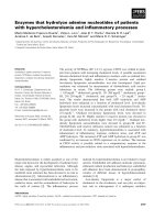
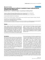
The crude proportion of clinical failure in the study
population is presented in Figure 1, split by pH value on
admission. The 95% confidence interval of the controls
group included the point estimate and most of the con-
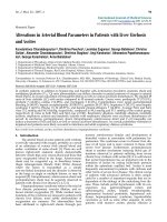
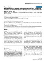
fidence intervals of the other groups. Figure 2 shows the
Aliberti et al. Critical Care 2010, 14:R196
/>Page 3 of 8
Table 1 Baseline characteristics on admission and before continuous positive airway pressure treatment
Variable Acidotic group (n = 290) Controls (n = 88) P value
Demographics
Male 143 (49) 36 (41) 0.167
a
Age (years) 80 ± 10 (n = 290) 81 ± 9.5 (n = 88) 0.360
b
Comorbidities
Chronic obstructive pulmonary disease 84 (29) 17/86 (20) 0.091
a
Essential hypertension 162 (56) 46/86 (54) 0.697
a
Diabetes mellitus 72 (25) 19/86 (22) 0.603
a
Congestive heart failure 165 (57) 51/86 (59) 0.692
a
Chronic renal failure 76 (26) 13/86 (15) 0.034
a
Severity of the disease
Simplified Physiologic Acute Score II 42 ± 6.7 (n = 258) 40 ± 8.1 (n = 74) 0.014
b
Physical findings
Systolic blood pressure (mmHg) 173 ± 30 (n = 286) 170 ± 31 (n = 87) 0.328
b
Diastolic blood pressure (mmHg) 99 ± 20 (n = 283) 97 ± 19 (n = 87) 0.391
b
Systolic <140 mmHg and diastolic <90 mmHg 32 (11) 9 (10) 0.802
b
Heart rate (beats/minute) 116 ± 22 (n = 283) 121 ± 22 (n = 87) 0.163
b
Heart rate >100 beats/minute 197/283 (70) 53/87 (61) 0.130
a
Respiratory rate (breaths/minute) 41 ± 6.1 (n = 175) 39 ± 6.9 (n = 64) 0.016
b
Respiratory rate ≥40 breaths/minute 120/175 (69) 30/64 (47) 0.002
a
Arterial blood gas analysis
pH 7.22 ± 0.09 (n = 290) 7.39 ± 0.03 (n = 88) Not applicable
PaCO
2
(mmHg) 53 ±16 (n = 290) 36 ±6.6 (n = 88) <0.001
b
Bicarbonates (mmol/l) 22 ± 5.3 (n = 288) 22 ± 3.8 (n = 88) 0.330
b
PaO
2
/FiO
2
ratio 178 ± 93 (n = 283) 222 ± 82 (n = 87) <0.001
b
PaO
2
/FiO
2
ratio <200 184/283 (65) 32/87 (37) <0.001
a
Acute myocardial infarction on admission 43 (15) 14 (16) 0.804
a
CPAP setting
Initial FiO
2
(%) 49.7 ± 12.1 (n = 288) 48.6 ± 11.4 (n = 88) 0.421
b
Initial PEEP (cmH
2
O) 9.7 ± 2.0 (n = 290) 9.7 ± 1.3 (n = 88) 0.927
b
Device
Face mask 38 (19) 15 (24) 0.475
a
Helmet 157 (81) 48 (76)
Information not available 95 29
Demographics, comorbidities, severity of the disease, clinical and laboratory findings on admission and before continuous positive airway pressure (CPAP)
treatment of the study population, according to the presence or absence of acidemia on admission. Data presented as number (%) or mean ± standard
deviation. PaCO
2
, partial pressure of carbon dioxide in arterial blood; PaO
2
/FiO
2
, partial pressure of oxygen in arterial blood/inspired oxygen fraction; PEEP,
positive end-expiratory pressure.
a
Chi-square test.
b
Unpaired t test.
Table 2 Clinical endpoints of the study population, according to presence or absence of acidemia on admission
Variable Acidotic group (n = 290, 77%) Controls (n = 88, 23%) P value (chi-square test)
Clinical failure 28 (9.7) 8 (9.1) 0.875
Change to bi-level 5 (1.7) 0 (0) 0.215
Change to intubation 6 (2.1) 0 (0) 0.174
ACPE-related mortality
a
6 (2.1) 1 (1.1) 0.484
Late mortality
b
17 (6.0) 7 (8.1) 0.488
In-hospital mortality
b
23 (8.2) 8 (9.3) 0.738
Length of hospital stay (days) 11 ± 6.9 11 ± 6.3 0.617
Data presented as number (%) or mean ± standard deviation. ACPE, acute cardiogenic pulmonary edema.
a
Two patients censored as by day 1.
b
Ten patients
censored as by day 1.
Aliberti et al. Critical Care 2010, 14:R196
/>Page 4 of 8
time co urse of the mean arterial pH in the study popu-
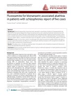
lation. Survival analysis indicates that, among acidotic
patients, the time at which 50% of patients reached
the 7.35 threshold was 173 minutes (95% confidence
interval = 153 to 193) (see Figure 3).
Respiratory, metabolic and mixed acidotic populations
Among the 290 acidotic patients, 13 could not be
further classified. Among the other 277 patients, 122
(44%) showed a respiratory acidosis, 89 (32% ) a meta-
bolic acidosis, and 66 (24%) a mixed acidosis on admis-
sion. The baseline characteristics and CPAP setting of
the acidotic population are summarized in the supple-
mental digital content in Additional file 1, according to
the type of acidemia on admission.
A total of 12 patients (10%) with respiratory acidosis,
11 patients (13%) with metabolic acidosis, four patients
(6.2%) with mixed acidosis and eight controls (9.3%)
experienced clinical failure (P = 0.613) (see Table 3).
The type of acido sis on admissio n did not affect clinical
failure after adjustment for age, history of acute myocar-
dial infarction, hypocapnia, normotension and PaO
2
/
FiO
2
ratio in a multivariable logistic regression model
(respiratory acidosis, P = 0.126; metabolic acidosis, P =
0.292; mixed acidosis P = 0.397).
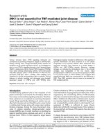
The time course of both pH and PaCO
2
values during
CPAP treatment in the acidotic groups, based on diag-
nosisatadmission,aswellasincontrolsisdepictedin
Figure 4, after replacing the missing values accordi ng to
the last observation carried forward technique and after
adjustment for age, sex and systolic blood pressure. An
increase in pH values was detected in all groups of
patients regardless of the type of acidosis, while a
decrease in PaCO
2
values was observed in mixed and
respiratory acidosis patients.
Discussion
The present study indicates that acidemia on admis sion
is not a risk factor for adverse outcomes in ACPE
patients treated with CPAP. Furthermore, not even the
Figure 1 Clinical failure r ate of the study p opulation by pH
value on admission. The 95% confidence intervals of the control
group are depicted with dashed horizontal lines.
Figure 2 Time course of pH during continuous positive airways
pressure treatment. The time course of mean arterial blood pH
during continuous positive airways pressure treatment in the
acidotic group and in controls. Adjusted for age and sex; missing
data replaced with the last observation carried forward technique.
Figure 3 Survival analysis of time to pH ≥7.350 among acidotic
patients. Dotted lines indicate the time at which 50% of the
sample reached the threshold pH (173 minutes).
Aliberti et al. Critical Care 2010, 14:R196
/>Page 5 of 8
type of acidosis on admission - respiratory, metabolic or
mixed - impacts clinical outcomes of ACPE patients
treated with CPAP.
Among our cohort of ACPE patients treated with
CPAP, more than three-quarters were acidotic on admis-
sion. Our acidotic patients showed similar clinical and
laboratory characteristics on admission in comparison
with the 346 ACPE acidotic patients treated with CPAP
enrolled in the randomized controlled trial by Gray and
coworkers [10]. The present study, however, reported
lower ACPE-related, late and inhospital mortality rates
than those reported in that trial. Possible explanations
could be found in the CPAP setting (ventilator with a
low initial PEEP), as well as the len gth of treatment used
in the study by Gray and colleagues. In this last study the
mean duration of CPAP treatment was 2 to 3 hours. We
showed that, while CPAP treatment in acidotic ACPE
patients did act ually bring 50% of patients to a pH value
above 7.35 within 3 (2.5 to 6) hours, the treatment never-
theless had to be protracted for at least 6 hours before
the mean pH crossed the threshold of 7.35.
We found that acidemia on admission is not a risk
factor for failure in ACPE patients treated with CPAP.
To date, no studies have evaluated the impact of the
degree of acidemia on admission on outcomes of ACPE
patients treated with CPAP. We found that the degree
of acidemia on admission seems not to be associated
with failure. This surprising finding could be explained
by the rapidity of the resolution of acidemia in our
ACPE patients during CPAP treatment. The increase of
pH seems to be particularly crucial during the first
hours of CPAP treatment, and thus the pH evaluat ion
during this timeframe would be a better marker of prog-
nosis rather than the single value of pH on admission.
One of the main implications of these findings is that
acidotic patients with ACPE undergoing CPAP treatment
should not be considered more severe t han those with a
normal pH value on admission. On the other hand, other
clinical and laboratory factors should be considered in the
severity assessment of the ACPE population treated with
CPAP, such as advanced age, normal-to -low blood pres-
sure, hypocapnia, or severe alteration of gas exchange [5].
Table 3 Clinical endpoints of the study population based on type of acidosis on admission
Variable Respiratory acidosis
(n = 122)
Metabolic acidosis
(n = 89)
Mixed acidosis
(n = 66)
Controls
(n = 88)
P value
(chi-square test)
Clinical failure 12 (10) 11 (13) 4 (6.2) 8 (9.3) 0.613
Change to bi-level 5 (4.1) 0 (0) 0 (0) 0 (0) 0.018
Change to intubation 1 (0.8) 2 (2.2) 2 (3.0) 0 (0) 0.341
ACPE-related mortality 1 (0.8) 4 (4.5) 1 (1.5) 1 (1.1) 0.237
Late mortality 8 (6.8) 7 (8) 2 (3.1) 7 (8.1) 0.595
In-hospital mortality 9 (7.6) (CI, 4.1 to 14.1) 11 (12.6) (CI, 7.4 to 21.7) 3 (4.6) (CI, 1.6 to 13.1) 8 (9.3) (CI, 4.9 to 17.7) 0.351
Length of hospital stay (days) 11 ± 7 11 ± 9 10 ± 5 13 ± 22 0.582
a
Data presented as number (%) or mean ± standard deviation. ACPE: acute cardiogenic pulmonary edema; CI, 95% confidence interval.
a
One-way analysis of
variance.
Figure 4 Time course of pH and PaCO
2
during continuous positive airways pressure treatment. Time course of pH and partial pressure of
carbon dioxide in arterial blood (PaCO
2
) during continuous positive airways pressure treatment in the controls and in the acidotic group
according to the diagnosis (after replacing the missing values according to the last observation carried forward technique and after adjustment
for age, sex and systolic blood pressure).
Aliberti et al. Critical Care 2010, 14:R196
/>Page 6 of 8
We found that the type of acidosis on admission
(respiratory, metabolic as well as mixed acidosis) does
not significantly modify the clinical outcomes in ACPE
patients treated with CPAP. ACPE patients with respira-
tory acido sis on admission undergoing CPAP treatment
seem to b enefit from this technique. In our study, we
found a decrease in PaCO
2
levels with a consequent
recovery of pH values during CPAP treatment in
respiratory acidotic patients. An explanation for this
finding could be identified in the rationale of the
increase of PaCO
2
during an episode of ACPE. The
etiology of hypoventilation as a sign of p ump failure is
twofold. On the one hand, such as among patients with
acute exacerbation of chronic bronchitis, hypercapnia,
often acute on chronic, occurs due to an increased load
of the respiratory system and reduced muscular fo rce
related to the presence of bronchial obstruction and
intrinsic PEEP. On the other hand, such as among
patients with ACPE without chronic pump failure, the
acute hypoventilation is strictly related to decreased
compliance due to parenchymal causes (interstitial/
alveolar flooding), and is thus easily reversed by the
alveolar recruitment induced by PEEP. Our findings
support data from Bellone and colleagues, who in an
elegant randomized controlled trial showed tha t CPAP
could be used in acidotic patients [11]. Based on these
data, excluding apriorithe use of CPAP in ACPE
patients who present respiratory acidosis on admission
could not be justified.
We also found an improvement in pH values in ACPE
patients with metabolic acidosis on admission under-
going CPAP treatment. This interesting finding could be
explained in light of beneficial effects of the application
of PEEP on the heart and hemodynamics, as well as tis-
sue perfusion in patients with ACPE. The most severe
ACPE patients treated with CPAP in our population
were those with mixed acidosis on admission who
showed the lowest pH values, mainly because of a dou-
ble effect on both the respiratory and metabolic systems.
During CPAP treatment, we found these patients to
have a quicker increase of pH values in comparison
with the other acidotic patients, in light of the double
action of CPAP on both respiratory mechanics and
hemodynamics.
In view of its retrospective design, a weakness of our
study could be a deficiency in accurately collecting some
history and clinical information. To our knowledge, the
present study is the first to evaluate the impact of differ-
ent acidosis patterns on admission in ACPE patients
treated with CPAP. This study is strengthened by a
large sample size of c onsecutive ACPE patients. More-
over, our findings are representative of an unselected
population, and our conclusions can thus be easily
generalized.
Conclusions
Neitheracidemianorthetypeofacidosisonadmission
should be considered a risk factor for adverse outcomes
in ACPE patients treated with CPAP. Furthermore, we
suggestthatnonacidoticpatien ts should be included in
future clinical trials, being at least as severe as the
acidotic population.
Key messages
• Acidemia on admission is not a risk factor for adverse
outcomes in patients with ACPE treated with CPAP.
• The type of acidosis on admission - respiratory,
metabolic or mixed - does not impact clinical outcomes
of ACPE patients treated with CPAP.
Additional material
Additional file 1: The acidotic population. A Word table presenting
demographics, comorbidities, severity of the disease, clinical and
laboratory findings on admission and before CPAP treatment of the
acidotic population, according to the type of acidemia on admission.
Abbreviations
ACPE: acute cardiogenic pulmonary edema; CPAP: continuous positive
airways pressure; ETI: endotracheal intubation; HCO
3
-
: bicarbonates; NIV: non-
invasive ventilation; PaCO
2
: partial pressure of carbon dioxide in arterial
blood; PaO
2
/FiO
2
: partial pressure of oxygen in arterial blood/inspired
oxygen fraction; PEEP: positive end-expiratory pressure.
Author details
1
Dipartimento toraco-polmonare e cardio-circolatorio, University of Milan,
IRCCS Fondazione Cà Granda Ospedale Maggiore Policlinico, via F. Sforza 35,
20122 Milan, Italy.
2
Emergency Medicine Department, IRCCS Fondazione Cà
Granda Ospedale Maggiore Policlinico, via F. Sforza 35, 20122 Milan, Italy.
3
School of Specialization in Hospital Pharmacy, University of Milan, Via
Colombo 71, 20133 Milan, Italy.
Authors’ contributions
SA contributed to the conception and design of the study, as well as the
acquisition, analysis and interpretation of data; he was involved in drafting
the manuscript and revising it critically for important intellectual content. RC
and AMB contributed to the conception and design of the study, the
analysis and interpretation of data; they were involved in revising the
manuscript. AAB contributed to the conception and design, analysis and
interpretation of data; he was involved in revising the manuscript. FP, TM
and VDR contributed to the acquisition, analysis and interpretation of the
data; they were involved in revising the manuscript critically. VM revised the
manuscript. All authors read and approved the final manuscript.
Competing interests
The authors declare that they have no competing interests.
Received: 18 June 2010 Revised: 5 October 2010
Accepted: 1 November 2010 Published: 1 November 2010
References
1. Nieminen MS, Böhm M, Cowie MR, Drexler H, Filippatos GS, Jondeau G,
Hasin Y, Lopez-Sendon J, Mebazaa A, Metra M, Rhodes A, Swedberg K,
Priori SG, Garcia MA, Blanc JJ, Budaj A, Cowie MR, Dean V, Deckers J,
Burgos EF, Lekakis J, Lindahl B, Mazzotta G, Morais J, Oto A, Smiseth OA,
Garcia MA, Dickstein K, Albuquerque A, Conthe P, et al: Executive summary
of the guidelines on the diagnosis and treatment of acute heart failure:
Aliberti et al. Critical Care 2010, 14:R196
/>Page 7 of 8
the Task Force on Acute Heart Failure of the European Society of
Cardiology. Eur Heart J 2005, 26:384-416.
2. Holt AW, Bersten AD, Fuller S, Piper RK, Worthley LI, Vedig AE: Intensive
care costing methodology: cost benefit analysis of mask continuous
positive airway pressure for severe cardiogenic pulmonary oedema.
Anaesth Intensive Care 1994, 22:170-174.
3. Plaisance P, Pirracchio R, Berton C, Vicaut E, Payen D: A randomized study
of out-of-hospital continuous positive airway pressure for acute
cardiogenic pulmonary oedema: physiological and clinical effects. Eur
Heart J 2007, 28:2895-2901.
4. Peter JV, Moran JL, Phillips-Hughes J, Graham P, Bersten AD: Effect of non-
invasive positive pressure ventilation (NIPPV) on mortality in patients
with acute cardiogenic pulmonary oedema: a meta-analysis. Lancet 2006,
367:1155-1163.
5. Cosentini R, Aliberti S, Bignamini A, Piffer F, Brambilla AM: Mortality in
acute cardiogenic pulmonary edema treated with continuous positive
airway pressure. Intensive Care Med 2009, 35:299-305.
6. Plant PK, Owen JL, Elliott MW: Non-invasive ventilation in acute
exacerbations of chronic obstructive pulmonary disease: long term
survival and predictors of in-hospital outcome. Thorax 2001, 56:708-712.
7. Meduri GU, Turner RE, Abou-Shala N, Wunderink R, Tolley E: Noninvasive
positive pressure ventilation via face mask. First-line intervention in
patients with acute hypercapnic and hypoxemic respiratory failure. Chest
1996, 109:179-193.
8. Confalonieri M, Garuti G, Cattaruzza MS, Osborn JF, Antonelli M, Conti G,
Kodric M, Resta O, Marchese S, Gregoretti C, Rossi A, Italian noninvasive
positive pressure ventilation (NPPV) study group: A chart of failure risk for
noninvasive ventilation in patients with COPD exacerbation. Eur Respir J
2005, 25:348-355.
9. Moretti M, Cilione C, Tampieri A, Fracchia C, Marchioni A, Nava S: Incidence
and causes of non-invasive mechanical ventilation failure after initial
success. Thorax 2000, 55:819-825.
10. Gray A, Goodacre S, Newby DE, Masson M, Sampson F, Nicholl J, 3CPO
Trialists: Noninvasive ventilation in acute cardiogenic pulmonary edema.
N Engl J Med 2008, 359:142-151.
11. Antonelli M, Conti G, Moro ML, Esquinas A, Gonzalez-Diaz G, Confalonieri M,
Pelaia P, Principi T, Gregoretti C, Beltrame F, Pennisi MA, Arcangeli A,
Proietti R, Passariello M, Meduri GU: Predictors of failure of noninvasive
positive pressure ventilation in patients with acute hypoxemic
respiratory failure: a multi-center study. Intensive Care Med 2001,
27:1718-1728.
12. Bellone A, Vettorello M, Monari A, Cortellaro F, Coen D: Noninvasive
pressure support ventilation vs. continuous positive airway pressure in
acute hypercapnic pulmonary edema. Intensive Care Med 2005,
31:807-811.
13. Kelly BJ, Matthay MA: Prevalence and severity of neurologic dysfunction
in critically ill patients. Influence on need for continued mechanical
ventilation. Chest 1993, 104:1818-1824.
14. Le Gall JR, Lemeshow S, Saulnier F: A new Simplified Acute Physiology
Score (SAPS II) based on a European/North American multicenter study.
JAMA
1993, 270:2957-2963.
doi:10.1186/cc9315
Cite this article as: Aliberti et al.: Acidemia does not affect outcomes of
patients with acute cardiogenic pulmonary edema treated with
continuous positive airway pressure. Critical Care 2010 14:R196.
Submit your next manuscript to BioMed Central
and take full advantage of:
• Convenient online submission
• Thorough peer review
• No space constraints or color figure charges
• Immediate publication on acceptance
• Inclusion in PubMed, CAS, Scopus and Google Scholar
• Research which is freely available for redistribution
Submit your manuscript at
www.biomedcentral.com/submit
Aliberti et al. Critical Care 2010, 14:R196
/>Page 8 of 8