Báo cáo y học: " Possible Cis-acting signal that could be involved in the localization of different mRNAs in neuronal axons" potx
Bạn đang xem bản rút gọn của tài liệu. Xem và tải ngay bản đầy đủ của tài liệu tại đây (315.13 KB, 10 trang )
BioMed Central
Page 1 of 10
(page number not for citation purposes)
Theoretical Biology and Medical
Modelling
Open Access
Research
Possible Cis-acting signal that could be involved in the localization
of different mRNAs in neuronal axons
Gonzalo E Aranda-Abreu*, Ma Elena Hernández, Abraham Soto and
Jorge Manzo
Address: Instituto de Neuroetología, Universidad Veracruzana, Av. Dos Vistas S/N, km 2.5 Carr. Xalapa-Veracruz. Col. Industrial-Animas. C.P.
91190. Xalapa, Ver. México
Email: Gonzalo E Aranda-Abreu* - ; Ma Elena Hernández - ; Abraham Soto - ;
Jorge Manzo -
* Corresponding author
mRNAU-rich regionaxon
Abstract
Background: Messenger RNA (mRNA) comprises three major parts: a 5'-UTR (UnTranslated
Region), a coding region, and a 3'-UTR. The 3'-UTR contains signal sequences involved in
polyadenylation, degradation and localization/stabilization processes. Some sequences in the 3'-
UTR are involved in the localization of mRNAs in (e.g.) neurons, epithelial cells, oocytes and early
embryos, but such localization has been most thoroughly studied in neurons. Neuronal polarity is
maintained by the microtubules (MTs) found along both dendrites and axon and is partially
influenced by sub-cellular mRNA localization. A widely studied mRNA is that for Tau protein,
which is located in the axon hillock and growth cone; its localization depends on the well-
characterized cis-acting signal (U-rich region) in the 3'-UTR.
Methods: We compared the cis-acting signal of Tau with mRNAs in the axonal regions of neurons
using the ClustalW program for alignment of sequences and the Mfold program for analysis of
secondary structures.
Results: We found that at least 3 out of 12 mRNA analyzed (GRP75, cofilin and synuclein) have a
sequence similar to the cis-acting signal of Tau in the 3'-UTR. This could indicate that these
messengers are localized specifically in the axon. The Mfold program showed that these mRNAs
have a similar "bubble" structure in the putative sequence signal.
Conclusion: Hence, we suggest that a U-rich sequence in the 3'-UTR region of the mRNA could
act as a signal for its localization in the axon in neuronal cells. Sequences homologous to the DTE
sequence of BC1 mRNA could direct the messenger to the dendrites. Messengers with
homologues of both types of sequence, e.g. β-actin, might be located in both dendrites and axon.
Background
A messenger RNA (mRNA) comprises three major parts, a
5'-UTR (UnTranslated Region), a coding region and a 3'-
UTR. The 3'-UTR contains signal sequences involved in
Published: 24 August 2005
Theoretical Biology and Medical Modelling 2005, 2:33 doi:10.1186/1742-4682-2-33
Received: 21 July 2005
Accepted: 24 August 2005
This article is available from: />© 2005 Aranda-Abreu et al; licensee BioMed Central Ltd.
This is an Open Access article distributed under the terms of the Creative Commons Attribution License ( />),
which permits unrestricted use, distribution, and reproduction in any medium, provided the original work is properly cited.
Theoretical Biology and Medical Modelling 2005, 2:33 />Page 2 of 10
(page number not for citation purposes)
polyadenylation, degradation and localization/stabiliza-
tion processes. Many studies have shown that certain
sequences in the 3'-UTR are involved in localizing the
mRNAs in different cells such as neurons, epithelial cells,
oocytes and early embryos [1,2]. Such localization has
been studied exhaustively in neurons. Neurons are polar
cells, with dendrites and axon; dendrites receive informa-
tion and the axon is specialized to transmit this informa-
tion to the next neuron [3]. The maintenance of neuronal
polarity depends on the microtubules (MTs) [3-6], which
are found along both axon and dendrites, and is partially
determined by subcellular mRNA localization. The mech-
anism responsible for creating the polarity involves syner-
gistic controls of translation, stabilization and association
with elements of the cytoskeleton.
The mRNA of tau has been studied in detail [7]. Tau is
located in the axon hillock and growth cone; the well-
characterized cis-acting signal (U-rich region) located in
the 3'-UTR of its mRNA is responsible for its localization
[8]. HuD protein interacts with this U-rich sequence to
form a mRNA-protein complex that is transported toward
the axon (axon hillock and growth cone) by interacting
with KIF3A, a kinesin responsible for anterograde move-
ment [9-11].
Recently, many mRNAs have been shown to be located in
neuronal axons: β-actin, tropomyosin 3 (Tpm3), cofilin,
vimentin, immunoglobulin heavy chain biding protein
(Bip), heat shock protein 60 (HSP60), heat shock protein
70 (HSP70), heat shock protein 90 (HSP90), glucose reg-
ulated protein (grp75) and synuclein [12]. The objective
of this paper is to determine, using bioinformatics tools,
whether there is a cis-acting signal in all the mRNAs that
are transported to the axon and whether this putative sig-
nal is similar to the U-rich region in the 3'-UTR of tau
mRNA.
Results
The 3'-UTR of tau mRNA contains 3884 bases; the U-rich
region (in bold) is responsible for the localization of this
mRNA in the axon hillock and growth cone.
UCAGGCCCCUGGGGCCGUCACUGAUCAUGGAGAGAAGAGAG
AGUGAGAGUGUGGAAAAAAAAAAAAAAAGAAUGACCUGGCC
CCUCACCCUCUGCCCUCCCCGCUGCUCCUCAUAGACAGGCU
GACCAGCUUGUCACCUAACCUGCUUUUGUGGCUCGGGUUUG
GCUCGGGACUUCAAAAUCAGUGAUGGGAAAAAGUAAAUUUC
AUCUUUCCAAAUUGAUUUGUGGGCUAGUAAUAAAAUAUUUU
UAAGGAAGGAAAAAAAAAACACGUAAAACCAUGGCCAAACA
AAACCCAACAUUUCCUUGGCAAUUGUUAUUGACCCCGCCCC
CCCCUCUGAGUUUUAGAGGGUGAAGGAGGCUUUGGAUAGAG
GCUGCUUCUGGGGAUUGGCUGAGGGACUAGGGCAACUAAUU
GCCCACAGCCCCAUCUUAGGGGCAUCAGGACAGCGGCAGAC
AUGAAAGACUUGGGACUUGGUGUGUUUGUGGAGCCGUAAGG
CGUAUGUUAACUUUGUGUGGGUUUGAGGGAGGACUGUGAUA
GUGAAGGCUGAGAGAUGGGUGGGCUGGGAGUCAGAGGAGAG
AGGUGAGGAAGACAGGUUGGGAGAGGGGGCAUUGCGUCCUU
GCCAAGGAGCUUGGGAAGCACAGGUAGCCCUGGCUGCAGCA
GUCUUAGCUAGCACAGAUGCCUGCCUGAGAAAGCACAGUGG
GGUACAGUGGGUGUGUGUGCCCCUUCUGAAGGGCAGCCCAU
GGGAGAAGGGGUAUUGGGCAGAAGGAAGGUA
GGCCCCAGAAGGUGGCACCUUGUAGAUUGGUUCUCUGAAGG
CUGACCUUGCCAUCCCAGGGCACUGCUCCCACCCUCCAGGA
GGAGGUCUGAGCUGAGGAGCUUCCUUUUCGAUCUCACAGGA
AAACCUGUGUUACUGAGUUCUGAAGUUUGGAACUACAGCCA
UGAUUUUGGCCACCAUACAGACCUGGGACUUUAGGGCUAAC
CAGUUCUUUGUAAGGACUUGUGCCUCUUGCGGGAACAUCUG
CCUGUUCUCAAGCCUGGUCCUCUGGCACUUCUGCAGUGGUG
AGGGAUGGGGGUGGUAUUCUGGGAUGUGGGUCCCAGGCCUC
CCAUCCCUCGCACAGCCACUGUAUCCCCUCUACCUGUCCUA
UCAUGCCCACGUCUGCCACGAGAGCCAGUCACUGCCGUCCG
UACAUCACGUCUCACCGUCCUGAGUGCCCAGCCUCCCAAGC
CCAAUCCCUGGACCCCUGGGUAGUUAUGGCCAAUCUGCUCU
ACACUAGGGGUUGGAGUCCAGGGAAGGCAAAGAUUUGGGCC
UUGGUCUCUAGUCCUACGUUGCCAGAAUCCAACCAGUGUGC
CUCCCACAAGGAACCUUACAACCUUGUUUGGUUUGCUCCAU
CAGGCGUUUGGCGCCAUCGUGGAUGGAGUCCGUGUGUGCCU
GGAGAUUACCCUGGACACCUCUGCUUUUUUUUUUUUUACUU
UAGCGGUUGCCUCCUAGGCCUGACUCCUUCCCAUGUUGAAC
UGGAGGCAGCCAAGUUAGGUGUCAAUGUCCUGGCAUCAGUA
UGAACAGUCAGUAGUCCCAGGGCAGGGCCACACUUCUCCCA
UCUUCUGCUUCCACCCCAGCUUGUGAUUGCUAGCCUCCCAG
AGCUCAGCCGCCAUUAAGUCCCCAUGCACGUAAUCAGCCCU
UCCAUACCCCAAUUUGGGGAACAUACCCCUUGAUUGAAAUG
UUUUCCCUCCAGUCCUAUGGAAGCGGUGCUGCCUGCCUGCU
GGAGCAGCCAGCCAUCUCAGAGACGCAGCCCUUUCUCUCCU
GUCCGCACCCUGCUGCGCUGUAGUCGGAUUCGUCUGUUUGU
CUGGGUUCACCAGAGUGACUAUGAUAGUGAAAAGAAAAAGA
AAAAGAAAAAAGAAAAAAGAAAAAAAAAAAAGGACGCAUGU
UAUCUUGAAAUAUUUGUCAAAAGGUUGUAGCCCACCGCAGG
GAUUGGAGGGCCUGAUAUUCCUUGUCUUCUUCGUGACUUAG
GUCCAGGCCGGUCGAGUGCUACCCUGCUGGACAUCCCAUGU
UUUGAAGGGUUUCUUCUUCAUCUGGGACCCCUGCAGACACU
GGAUUGUGACAUUGGAGGUCUAUACAUUGGCCAAGGCUGAA
GCACAGGACCCGUUAGAGGCAGCAGGCUCCGACUGUCAGGG
AGAGCUUGUGGCUGGCCUGUUUCUCUGAGUGAAGAUGGUCC
UCUCUAAUCACAACUUCAAGUCCCACAGCAGCCCUGGCAGA
CAUCUAAGAACUCCUGCAUCACAAGAGAAAAGGACACUAGU
ACCAGCAGGGAGAGCUGUGGCCCUAGAAAUUCCAUGACUCU
CCACUACUAUCCGUGGGUCCUUUCCAAGCCUUGCCUCGUCA
CCAAGGGCUUGGGAUGGACUGCCCCACUGAUGAAAGGGACA
UCUUUGGAGACCCCCUUGGUUUCCAAGGCGUCAGCCCCCUG
ACCUUGCAUGACCUCCUACAGCUGAAGGAUGAGGCCUUUAA
AGAUUAGGAACCUCAGGCCCAGGUCGGCCACUUUGGGCUUG
GGUACAGUUAGGGACGAUGCGGUAGAAGGAGGUGGCCAACC
UUUCCAUAUAAGAGUUCUGUGUGCCCAGAGCUACCCUAUUG
UGAGCUCCCCACUGCUGAUGGACUUUAGCUGUCCUUAGAAG
UGAAGAGUCCAACGGAGGAAAAGGAAGUGUGGUUUGAUGGU
Theoretical Biology and Medical Modelling 2005, 2:33 />Page 3 of 10
(page number not for citation purposes)
CUGUGGUCCCUUCAUCAUGGUUACCUGUUGUGGUUUUCUCU
GUAUACCCCCAUUUACCCAUCCUGCAGUUCCUGUCCUUGAA
UAGGGGUGGGGGUACUCUGCCAUAUCUCUUGUAGGCAGUCA
GCCCCCAAGUCAUAGUUUGGAGUGAUCUGGUCAGUGCUAAU
AGGCAGUUUACAAGGAAUUCUGGCUUGUUACUUCAGUGAGG
ACAAUCCCCCAAGGCCCUGGCACCUGUCCUGUCUUUCCAUG
GCUCUCCACUGCAGAGCCAAUGUCUUUGGGUGGGCUAGAUA
GGGUGUACAAUUUGCCUGGAACCUCCAAGCUCUUAAUCCAC
UUUAUCAAUAGUUCCAUUUAAAUUGACUUCAAUAUAAGAGU
GUAUCCAUUUGAGAUUGCUUGUGUUGUGGGGUAAAGGGGGG
AGGAGGAACAUGUUAAGAUAAUUGACAUGGGCAAGGGGAAG
UCUUGAAGUGUAGCAGUUAAACCAUCUUGUAGCCCCAUUCA
UGAUGUUGACCACUUGCUAGAGAGAAGAGGUGCCAUAAGGC
UAGAACCUAGAGGCUUGGCUGUCCACCAACAGGCAGGCUUU
UGCAAGGCAGAGGCAGCCAGCUAGGUCCCUGACUUCCCAGC
CAGGUGCAGCUCUAAGAACUGCUCUUGCCUGCUGCCUUCUU
GUGGUGUCCAGAGCCCACAGCCAAUGCCUCCUCAAAACCCU
GGCUUCCUUCCUUCUAAUCCACUGGCACAUCAGCAUCACCU
CCGGAUUGACUUCAGAUCCACAGCCUACACUACUAGCAGUG
GGUAAGACCACUUCCUUUGUCCUUGUCUGUUCUCCAGAAAA
GUGGGCAUGGAGGCGGUGUUAAUAACUAUAGGUCUGUGGCU
UUAUGAGCCUUCAAACUUCUCUCUAGCUUCUGAAAGGGUUA
CUUUUGGGCAGUAUUGCAGUCUCACCCUCCGAUGGCUGUAG
CCUGUGCAGUUGCUGUACUGGGCAUGAUCUCCAGUGCUUGC
AAGUCCCAUGAUUUCUUUGGUGUUUUGAGGGUGGGGGGAGG
GACAUGAAUCAUCUUAGCUUAGCUUCCUGUCUGUGAAUGUC
CAUAUAGUGUACUGUGUUUUAACAAACGAUUUACACUGACU
GUUGCUGUACAAGUGAAUUUGGAAAUAAAGUUAUUACUCUG
AUUAAACAAAAAAAAAAAAAAAAAAAAAAAAAAAAAAAA
This cis-acting signal of tau was compared base by base
with the other afore mentioned mRNAs using simple
alignment. We also made comparisons with another
sequence that is specific for the localization of BC1 mRNA
in dendrites, the Dendritic Target Element (DTE) [13].
In the β-actin mRNA of chicken a cis-acting signal "zip-
code" has been described; a zipcode binding protein
binds to this sequence and this is a prerequisite for the
localization of the mRNA. The sequence is a tandem
repeat of an ACACCCACACCC motif. The mRNA of β-
actin has been located in the axon of the neuron and in
dendritic spines [12,14]. β-actin mRNA has a sequence
closely similar to the tau signal in the first part of its 3'-
UTR, but there is also another sequence that could partic-
ipate in its localization in dendrites; this sequence is very
similar to the DTE [13]. The protein tropomyosin 3 has
been located in the growth cone of the neuron; its mRNA
has also been detected in axons during development [15].
Both tropomyosin 3 and β-actin form parts of the
cytoskeleton. No well-defined sequence signal that could
be involved in the specific localization of these messen-
gers in the axon (tpm3 and β-actin) has been identified, so
it is likely that β-actin and tropomyosin are not exclusive
to the axon and could be also found in dendrites.
Cofilin is a cytoskeleton modulating protein; it is also
known as actin depolymerizing factor (ADF). The poten-
tial role of cofilin is to modulate the changes of actin
organization that accompany neurite initiation, axono-
genesis and growth cone guidance [16]. The possible sig-
nal sequence found in the 3'-UTR of cofilin mRNA is very
similar to that of tau; they share a U-rich region, which
indicates that this messenger might be transported to the
growth cone of the developing neuron. However, a possi-
ble DTE sequence is also present, located upstream of the
U-signal.
Vimentin has been located by RT-PCR in the axons of dor-
sal root ganglia (DRG) neurons. A possible sequence sig-
nal in vimentin mRNA shares some U with tau but also
contains more purines, which might indicate that the pro-
tein is not exclusive to the axonal region [12].
Bip is a protein that binds to the immunoglobin heavy
chains in pre-β cells. Its mRNA shares some U with the tau
sequence; nevertheless, its sequence suggests that this
mRNa, like vimentin, is probably not exclusive to the
axon [17].
tau UUUUUUUUUUUUU 13
A
ctb GCGGACUGUUACUGAGCUGCGUUUUACACCCUUUCUUUGACAAAACCUAACUUGCGCAAA 60
**** ** GAGGUUGGGGAU DTE
* *** * *
tau
A
ctb AAAAAAAAAAAAAAAAAAAAAAAAAAA 87
tau
Tpm3 AAGAGAUUGUGGGUGAUGAAGAUGGGGCCUGGGAGGUUUAGUGCAGAACUUGAAAACCGU 240
GAGGUUGGGGAU DTE
*** *** ** *
tau UUUUUUUUU 9
Tpm3 UAGCUGCAGCCCUCUCACCUGUAUACUGACUGUAGGGUUUGCUCACCUGCAUGGUUAUUU 300
* ** ***
tau U 13
Tpm3 UCUAACAAUAAAAACA 316
*
tau
cofilin GCCACCUCCAGCCCCCUGCCUGGAGCAUCUAGCAGCCCCAGACCUGCUCUUGGGUGUUGC 60
tau
cofilin AGGCUGCCCUUUUCCUGCCAGACCGGAGGGGCUGGGGGGGUUCCAGCAGGGGGAGGGUUU 120
GAGGUUGGGGAU DTE
* ** *****
tau
cofilin UCCCUUCACCCCAGUUGCCAAACAUCCCUCCCACCCCCUGGACCGUCCUUUUCCCUCCAU 180
tau
cofilin CCCUGACGGUUCUGGCCUUCCCAAACUGCUUUUGAUCUUCUGAUUCCUCUUGGGUUGAAG 240
tau UUUUUUUUUUUUU 13
cofilin CAGACCAAGUCCCGUCCUAGGCACCCAGUUUGGGGGGAGCCUGUAUUUUUUUUUUUUAAC 300
************
tau 18
cofilin GACACCCCUACUCCUCAUCUGUCCCAUCCCAUGCUGCCAACUUCUAACCACAAUAGUGAC 360
Tau
Vim UUAGAAAAAAGAGCUUUCAAGUGCCUUUACUGCAGUUUUCAGGAGCGCAAGAUAGAUCUG 120
GAGGUUGG
** * *
Tau
Vim GGAUAGAAACGAGCUCAGCACAUAACAACUGACACCCCCAAAAGGCGUAGAAAAGGUUUA 180
GGAU DTE
****
Tau UUUUUUUUUUUUU 13
Vim CAAAAUAAUCUAGUUUUACGAAGAAAUCUUGUGCUAGAAUACUUUUUAAAGUAUUUUUGA 240
* ***** *
Theoretical Biology and Medical Modelling 2005, 2:33 />Page 4 of 10
(page number not for citation purposes)
The heat shock proteins and grp75 messengers have simi-
larities with the tau sequence, but once again they are
probably not exclusive to the axon. They could interact
with other proteins in different parts of the cell.
Synuclein is a soluble unfolded protein that can aggregate
into insoluble fibrils under several pathological condi-
tions including Parkinson's and Alzheimer's diseases [12].
The possible cis-acting signal of the mRNA for this protein
is very similar to the tau signal, with only a single U to C
substitution, suggesting that the synuclein messenger may
be transported to the axon by a similar mechanism to the
tau messenger and that aggregation and precipitation of
the synuclein protein within the axon contributes to neu-
rodegenerative disease.
These analyses carried out by alignment allowed us to
show that the cis-acting signals of the mRNAs examined
have some homology with that of the tau messenger.
The highest homology scores are:
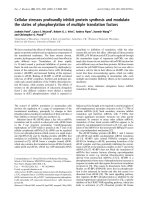
The secondary structures of these four mRNAs, which
showed the closest homologies to the tau sequence, were
analyzed using the program Mfold (Figs. 1, 2, 3, 4). Fig. 5
shows a model of U-rich mRNAs that could be trans-
ported to the axon. The results show that the secondary
structures of synuclein and cofilin mRNAs are very similar
to that of the tau messenger, and the cis-acting signal
sequence is inside the "bubble" according to the Mfold
program. This indicates to us that both the signal
sequence and the secondary structure could be determin-
ing factors in the location of these messengers in the axon
region.
Discussion
The first messenger to be analyzed in the 3'-UTR with
respect to its localization/stabilization was tau [9,10]. The
Tau
Bip UGGGGUCAGGGAGAGGAGGAAUUGGCUAUUUUAAAAUUGGGGAAAAGCUGGGUCAGGGUG 240
GAGGUUGGGGAU DTE
* *******
Tau
Bip UGUGUUCACCUUGGAUAUGUUCUAUUUAACGGUUGGGUCAUGCACAUCUGGUGUAGGAAC 300
Tau UUUUUUUUUUUUU 13
Bip UUUUUUCUACCAUAAGUGACACCAAUAAAUGUUUGUUAUUUACACUGGUCUAGUUUUUGU 360
* *** ** ***
Tau UUUUUUUUUUUUU 13
HSP60 GUCCAUGCCUACAGAUAAUUUAUUUUGUAUUUUUGAAUAAAGACAUUUGUACAUUCCUGA 240
* *** **** *
Tau
HSP60 UCUGUUAGCAUCAGGACUGUAGCGCUGUGUCACCACAUGAGAAGUUCAGAAGCAGCCUUU 360
Tau
HSP60 CUGUGGAGGGUGAGAAUGAUUGUGUACAGAGUAGAGAAGUAUCCAAUUAUGUGACAACCU 420
GAGGUUGGGGAU DTE
**** ** * **
Tau UUUUUUUUUUUUU 13
HSP70 GGUAAUUGAUUUGAGUUUGUUACAUUUUGUAUGCUCGUGGGUUUUUUAUAUAUUCAAAUU 180
***** * * **
Tau
HSP70 AAGGUUGCAUGUUCUUUGCGUUUAAUCUAAGUAGCUGUGUAAAAAUGGUGUUUCCUUCCU 240
GAGGUUGGGGAU DTE
****** *
Tau
HSP90 UCCUUGUGCCUUAAGGCAGGAAGAUCCCCUCCCACAGAUAGCAGGGUUGGGUGUUGUGUA 180
GAGGUUGGGGAU DTE
******* *
Tau UUUUUUUUUUUUU 18
HSP90 UUGUGUUUUUUUGUUUGUUUUAUUUUGUUCUGAAAUUAAAAGUAUGCAAAAUAAAGAUGA 240
**** *** ****
Tau
Grp75 UAAUAGUGGCAGUGCAUUGUGGAGCUAGGACGACAUACUAUGAAGCUUGGGAGUAAAGGA 60
GAGGUUGGGGAU DTE
** ***** *
Tau
Grp75 ACUUCCUGAGCAGAAAAGGGGCAAACUUCAGUCUUUUUACUGUAUUUUUGCAGUAUUCUA 120
Tau
Grp75 UAUAUAAUUUCCUUAAUAUAUAAAUCCAGUGACAAUAUAUAAAUCCAGUGACAAUAGCUA 180
Tau
Grp75 UAACUCAUUUAAUGGUAAUAAAGUCAGCAAUAGCAGGUUCACACUUCUAUAACUAGCCUG 240
Tau
Grp75 CUGUUUUCAGCUGCACGUAAAGGGGUGGGAUGGGGCUGUGUACCAAUCAUUAUUAGGUAA 300
Tau
Grp75 AUCUGGUUUGUGCUGAAGUAGCUAUGUUUUCGAGAUGGAAGCCCAUUUCACAUGCAGUAG 360
Tau
Grp75 AGGUAAUCUGUCAUGGACCUUGAAUUGAGGUUCAUAUGCAGAUGCUUGUUGACCAAGAGC 420
Tau
Grp75 ACUGCUAUAAAUGACCUGUGUGUACAUUUGCUCCUUCAACUGAUGCCUUGCAAGACUAAG 480
Tau
Grp75 CUCUCUGUGUCAUGGUCUAUAGGUACAGAAGUUAGGUCAAUGGAUAACAGCUGUGUUAGC 540
Tau
Grp75 CAUAGCUUAAAGUGAUCUAUCAAGAAUUAUACAAGCCUCUCAUGGGCCUAAGGCAUACUU 600
Tau
Grp75 CUCCAGCUACCCUCUUGGGUGGCCAAUGUCUGACAUCUAUAUUCUUGAUGAUUGUUCCUU 660
Tau UUUUUUUUUUUUU 18
Grp75 UUUCAUCCAUUCUGGAUUUUUUUUUUUUUUAAUAAAAUUCUGAAAGCCUCUUGAUCUCCU 720
*************
Tau U 1
Synuclein CCUGCUGGCUCAUUUUACCCCAUGGUCCUUCGGAUCACCUUCCAGACGCUGCUGUGAAUU 240
*
Tau UUUUUUUUUUUU 18
Synuclein UUUUCUUUUUUUAAUGAUUCCAAAUAAAACCUGAGUCCUAAUCCAAAAAAAAAAAAAAAA 300
**** *******
Tau
Synuclein AAAAAAAAAA 310
Tau UUUUUUUUUUUUU 13
Grp75 UUUUUUUUUUUUUAAUAA- 18
Synuclein UUUUUCUUUUUUUAAUGA- 18
Cofilin -UUUUUUUUUUUUAACGAC 18
HSP90 -UUUUGUUUGUUUUAUUUU 18
Tpm3 -UGGUUAUUUUCUAACAAU 18
Bip AUGUUUGUUAUUUA-CACU 18
HSP70 -UUUUUAUAUAUUCAAAUU 18
Vim -UACUUUUUAAAGUAUUUU 18
HSP60 -UAAUUUAUUUUGUAUUUU 18
B-Act -GUUUUACACCCUUUCUUU 18
*
Tau UUUUUUUUUUUUU 13
Grp75 UUUUUUUUUUUUUAAUAA- 18
Synuclein UUUUUCUUUUUUUAAUGA- 18
Cofilin UUUUUUUUUUUUAACGAC 18
*** *******
Theoretical Biology and Medical Modelling 2005, 2:33 />Page 5 of 10
(page number not for citation purposes)
U-rich region in its signal sequence enables the formation
of a complex with HuD, a prerequisite for transport to the
axon, and increases the stability of the mRNA. The basic
function of tau protein is to stabilize the microtubules; it
prevents depolymerization and consequent loss of neuro-
nal polarity. Recently, several other messengers have been
shown by RT-PCR to be localized in neuronal axon of the
neuron, but the possibility that these mRNAs are also
located in the dendrites has not been excluded.
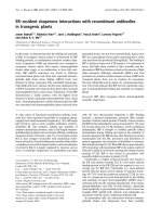
GRP75
Glucose regulated protein 75 (GRP75) is an important
molecular chaperon belonging to the heat shock protein
(HSP) family. It is highly expressed in conditions of glu-
cose deprivation of glucose. Its messenger was located in
the axon and it has a U-rich region. It might not be con-
fined exclusively to the axon because this protein
responds to a metabolic stress [18].
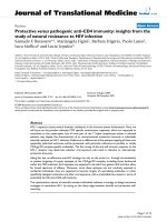
Synuclein
Alfa-synuclein is involved in neurodegenerative diseases
and its presence has been observed in the pre-synaptic and
nuclear compartments, though the location in the nucleus
has not been well documented. The synuclein messenger
possesses a U-rich region; nevertheless a C interrupts the
potential signal sequence. When it is wrongly folded, this
protein may aggregate in the cell forming fibrils, typical of
Alzheimer's and Parkinson's diseases. The aggregation of
synuclein is similar to tau in Alzheimer patients, which
could indicate similar intracellular behavior by both pro-
teins [19].
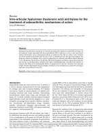
Cofilin
The 3'-UTR of the cofilin messenger has a U-rich region
very similar to the signal sequence of tau, which on the
face of it suggests that it might be located exclusively in the
axon. Nevertheless, recent studies demonstrate that it par-
ticipates in the shrinkage of dendritic spines associated
with the long-term depression of hippocampal synapses,
suggesting that it is also found in dendrites. Moreover, it
is involved in neuronal development, axogenesis, guid-
ance of the growth cone and dendrite formation.
Although the cofilin messenger is present in axons, the
possible participation of the protein in events related to
the unplugging of synapses because of its association with
actin further suggests that it is not confined to the axon
but also occurs in the dendrites [16].
β
-actin
The 3'-UTR of the β-actin messenger is very short and
shows low homology when aligned with the tau cis-acting
signal. However, when it was aligned with the dendritic
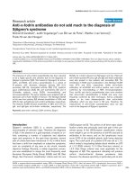
RNA secondary structure of 3'-UTR of tau mRNAFigure 1
RNA secondary structure of 3'-UTR of tau mRNA. The arrow indicates the "bubble" where the HuD binds to stabilize the
messenger.
Signal Sequence
Theoretical Biology and Medical Modelling 2005, 2:33 />Page 6 of 10
(page number not for citation purposes)
target element, it showed better homology. The β-actin
messenger was shown to possess a zipcode that leads it
towards the dendrites instead of the axon [14].
HSP70 and HSP90
Molecular chaperones and their functions in protein fold-
ing have been implicated in several neurodegenerative
conditions, including Parkinson's and Huntington's dis-
eases, which are characterized by accumulation of protein
aggregates (e.g. α-synuclein and huntingtin, respectively).
These aggregates have been shown in various experimen-
tal systems to respond to changes in levels of molecular
chaperones, suggesting the possibility of therapeutic inter-
vention and a role for chaperones in disease pathogenesis.
It remains unclear whether chaperones also play a role in
Alzheimer's disease, a neurodegenerative disorder charac-
terized by β-amyloid and tau protein aggregates. In
various cellular models, increased levels of Hsp70 and
Hsp90 promote tau solubility and tau binding to micro-
tubules, reduce insoluble tau and cause reduced tau phos-
phorylation. Conversely, lowered levels of Hsp70 and
Hsp90 result in the opposite effects. A direct association
between the chaperones and tau protein has been demon-
strated. Many results suggest that the up-regulation of
molecular chaperones may suppress the formation of
neurofibrillary tangles by partitioning tau into a produc-
tive folding pathway and thereby preventing tau aggrega-
tion [20]. When we compared the 3'-UTRs of the
messengers for these chaperones, they showed some hom-
ology with the cis-acting signal of tau because each pos-
sesses a U-rich region, which could indicate that they are
found in axons.
The model
On the basis of the results we suggested a model for
mRNA localization in the axon (Fig. 5).
RNA secondary structure of 3'-UTR of GRP75 mRNAFigure 2
RNA secondary structure of 3'-UTR of GRP75 mRNA. The arrow indicates the U-rich signal sequence that could be involved
in the localization of the messenger.
Signal Sequence
Theoretical Biology and Medical Modelling 2005, 2:33 />Page 7 of 10
(page number not for citation purposes)
RNA secondary structure of 3'-UTR of synuclein mRNAFigure 3
RNA secondary structure of 3'-UTR of synuclein mRNA. The arrow indicates the U-rich signal sequence that could be
involved in the localization of the messenger.
Signal Sequence
Theoretical Biology and Medical Modelling 2005, 2:33 />Page 8 of 10
(page number not for citation purposes)
The mRNAs containing the U-rich region could be com-
plexed with a protein responsible for transport toward the
axon, just as HuD complexes with and stabilizes the tau
messenger [9]. HuD itself has the capacity to bind to dif-
ferent mRNAs such as GAP-43 [21], neuroserpin [22], ace-
tylcholinesterase [23] and c-myc [24], so it might interact
with other messengers with a U-rich signal, stabilizing the
messenger and facilitating transport to the axon. The
motor protein that translocates the complex along the
axonal microtubules could belong to the kinesin family,
by analogy with the translocation of the tau messenger by
the kinesin KIF3A [11]. When the complex reaches the
correct destination, the mRNA is translated. mRNAs that
lack the U-rich sequence presumably go to another
cellular compartment in the neuron; those with DTE-like
signals might preferentially accumulate in the dendrites.
The mechanisms determining whether a messenger such
as β-actin is transported preferentially to the axon or the
dendrites are poorly understood. The existence of two
potentially conflicting location signals in the 3'-UTR (one
U-rich and tau-like, the other DTE-like) raises questions
about how the final destination of such mRNAs is deter-
mined within the neuron.
Conclusion
In the 3'-UTRs of some mRNAs in neurons there are cis-
acting signals that direct mRNAs such as tau and GAP-43
RNA secondary structure of 3'-UTR of cofilin mRNAFigure 4
RNA secondary structure of 3'-UTR of cofilin mRNA. The arrow indicates the U-rich signal sequence that could be involved in
the localization of the messenger.
Signal Sequence
Theoretical Biology and Medical Modelling 2005, 2:33 />Page 9 of 10
(page number not for citation purposes)
to the axon. In general, these signals are rich in uridine
and do not contain guanidine. Comparison of the Den-
dritic Target Element (DTE) with the 3'-UTRs of several
axon-located messengers showed some homology in a
specific region of the 3'-UTR. Most of the 3'-UTRs studied
possess homologies with the signals involved in the local-
ization of mRNAs in axons and dendrites. This might
explain why as much β-actin is present in dendrites as in
axons, though the distribution mechanisms in such cases
are not understood. In addition, we found a DTE hom-
ology in the 3'-UTR of HSP70 and 90. The significance of
this is not clear; some messengers are transported towards
the axon or towards the dendrites as required.
A sequence homologous to DTE in tau occurs near the end
of the 3'-UTR, next to the polyadenylation site, which
indicates that only the axon signal sequence (not the den-
drite signal sequence) is functional, because mRNA degra-
dation starts at the poly(A) site. The 3'-UTR of MAP2 [25]
possesses no homology with the axon signal sequence,
suggesting that as many tau as MAP2 mRNAs are trans-
ported exclusively to their respective regions inside the
neuron.
Very U-rich sequences in the 3'-UTR might be signals that
direct some mRNAs exclusively to the axon. If we under-
stand which signals/sequences the neuronal cell uses for
A model of the 3'-UTR/U-rich region by virtue of which the mRNA could be transported toward the axonFigure 5
A model of the 3'-UTR/U-rich region by virtue of which the mRNA could be transported toward the axon. The mRNAs that
contain a U-rich sequence in the 3'-UTR are candidates for transport toward the axon. The model suggests that an mRNA
binding protein intreacts with the signal-sequence forming a putative complex that is anchored to a kinesin protein. The
mRNAs that do not contain such a U-sequence might remain in the cell body or to migrate towards the dendrites.
Kinesin
mRNA
Microtubules
+
+
-
-
A Protein could be Involved in the mRNA transport
U-rich region
Publish with BioMed Central and every
scientist can read your work free of charge
"BioMed Central will be the most significant development for
disseminating the results of biomedical research in our lifetime."
Sir Paul Nurse, Cancer Research UK
Your research papers will be:
available free of charge to the entire biomedical community
peer reviewed and published immediately upon acceptance
cited in PubMed and archived on PubMed Central
yours — you keep the copyright
Submit your manuscript here:
/>BioMedcentral
Theoretical Biology and Medical Modelling 2005, 2:33 />Page 10 of 10
(page number not for citation purposes)
the correct location of its mRNAs, it might become possi-
ble to determine which factors lead to mislocalization of
messengers and of proteins, as has recently been suggested
in relation to certain neurodegenerative diseases such as
Alzheimer's.
Methods
All the mRNAs analyzed in this study belong to the Rattus
norvegicus genome and were located using the following
GeneBank accession numbers. β-actin; NM_031144, tro-
pomyosin 3 (Tpm3); NM_057208, cofilin; NM_017147,
vimentin; NM_031140, immunoglobulin heavy chain
biding protein (Bip); M14050, heat shock protein 60
(HSP60); X53585, heat shock protein 70 (HSP70);
L16764, heat shock protein 90 (HSP90); S45392, glucose
regulated protein (grp75); s78556, synuclein;
NM_031688; NM_057114 and NM_053576 and tau;
X79321. The 3'-UTRs of the mRNAs were analyzed using
the program ClustalW [26], and the secondary structures
were generated by the Mfold program [27].
Competing interests
The author(s) declare that they have no competing
interests.
References
1. Wilhelm JE, Vale RD: RNA on the move: the mRNA localization
pathway. J Cell Biol 1993, 123:269-274.
2. St Johnston D: The intracellular localization of messenger
RNAs. Cell 1995, 81:161-170.
3. Black MM, Baas PW: The basis of polarity in neurons. TINS 1989,
12:211-214.
4. Daniels M: The role of microtubules in the growth ans stabili-
zation of nerve fibres. Ann NY Acad Sci 1975, 253:535-544.
5. Sargent BP: What distinguishes axons from dendrites? Neuron
knows more than we do. TINS 1989, 12:203-205.
6. Yamada KM, Spooner BS, Wessells NK: Axon growth: Roles of
microfilaments and microtubules. Proc Natl Acad Sci 1970,
66:1206-1212.
7. Litman P, Barg J, Rindzoonski L, Ginzburg I: Subcellular localiza-
tion of tau mRNA in differentiating neuronal cell culture:
Implications for neuronal polarity. Neuron 1993, 10:627-638.
8. Sadot E, Marx R, Barg J, Behar L, Ginzburg I: Complete sequence
of 3'-untranslated region of tau from rat central nervous sys-
tem: implications for mRNA heterogeneity. J Mol Biol 1994,
241:325-331.
9. Aranda-Abreu GE, Behar L, Chung S, Furneaux H, Ginzburg I:
Embryonic lethal abnormal vision-like RNA-binding proteins
regulate neurite outgrowth and tau expression in PC12 cells.
J Neurosci 1999, 19:6907-6917.
10. Aronov S, Aranda G, Behar L, Ginzburg I: Axonal tau mRNA local-
ization coincides with tau protein in living neuronal cells and
depends on axonal targeting signal. J Neurosci 2001,
21:6577-6587.
11. Aronov S, Aranda G, Behar L, Ginzburg I: Visualization of trans-
lated tau protein in the axons of the neuronal P19 cells and
characterization of tau RNP granules. J Cell Sci 2002,
115:3817-3827.
12. Willis D, Ka WL, Jun-Qi Z, Chang JH, Smith A, Kelly T, Merianda TT,
Sylvester J, Minnen JV, Twiss J: Differential Transport and Local
Translation of Cytoskeletal, Injury-Response, and Neurode-
generation Protein mRNAs in Axons. J Neurosci 2005,
25:778-791.
13. Muslimov IA, Santi E, Homel P, Perini S, Higgins D, Tiedge H: RNA
transport in dendrites: a cis-acting targeting element is con-
tained within neuronal BC1 RNA. J Neurosci 1997,
17:4722-4733.
14. Dhanrajan MT, Oleynikov Y, Kelič S, Shenoy SM, Hartley A, Stanton
PK, Singer R, Bassell GJ: Activity-Dependent Trafficking and
Dynamic Localization of Zipcode Binding Protein 1 and β-
Actin mRNA in Dendrites and Spines of Hippocampal
Neurons. J Neurosci 2003, 23:3251.
15. Hannan AJ, Gunning P, Jeffrey PL, Weinberger RP: Structural com-
partments within neurons: developmentally regulated
organization of microfilament isoform mRNA and protein.
Mol Cell Neurosci 1998, 11:289-304.
16. Sarmiere PD, Bamburg JR: Regulation of the neuronal actin
cytoskeleton by ADF/cofilin. J Neurobiol 2004, 58:103-117.
17. Munro S, Pelham HR: An Hsp70-like protein in the ER: identity
with the 78 kd glucose-regulated protein and immunoglobu-
lin heavy chain binding protein. Cell 1986, 46:291-300.
18. Liu Y, Liu W, Song XD, Zuo J: Effect of GRP75/mthsp70/PBP74/
mortalin overexpression on intracellular ATP level, mito-
chondrial membrane potential and ROS accumulation fol-
lowing glucose deprivation in PC12 cells. Mol Cell Biochem 2005,
268:45-51.
19. Specht CG, Tigaret CM, Rast GF, Thalhammer A, Rudhard Y, Schoep-
fer R: Subcellular localisation of recombinant alpha- and
gamma-synuclein. Mol Cell Neurosci 2005, 28:326-334.
20. Dou F, Netzer WJ, Tanemura K, Li F, Hartl FU, Takashima A, Gouras
GK, Greengard P, Xu H: Chaperones increase association of tau
protein with microtubules. Proc Natl Acad Sci 2003, 100:721-726.
21. Mobarak CD, Anderson KD, Morin M, Beckel-Mitchener A, Rogers
SL, Furneaux H, King P, Perrone-Bizzozero NI: The RNA-binding
protein HuD is required for GAP-43 mRNA stability, GAP-
43 gene expression, and PKC-dependent neurite outgrowth
in PC12 cells. Mol Biol Cell 2000, 11:3191-3203.
22. Cuadrado A, Navarro-Yubero C, Furneaux H, Kinter J, Sonderegger
P, Munoz A: HuD binds to three AU-rich sequences in the 3'-
UTR of neuroserpin mRNA and promotes the accumulation
of neuroserpin mRNA and protein. Nucleic Acids Res 2002,
0:32202-2211.
23. Deschenes-Furry J, Belanger G, Perrone-Bizzozero N, Jasmin BJ:
Post-transcriptional regulation of acetylcholinesterase
mRNAsin nerve growth factor-treated PC12 cells by the
RNA-binding protein HuD. J Biol Chem 2003, 278:5710-5717.
24. Manohar CF, Short ML, Nguyen A, Nguyen NN, Chagnovich D, Yang
Q, Cohn SL, Cohn : HuD, a neuronal-specific RNA-binding pro-
tein, increases the in vivo stability of MYCN RNA. J Biol Chem
2002, 277:1967-1973.
25. Blichenberg A, Schwanke B, Rehbein M, Garner CC, Richter D, Kin-
dler S: Identification of a cis-acting dendritic targeting ele-
ment in MAP2 mRNAs. J Neurosci 1999, 19:8818-8829.
26. EMBL-EBI, European Bioinformatics Institute [http://
www.ebi.ac.uk/clustalw/#]
27. MFOLD: Prediction of RNA secondary structure (M. Zuker)
[ />]