Báo cáo y học: "Common angiotensin receptor blockers may directly modulate the immune system via VDR, PPAR and CCR2b" ppt
Bạn đang xem bản rút gọn của tài liệu. Xem và tải ngay bản đầy đủ của tài liệu tại đây (2.76 MB, 33 trang )
BioMed Central
Page 1 of 33
(page number not for citation purposes)
Theoretical Biology and Medical
Modelling
Open Access
Research
Common angiotensin receptor blockers may directly modulate the
immune system via VDR, PPAR and CCR2b
Trevor G Marshall*
1
, Robert E Lee
2
and Frances E Marshall
3
Address:
1
Autoimmunity Research Foundation, Thousand Oaks, California 91360, USA,
2
Black Hawk College, Moline, Illinois 61443, USA and
3
Los Robles Regional Medical Centre, Thousand Oaks, California 91360, USA
Email: Trevor G Marshall* - ; Robert E Lee - ; Frances E Marshall -
* Corresponding author
Abstract
Background: There have been indications that common Angiotensin Receptor Blockers (ARBs)
may be exerting anti-inflammatory actions by directly modulating the immune system. We decided
to use molecular modelling to rapidly assess which of the potential targets might justify the expense
of detailed laboratory validation. We first studied the VDR nuclear receptor, which is activated by
the secosteroid hormone 1,25-dihydroxyvitamin-D. This receptor mediates the expression of
regulators as ubiquitous as GnRH (Gonadatrophin hormone releasing hormone) and the
Parathyroid Hormone (PTH). Additionally we examined Peroxisome Proliferator-Activated
Receptor Gamma (PPARgamma), which affects the function of phagocytic cells, and the C-
CChemokine Receptor, type 2b, (CCR2b), which recruits monocytes to the site of inflammatory
immune challenge.
Results: Telmisartan was predicted to strongly antagonize (Ki≈0.04nmol) the VDR. The ARBs
Olmesartan, Irbesartan and Valsartan (Ki≈10 nmol) are likely to be useful VDR antagonists at typical
in-vivo concentrations. Candesartan (Ki≈30 nmol) and Losartan (Ki≈70 nmol) may also usefully
inhibit the VDR. Telmisartan is a strong modulator of PPARgamma (Ki≈0.3 nmol), while Losartan
(Ki≈3 nmol), Irbesartan (Ki≈6 nmol), Olmesartan and Valsartan (Ki≈12 nmol) also seem likely to
have significant PPAR modulatory activity. Olmesartan andIrbesartan (Ki≈9 nmol) additionally act
as antagonists of a theoretical modelof CCR2b. Initial validation of this CCR2b model was
performed, and a proposed model for the AngiotensinII Type1 receptor (AT2R1) has been
presented.
Conclusion: Molecular modeling has proven valuable to generate testable hypotheses concerning
receptor/ligand binding and is an important tool in drug design. ARBs were designed to act as
antagonists for AT2R1, and it was not surprising to discover their affinity for the structurally similar
CCR2b. However, this study also found evidence that ARBs modulate the activation of two key
nuclear receptors-VDR and PPARgamma. If our simulations are confirmed by experiment, it is
possible that ARBs may become useful as potent anti-inflammatory agents, in addition to their
current indication as cardiovascular drugs.
Published: 10 January 2006
Theoretical Biology and Medical Modelling 2006, 3:1 doi:10.1186/1742-4682-3-1
Received: 07 December 2005
Accepted: 10 January 2006
This article is available from: />© 2006 Marshall et al; licensee BioMed Central Ltd.
This is an Open Access article distributed under the terms of the Creative Commons Attribution License ( />),
which permits unrestricted use, distribution, and reproduction in any medium, provided the original work is properly cited.
Theoretical Biology and Medical Modelling 2006, 3:1 />Page 2 of 33
(page number not for citation purposes)
Background
Why would ARBs have dose-dependent efficacy?
Angiotensin Receptor Blockers (ARBs) act as antagonists
of the AngiotensinII Type1 receptor (AT2R1) [Swiss-
Prot:P30556], and were designed to treat moderate hyper-
tension. Although ARBs have been marketed for nearly a
decade, their mode of action is not fully understood, and
debate still rages whether Angiotensin Converting
Enzyme Inhibitors (ACEI) or ARBs are superior at reduc-
ing ultimate mortality due to cardiovascular dysfunction.
An editorial in the New England Journal of Medicine con-
cluded [1]:
"in two recently reported clinical trials in which the investiga-
tors were allowed to increase the dose of Losartan gradually to
100 mg per day, there was a significant reduction in the inci-
dence of heart failure among high-risk patients; this finding
raises the important question of whether higher doses of Losar-
tan might have been more effective in reducing the rates of car-
diovascular events"
Yet in-vitro studies [2] have shown that the ARBs produce
an efficient and total blockade of the Angiotensin II Type
1 receptor (AT2R1) at doses much lower than this edito-
rial was contemplating. There should be no dose related
effects once a total receptor blockade is place, so the obvi-
ous question arises "how can an ARB have dose-depend-
ent efficacy?"
It is accepted that diabetic nephropathy is beneficially
affected by ARBs [3-6], yet again the mechanisms, and
optimal dosage, remain elusive. A study using Irbesartan
noted dosage-dependant efficacy, with significantly
greater protection at 300 mg/day versus 150 mg/day [4].
Schieffer, et.al. [7], found that ARBs appeared to exert
stronger systemic anti-inflammatory and anti-aggregatory
effects compared with ACEIs in Atherosclerosis. Luno,
et.al. [8], recently reviewed studies which have shown that
ACE Inhibitors (ACEI) did not always lead to the same
clinical outcome as ARBs, especially where the patient was
suffering from inflammatory diseases such as diabetes.
The reason for this is not immediately obvious, as ACE's
function is to cleave the octapeptide Angiotensin II from
Angiotensin I. The AngiotensinII then binds to AT2R1
receptors on the activated phagocytes, an action inhibited
by the ARBs. Interrupting either pathway, with either ACEI
or ARBs, should have the same effect – the activated
phagocytes will be denied Angiotensin II bound at their
receptors.
Waterhouse, et.al. [9], and Marshall, et.al. [10], noted that
patients with autoimmune disease were anecdotally
reporting that ARBs prescribed for hypertension caused a
noticeable change in their perceived immune disease
symptoms, a change not easily explained in terms of
hypertension, or hypotension, alone. We consequently
decided to investigate whether molecular modelling
could help define precise mechanism(s) of action of the
ARBs upon inflammatory disease. Do they perhaps act as
antagonists for receptors other than AT2R1? Immune sys-
tem receptors, for example?
Identifying target nuclear and transmembrane receptors
1. The VDR
The T-helper Type 1 (Th1) immune response is usually
defined as one which generates significant quantities of
the cytokine Interferon-gamma [11]. Many chronic dis-
eases are associated with Th1 inflammation [12], includ-
ing atherosclerosis [13], diabetes [14], and perhaps even
asthma [15].
Generation of Interferon-gamma in a Th1 activated mac-
rophage catalyzes its mitochondrial production of the
secosteroid hormone 1,25-dihydroxyvitamin-D (1,25-D)
by as much as 30-fold [16]. 1,25-D is the active secoster-
oid of the Vitamin-D metabolism [9]. This steroid's pres-
ence is often ignored by clinical medicine, since it
circulates in low concentrations (typically 75 picomoles/
Litre, 29 pg/ml), which are very difficult to measure. Yet
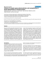
Table 1: Estimated Inhibition Constant, Ki (nmol), for ARBs docking into several immune system receptors.
Olmesartan Telmisartan Valsartan Irbesartan Candesartan Losartan
VDR,1DB1 12, 27 0.038 14 10 35 77
VDR,1TXI 10,34 0.039 14 12 30 74
PPAR120.2912 6 61 3
CCR2b * 9* 25* 22* 9* 39* 25*
AT2R1 * 0.10* 0.10* 0.3* 0.17* 1.5* 0.50*
*Note 1: CCR2b and AT2R1 are theoretical models, and may not be reliable (see text)
Note 2: (conventional ligand binding data): 1,25-dihydroxyvitamin-D docks into VDR (PDB:1DB1
) with Ki = 0.029 nmol and into VDR (PDB:1TXI)
with Ki= 0.059 nmol
TX522 docks into VDR (PDB:1DB1
) with Ki = 0.071 nmol and VDR (PDB:1TXI) with Ki = 0.12 nmol
TAK779 docks into putative CCR2b with Ki = 10 nmol
GI262570 docks into PPAR (PDB:1FM9
) with Ki = 0.040 nmol.
Theoretical Biology and Medical Modelling 2006, 3:1 />Page 3 of 33
(page number not for citation purposes)
1,25-D and its receptor, the Vitamin-D Receptor (VDR)
[Swiss-Prot:P11473], are expressed in over 30 target tis-
sues, and their expression is tightly coupled with regula-
tors as ubiquitous as GnRH (Gonadatrophin hormone
releasing hormone) [17], and the Parathyroid Hor-
mone(PTH) [18].
Ripple-down effects of VDR activation include changes
not only to the androgens and thyroid hormones, but also
to ACTH, Insulin Receptors, P450C1, and many other bio-
logically important metabolites [18,46].
In patients with severe Th1 immune disease, clinical
observations [9,10] indicated that the administration of
the ARB Olmesartan, at a concentration in excess of that
needed for full AT2R1 antagonism, often causes the level
of circulating 1,25-D to drop.
We therefore decided to target the VDR nuclear receptor
[19] for further study.
2. Peroxisome Proliferator Activated Receptors (PPARs)
Benson, et.al. reported [20] that the ARB 'Telmisartan'
seems to act both as an agonist and antagonist of Peroxi-
some Proliferator Activated Receptor gamma (PPAR-
gamma) [Swiss-Prot:P37231], a nuclear hormone
receptor from the same 'NR1' subfamily as VDR. The
PPARs act as anti-inflammatory transcription factors [21].
Part of this anti-inflammatory regulation is mediated
through negative interference between PPARs and nuclear
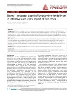
VDR binding pocket showing primary 1,25-D docking resi-duesFigure 4
VDR binding pocket showing primary 1,25-D docking
residues. Note: 1,25-D depicted with yellow backbone for
visual clarity. Carbon atoms shown as grey, oxygen as red,
nitrogen as blue, polar hydrogen as blue-white. Non-polar
hydrogens not displayed. Residues displayed as 'CPK' charge
spheres, ligand in 'ball and stick' format.
VDR-docked configurations for 1,25-D and Telmisartan, sep-arately and superimposedFigure 2
VDR-docked configurations for 1,25-D and Telmisar-
tan, separately and superimposed. Note: Models
depicted as "thick" and "thin" solely for visual clarity. Carbon
atoms shown as grey, oxygen as red, nitrogen shown as blue,
polar hydrogen as blue-white. Non-polar hydrogens not dis-
played.
1,25-D and TX522 with superimposed X-ray and VDR-docked configurationsFigure 1
1,25-D and TX522 with superimposed X-ray and
VDR-docked configurations. Note: Carbon atoms shown
as grey, oxygen as red. Hydrogens not displayed.
VDR-docked configurations for 1,25-D and Olmesartan, with superimposition showing both conformationsFigure 3
VDR-docked configurations for 1,25-D and Olme-
sartan, with superimposition showing both confor-
mations. Note: Models depicted as "thick" and "thin" solely
for visual clarity. Carbon atoms shown as grey, oxygen
shown as red, nitrogen as blue, polar hydrogen as blue-white.
Non-polar hydrogens not displayed.
Theoretical Biology and Medical Modelling 2006, 3:1 />Page 4 of 33
(page number not for citation purposes)
2D LigPlot of 1,25-D bound into the VDR ligand binding pocketFigure 5
2D LigPlot of 1,25-D bound into the VDR ligand binding pocket. Note: The core structure of the hydrogen-bonded
residues is expanded to a 'ball-and-stick' format, so as to show the atoms involved in hydrogen bond formation.
Key
Ligand bond
Non-ligand bond
3.0
Hydrogen bond & length
His 53
Non-ligand residues involved in hydrophobic
contact
Atoms involved in hydrophobic contact
2.99
2.98
2.89
3.19
3.34
2.64
C20
C21
C17
C22
C13
C16
C12
C14
C18
C15
C11
C8
C9
C7
C6
C5
C4
C10
C3
C1
C19
C2
O3
O1
C23
C24
C25
C26
C27
O25
N
CA
C
CB
O
CG
CD1
CD2
CE1
CE2
CZ
OH
N
CA
C
CB
O
CG
CD
NE
CZ
NH1
NH2
N
CA
C
CB
O
CG
ND1
CD2
CE1
NE2
N
CA
C
CB
O
OG
N
CA
C
CB
O
OG
N
CA
C
CB
O
CG
ND1
CD2
CE1
NE2
Trp 286
Ser 275
Tyr 295
Val 234
Phe 422
Leu 230
Ile 271
Cys 288
Val 300
Leu 313
1,25-D
Tyr 143
Arg 274
His 397
Ser 278
Ser 237
His 305
Theoretical Biology and Medical Modelling 2006, 3:1 />Page 5 of 33
(page number not for citation purposes)
The VDR agonist TX522 in the VDR ligand binding pocketFigure 6
The VDR agonist TX522 in the VDR ligand binding pocket. Note: The core structure of the hydrogen-bonded resi-
dues is expanded to a 'ball-and-stick' format, so as to show the atoms involved in hydrogen bond formation.
Key
Ligand bond
Non-ligand bond
3.0
Hydrogen bond & length
His 53
Non-ligand residues involved in hydrophobic
contact
Atoms involved in hydrophobic contact
3.02
2.88
2.80
3.22
2.71
C20
C21
C17
C22
C13
C16
C12
C14
C18
C15
C11
C8
C9
C7
C6
C5
C4
C10
C3
C1
C2
O3
O1
C23
C24
C27
C26
C25
O25
N
CA
C
CB
O
CG
CD1
CD2
CE1
CE2
CZ
OH
N
CA
C
CB
O
CG
ND1
CD2
CE1
NE2
N
CA
C
CB
O
CG
CD
NE
CZ
NH1
NH2
N
CA
C
CB
O
OG
N
CA
C
CB
O
CG
ND1
CD2
CE1
NE2
Ser 275
Trp 286
Tyr 295
Val 234
Ser 237
Phe 422
Leu 230
Cys 288
Val 300
Leu 313
TX522
Tyr 143
His 397
Arg 274
Ser 278
His 305
Theoretical Biology and Medical Modelling 2006, 3:1 />Page 6 of 33
(page number not for citation purposes)
Olmesartan bound into the sterol terminus of the VDR binding pocketFigure 7
Olmesartan bound into the sterol terminus of the VDR binding pocket. Note: This is the 12 nanomolar conforma-
tion of Olmesartan in the binding pocket. The core structure of the hydrogen-bonded residues is expanded to a 'ball-and-stick'
format, so as to show the atoms involved in hydrogen bond formation.
Key
Ligand bond
Non-ligand bond
3.0
Hydrogen bond & length
His 53
Non-ligand residues involved in hydrophobic
contact
Atoms involved in hydrophobic contact
3.35
C1
C2
C6
C18
C3
C4
C5
C7
N1
C8
C9
N2
C10
C13
C14
C11
C12
C15
O1
O2
C16
C17
O3
C19
C20
C21
C23
C24
C22
N3
N4
N5
N6
N
CA
C
CB
O
CG
CD
NE
CZ
NH1
NH2
Ser 275
Tyr 143
Val 300
Tyr 295
Ser 237
Leu 230
Leu 233
Val 234
Trp 286
Ile 271
His 305
Ser 278
Leu 313
Olmesartan
Arg 274
Theoretical Biology and Medical Modelling 2006, 3:1 />Page 7 of 33
(page number not for citation purposes)
Telmisartan docked into the VDR ligand binding pocketFigure 8
Telmisartan docked into the VDR ligand binding pocket. Note: Telmisartan is a strong antagonist of the VDR's activa-
tion.
Key
Ligand bond
Non-ligand bond
3.0
Hydrogen bond & length
His 53
Non-ligand residues involved in hydrophobic
contact
Atoms involved in hydrophobic contact
3.18
3.29
2.52
2.97
C1
C2
C3
C26
C4
C6
C5
C7
N1
N2
C8
C9
C23
C10
C11
C12
C13
C15
C14
C16
C17
C18
C19
C22
C21
C20
O1
O2
C24
C25
N3
N4
C27
C28
C30
C29
C31
C33
C32
N
CA
C
CB
O
CG1
CG2
CD1
N
CA
C
CB
O
CG
CD
NE
CZ
NH1
NH2
N
CA
C
CB
O
CG
ND1
CD2
CE1
NE2
N
CA
C
CB
O
OG
Ser 275
Leu 233
Tyr 143
Ala 231
Met 272
Leu 313
Trp 286
Tyr 147
Phe 150
His 305
Leu 227
Leu 230
Cys 288
Leu 309
Leu 414
Val 418
Val 234
Val 300
Tyr 401
Telmisartan
Ile 271
Arg 274
His 397
Ser 237
Theoretical Biology and Medical Modelling 2006, 3:1 />Page 8 of 33
(page number not for citation purposes)
Irbesartan docked into the VDR ligand binding pocketFigure 9
Irbesartan docked into the VDR ligand binding pocket. Note: The core structure of the hydrogen-bonded residues is
expanded to a 'ball-and-stick' format, so as to show the atoms involved in hydrogen bond formation.
Key
Ligand bond
Non-ligand bond
3.0
Hydrogen bond & length
His 53
Non-ligand residues involved in hydrophobic
contact
Atoms involved in hydrophobic contact
3.23
C1
C2
C6
C19
C3
C4
C5
C7
N1
C8
C9
N2
C10
C14
O1
C11
C12
C13
C15
C16
C17
C18
C20
C21
C22
C24
C25
C23
N3
N4
N5
N6
N
CA
C
CB
O
CG
CD
NE
CZ
NH1
NH2
Trp 286
Ser 237
Ile 271
Ser 275
Tyr 143
Tyr 295
Met 272
Leu 233
Leu 230
Tyr 147
Phe 150
Val 300
Ser 278
Cys 288
Irbesartan
Arg 274
Theoretical Biology and Medical Modelling 2006, 3:1 />Page 9 of 33
(page number not for citation purposes)
Valsartan docked into the VDR ligand binding pocketFigure 10
Valsartan docked into the VDR ligand binding pocket.
Key
Ligand bond
Non-ligand bond
3.0
Hydrogen bond & length
His 53
Non-ligand residues involved in hydrophobic
contact
Atoms involved in hydrophobic contact
C1
C2
C6
C18
C3
C4
C5
C7
N1
C8
C13
O1
C9
C10
C11
C12
C14
C15
O2
O3
C16
C17
C19
C20
C21
C23
C24
C22
N2
N3
N4
N5
Ile 271
Arg 274
Ser 275
Leu 233
Tyr 143
Tyr 295
Ser 237
Ile 268
Ser 278
Trp 286
Leu 230
Met 272
Val 300
Tyr 147
Cys 288
Asp 299
Leu 313
Valsartan
Theoretical Biology and Medical Modelling 2006, 3:1 />Page 10 of 33
(page number not for citation purposes)
Candesartan docked into the VDR ligand binding pocketFigure 11
Candesartan docked into the VDR ligand binding pocket. Note: The core structure of the hydrogen-bonded residues
is expanded to a 'ball-and-stick' format, so as to show the atoms involved in hydrogen bond formation.
Key
Ligand bond
Non-ligand bond
3.0
Hydrogen bond & length
His 53
Non-ligand residues involved in hydrophobic
contact
Atoms involved in hydrophobic contact
3.07
3.18
C1
C2
C6
C7
C3
C4
C5
C18
N1
C8
C9
C10
C11
N2
O1
C12
C17
C14
C13
C15
C16
O2
O3
C19
C20
C21
C23
C24
C22
N3
N4
N5
N6
N
CA
C
CB
O
CG
CD
NE
CZ
NH1
NH2
N
CA
C
CB
O
SG
Trp 286
Tyr 295
Ser 237
Leu 233
Tyr 143
Leu 230
Met 272
Val 234
Ser 278
Tyr 147
Ile 268
His 305
Ile 271
Leu 313
Ser 275
Candesartan
Arg 274
Cys 288
Theoretical Biology and Medical Modelling 2006, 3:1 />Page 11 of 33
(page number not for citation purposes)
Losartan docked into the VDR ligand binding pocketFigure 12
Losartan docked into the VDR ligand binding pocket. Note: The core structure of the hydrogen-bonded residues is
expanded to a 'ball-and-stick' format, so as to show the atoms involved in hydrogen bond formation.
Key
Ligand bond
Non-ligand bond
3.0
Hydrogen bond & length
His 53
Non-ligand residues involved in hydrophobic
contact
Atoms involved in hydrophobic contact
2.72
C1
C2
C6
C7
C3
C4
C5
C16
N1
C8
C9
C10
C11
N2
C12
CL1
O1
C13
C14
C15
C17
C18
C19
C20
C22
C21
N3
N4
N5
N6
N
CA
C
CB
O
OG
Ile 271
Tyr 143
Leu 233
Val 234
Met 272
Leu 230
Trp 286
Ile 268
Ser 278
Tyr 295
Arg 274
Ser 275
Phe 150
Tyr 236
Cys 288
Tyr 147
Losartan
Ser 237
Theoretical Biology and Medical Modelling 2006, 3:1 />Page 12 of 33
(page number not for citation purposes)
factors such as NF-kappaB. Ligands of PPAR may affect the
inflammatory response in diseases as wide-ranging as
Inflammatory Bowel Diseases, Atherosclerosis, Parkin-
son's Disease and Alzheimer's [22]. Clearly, it is impor-
tant to know exactly how the ARBs might affect
PPARgamma.
3. C-C chemokine receptor type 2 (CCR2b)
Monocyte chemotactic protein-1 (MCP-1) binding to its
receptor, CCR2b [EMBL:BC095540], plays an important
role in a variety of diseases involving infection, inflamma-
tion, and/or injury [23,24]. CCR2b recruits monocytes to
the sites of tissue damage. The monocytes later differenti-
ate to macrophages and/or polymorphonucleated 'giant'
cells.
CCR2b belongs to the same family of 7-Transmembrane
G-Protein Coupled Receptors (GPCRs) [25] as does
AT2R1, and the similarities between these two GPCRs,
together with the clinical observations [9,10], supported
the addition of CCR2b to this study.
Results
Validation of 'AutoDock' simulation software
It was decided to use automated docking of the ligands so
as to minimize subjective factors which might arise if the
ligands were fitted into the binding pockets manually. The
Scripps' package, AutoDock [26-28], was selected for this
task. Toprakci, et.al. [29], recently compared the Ki values
estimated by AutoDock for ten inhibitors of human
monoamine oxidase-B, with the values of Ki which had
been determined by experiment. In every case, there was
less than one order of magnitude difference between the
experimentally determined Ki, and the value estimated by
computer simulation of the ligand-bound enzyme. Chen,
et.al. [30], also concluded that AutoDock provided accu-
rate estimation of ligand-DNA binding parameters.
We were able to compare calculated Ki for some of our
docking experiments with published values, and similarly
found excellent agreement. For example, we validated our
PPARgamma model by docking the ligand GI262570
(Farglitazar), essentially as predicted by the data of Xu,
et.al. [31].
Table 2: Multiple sequence alignment for AT2R1 and Bovine Rhodopsin (PDB:1L9H)
sp|P30556|AGTR1_HUMAN
gi|21465997|pdb|1L9H|A
sp|P30556|AGTR1_HUMAN
gi|21465997|pdb|1L9H|A
sp|P30556|AGTR1_HUMAN
gi|21465997|pdb|1L9H|A
sp|P30556|AGTR1_HUMAN
gi|21465997|pdb|1L9H|A
sp|P30556|AGTR1_HUMAN
gi|21465997|pdb|1L9H|A
sp|P30556|AGTR1_HUMAN
gi|21465997|pdb|1L9H|A
sp|P30556|AGTR1_HUMAN
gi|21465997|pdb|1L9H|A
sp|P30556|AGTR1_HUMAN
gi|21465997|pdb|1L9H|A
SeqA Name
1
sp|P30556|AGTR1_HUMAN
MILNSSTEDGIKRIQDDCPKAGRHN-YIFVMIPTLYSIIFV 40
XMNGTEGPNFYVPFSNKTGVVRSPFEAPQYYLAEPWQFSMLAAYMFLLIM 50
: : *. : *: * :.*: : : * * :. : ::::
VGIFGNSLVVIVIYFYMKLKTVASVFLLNLALADLCFLLTLPLWAVYTAM 90
LGFPINFLTLYVTVQHKKLRTPLNYILLNLAVADLFMVFGGFTTTLYTSL 100
:*: * *.: * : **:* . :*****:*** : : : : :**::
EYRWPFGNYLCKIASASVSFNLYASVFLLTCLSIDRYLAIVHPMKSRLRR 140
HGYFVFGPTGCNLEGFFATLGGEIALWSLVVLAIERYVVVCKPMSN-FRF 149
. : ** *:: . .::. : : : *. * : *:** : . : :** :*
TMLVAKVTCIIIWLLAGLASLPAIIHRNVFFIENTNITVCAFHYESQNST 190
GENHAIMGVAFTWVMALACAAPPLVGWSRYIPEGMQCSCGIDYYTPHEET 199
* : : *::* .: *.:: . :: *. : : :* .::.*
LPIGLGLTKNILGFLFPFLIILTSYTLIWKALKKAYEIQKN KPRND 236
NNESFVIYMFVVHFIIPLIVIFFCYGQLVFTVKEAAAQQQESATTQKAEK 249
. : : : : *::*:::*: .* : : : *:* * : : : : .
DIFKIIMAIVLFFFFSWIPHQIFTFLDVLIQLGIIRDCRIADIVDTAMPI 286
EVTRMVIIMVIAFLICWLPYAGVAFYIFTHQG SDFGPIFMTI 291
: : : : : : : *: *:: . * : *: . : * . * . : * : * . *
T
ICIAYFNNCLNPLFYGFLGKKFKRYFLQLLKYIPPKAKSHSNLSTKMST 336
PAFFAKTSAVYNPVIYIMMNKQFRNCMVTTLCCG KNPLGDDEASTT 337
. :* . **: :* : : . * : * : . : : * :* . . . :*
LSYRPSDNVSSSTKKPAPCFEVE 359
VSKTETSQVAPA 349
: * : . : * : . : : : . .
Len(aa) SeqB Name Len(aa) Score
359 2 gi|21465997|pdb|1L9H|A 349 17
Theoretical Biology and Medical Modelling 2006, 3:1 />Page 13 of 33
(page number not for citation purposes)
It is important to understand that the 'Lamarckian genetic
algorithm' used by AutoDock does not guarantee conver-
gence to an optimal solution. The existence of the 'opti-
mal' solution, amongst any set of docking results, only
becomes assured as the number of docking attempts tends
to infinity. Considerable computing power was expended
in order to maximize the likelihood that this study identi-
fied the lowest energy docking configurations. Addition-
ally, the algorithm's convergence parameters were
manually adjusted whenever successive docking runs were
not returning consistent minima.
ARBs exhibit a strong affinity for VDR ligand binding
In order to maximize reliability, two discrete models were
used for the ligand binding pocket of the VDR, extracted
from two separate X-ray generated structures. The first
model was "The crystal structure of the nuclear receptor
for vitamin D bound to its natural ligand" [32]
[PDB:1DB1
], while the second was the VDR bound to the
agonist TX522 [33] [PDB:v
].
There was no significant difference between the results
obtained from either VDR structure. Table 1 shows the
predicted inhibition constants (Ki), in nanomoles, for
each of the ARBs binding into [PDB:1DB1
] and
[PDB:1TXI
].
As a further check of model validity, 1,25-D was initially
docked into [PDB:1DB1
] with a Ki = 0.03 nmol and into
[PDB:1TXI
] with Ki = 0.06 nmol. TX522 was then docked
into [PDB:1DB1
] with Ki = 0.07 nmol and [PDB:1TXI]
with Ki = 0.12 nmol. The difference between the crystal
structure of the ligands and the predicted docked confor-
mations was very small (Figure 1), and seems primarily
due to AutoDock's reliance upon grid-based energy calcu-
lations.
The ARB 'Telmisartan' had a strong affinity for the VDR,
with Ki≈0.04 nmol into either structure. This value is close
to that achieved by 1,25-D itself, which yielded Ki≈0.03
nmol into [PDB:1DB1
] and Ki≈0.09 nmol into
[PDB:1TXI
]. Telmisartan docked with a conformation
uncannily similar to 1,25-D (see Figure 2).
Irbesartan and Valsartan gave predicted Ki values in the
10–14 nanomolar region, probably indicating significant
Table 3: Multiple sequence alignment for CCR2b and Bovine Rhodopsin (PDB:1L9H)
1kp1_A (CCR2b)
gi|21465997|pdb|1L9H|A
1kp1_A (CCR2b)
gi|21465997|pdb|1L9H|A
1kp1_A (CCR2b)
gi|21465997|pdb|1L9H|A
1kp1_A (CCR2b)
gi|21465997|pdb|1L9H|A
1kp1_A (CCR2b)
gi|21465997|pdb|1L9H|A
1kp1_A (CCR2b)
gi|21465997|pdb|1L9H|A
1kp1_A (CCR2b)
gi|21465997|pdb|1L9H|A
1kp1_A (CCR2b)
gi|21465997|pdb|1L9H|A
SeqA Name
2
gi|21465997|pdb|1L9H|A
MLSTSRSR FIRNTNESGEEVTTFFDYDYGAPCHKFDVKQIGAQLLPPL 48
XMNGTEGPNFYVPFSNKTGVVRSPFEAPQY YLAEPWQFSMLAAY 44
: . : . . : : : * : : * : .* : *.: . : . : . : * . .
Y
SLVFIFGFVGNMLVVLILINCKKLKCLTDIYLLNLAISDLLFLIT LP 96
MFLLIMLGFPINFLTLYVTVQHKKLRTPLNYILLNLAVADLFMVFGGFTT 94
* : : : : * * * : *. : : : : * * *: : ***** : : * *: : : : .
LWAHSAANEWVFGNAMCKLFTGLYHIGYFGGIFFIILLTIDRYLAIVHAV 146
T
LYTSLHGYFVFGPTGCNLEGFFATLGGEIALWSLVVLAIERYVVVCKPM 144
* . : *** : * : * : : * . : : : : : * :*:** : . : : . :
FALKARTVTFGVVTSVITWLVAVFASVPGII-FTKCQKEDSVYVCGP Y 193
SNFRFG-ENHAIMGVAFTWVMALACAAPPLVGWSRYIPEGMQCSCGIDYY 193
: : : . . . : : . :** : : * : . : . * : : : : : * . * * *
FPRGWNN FHTIMRNILGLVLPLLIMVICYSGILKTLLRCRNEKKRHRA 241
T
PHEETNNESFVIYMFVVHFIIPLIVIFFCYGQLVFTVKEAAAQQQESAT 243
* : . * . . * : : : : : **: : : . : ** . : : * : . . : : : . :
VRVIFTIMIVYFLFWTPYNIVILLNTFQEFFGLSNCESTSQLDQATQVTE 291
TQKAEKEVTRMVIIMVIAFLICWLPYAGVAFYIFTHQGSDFGPIFMTIPA 293
. : . : . : : . : : * * : . : . : . : .
T
LGMTHCCINPIIYAFVGEKFRRYLSVFFRKHITKRFCKQCPVFYRETVD 341
FFAKTSAVYNPVIYIMMNKQFR NCMVTTLCCGKNPLGDDEAST 336
: . * . * * :** : : . : : ** : . : * . * : * : *:
GVTSTNTPSTGEQEVSAGL 360
T
VSKTETSQVAPA 349
* : . * : * . . . . . : : .
Len(aa) SeqB Name Len(aa) Score
349 3 1kp1_A 360 17
Theoretical Biology and Medical Modelling 2006, 3:1 />Page 14 of 33
(page number not for citation purposes)
antagonistic action at concentrations safely achievable in-
vivo.
Olmesartan similarly predicted useful Ki values, ranging
from 10 to 34 nmol. Particularly interesting is that two
distinct conformations were identified.
Figure 3 shows that Olmesartan docked in each conforma-
tion, one with its imidazole terminus near the triol of
1,25-D. The second focused on the seco terminus of 1,25-
D.
Losartan docked with a Ki around 70 nanomolar, Cande-
sartan around 30 nanomolar. These are likely also signifi-
cant antagonists, but higher dosage levels would be
necessary.
Hydrogen bonds and hydrophobic contacts during docking
with the VDR
Figure 4 shows the ligand binding pocket of the VDR with
1,25-D docked into it, highlighting those residues with
which 1,25-D forms hydrogen-bonds.
Figure 5 is a 2D representation of the 3D structure of Fig-
ure 4, created with Ligplot [53,54]. The hydrogen bonds
were identified with HBPLUS [55,56], as were the hydro-
phobic contacts formed between 1,25-D and the VDR res-
idues. The core structure of the hydrogen-bonded residues
is expanded to a 'ball-and-stick' format so as to show
which atoms are involved in hydrogen bond formation.
A double hydrogen bond was formed from the oxygen of
the triol group of 1,25-D, both to the imidazole nitrogen
of HIS305, and to the imidazole nitrogen of HIS397.
Another hydrogen bond extends from the 1-hydroxyl oxy-
gen to the aminoacetal of ARG274 and the hydroxyl of
SER237, and another pair from the ligand's O3 oxygen to
SER278 and TYR143.
Figure 6 shows that the VDR agonist TX522 [42] also
forms a double hydrogen bond between the oxygen of its
triol group, the imidazole of HIS397, and the imidazole
of HIS305. The 3-hydroxyl-oxygen is hydrogen-bonded to
TYR 143 and SER278, while the 1-hydroxyl-oxygen forms
a hydrogen bond with the aminoacetal of ARG274. No
hydrogen bond is formed with SER237, presumably due
Table 4: Multiple sequence alignment for AT2R1 and CCR2b
gi|4757938|ref|NP_000639.1|CCR2b
gi|231519|sp|P30556|AGTR1_HUMA
gi|4757938|ref|NP_000639.1|CCR2b
gi|231519|sp|P30556|AGTR1_HUMA
gi|4757938|ref|NP_000639.1|CCR2b
gi|231519|sp|P30556|AGTR1_HUMA
gi|4757938|ref|NP_000639.1|
gi|231519|sp|P30556|AGTR1_HUMA
gi|4757938|ref|NP_000639.1|CCR2b
gi|231519|sp|P30556|AGTR1_HUMA
gi|4757938|ref|NP_000639.1|CCR2b
gi|231519|sp|P30556|AGTR1_HUMA
gi|4757938|ref|NP_000639.1|CCR2b
gi|231519|sp|P30556|AGTR1_HUMA
gi|4757938|ref|NP_000639.1|CCR2b
gi|231519|sp|P30556|AGTR1_HUMA
SeqB Name
1
gi|4757938|ref|NP_000639.1| 360
-MLSTSRSRFIRNTNESGEEVTTFFDYDYGAPCHKFDVK QIGAQLLPPLY 49
MILNSSTEDGIKRIQDD
CPKAGRHNYIFVMIPTLY 35
: * . : * . * : . : : . . . . : : . . . : . * * . : : : :* . **
SLVFIFGFVGNMLVVLILINCKKLKCLTDIYLLNLAISDLLFLITLPLWA 99
SIIFVVGIFGNSLVVIVIYFYMKLKTVASVFLLNLALADLCFLLTLPLWA 85
*::*:.*:.** ***::: *** ::.::*****::** **:******
HSAANE WVFGNAMCKLFTGLYHIGYFGGIFFIILLTIDRYLAIVHAVF 147
VYTAMEYRWPFGNYLCKIASASVSFNLYASVFLLTCLSIDRYLAIVHPMK 135
: * * * * * * : * *: : . : . : . . : * : : *:********* . :
ALKARTVTFGVVTSVITWLVAVFASVPGIIFTKCQKED SVYVCGPYFP 195
SRLRRTMLVAKVTCIIIWLLAGLASLPAIIHRNVFFIENTNITVCAFHYE 185
: ** : . . ** .: * * *:* :**: * .** . : : . : * * . : :
RGWNNFHT IMRNILGLVLPLLIMVICYSGILKTLLRCRNEKKR 238
SQNSTLPIGLGLTKNILGFLFPFLIILTSYTLIWKALKKAYEIQKNKPRN 235
. . : : : *** *: : : *:**: : .* : * * : * : . : : * .
HRAVRVIFTIMIVYFLFWTPYNIVILLNTFQEFFGLSNCESTSQLDQATQ 288
DDIFKIIMAIVLFFFFSWIPHQIFTFLDVLIQLGIIRDCRIADIVDTAMP 285
. . : :* : : *: : . : * : * * : : *. :* : . : : : : : * . : . : * *
VTETLGMTHCCINPIIYAFVGEKFRRYLSVFFRKHITKRFCKQCPVFYRE 338
ITICIAYFNNCLNPLFYGFLGKKFKRYFLQLLKYIPPKAKSHSNLSTKMS 335
:* : . : * : * * : : * . * : * : * * : * * : : : : .* . : . .
T
VDGVTSTNTPSTGEQEVSAGL 360
T
LSYRPSDNVSSSTKKPAPCFEVE 359
* : . . * * . .* : : : . . .
Len(aa) SeqB Name Len(aa) Score
360 2 gi|231519|sp|p30556|AGTR1_HUMA 359 27
Theoretical Biology and Medical Modelling 2006, 3:1 />Page 15 of 33
(page number not for citation purposes)
to a lowered affinity consequent upon the removal of the
C19 position carbon from 1,25-D(cf.Figure 4).
The Ki = 12E-9 configuration of Olmesartan (Figure 7),
forms a hydrogen bond from its imidazole terminal
hydroxyl to ARG274. Olmesartan forms only hydropho-
bic contacts with the key VDR binding residues TYR143,
SER237, SER278 and HIS305. TYR143 is especially impor-
tant. It is part of the 'hinge region,' and key for VDR tran-
scriptional activity [51,57]. It is thus almost certain that
Olmesartan will function as a VDR antagonist.
Telmisartan docks with a Ki of 0.04 nmol, so that typical
in-vivo concentrations of the ARB should be sufficient to
displace 1,25-D from the ligand binding domain. Figure 8
shows that that hydrogen bonds are formed to SER237,
ARG274, HIS397 and ILE271, but not to TYR143. SER278
or HIS305. Telmisartan would thus seem likely to act as a
very strong antagonist of the VDR, with an affinity signif-
icantly stronger than the other ARBs.
Irbesartan (Figure 9) formed a hydrogen bond between its
tetrazole group and the amino of ARG274. The lack of
hydrogen bonds to TYR143 and SER278 indicate that
Irbesartan will be a VDR antagonist.
Valsartan, although it exhibits a potentially useful affinity
as a VDR antagonist, failed to form hydrogen bonds with
any key residue (Figure 10).
The imidazole of Candesartan formed a bond with the
sulphur of CYS288 (Figure 11), and the imidazole termi-
nus oxygen of Losartan hydrogen-bonded with
SER237(Figure 12). Both are indicative of actions antago-
nistic to VDR activation.
ARBs exhibit an affinity for PPARgamma
We extracted the coordinate data for PPARgamma from
[PDB:1FM9
], an X-ray structure. As model validation, the
PPARgamma agonist GI262570 (Farglitazar) was docked
with Ki≈0.04 nmol, close to the (approx.) 0.01 nmol pre-
dicted by the inhibition curve in figure 1A of Xu, et.al.
[31].
Table 1 shows that the ARBs exhibited a strong affinity for
the ligand binding pocket of PPARgamma, with Ki rang-
ing from 0.29 to 61 nanomoles.
Telmisartan is the strongest modulator of PPARgamma
(Ki≈0.3 nmol), while Losartan (Ki≈ 3 nmol), Olmesartan
(Ki≈12 nmol), Irbesartan (Ki≈6 nmol) and Valsartan
(Ki≈12 nmol) also seem likely to have significant PPAR
modulatory activity. Candesartan (Ki≈ 61 nmol) may also
have useful activity at a higher dosage.
ARBs exhibit a strong affinity for CCR2b
The ARBs are designed as antagonists for the Angiotensin
II Type 1 Receptor (AT2R1). This is a GPCR [36] of the
"Class A (Rhodopsin-like) 7-transmembrane receptors."
CCR2b is another Class A GPCR, with surprising similar-
ity to AT2R1.
Table 2 shows the multiple sequence alignment between
AT2R1 and Bovine Rhodopsin [PDB:1L9H
], the prototype
structure for Class A GPCRs. Table 3 shows an alignment
for CCR2b vs. Rhodopsin, while Table 4 compares AT2R1
and CCR2b. It is interesting to note that CCR2b and
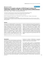
Overview of the ligand binding pocket identified in CCR2b (PDB:1KP1
)Figure 13
Overview of the ligand binding pocket identified in CCR2b
(PDB:1KP1
). Olmesartan is shown docked into pocket.
Theoretical Biology and Medical Modelling 2006, 3:1 />Page 16 of 33
(page number not for citation purposes)
AT2R1 both exhibit only 17% homology with Bovine
Rhodopsin, while the score between them is much higher,
at 27%.
There are no complete X-ray or NMR structures of Homo
sapiens' Class A GPCRs in PDB, or any other public data-
base. However, Shi, et.al. [37] had derived a theoretical
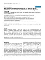
Perspective view showing how pocket is located underneath Extracellular 'loop' 1. Olmesartan is shown docked into pocketFigure 14
Perspective view showing how pocket is located underneath Extracellular 'loop' 1. Olmesartan is shown
docked into pocket. Note: Residues displayed as 'CPK' charge spheres. Ligand displayed as stick and ball model. Left is view
from front of pocket, facing helices 7 and 1, right view is from the top, looking across the top of helices 1 and 2.
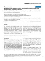
CCR2b residues highlighted alongside docked TAK779. From left: front of pocket, rear of pocketFigure 15
CCR2b residues highlighted alongside docked TAK779. From left: front of pocket, rear of pocket. Note: Carbon
atoms shown as grey, oxygen as red, nitrogen as blue, polar hydrogen as blue-white, sulphur as yellow. Non-polar hydrogens
not displayed. Residues displayed as 'CPK' charge spheres, ligand as 'ball and stick' models.
Theoretical Biology and Medical Modelling 2006, 3:1 />Page 17 of 33
(page number not for citation purposes)
TAK779 docked into the CCR2b binding pocketFigure 16
TAK779 docked into the CCR2b binding pocket.
Key
Ligand bond
Non-ligand bond
3.0
Hydrogen bond & length
His 53
Non-ligand residues involved in hydrophobic
contact
Atoms involved in hydrophobic contact
C1
N1
O1
C2
C20
C3
C4
C5
C19
C6
C7
C8
C9
C11
C10
C12
C13
C14
C15
C18
C16
C17
C21
C22
C23
C25
C24
C26
N2
C27
C28
C29
C30
C31
C32
C33
O2
Leu 45
Thr 292
Gly 41
Tyr 188
Ser 186
Val 37
Thr 296
Ile 300
His 297
Pro 31
Cys 32
Ile 304
Leu 44
Asn 301
His 33
Leu 293
Tak779
Theoretical Biology and Medical Modelling 2006, 3:1 />Page 18 of 33
(page number not for citation purposes)
model, [PDB:1KP1], which provided a basis for us to
study. We tried to improve upon [PDB:1KP1] by using,
inter alia, Truncated Newton energy minimization with
Ponder's TINKER Tools [38,39] and homology modelling
with Sali's 'Modeller' [40,41]. However, even extensive
homology modelling against the Bovine Rhodopsin X-ray
structure [PDB:1L9H], and other theoretical models, such
as [PDB:1KPX], failed to improve upon [PDB:1KP1].
We accepted that [PDB:1KP1
] was probably a valid model
for CCR2b based on the detailed nature of Shi, et.al's stud-
ies [37], our failed attempts to improve upon it, and the
manner in which it docked, exactly as predicted, with the
CCR2b antagonist, TAK779.
A binding pocket exists between helices seven and one of
[PDB:1KP1
], extending back to extracellular regions one
and three. Baba, et.al. [42] had measured the inhibitory
effects of Tak779 on CCR2b in their laboratory, showing
an experimental Ki≈9 nmol. When we docked TAK779
into our putative binding pocket, it predicted a Ki≈10
nmol, essentially identical with this experimental value.
Figure 13 shows the location of this binding pocket, and
Figure 14 an overview of the pocket structure, running
between GPCR helices seven and one, beneath the extra-
cellular regionone, and bounded at the rear by extracellu-
lar region three.
Figure 15 shows the residues binding TAK779 into the
putative pocket. Hydrophobic interactions with LEU45,
HIS297, ILE300, TYR188, PRO31 and CYS32, help to sta-
bilize the ligand. The 2D LigPlot of residue interactions
can be seen at Figure 16.
Olmesartan and Irbesartan each showed excellent affinity
(Ki≈9 nmol) for this binding pocket, while Valsartan, Tel-
misartan, Candesartan and Losartan exhibited slightly less
(Kifrom 22 to 40 nmol).
Figure 17 shows the residues which interact with Olme-
sartan. A hydrogen bond is formed with the imidazole of
HIS297, while ILE300, ALA42, LEU45, THR292, TYR188,
CYS32 and PRO31 all help to stabilize the ligand. Figure
18 shows the 2D LigPlot of these interactions.
Figure 19 shows the docked position of TAK779 and Olm-
esartan superimposed, to enable easier comparison of the
final location of each ligand.
Irbesartan forms hydrophobic contacts with a set of resi-
dues similar to that of Olmesartan (see Figure 20).
The ARBs, and TAK779, not only fill space within this
binding pocket, but also 'anchor' the top of helices seven
and one to extracellular regions three and one, restraining
the motion of GPCR elements, and, most probably, inhib-
iting its activation [43].
A putative AT2R1 receptor model
A primary goal set for this study had been the validation
of every structure and tool we used. It had therefore been
decided to ensure that the ARBs would dock into AT2R1
with inhibition constants close to the values measured in-
vitro, as documented in the various FDA New Drug Appli-
cations (NDAs). For example, NDA21-286 [2], indicates a
Ki for Olmesartan and Candesartan of approx. 0.1
nanomolar, and for Losartan about 3 times higher.
This validation task proved to be the most difficult of the
study. There was no AT2R1 X-ray structure publicly avail-
able, nor any comprehensive theoretical model. Addition-
ally, there was very little comparative experimental ARB
data available (FDA NDA21-286 is the exception to this).
Most authors studied only one commercial ARB product
in isolation.
We tried to use the theoretical model published by Mar-
tin, et.al. [43] [PDB:1ZV0
] for an activated AT2R1. But no
ARB would bind to that receptor configuration, even after
the extensive energy optimization required to move helix
seven back into its un-activated position.
CCR2b residues highlighted alongside docked Olmesartan, viewed from the front of the binding pocketFigure 17
CCR2b residues highlighted alongside docked Olme-
sartan, viewed from the front of the binding pocket.
Note: Carbon atoms shown as grey, oxygen as red, nitrogen
as blue, polar hydrogen as blue-white, sulphur as yellow.
Non-polar hydrogens not displayed. Residues displayed as
'CPK' charge spheres, ligand as 'ball and stick' models.
Theoretical Biology and Medical Modelling 2006, 3:1 />Page 19 of 33
(page number not for citation purposes)
Olmesartan docked into the CCR2b binding pocketFigure 18
Olmesartan docked into the CCR2b binding pocket. Note: The core structure of the hydrogen-bonded residues is
expanded to a 'ball-and-stick' format, so as to show the atoms involved in hydrogen bond formation.
Key
Ligand bond
Non-ligand bond
3.0
Hydrogen bond & length
His 53
Non-ligand residues involved in hydrophobic
contact
Atoms involved in hydrophobic contact
3.29
C1
C2
C6
C18
C3
C4
C5
C7
N1
C8
C9
N2
C10
C13
C14
C11
C12
C15
O1
O2
C16
C17
O3
C19
C20
C21
C23
C24
C22
N3
N4
N5
N6
N
CA
C
CB
O
CG
ND1
CD2
CE1
NE2
Tyr 188
Cys 32
Thr 296
Val 37
Thr 292
Leu 45
Ile 300
Gly 41
Asn 104
Pro 31
Ala 42
Ala 102
Ser 186
Leu 293
His 33
Lys 38
Glu 291
Olmesartan
His 297
Theoretical Biology and Medical Modelling 2006, 3:1 />Page 20 of 33
(page number not for citation purposes)
We then decided to produce an AT2R1 model by compar-
ative homology [40] with Bovine Rhodopsin
[PDB:1L9H
], but still could not produce a model which
would dock the known ARBs, even after extensive energy
minimization. Eventually we used the putative CCR2b,
[PDB:1KP1
] as the comparative model. Surprisingly,
straight out of the 'Modeller' [41], all the ARBs docked
into a pocket on the opposite side of the GPCR from the
binding pocket which had been located on CCR2b. The Ki
for the ARBs ranged from 0.10 to 1.5 nmol, as detailed in
Table 1.
It is interesting to note that although the comparative
homology between AT2R1 and Rhodopsin is only 17%
(Table 2) the AT2R1 sequence is much closer to that of
CCR2b (Table 4). Our failure to produce a usable receptor
by comparative homology with Bovine Rhodopsin would
seem to caste doubt on its utility as a prototype for the
Class A 7-transmembrane GPCR structures.
Figure 21 shows the primary residues involved in docking
the ARBs, and a superimposition of the docked conforma-
tions of Olmesartan and Losartan, demonstrating the
homogeneity of location of the imidazole group into the
binding pocket, even amongst ARBs with significant struc-
tural differences.
The hydrophobic interactions between Olmesartan and
our AT2R1 is shown in Figure 22. Olmesartan forms two
hydrogen bonds, with GLY194 and LEU197, as does Losa-
rtan (Figure 23). Candesartan binds to quite different res-
idues, in particular, making 6 hydrophobic contacts with
ILE193(Figure 24).
Discussion
Models provided to ease visualization of nuclear receptors
It is evident from the lack of clarity in Figure 4 that it is
extremely difficult to visualize ligand conformation in the
binding pockets of nuclear receptors using two dimen-
sional media. For this reason we have provided, as an
attached file, an archive of the receptor configurations
used in this study, in addition to the most significant
bound ligand conformations. The models can be loaded
into, for example, the Python Molecular Viewer [35], and
3D analysis performed.
This archive will also facilitate the testability of our results.
Does telmisartan selectively modulate PPARgamma?
Benson, et.al. [20], presented the ARBs as suited to PPAR-
gamma modulation. Their primary conclusion was that
Telmisartan's structure allowed it to exhibit selective mod-
ulation, exhibiting in-vitro PPARgamma agonistic activity
at low concentrations, changing to antagonistic activity at
higher concentrations.
Figure 25 shows the key binding pocket for the agonist
Farglitazar (GI262570) in the PPAR ligand binding
domain. Figure 26, the LigPlot of this conformation,
shows two key hydrogen bonds between Farglitazar's O1,
HIS449 and TYR473, and two more between O2, SER289
and HIS323.
Tsukahara, et.al. [52] recently studied a number of PPAR
agonists. They found that agonistic activity disappears
when TYR473 is mutated, and noted the importance of
HIS323 and HIS449.
Figures 27 and 28 show the residues which contact PPAR-
gamma when Irbesartan and Losartan are docked into
their minimum energy conformations. Although Irbe-
sartan hydrogen-bonds TYR473 and HIS449, Losartan
only contacts these residues, and forms its sole hydrogen-
bond to ALA278. It would thus seem likely that Losartan
is an effective PPAR antagonist. Irbesartan does not hydro-
gen-bond to HIS323, a residue found critical to Rosiglita-
zar's agonism [52], and probably is more likely an
antagonist than agonist.
Figure 29 shows that Telmisartan does not form any
hydrogen bonds with the PPARgamma residues identified
by Tsukahara, et.al., as critical to the agonistic activity of
Rosglitazar. Any molecular mechanism which could result
in 'partial agonism' of PPARgamma by Telmisartan is still
to be elucidated
CCR2b-docked configurations for TAK779 and Olmesartan, individually and with superimpositionFigure 19
CCR2b-docked configurations for TAK779 and Olm-
esartan, individually and with superimposition. Note:
Ligands depicted as "thick" and "thin" solely for visual clarity.
Carbon atoms shown as grey, oxygen as red, nitrogen as
blue, polar hydrogen as blue-white. Non-polar hydrogens not
displayed.
Theoretical Biology and Medical Modelling 2006, 3:1 />Page 21 of 33
(page number not for citation purposes)
Irbesartan docked into the CCR2b binding pocketFigure 20
Irbesartan docked into the CCR2b binding pocket.
Key
Ligand bond
Non-ligand bond
3.0
Hydrogen bond & length
His 53
Non-ligand residues involved in hydrophobic
contact
Atoms involved in hydrophobic contact
C1
C2
C6
C19
C3
C4
C5
C7
N1
C8
C9
N2
C10
C14
O1
C11
C12
C13
C15
C16
C17
C18
C20
C21
C22
C24
C25
C23
N3
N4
N5
N6
Tyr 188
Val 37
Cys 32
Leu 45
Thr 292
Thr 296
Pro 31
Asn 104
Ala 42
Leu 44
Gly 41
Ala 102
Ser 186
Ile 300
Lys 38
Glu 291
Irbesartan
Theoretical Biology and Medical Modelling 2006, 3:1 />Page 22 of 33
(page number not for citation purposes)
We would note, however, that the extreme affinity which
Telmisartan exhibits for the ubiquitous VDR might well
alter expression of many hormones at concentrations
lower than those at which Telmisartan begins to modulate
PPARgamma. This may make it very difficult to evaluate
cause and effect in the cascade of metabolic changes
which will result from Telmisartan's blockade of the VDR.
Bovine and guinea pig AT2R1 for FDA in-vitro ARB studies
While modelling the ARBs docking into the AT2R1 recep-
tor, we were struck by data in United States Food and Drug
Administration (US FDA) documents which did not
exactly match our own observations.
For example, there are inconsistencies between our pre-
dictions for the relative efficacies of Olmesartan, Cande-
sartan and Losartan; and those of Figure 1.1.1.4 of FDA
NDA21-286 [2]. The NDA's in-vitro experiments, using
Cavia porcellus, showed Olmesartan as having the highest
ARB efficacy, as we did, but found Candesartan close in
efficacy to Olmesartan (1.2×) and Losartan to be less
effective (3.4×). Our study found Losartan (Ki≈0.5 nmol)
to be a better antagonist of AT2R1 than was Candesartan
(Ki≈1.5 nmol).
The answer may well lie in sequence divergence between
the AT2R1 proteins from human, bovine, and guinea pig
sources. The multiple sequence alignment showing differ-
ences between AT2R1 from Homo sapiens, Cavia porcellus
and Bos taurus is shown in Table 5.
Our model predicts that the primary residues involved in
docking most of the ARBs are GLN15, GLY194, GLY196,
THR198 and GLY203.
The binding pocket around GLN15 is conserved in all
three homologies.
However, in Bos taurus, the Isoleucine residue 193 is
mutated to Valine. Candesartan has 6 hydrophobic con-
tacts with ILE193, while Losartan and Olmesartan have
only one. It is thus very likely that substitution of ILE193
will differentially effect the degree of Candesartan's antag-
onism of Bos taurus AT2R1 receptors, when compared
with that of other ARBs, less dependent on contacts with
ILE193.
Additionally, there is a mutation in Leucine 205, structur-
ally adjacent to GLY203. GLY203 has seven hydrophobic
contacts with Olmesartan, eight with Losartan, and six
with Candesartan. In Cavia porcellus, this Glycine is
mutated to Methionine.
The authors consequently believe that the FDA should re-
examine the acceptability of Bos taurus and Caviaporcellus
tissues for demonstration of the efficacy of ARBs.
It was beyond the scope of this study to model AT2R1
receptors for all three species used in the FDA in-vitro
data. This should form a topic for ongoing research.
Putative AT2R1 with (from left) Olmesartan, and Losartan docked, showing primary residues. Ligands are also shown superim-posedFigure 21
Putative AT2R1 with (from left) Olmesartan, and Losartan docked, showing primary residues. Ligands are also
shown superimposed. Note: Carbon atoms shown as grey, oxygen as red, nitrogen as blue, polar hydrogen as blue-white,
and chlorine as green. Non-polar hydrogens not displayed. Residues displayed as 'CPK' charge spheres, ligands as 'ball and stick'
models. Thick and thin ligand backbones displayed solely for visual clarity.
Theoretical Biology and Medical Modelling 2006, 3:1 />Page 23 of 33
(page number not for citation purposes)
Olmesartan docked into the putative AT2R1 binding pocketFigure 22
Olmesartan docked into the putative AT2R1 binding pocket. Note: The core structure of the hydrogen-bonded resi-
dues is expanded to a 'ball-and-stick' format, so as to show the atoms involved in hydrogen bond formation.
Key
Ligand bond
Non-ligand bond
3.0
Hydrogen bond & length
His 53
Non-ligand residues involved in hydrophobic
contact
Atoms involved in hydrophobic contact
2.65
2.88
C1
C2
C6
C18
C3
C4
C5
C7
N1
C8
C9
N2
C10
C13
C14
C11
C12
C15
O1
O2
C16
C17
O3
C19
C20
C21
C23
C24
C22
N3
N4
N5
N6
N
CA
C
O
N
CA
CB
C
CG
CD1
CD2
O
Gly 196
Asn 200
Gly 203
Leu 195
Thr 198
Leu 202
Gln 15
Lys 199
Gln 267
Glu 8
Thr 175
Phe 206
Thr 178
Ile 193
Olmesartan
Gly 194
Leu 197
Theoretical Biology and Medical Modelling 2006, 3:1 />Page 24 of 33
(page number not for citation purposes)
Losartan docked into the putative AT2R1 binding pocketFigure 23
Losartan docked into the putative AT2R1 binding pocket. Note: The core structure of the hydrogen-bonded residues
is expanded to a 'ball-and-stick' format, so as to show the atoms involved in hydrogen bond formation.
Key
Ligand bond
Non-ligand bond
3.0
Hydrogen bond & length
His 53
Non-ligand residues involved in hydrophobic
contact
Atoms involved in hydrophobic contact
2.96
2.49
C1
C2
C6
C7
C3
C4
C5
C16
N1
C8
C9
C10
C11
N2
C12
CL1
O1
C13
C14
C15
C17
C18
C19
C20
C22
C21
N3
N4
N5
N5
N
CA
CB
C
CG
CD1
CD2
O
N
CA
C
O
Gly 196
Asn 200
Thr 198
Gly 203
Leu 195
Leu 202
Lys 199
Gln 267
Thr 175
Phe 206
Gln 15
Thr 178
Ile 193
Losartan
Leu 197
Gly 194
Theoretical Biology and Medical Modelling 2006, 3:1 />Page 25 of 33
(page number not for citation purposes)
Candesartan docked into the putative AT2R1 binding pocketFigure 24
Candesartan docked into the putative AT2R1 binding pocket. Note: The core structure of the hydrogen-bonded res-
idues is expanded to a 'ball-and-stick' format, so as to show the atoms involved in hydrogen bond formation.
Key
Ligand bond
Non-ligand bond
3.0
Hydrogen bond & length
His 53
Non-ligand residues involved in hydrophobic
contact
Atoms involved in hydrophobic contact
3.17
C1
C2
C6
C7
C3
C4
C5
C18
N1
C8
C9
C10
C11
N2
O1
C12
C17
C14
C13
C15
C16
O2
O3
C19
C20
C21
C23
C24
C22
N3
N4
N5
N6
N
CA
CB
C
CG
CD
OE1
NE2
O
Gly 196
Gly 203
Ile 270
Asp 263
Glu 8
Ile 193
Leu 195
Gly 194
Leu 202
Phe 171
Tyr 184
Lys 199
Phe 206
Asn 200
Asp 273
Thr 175
Thr 178
Leu 197
Thr 198
Candesartan
Gln 267