Báo cáo y học: "Protein-lipid interactions: correlation of a predictive algorithm for lipid-binding sites with three-dimensional structural data" ppt
Bạn đang xem bản rút gọn của tài liệu. Xem và tải ngay bản đầy đủ của tài liệu tại đây (11.67 MB, 14 trang )
BioMed Central
Page 1 of 14
(page number not for citation purposes)
Theoretical Biology and Medical
Modelling
Open Access
Review
Protein-lipid interactions: correlation of a predictive algorithm for
lipid-binding sites with three-dimensional structural data
David L Scott*
1
, Gerold Diez
2
and Wolfgang H Goldmann*
1,2
Address:
1
Renal Unit, Leukocyte Biology & Inflammation Program, Structural Biology Program and the Massachusetts General Hospital/Harvard
Medical School, 149 13th Street, Charlestown, MA 02129, USA and
2
Friedrich-Alexander-University of Erlangen-Nuremberg, Center for Medical
Physics and Technology, Biophysics Group, Henkestrasse 91, 91052 Erlangen, Germany
Email: David L Scott* - ; Gerold Diez - ;
Wolfgang H Goldmann* -
* Corresponding authors
Abstract
Background: Over the past decade our laboratory has focused on understanding how soluble
cytoskeleton-associated proteins interact with membranes and other lipid aggregates. Many
protein domains mediating specific cell membrane interactions appear by fluorescence microscopy
and other precision techniques to be partially inserted into the lipid bilayer. It is unclear whether
these protein-lipid-interactions are dependent on shared protein motifs or unique regional
physiochemistry, or are due to more global characteristics of the protein.
Results: We have developed a novel computational program that predicts a protein's lipid-binding
site(s) from primary sequence data. Hydrophobic labeling, Fourier transform infrared spectroscopy
(FTIR), film balance, T-jump, CD spectroscopy and calorimetry experiments confirm that the
interfaces predicted for several key cytoskeletal proteins (alpha-actinin, Arp2, CapZ, talin and
vinculin) partially insert into lipid aggregates. The validity of these predictions is supported by an
analysis of the available three-dimensional structural data. The lipid interfaces predicted by our
algorithm generally contain energetically favorable secondary structures (e.g., an amphipathic alpha-
helix flanked by a flexible hinge or loop region), are solvent-exposed in the intact protein, and
possess favorable local or global electrostatic properties.
Conclusion: At present, there are few reliable methods to determine the region of a protein that
mediates biologically important interactions with lipids or lipid aggregates. Our matrix-based
algorithm predicts lipid interaction sites that are consistent with the available biochemical and
structural data. To determine whether these sites are indeed correctly identified, and whether use
of the algorithm can be safely extended to other classes of proteins, will require further mapping
of these sites, including genetic manipulation and/or targeted crystallography.
Background
Signal transduction, vesicle trafficking, retroviral assem-
bly, and other central biological processes involve the
directed binding of proteins to membranes. Soluble pro-
teins may associate with membranes through well-
defined structural domains (e.g., pleckstrin-homology, PX
(phox), C2, amphipathic helices and/or unstructured
motifs that interact through non-specific electrostatic and
Published: 28 March 2006
Theoretical Biology and Medical Modelling 2006, 3:17 doi:10.1186/1742-4682-3-17
Received: 21 November 2005
Accepted: 28 March 2006
This article is available from: />© 2006 Scott et al; licensee BioMed Central Ltd.
This is an Open Access article distributed under the terms of the Creative Commons Attribution License ( />),
which permits unrestricted use, distribution, and reproduction in any medium, provided the original work is properly cited.
Theoretical Biology and Medical Modelling 2006, 3:17 />Page 2 of 14
(page number not for citation purposes)
non-polar interactions [1-3]. Post-translational modifica-
tions, such as myristylation or palmitoylation, may also
play critical roles in regulating membrane association.
Many cytoskeleton-associated proteins interact, at least
transiently, with membranes [4-6]. The application of
biophysical techniques including Fourier-transformed
infrared spectroscopy (FTIR), neutron reflection, electron
spin resonance (ESR), nuclear magnetic resonance (NMR)
and X-ray crystallography has been helpful in characteriz-
ing protein and membrane structure [7,8]. Unfortunately,
the mechanism(s) and structural consequences of mem-
brane association remain poorly understood [9,10].
In previous papers, we have used a purpose-written
matrix-based computational program to predict potential
lipid interfaces for several key cytoskeletal proteins
(alpha-actinin, Arp2, CapZ, talin, and vinculin) [11].
Although there is no direct biochemical evidence to sup-
port the CapZ sites, the locations proposed for alpha-
actinin, Arp2, talin, and vinculin are supported by in vitro
experiments, including hydrophobic labeling, differential
scanning calorimetry, film balance, T-jump, CD spectros-
copy, and isothermal titration calorimetry [12-16]. In this
paper we correlate the results of our predictive algorithm
with the respective high-resolution three-dimensional
crystal structures.
Method
Our algorithm for predicting a protein's lipid interface
identifies highly hydrophobic or amphipathic amino acid
segments while discriminating between surface-seeking
and transmembrane configurations [11,17-19]. An
amphipathic helix, defined as an alpha- helix with oppos-
ing polar and nonpolar surfaces oriented along its long
axis, is a common secondary structural motif that reversi-
bly associates with lipids and displays detergent proper-
ties. Based on analysis of the lipid-binding properties of
apolipoproteins, polypeptide hormones and lytic
polypeptides, we designed our algorithm to classify
amino acids into five physiochemical groups (hydropho-
bic, polar, positive, negative and neutral) and divide
amphipathic helices spatially into three sectors (hydro-
phobic, interface and polar). The composition of an ide-
alized amphipathic helix is mathematically defined by a
matrix motif (M
ij
) consisting of five rows (representing
the physiochemical groups) with the number of columns
equal to the number of residues within the idealized helix.
A comparison matrix (C
ik
) is calculated by multiplying
together the matrix motif (M
ij
) and a second matrix deter-
mined for a segment of residues from the test protein (S
jk
).
Summation over all components of C
ik
generates a con-
sensus score that estimates the compatibility between a
given amino acid segment and the amphipathic motif.
Higher scores indicate increasing probabilities that the
residues of a segment do not form an amphipathic struc-
ture by chance. The algorithm generally identifies several
candidate sites per protein species.
In this study, the computationally predicted lipid-binding
sites for alpha-actinin, Arp2, CapZ, talin, and vinculin are
examined in the context of the respective high-resolution
three-dimensional coordinates obtained from the Protein
Data Bank (Tables 1, 2, 3) [20]. Qualitative graphical
analysis, performed with the display programs SPDBV
and PYMOL, include examination of secondary and terti-
ary structure, solvent accessibility and electrostatic field
potentials [21,22]. The electrostatic calculations were per-
formed by SPDBV subroutines using the Coulomb
method with the dielectric constants for the solvent and
protein set to 80.0 and 4.0, respectively, and incorporat-
ing only charged residues.
Results
Alpha-actinin
Dynamic turnover of the actin network drives cell motility
and muscle contraction. Alpha-actinin, one of several
actin-binding proteins essential for cytoskeletal function,
Table 1: Characteristics of the three-dimensional structures. Coordinate files were obtained from the Protein Data Bank [20]; 1HCI
[28]; 1K8K [49]; 1IZN [61]; 1MIX [83]; 1MIZ [83]; 1QKR [93]; 1TR2 [92]; 1ST6 [94].
# Protein Crystal Organism Sequence
Included
Resolution (Å) Refinement
(R-value)
PDB ID
1 α-actinin Rod domain: spectrin-like repeats 1–4 Homo sapiens 274–746 2.8 0.270 1HCI
2 Arp2 Arp2/3 complex Bos taurus 154–343
1
2.0 0.216 1K8K
3CapZβ-1 CapZ Gallus gallus 2–271 2.1 0.222 1IZN
4 Talin FERM domain (subdomains 2 and 3) Gallus gallus 196–400 1.75 0.199 1MIX
FERM domain/Integrin β3 tail fragment (739–743) Complex Gallus gallus 200–400 1.9 0.204 1MIZ
5 Vinculin Tail Domain Gallus gallus 881–1061
2
1.8 0.200 1QKR
Full length (Selenium-methionine derivative) Homo sapiens 1–1066 2.85 0.251 1TR2
Full length Gallus gallus 1–1065 3.1 0.316 1ST6
•
1
Subdomains 1 and 2 are partially disordered and not included in the refined model.
•
2
Residues 856–874 could not be adequately modeled or refined and are not included in the PDB coordinates.
Theoretical Biology and Medical Modelling 2006, 3:17 />Page 3 of 14
(page number not for citation purposes)
is a ubiquitous protein that cross-links actin filaments in
muscle and non-muscle cells [23-27]. The protein is
found at cell adhesion sites, focal contacts, and along
actin stress-fibers in migrating cells. Alpha-actinin can
localize to the plasma membrane, where it cross-links the
cortical actin, aids in membrane displacement, and links
transmembrane receptors with the cytoskeleton. Alpha-
actinin is the major thin filament cross-linking protein in
the muscle Z-discs. Mutations to the Drosophila mela-
nogaster alpha-actinin gene disrupt the Z-discs and are
generally lethal [26]. Translocation of alpha-actinin from
the cytosol to the plasma membrane may occur indirectly
by interactions with the cytoplasmic tails of transmem-
brane receptors. Alpha-actinin associates with several
plasma membrane associated proteins including ICAM-1,
ICAM-2, beta1-integrin, beta2-integrin, L-selectin, vincu-
lin, and zyxin. The peptides that interact with alpha-
actinin tend to be basic, alpha-helical, and appear to inter-
act with the conserved acidic surface of the alpha-actinin
rod [28].
Alpha-actinin may interact with phospholipid mem-
branes directly [29]. Static light scattering experiments,
employing monolayers and bilayers of varied charge com-
position, demonstrate that alpha-actinin reconstitutes
into the hydrophobic core of lipid bilayers containing
negatively charged phospholipids [30]. Phosphoi-
nositides, such as phosphatidylinositol 3,4,5-trisphos-
phate (PIP
3
) and phosphatidylinositol 4,5-bisphosphate
(PIP
2
), differentially regulate alpha-actinin flexibility and
function [27,31-34]. Binding of phosphoinositides to
alpha-actinin occurs through the calponin homology
domain and has been localized to amino acids 168–184
of striated muscle species [32]. Phosphatidylinositol 3-
kinase may directly bind to alpha-actinin through its p85
subunit [35]. In the presence of diacylglycerol and pal-
mitic acid, alpha-actinin can form microfilament-like
complexes with actin [36].
Alpha-actinin is an anti-parallel homodimeric rod with
extensive homology to spectrin and dystrophin [28,37].
The 30–40 nm long dimer consists of two identical
polypeptide chains, divided into three functional
domains: an actin-binding region at the amino-terminus,
a central alpha-actinin segment (rod), and a carboxyl-ter-
minus containing two EF hands (generally a 12 residue
loop flanked on both sides by a 12 residue alpha helix)
(Figure 1). The actin-binding region contains the amino
terminal calponin-homology (CH) domain and the car-
boxyl-terminal calmodulin-homology (CaM) domain.
The relatively rigid central rod domain (242 × 31–49 Å),
derived from four spectrin repeats, defines the distance
between cross-linked actin filaments and mediates inter-
actions with receptors and signaling proteins.
Electron and cryo-electron microscopy have provided
low-resolution (15 Å) images of the intact alpha-actinin
molecule [38,39]. Unfortunately, only the rod domain
(residues 274–746, Table 1) has been successfully crystal-
lized for high-resolution structural studies [28]. The seg-
ments implicated in lipid-binding by our algorithm,
amino acid residues 281–300 (1st spectrin repeat) and
residues 720–739 (4th spectrin repeat), lie at the head/tail
junctions of opposite ends of the isolated monomer in the
crystallized rod domain (Figure 1; Table 2) [14]. The site
experimentally implicated in phosphatidylinositide bind-
ing, amino acids 168–184, is absent from the crystallized
construct [28,31]. This segment was not identified as a
Table 2: Computationally determined sites of probable lipid binding. A matrix algorithm [11] was used to identify probable lipid-
binding sites in the following cytoskeletal proteins; α-actinin [14], Arp2 [16], CapZβ-1 (submitted, TBMM), Talin [12-13, 121] and
Vinculin [14]. In-vitro experimental support for the computationally predicted sites for α-Actinin, Arp2, Talin, and Vinculin (site 935–
978) was obtained from a variety of techniques including hydrophobic labeling, differential scanning calorimetry (DSC), Langmuir
Blodgett (film balance), T-jump, CD spectroscopy, cryo-electron microscopy (EM), FTIR, and isothermal titration calorimetry.
Protein Sequence
Residues
Species Sequence Experimental (in-vitro) Validation
α-actinin 281–300 Gallus gallus EKLASDLLEWIRRTIPWLEN Residues (287–306) of 1HCI DSC, Centrifugation, SDS-PAGE [14]
720–739 Gallus gallus QLLTTIARTINEVENQILTR Residues (726–745) of 1HCI DSC, Centrifugation, SDS-PAGE [14]
Arp2 185–202 A. castellanii RDVTRYLIKLLLLRGYVF DSC, Film Balance, Temperature Jump
[16]
CapZβ-1 134–151 Homo sapiens IKKAGDGSKKIKGCWDSI No data
215–232 Homo sapiens RLVEDMENKIRSTLNEIY No data
Talin 385–406 M. musculatus GEQIAQLIAGYIDIILKKKKSK Isothermal Titration Calorimetry,
Monolayer Expansion, CD-spectroscopy
[15]; FTIR [86] Resonance energy
transfer, Cryo-EM [90]
Vinculin 935–978 Gallus gallus RLVRGGSGNKRALIQCAKDIAKASDEVT RLAKEVAKQCTDKRIR Co-sedimentation, Hydrophobic
Photolabeling [102]
1020–1040 Gallus gallus TEMLVHNAQNLMQSVKETVRE No data
1052–1066 Homo sapiens AGFTLRWVRKTPWYQ No data
Theoretical Biology and Medical Modelling 2006, 3:17 />Page 4 of 14
(page number not for citation purposes)
Table 3: Characteristics of sequences implicated in lipid binding. The isolelectric point for the isolated peptide was calculated and the
percent alpha-helix determined from the relevant crystal structure. The symbols for electrically positive residues are underlined
( ); those corresponding to electrically negative residues are underlined ( ). The characters under the amino acid
sequence refer to the secondary structure; H = helix, T = hydrogen-bonded turn, S = bend, E = extended beta-strand, and B = residue
in isolated beta-bridge. Residues 401–406 (KKKKSK) are not present in talin crystal structures. Helical residues are underlined
().
Protein Residues Sequence Number
Residues
Isoelectric
Point
Helix
Content
Sequence Site in Protein
α-actinin 281–300 20 4.49 15/20
(75%)
Helices 1–2
720–739 20 4.66 16/20
(80%)
Carboxyl-terminal portion of
Helix 16
Arp2 185–202 18 10.0 13/18
(72%)
Helix 1 of Actin-like Subdomain
4
CapZβ-1 134–151 18 9.62 0/18 (0%) Contains portion of β strand 6
215–232 18 4.49 18/18
(100%)
Helix 5
Talin 385–406 22 8.61 9/22
(41%)
Helix 5 of Subdomain F3 of
Talin-H
Vinculin 935–978 44 9.73 31/44
(70%)
Domain 5, Helices 2–3 + amino-
terminal portion of Helix 4
1020–1040 21 4.47 20/21
(95%)
Domain 5, Helix 5
1052–1066 15 Hairpin [122]
double single
dashed
EKLASDLLEWIRRTIPWLEN
-
-
HHHHHHHHTHHHHHHHTTSS
-
-
-
-
-
-
QLLTTIARTINEVENQILTR
HHHHHHHHHHHHHHHHTTTT
-
-
-
-
-
-
-
RDVTRYLIKLLLLRGYVF
HHHHHHHHHHHHHTT
-
-
-
-
IKKAGDGSKKIKGCWDSI
EEEE SSSSEEEEEEEE
-
-
-
-
-
-
-
-
RLVEDMENKIRSTLNEIY
HHHHHHHHHHHHHHHHHH
-
-
-
-
GEQIAQLIAGYIDIILKKKKSK
HHHHHHHHHTTS
-
-
-
-
-
-
RLVRGGSGNKRALIQCAKDIAKA
HTTTS-SSTTHHHHHHHHHTHHH
-
-
-
-
-
-
SDEVTRLAKEVAKQCTDKRIR
HHHHHHHHHHHHHHB-HHHH
-
-
-
-
-
TEMLVHNAQNLMQSVKETVRE
HHHHHHTHHHHHHHHHHHHHH
-
-
AGFTLRWVRKTPWYQ
HHHHH-HH HHHHH
Theoretical Biology and Medical Modelling 2006, 3:17 />Page 5 of 14
(page number not for citation purposes)
lipid-binding candidate by our computer algorithm, pre-
sumably because the amino acid sequence (TAPYRNV-
NIQNFHLSWK) forms an extended loop or coil [40].
In the dimeric rod, the predicted lipid-binding regions
from constituent monomers lie close, but not confluent,
to one another. The left-handed ninety-degree trans-rod
twist places the dimer's two amino-terminal lipid-binding
segments, residues 281–300, on a common face while
separating the carboxyl-terminal segments. Amino acid
residues 281–300 and 720–739 are largely alpha-helical
and solvent exposed. Whether this accessibility is main-
tained in the intact alpha-actinin molecule is not clear
from the low-resolution structural studies since the region
of the protein that joins the 47 kDa head to the rod
domain appears to be quite flexible [38].
Alpha-actinin is an acidic protein with a pI of 6.0. Mem-
brane binding is not calcium-dependent but the protein
may undergo conformational changes in response to salts,
cations, and lipids [30,41]. The native alpha-actinin rod is
globally electrostatically negative; however, the ends con-
taining the predicted lipid-binding sites are less acidic
than the middle core (Figure 1, panel c
). This suggests that
the dimer ends would be the most likely candidates to
interact with the negatively charged phospholipids at the
bilayer interface. The relatively low isoelectric points of
the computationally predicted sites (Table 3) and the pre-
ponderance of surrounding negative charge in the intact
rod implies a relatively weak attraction between alpha-
actinin and negatively charged phospholipids in the
absence of neutralizing cofactors or a significant confor-
mational change. Surprisingly, not only do the isolated
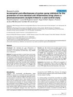
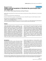
The predicted lipid-binding site of the alpha-Actinin dimerFigure 1
The predicted lipid-binding site of the alpha-Actinin dimer. The coordinates of the alpha-actinin rod domain (PDB
1HCI) are displayed with one monomer of the dimer shown in silver and the other in gold. The predicted lipid-binding sites are
colored yellow. Amino and carboxyl termini are indicated in blue and red, respectively. (a) Ribbon model, (b) Space-filling rep-
resentation, and (c) Electrostatic field potentials (orientation of the protein is identical to that viewed in (a) and (b)). The
colors red, white and blue are used to indicate negative, neutral and positive field potentials (c), respectively.
(a)
(b)
(c)
Theoretical Biology and Medical Modelling 2006, 3:17 />Page 6 of 14
(page number not for citation purposes)
computationally identified lipid-binding fragments read-
ily insert into lipid aggregates, but intact smooth muscle
alpha-actinin preferentially binds in-vitro to membranes
containing negatively charged phospholipids [30].
Arp2
Arp2 (actin-related-protein), in a complex with six other
proteins including Arp3, promotes branched growth of
actin filaments. Immunoelectron microscopy localizes the
Arp2/3 complex to the Y-branch, the point where a daugh-
ter actin filament branches off at a seventy-degree angle
from the parent filament [42-44]. The Arp2/3 complex
attaches to the side of the parent actin filament through
the interactions between three of its five ancillary proteins
(p16, p34 and p40) and actin subunits. Activation of the
Arp2/3 complex requires the presence of nucleation-pro-
moting factors and a pre-existing filament [45,46]. Nucle-
ation factors such as WASP/Scar (Wiskott-Aldrich
Syndrome), in turn, require activation through chemotac-
tic signaling pathways that guide cellular movement.
WASP promotes the binding of the Arp2/3 complex to the
side of a pre-existing filament and may transfer the first
actin subunit to the nascent filament's rapidly growing
barbed end. Vinculin may also bind to the Arp2/3 com-
plex, in a phosphatidylinositol-dependent manner, dur-
ing membrane protrusion [47].
The Arp2/3 complex is a 220 kDa stable assembly of two
actin-related proteins and five novel protein subunits
[48,49]. Arp protein sequences are homologous to actin,
and subunit p40 (gene name ARPC1) resembles a beta-
propeller protein. The other 4 subunits of the complex
(gene names ARPC2 through ARPC5) share little sequence
homology to known proteins. The maximum dimensions
of the complex are 150 × 140 × 100 Å (Figure 2) [49].
The low-resolution 'kidney bean' structure revealed for the
Arp2/3 complex by electron microscopy is in general
agreement with the inactive crystallographic complex
[48,49]. It is thought that ATP binding induces a modest
rigid body rotational conformational change, together
with a more dramatic translation, that activates the Arp2/
3 complex (Figure 2, panel d
) [48,49]. Unfortunately,
since the electron densities for subdomains 1 and 2 of
Arp2 are weak, preventing accurate refinement of this
region, the three-dimensional coordinates available from
the Protein Data Bank are a synthesis of refined structure
and molecular modeling. Subdomains 1 and 2 are mod-
eled by the polyalanine trace of the highly homologous
protein actin. Subdomains 3 and 4 of Arp2, which are ade-
quately visualized and refined, also resemble actin.
Our algorithm predicts that amino acid residues 185–202
of Arp2 are involved in mediating lipid interactions. The
isolated segment partially inserts into lipid aggregates
with an apparent K
d
of 1.1 µM [16]. In the crystal struc-
ture, this segment is primarily alpha-helical (72 %) and
lies near the center of the Arp2/3 complex (Figure 2, panel
d) [49]. The helix is relatively recessed within Arp2 and
solvent access is further limited by the presence of adja-
cent proteins in the complex. It is likely that subdomains
1 and 2 of Arp2, which are missing from the refined struc-
ture, would further limit the ability of residues 185–202
to interact directly with lipids in the absence of a substan-
tial rearrangement of the ternary complex.
Both p21 and p40 have substantial areas of positive sur-
face charge. These regions are relatively remote from the
Arp2's predicted lipid interface in the inactive complex.
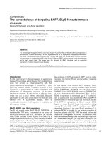
The predicted lipid-binding site of Arp2 and the Arp2/3 com-plexFigure 2
The predicted lipid-binding site of Arp2 and the
Arp2/3 complex. The coordinates of subdomains 3 and 4
of Arp2 (PDB 1K8K) are displayed as they appear in the inac-
tive crystallized Arp2/3 complex. The predicted lipid-binding
site is colored yellow. Amino and carboxyl termini are indi-
cated in blue and red, respectively. Arp2 subdomains 3 and 4;
(a) Ribbon model, (b) Space-filling representation, and (c)
Electrostatic field potentials (orientation of the protein is
identical to that viewed in (a) and (b)). The crystallized Arp2/
3 complex is shown as; (d) Space-filling representation (Arp2
(white), Arp3 (gold), p21 (blue), p40 (green); p34 (purple);
p20 (red), p16 (brown)), and (e) Electrostatic field potentials
(orientation of the protein is identical to that viewed in (d)).
The colors red, white and blue are used to indicate negative,
neutral and positive field potentials (e), respectively.
(a)
(b)
(c)
(e)
(d)
Theoretical Biology and Medical Modelling 2006, 3:17 />Page 7 of 14
(page number not for citation purposes)
The computationally predicted lipid interaction site is
itself electrostatically neutral but surrounded by strong
negative potentials in the assembled complex (Figure 2,
panel e
). Thus, the interaction of Arp2 with lipids is likely
to occur either prior to assembly of the complex or after a
significant conformational change (as postulated for acti-
vation) that reduces local charge barriers and improves
solvent access.
CapZ
β
1
Capping protein is crucial for actin filament assembly.
Activated Cap binds to the barbed end of actin with high
affinity (K
d
= 1nM) and at a 1:1 stoichiometry forming a
mechanical 'cap' that prevents the addition or loss of actin
monomers [50,51]. The sarcomeric isoform of capping
protein, which is composed of two polypeptide chains
(CapZ α1-β1), localizes to the Z-line of muscle through an
interaction with alpha-actinin [52]. The non-sarcomeric
isoforms are localized at the sites of membrane-actin con-
tact [53-56]. Capping protein 'caps' the Arp1 mini-fila-
ment in the dynactin complex, directly interacts with
twinfillin, and indirectly affects the Arp2/3 complex via
the CARMIL protein [57-60]. Residues at the carboxyl-ter-
mini of each CapZ chain (α 259–286 and β 266–277) are
essential for actin binding.
CapZ is an elongated, tightly assembled, heterodimeric
alpha/beta protein with overall dimensions of 90 × 50 ×
55 Å [61]. The two subunits, which may have arisen from
gene duplication, are structurally homologous creating a
pseudo two-fold symmetry perpendicular to the long axis
of the molecule (Figure 3). Each subunit contains three
domains and an additional carboxyl-terminal extension.
Three anti-parallel helices in an up-down-up arrangement
(helices 1–3) form the amino-terminal domain. The mid-
dle domain is composed of four beta strands (strands 1–4
for the alpha subunit; three beta strands 1–3 for the beta
subunit), containing two reverse turns. The carboxyl-ter-
minal domain comprises an anti-parallel beta sheet
formed by five consecutive beta strands (strands 5–9),
flanked on one side by a short amino-terminal helix (helix
4) and a long carboxyl-terminal helix (helix 5). The beta
strands of each subunit form a single 10-stranded anti-
parallel beta-sheet in the center of the molecule. A 'jelly-
fish' model has been proposed for Cap function in which
the carboxyl-terminal helical regions of the protein are
mobile and extend outward to engage the barbed end of
actin [61].
Phosphatidylinositol 4,5-bisphosphate (PIP
2
) regulates
CapZ function by dissociating the protein from the
barbed ends of actin filaments [59,62]. This effect appears
to be due to the direct binding of dispersed PIP
2
to CapZ.
High concentrations of other anionic phospholipids also
inhibit the ability of CapZ to effect actin polymerization
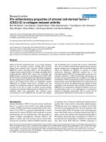
The predicted lipid-binding site of CapZbeta-1Figure 3
The predicted lipid-binding site of CapZbeta-1. The
coordinates of CapZ (PDB 1IZN) are displayed with the
alpha subunit shown in gold and the beta subunit in silver.
The predicted lipid-binding sites are colored yellow. Amino
and carboxyl termini are indicated in blue and red, respec-
tively. (a) Ribbon model, (b) Space-filling representation, and
(c) Electrostatic field potentials (orientation of the protein is
identical to that viewed in (a) and (b)). The colors red, white
and blue are used to indicate negative, neutral and positive
field potentials, respectively.
(a)
(b)
(c)
Theoretical Biology and Medical Modelling 2006, 3:17 />Page 8 of 14
(page number not for citation purposes)
[63]. In some phosphatase and kinase structures, nitrate
ions have been found near the phosphate binding sites
mimicking the transition state [64-68]. Sulfate ion also
may serve as a marker for phospholipid binding sites. The
crystal structure of CapZ beta-1 contains four nitrate ions
[61]. Only two nitrate ions appear to bind to the protein
with high specificity; one nitrate is associated with Lys95
while the other interacts with the dipole of helix 5 (Figure
3, panel a
). These nitrate-binding sites, located near the
actin-binding carboxyl-terminal extension of the Z subu-
nit, suggest a potential mechanism for PIP
2
regulation of
CapZ – actin association.
The sequences predicted to mediate lipid binding by our
algorithm, amino acid residues 134–151 and 215–232 of
the CapZ-β1 subunit, lie adjacent to one another in the
crystal structure [61]. Residues 134–151 primarily form
beta-sheet whereas residues 215–232 are part of Helix 5.
Both segments are solvent-accessible despite contributing
residues to the strong dimer interface (e.g., Lys136,
Glu221 and Asn222). Although CapZ is predominantly
electrostatically negative, the proposed lipid-binding
interface varies from neutral to positive (Figure 3, panel
c).
Talin
Talin is an abundant cytoskeletal protein that binds to the
cytoplasmic tails of integrin beta subunits, to actin fila-
ments, to other actin-binding proteins, and to phospholi-
pids [12,69-76]. In fibroblasts, the binding of talin to
membranes may induce the formation of focal adhesions
or trigger actin assembly by activating integrins or layilin,
respectively. In platelets, activated talin translocates from
the cytoplasm to the membrane where it co-localizes with
the GPIIb/IIIa complex [76].
Talin is a member of the 4.1 superfamily of FERM pro-
teins, a group of membrane-associated proteins that
includes the erythrocyte membrane protein 4.1, the ezrin,
radixin, moesin, and merlin proteins, and some tyrosine
phosphatases [77]. A common feature of FERM domain
proteins is extensive intramolecular head-tail interactions
that mask binding sites on the head [78,79]. Association
of extracellular matrix ligands with integrins triggers the
binding of the second messenger phosphatidylinositol
4,5-bisphosphate (PIP
2
) to the head domain, altering its
conformation to allow talin to bind to the cytoplasmic
tails of integrin receptors [78]. Binding occurs through a
largely hydrophobic area centered on the b5 strand and
also involves residues of the b6 strand, the carboxyl-termi-
nal half of helix H5 and the b4-b5 loop. During outside-
in integrin signaling, talin binds to other partners on the
cytoplasmic face of adhesion complexes, and in particular
vinculin, which then binds directly to actin and induces
actin bundling [80,81]. The incorporation of talin into
zwitterionic phospholipid bilayers is low but improved in
the presence of negatively charged phospholipids (K = 2.9
× 10
6
M
-1
) [13]. Talin is able to bind in vitro to phosphati-
dylinositol, phosphatidylinositol 4-monophosphate, and
PIP
2
. However, within a phospholipid bilayer, binding is
restricted to PIP
2
.
Talin is a flexible 235 kDa 51 nm dumbbell-shaped
homodimer (Figure 4) [82,83]. Calpain cleavage before
amino acid residue 434 yields 2 major domains, an N-ter-
minal 47 kDa FERM head and a carboxyl 190-kDa rod
domain. The rod domain, which is responsible for actin
interaction and nucleation, contains low-affinity integrin
binding sites as well as actin and vinculin binding sites
[84,85]. The isolated 47 kDa FERM-containing domain
retains the lipid-binding capacity of intact talin and
includes a primary integrin-binding site [71]. Talin binds
to phospholipids using both hydrophobic and electro-
static forces with a strong preference for negatively
charged aggregates [86].
FERM domains are cysteine-rich modules that bind phos-
phoinositides via amino acid sequences with a high per-
centage of basic and polar amino acids. FERM domains
contain three modules arranged in a clover shape: F1, F2
and F3 [87]. The F3 module of talin, which structurally
resembles a phosphotyrosine-binding domain, is formed
by a single carboxyl-terminal helix that partly encloses
one edge of an internally hydrophobic beta sandwich
[88]. A consensus sequence for PIP
2
binding has been
described (K/R)XXXKX(K/R)(K/R) but exceptions are fre-
quent [89].
The computationally predicted lipid-binding site, amino
acids 385–406, has a calculated hydrophobicity of 0.029,
high amphipathicity, and a hydrophobic moment of 0.3
[13,15,90]. At pH 7.4 the total free energy of binding
(∆G
0
) is approximately -9.4 kcal/mol, a value that com-
pares favorably with that determined for myristylolated
membrane-anchoring peptides. Residues 385–406 lie
within helix 5 and thus contribute substantially to the
binding site for the integrin beta3 tail. This proximity sug-
gests a mechanism for the PIP2 induced conformational
change that permits tail binding [78].
Vinculin
Vinculin is a conserved regulator of cell-cell adhesion
(cadherin-mediated) and cell-matrix focal adhesions
(integrin/talin-mediated). In its resting state, vinculin is
held in a closed conformation through interactions
between its head (Vh) and tail (Vt) domains. Vinculin
activation, associated with junctional signaling, generates
an open conformation that binds in vitro to talin, alpha-
actinin, paxillin, actin, the Arp2/3 complex, and to itself
[47,91-95].
Theoretical Biology and Medical Modelling 2006, 3:17 />Page 9 of 14
(page number not for citation purposes)
Talin and phospholipids activate vinculin. Talin binds to
Vh through high-affinity vinculin-binding sites present in
its central rod domain. Talin binding stimulates confor-
mational changes in the amino-terminal helical bundle of
Vh, displacing the tightly bound Vt [95]. Talin also
increases the activity of phosphatidylinositol phosphate
kinase-1 γ, generating PIP
2
[96-99]. The binding of phos-
phatidylinositol 4,5-bisphosphate to Vt, in turn, disrupts
the Vh-Vt interaction freeing vinculin to bind talin, actin,
VASP or the Arp2/3 complex [100]. Vinculin can readily
insert into the hydrophobic core of mono/bilayers con-
taining acidic (phosphatidic acid, phosphatidylinositol
and phosphati-dylglycerol), but not neutral (phosphati-
dylcholine and phosphatidylethanolamine), lipids
[101,102]. Vinculin can also undergo covalent modifica-
tion by lipids in vivo or bind acidic phospholipids through
its carboxyl-terminal domain (amino acids 916–970)
[103-106]. The latter process may inhibit the intramolecu-
lar association between the amino and carboxyl terminal
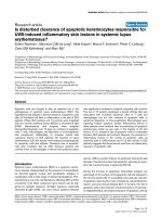
The predicted lipid-binding site of TalinFigure 4
The predicted lipid-binding site of Talin. The coordinates of talin are displayed either in (I) isolation (1MIX); or (II), in a
complex with an integrin beta3 tail fragment (residues 739–743) (1MIZ). The predicted lipid-binding sites are colored yellow
and the integrin beta3 tail fragment gold. Amino and carboxyl termini are indicated in blue and red, respectively. (a) Ribbon
model, (b) Space-filling representation, and (c) Electrostatic field potentials (orientation of the protein is identical to that
viewed in (a) and (b)). The colors red, white and blue are used to indicate negative, neutral and positive field potentials (c),
respectively.
I(a) I(b)
II(a) II(b) II(c)
Theoretical Biology and Medical Modelling 2006, 3:17 />Page 10 of 14
(page number not for citation purposes)
regions of vinculin and/or expose a binding site for pro-
tein kinase C [107,108].
Vinculin is a large (1,066 amino acid), structurally
dynamic protein with overall dimensions of 100 × 100 ×
50 Å in its autoinhibited conformation (Figure 5) [92-95].
The protein is composed of eight four-helix bundles that
divide the protein into five distinct domains; an 850
amino acid head (Vh), a 200 amino acid tail (Vt) and 3
intervening linkers (Vh2, Vh3, Vt2). The sequences impli-
cated in lipid binding by our algorithm, amino acid resi-
dues 935–978 and 1020–1040, contribute to helices 2
through 5 of Vt. Segment 935–978 includes residues
involved in Vt-Vh interactions (Arg 945, Arg 978) as well
as those mediating phosphatidylinositol binding. Phos-
phatidylinositol 4,5-bisphosphate appears to bind to a
basic "collar" surrounding the carboxyl-terminal arm (res-
idues 910, 911, 1039, 1049, 1060 and 1061), and a basic
'ladder' along the edge of helix 3 (residues 944, 945, 952,
956, 963, 966, 970, 978, 1008 and 1049) (Figure 5, panel
a). Point mutations in the collar (Lys911Ala and
Lys924Ala) or ladder (Lys952Ala) reduce PIP
2
binding by
50%. The ladder is largely solvent exposed, although at its
amino-terminal end Lys944 and Arg945 make salt bridges
to acidic residues on the head. His906, which lies adjacent
to the computationally predicted lipid-binding site, is
essential for PIP
2
induced conformational changes [110].
Binding of 10% PIP
2
in phosphatidylcholine vesicles to Vt
occurs in the micromolar range, but in combination with
PIP
2
miscelles and talin, vinculin appears to form a ter-
nary activation complex.
Discussion
Intracellular signaling and trafficking are regulated by
selective protein-membrane interactions. Transfer of
cytosolic proteins to the membrane presumably occurs in
two steps: an initial approach based on electrostatic attrac-
tion followed by lipid-induced protein refolding and/or
insertion [110]. Potential control mechanisms include:
(1) modulating the protein's affinity for lipid (e.g., cal-
cium-binding promotes the membrane association of C2
domains by enhancing electrostatic forces), (2) sequester-
ing the lipid at specific locations, and/or (3) restricting
access to the lipid in the absence of specific stimuli
[10,111-113].
In-vitro experimental support for the computationally pre-
dicted lipid-binding sites of α-Actinin, Arp2, Talin, and
Vinculin (site 935–978) was obtained using standard
techniques such as hydrophobic labeling, differential
scanning calorimetry (DSC), Langmuir Blodgett (film bal-
ance), FTIR, T-jump, CD spectroscopy, cryo-electron
microscopy (EM), and isothermal titration calorimetry.
Similar data are not yet available to gauge the in-vitro
binding characteristics of the sites predicted by our algo-
The predicted lipid-binding site of VinculinFigure 5
The predicted lipid-binding site of Vinculin. The coordinates of vinculin (PDB 1ST6) are displayed with the predicted
lipid-binding sites colored yellow (residues 935–978) and brown (residues 1020–1040). Phosphatidylinositol 4,5-bisphosphate
appears to bind to a basic "collar" surrounding the carboxyl-terminal arm (residues 910, 911, 1039, 1049, 1060, 1061), and a
basic 'ladder' along the edge of helix 3 (residues 944, 945, 952, 956, 963, 966, 970, 978, 1008, and 1049). These residues are
shown in gold. Note: the overlap of the computationally derived site and the experimentally discovered phosphatidylinositol
site. Amino and carboxyl termini are indicated in blue and red, respectively. Residues 856 through 874 are disordered in the
vinculin electron-density map and are not shown, the start (residue 855) and stop site (residue 874) for this region are shown
in green. (a) Ribbon model, (b) Space-filling representation, and (c) Electrostatic field potentials (orientation of the protein is
identical to that viewed in (a) and (b)). The colors red, white and blue are used to indicate negative, neutral and positive sfield
potentials (c), respectively.
(a) (b) (c)
Theoretical Biology and Medical Modelling 2006, 3:17 />Page 11 of 14
(page number not for citation purposes)
rithm for CapZbeta-1 or the vinculin sites (residues 1020–
1040 and 1052–1066).
The three-dimensional structures of the computationally
predicted lipid-binding sites described here are, with the
exception of Site 1 of CapZbeta-1, predominantly or
exclusively alpha-helical. The energy required to insert a
polypeptide into a membrane is minimized by the pres-
ence of favorable secondary structure [114]. Membrane-
spanning or surface associated amphipathic alpha-helices
and beta-strands/sheets are common in biologically active
peptides and proteins. Amphipathic alpha-helices may
reversibly associate with lipids and function as peptide
detergents [115-117]. Amphipathic beta-sheets, in con-
trast, interact with lipids in an essentially irreversible
manner, and lack detergent properties. Unfavorable
energy costs associated with individual amphipathic beta-
strands are likely to drive coalesence into beta-sheets on
lipid surfaces. When the axis of an amphipathic helix lies
parallel to the membrane surface and partially inserted
into the membrane, the polar and non-polar protein sur-
faces may interact simultaneously with the charged head
groups and hydrophobic side chains, respectively.
Four of the five cytoskeletal proteins studied here show a
strong preference for acidic phospholipids in vitro (alpha-
actinin, Arp2, talin, and vinculin). The mechanism by
which these soluble cytoplasmic proteins become mem-
brane associated is unclear. Only one of the five proteins
is known to undergo covalent lipid modification (i.e., vin-
culin). Although myristoylation and palmitoylation
increase hydrophobicity, myristate alone may be insuffi-
cient to anchor proteins to the plasma membrane
[1,118,119]. The clustering of basic residues adjacent to
lipid modification sites found among proteins such as K-
ras4B and HIV-1 Gag enhances favorable electrostatic
interactions with acidic lipids [19,66]. Other peripheral
proteins (e.g., type II beta-phosphatidyinositol-3-kinase,
AKAP79, myelin basic protein, and a number of proteins
containing C2 domains), in the absence of lipophilic
modifications, depend solely upon basic groups to bind
to membrane surfaces [112]. The three-dimensional struc-
tures of pleckstrin homology domains reveal large posi-
tively charged electrostatic patches surrounding the
ligand-binding sites, suggesting that the excess charge is
useful in improving initial attraction and orientation to
the predominantly negatively charged plasma membrane
[113]. Most of the predicted lipid interface sites in this
study are either intrinsically electrostatically positive
(Table 3) or are located in regions that are relatively basic.
Many critical biological pathways are regulated by pro-
tein-lipid interactions. Understanding this biology is diffi-
cult given the complexity and heterogeneity of the
interface. Computational methods, such as our matrix
algorithm, provide a potentially powerful means for pre-
dicting the region, and orientation, of a protein as it asso-
ciates with lipid aggregates. Further experimental work
will be required to validate and refine this algorithm.
However, based on experience with the protein hunting-
tin, it appears that the methods may be applicable to mul-
tiple protein classes [120].
Acknowledgements
This work was funded by the Deutsche Forschungsgemeinschaft (DFG;
Is25/8-1 to WHG) and North Atlantic Treaty Organization (NATO; CLG
978417 to WHG). We thank Dr. H. Banfic for helpful comments. WHG is
currently on sabbatical in Germany.
This paper is dedicated to Prof. Dr. Gerhard Isenberg for his lifetime
achievement in the field of cytoskeletal proteins and plasma membranes.
Dr. Isenberg retired on 31
st
July 2005 after a very productive scientific
career. We wish him all the best for the future. WHG.
References
1. Resh MD: Fatty acylation of proteins: new insights into mem-
brane targeting of myristoylated and palmitoylated pro-
teins. Biochim Biophys Acta 1999, 1451:1-16.
2. Hurley JH, Misra S: Signaling and subcellular targeting by mem-
brane-binding domains. Annu Rev Biophys Biomol Struct 2000,
29:49-79.
3. Simon SA, McIntosh TJ, (Eds): Peptide-Lipid Interactions. In Cur-
rent Topics in Membranes Volume 52. Academic Press; 2002:1-581.
4. Isenberg G: Actin binding proteins – lipid interactions. J Muscle
Res Cell Mot 1991, 12:136-144.
5. Isenberg G: New concepts for signaling perception and trans-
duction by the actin cytoskeleton at cell boundaries. Sem Cell
Dev Biol 1996, 7:707-15.
6. Niggli V: Structural properties of lipid-binding sites in
cytoskeletal proteins. Trends Biochem Sci 2001, 26:604-11.
7. Isenberg G, Niggli V: Interaction of cytoskeletal proteins with
membrane lipids. Int Rev Cytol 1998, 178:73-125.
8. Marsh D, Pali T: The protein-lipid interface: perspectives from
magnetic resonance and crystal structures. Biochim Biophys
Acta 2004, 1666:118-141.
9. Bottomley MJ, Salim K, Panayotou G: Phospholipid-binding pro-
tein domains. Biochim Biophys Acta 1998, 1436:165-83.
10. DiNitto JP, Cronin TC, Lambright DG: Membrane recognition
and targeting by lipid-binding domains. Sci STKE 2003,
213:re16.
11. Tempel M, Goldmann WH, Isenberg G, Sackmann E: Interaction of
the 47-kDa talin fragment and the 32-kDa vinculin fragment
with acidic phospholipids: a computer analysis. Biophys J 1995,
69:228-41.
12. Dietrich C, Goldmann WH, Sackmann E, Isenberg G: Interaction of
NBD-talin with lipid monolayers: a film balance study. FEBS
Lett 1993, 324:37-40.
13. Goldmann WH, Senger R, Kaufmann S, Isenberg G: Determination
of the affinity of talin and vinculin to charged lipid vesicles: a
light scatter study. FEBS Lett 1995, 368:516-8.
14. Goldmann WH, Teodoridis JM, Sharma CP, Alonso JL, Isenberg G:
Fragments from alpha-actinin insert into reconstituted lipid
bilayers. Biochem Biophys Res Commun 1999, 264:225-9.
15. Seelig A, Blatter XL, Frentzel A, Isenberg G: Phospholipid binding
of synthetic talin peptides provides evidence for an intrinsic
membrane anchor of talin. J Biol Chem 2000, 275:17954-61.
16. Goldmann WH, Isenberg G: Actin-related protein (Arp2)
inserts into artificial lipid membranes. Cell Biol Int 2002,
26:1073-8.
17. Kyte J, Doolittle RF: A simple method for displaying the hydro-
pathic character of a protein. J Mol Biol 1982, 157:105-32.
18. Eisenberg D, Schwarz E, Komaromy M, Wall R: Analysis of mem-
brane and surface protein sequences with the hydrophobic
moment plot. J Mol Biol 1984, 179:125-42.
Theoretical Biology and Medical Modelling 2006, 3:17 />Page 12 of 14
(page number not for citation purposes)
19. Deber CM, Liu L-P, Wang C, Goto N, Reithmeier RAF: The Hydro-
phobicity Threshold for Peptide Insertion into Membranes.
Current Topics in Membranes 2002, 52:459-473.
20. Berman HM, Westbrook J, Feng Z, Gilliland G, Bhat TN, Weissig H,
Shindyalov IN, Bourne PE: The Protein Data Bank. Nucleic Acids
Res 2000, 28:235-42.
21. Guex N, Peitsch MC: SWISS-MODEL and the Swiss-Pdb-
Viewer: an environment for comparative protein modeling.
Electrophoresis 1997, 18:2714-23.
22. DeLano WL: The PyMOL Molecular Graphics System. DeLano
Scientific, San Carlos, CA, USA; 2002.
23. Blanchard A, Ohanian V, Critchley D: The structure and function
of alpha-Actinin. J Muscle Res Cell Motil 1989, 10:280-9.
24. Fritz M, Zimmermann RM, Bärmann M, Gaub HE: Actin binding to
lipid-inserted alpha-Actinin. Biophys J 1993, 65:1878-85.
25. Gunst SJ, Tang DD, Opazo Saez A: Cytoskeletal remodeling of
the airway smooth muscle cell: a mechanism for adaptation
to mechanical forces in the lung. Respir Physiol Neurobiol 2003,
137:151-68.
26. Fyrberg C, Becker J, Barthmaier P, Mahaffey J, Fyrberg E: Character-
ization of lethal Drosophila melanogaster alpha-Actinin
mutants. Biochem Genet 1998, 36:299-310.
27. Fukami K, Endo T, Imamura M, Takenawa T: alpha-Actinin and vin-
culin are PIP2-binding proteins involved in signaling by tyro-
sine kinase. J Biol Chem 1994, 269:1518-22.
28. Ylanne J, Scheffzek K, Young P, Saraste M: Crystal structure of the
alpha-Actinin rod reveals an extensive torsional twist. Struc-
ture (Camb) 2001, 9:597-604.
29. Meyer RK, Schindler H, Burger MM: alpha-Actinin interacts spe-
cifically with model membranes containing glycerides and
fatty acids. Proc Natl Acad Sci USA 1982, 79:4280-4.
30. Han X, Li G, Lin K: Interactions between smooth muscle alpha-
Actinin and lipid bilayers. Biochemistry 1997, 35:10364-71.
31. Fukami K, Furuhashi K, Inagaki M, Endo T, Hatano S, Takenawa T:
Requirement of phosphatidylinositol 4,5-bisphosphate for
alpha-Actinin function. Nature 1992, 359:150-2.
32. Fraley TS, Tran TC, Corgan AM, Nash CA, Hao J, Critchley DR,
Greenwood JA: Phosphoinositide binding inhibits alpha-
Actinin bundling activity. J Biol Chem 2003, 278:24039-45.
33. Fraley TS, Pereira CB, Tran TC, Singleton C, Greenwood JA: Phos-
phoinositide binding regulates alpha -Actinin dynamics:
Mechanism for modulating cytoskeletal remodeling. J Biol
Chem 2005, 280:15479-82.
34. Corgan AM, Singleton C, Santoso CB, Greenwood JA: Phosphoi-
nositides differentially regulate alpha-Actinin flexibility and
function. Biochem J 2004, 378:1067-72.
35. Shibasaki F, Fukami K, Fukui Y, Takenawa T: Phosphatidylinositol
3-kinase binds to alpha-Actinin through the p85 subunit. Bio-
chem J 1994, 302:551-7.
36. Burn PA: Diacylglycerol in large alpha-Actinin/actin com-
plexes and in the cytoskeleton of activated platelets. Nature
1985, 314:469-72.
37. Meyer RK, Aebi U: Bundling of actin filaments by alpha-Actinin
depends on its molecular length. J Cell Biol 1990, 110:2013-24.
38. Tang J, Taylor DW, Taylor KA: The three-dimensional structure
of alpha-Actinin obtained by cryoelectron microscopy sug-
gests a model for Ca(2+)-dependent actin binding. J Mol Biol
2001, 310:845-58.
39. Djinovic-Carugo K, Young P, Gautel M, Saraste M: Structure of the
alpha-Actinin rod: molecular basis for cross-linking of actin
filaments. Cell 1999, 98:537-46.
40. Rost B, Yachdav G, Liu J: The Predict Protein Server. Nucleic
Acids Res 2004, 32:W321-W326.
41. Kuroda M, Kohira Y, Sasaki M: Conformational change of skele-
tal muscle alpha-Actinin induced by salt. Biochim Biophys Acta
1994, 1205:97-104.
42. Mullins RD, Stafford WF, Pollard TD: Structure, subunit topol-
ogy, and actin-binding activity of the Arp2/3 complex from
Acanthamoeba. J Cell Biol 1997, 136:331-43.
43. Machesky LM, May R: Arps: actin-related proteins. Results Probl
Cell Differ 2001, 32:213-29.
44. Weeds A, Yeoh S: Structure. Action at the Y-branch. Science
2001, 294:1660-1.
45. Higgs HN, Pollard TD: Regulation of actin filament network for-
mation through Arp2/3 complex: activation by a diverse
array of proteins. Annu Rev Biochem 2001, 70:649-76.
46. Marchand JB, Kaiser DA, Pollard TD, Higgs HN: Interaction of
WASP/Scar proteins with actin and vertebrate Arp2/3 com-
plex. Nat Cell Biol 2001, 3:76-82.
47. DeMali KA, Barlow CA, Burridge K: Recruitment of the Arp2/3
complex to vinculin: coupling membrane protrusion to
matrix adhesion. J Cell Biol 2002, 159:881-91.
48. Volkmann N, Amann KJ, Stoilova-McPhie S, Egile C, Winter DC,
Hazelwood L, Heuser JE, Li R, Pollard TD, Hanein D: Structure of
Arp2/3 complex in its activated state and in actin filament
branch junctions. Science 2001, 293:2456-9.
49. Robinson RC, Turbedsky K, Kaiser DA, Marchand JB, Higgs HN,
Choe S, Pollard TD: Crystal structure of Arp2/3 complex. Sci-
ence 2001, 294:1679-84.
50. Caldwell JE, Heiss SG, Mermall V, Cooper JA: Effects of CapZ, an
actin capping protein of muscle, on the polymerization of
actin. Biochemistry 1989, 28:8506-14.
51. Schafer DA, Waddle JA, Cooper JA: Localization of CapZ during
myofibrillogenesis in cultured chicken muscle. Cell Motil
Cytoskeleton 1993, 25:317-35.
52. Casella JF, Craig SW, Maack DJ, Brown AE: Cap Z(36/32), a barbed
end actin-capping protein, is a component of the Z-line of
skeletal muscle. J Cell Biol 1987, 105:371-9.
53. Schafer DA, Gill SR, Cooper JA, Heuser JE, Schroer TA: Ultrastruc-
tural analysis of the dynactin complex: an actin-related pro-
tein is a component of a filament that resembles F-actin. J
Cell Biol 1994, 126:403-12.
54. Schafer DA, Korshunova YO, Schroer TA, Cooper JA: Differential
localization and sequence analysis of capping protein beta-
subunit isoforms of vertebrates. J Cell Biol 1994, 127:453-65.
55. Papa I, Astier C, Kwiatek O, Raynaud F, Bonnal C, Lebart MC, Rous-
tan C, Benyamin Y: Alpha actinin-CapZ, an anchoring complex
for thin filaments in Z-line. J Muscle Res Cell Motil 1999,
20:187-97.
56. Amatruda JF, Cooper JA: Purification, characterization, and
immunofluorescence localization of Saccharomyces cerevi-
siae capping protein. J Cell Biol 1992, 117:1067-76.
57. Palmgren S, Ojala PJ, Wear MA, Cooper JA, Lappalainen P: Interac-
tions with PIP2, ADP-actin monomers, and capping protein
regulate the activity and localization of yeast twinfilin. J Cell
Biol 2001, 155:251-60.
58. Jung G, Remmert K, Wu X, Volosky JM, Hammer JA 3rd: The Dic-
tyostelium CARMIL protein links capping protein and the
Arp2/3 complex to type I myosins through their SH3
domains. J Cell Biol 2001, 153:1479-97.
59. Schafer DA, Jenning PB, Cooper CA: Dynamics of capping pro-
tein and actin assembly in vitro: uncapping barbed ends by
polyphosphoinositides. J Cell Biol 1996, 135:169-79.
60. Kim K, Yamashita A, Wear MA, Maeda Y, Cooper JA: Capping pro-
tein binding to actin in yeast: biochemical mechanism and
physiological relevance. J Cell Biol 2004, 164:567-80.
61. Yamashita A, Maeda K, Maeda Y: Crystal structure of CapZ:
structural basis for actin filament barbed end capping. EMBO
J 2003, 22:1529-38.
62. Czech MP: PIP2 and PIP3: complex roles at the cell surface.
Cell 2000, 100:603-6.
63. Heiss SG, Cooper JA: Regulation of CapZ, an actin capping pro-
tein of chicken muscle, by anionic phospholipids. Biochemistry
1991, 30:8753-8.
64. Fauman EB, Yuvaniyama C, Schubert HL, Stuckey JA, Saper MA: The
X-ray crystal structures of Yersinia tyrosine phosphatase
with bound tungstate and nitrate: Mechanistic implications.
J Biol Chem 1996, 271:18780-8.
65. Poland BW, Fromm HJ, Honzatko RB: Crystal structures of ade-
nylosuccinate synthetase from Escherichia coli complexed
with GDP, IMP hadacidin, NO3-, and Mg2+. J Mol Biol 1996,
264:1013-27.
66. Zhau W, Parent LJ, Wills JW, Resh MD: Identification of a mem-
brane-binding domain within the amino-terminal region of
human immunodeficiency virus type 1 Gag protein which
interacts with acidic phospholipids. J Virol 1994, 68:2556-69.
67. Cook A, Lowe ED, Chrysina ED, Skamnaki VT, Oikonomakos NG,
Johnson LN: Structural studies on phospho-CDK2/cyclin A
bound to nitrate, a transition state analogue: implications
for the protein kinase mechanism. Biochemistry 2002,
41:7301-11.
Theoretical Biology and Medical Modelling 2006, 3:17 />Page 13 of 14
(page number not for citation purposes)
68. Karathanassis D, Stahelin RV, Bravo J, Perisic O, Pacold CM, Cho W,
Williams RL: Binding of the PX domain of p47(phox) to phos-
phatidylinositol 3,4-bisphosphate and phosphatidic acid is
masked by an intramolecular interaction. EMBO J 2002,
21:5057-68.
69. Goldmann WH, Niggli V, Kaufmann S, Isenberg G: Probing actin
and liposome interaction of talin and talin-vinculin com-
plexes: a kinetic, thermodynamic and lipid labeling study.
Biochemistry 1992, 31:7665-71.
70. Dietrich C, Goldmann WH, Sackmann E, Isenberg G: Interaction of
NBD-talin with lipid monolayers: a film balance study. FEBS
Lett 1993, 324:37-40.
71. Niggli V, Kaufmann S, Goldmann WH, Weber T, Isenberg G: Identi-
fication of functional domains in the cytoskeletal protein
talin. Eur J Biochem 1994, 224:951-7.
72. Calderwood DA, Fujioka Y, de Pereda JM, Garcia-Alvarez B,
Nakamoto T, Margolis B, McGlade CJ, Liddington RC, Ginsberg MH:
Integrin beta cytoplasmic domain interactions with phos-
photyrosine-binding domains: a structural prototype for
diversity in integrin signaling. Proc Natl Acad Sci USA 2003,
100:2272-7.
73. Isenberg G, Goldmann WH: Actin-membrane coupling: a role
for talin. J Muscle Res Cell Motil 1992, 13:587-9.
74. Kaufmann S, Käs J, Goldmann WH, Sackmann E, Isenberg G: Talin
anchors and nucleates actin filaments at lipid membranes: a
direct demonstration. FEBS Lett 1992, 314:203-5.
75. Goldmann WH, Ezzell RM, Adamson ED, Niggli V, Isenberg G: Vin-
culin, talin and focal adhesions. J Muscle Res Cell Motil 1996,
17:1-5.
76. Knezevic I, Leisner TM, Lam SC: Direct binding of the platelet
integrin alphaIIbbeta3 (GPIIb-IIIa) to talin. Evidence that
interaction is mediated through the cytoplasmic domains of
both alphaIIb and beta3. J Biol Chem 1996, 271:16416-21.
77. Chishti AH, Kim AC, Marfatia SM, Lutchman M, Hanspal M, Jindal H,
Liu SC, Low PS, Rouleau GA, Mohandas N, Chasis JA, Conboy JG,
Gascard P, Takakuwa Y, Huang SC, Benz EJ Jr, Bretscher A, Fehon
RG, Gusella JF, Ramesh V, Solomon F, Marchesi VT, Tsukita S, Tsukita
S, Hoover KB, et al.: The FERM domain: a unique module
involved in the linkage of cytoplasmic proteins to the mem-
brane. Trends Biochem Sci 1998, 23:281-2.
78. Martel V, Racaud-Sultan C, Dupe S, Marie C, Paulhe F, Galmiche A,
Block MR, Albiges-Rizo C: Conformation, localization, and
integrin binding of talin depend on its interaction with phos-
phoinositides. J Biol Chem 2001, 276:21217-27.
79. Bretscher A, Edwards K, Fehon RG: ERM proteins and merlin:
integrators at the cell cortex. Nat Rev Mol Cell Biol 2002,
3:586-99.
80. Gilmore AP, Burridge K: Regulation of vinculin binding to talin
and actin by phosphatidyl-inositol-4-5-bisphosphate. Nature
1996, 381:531-5.
81. Bass MD, Patel B, Barsukov IG, Fillingham IJ, Mason R, Smith BJ, Bag-
shaw CR, Critchley DR: Further characterization of the inter-
action between the cytoskeletal proteins talin and vinculin.
Biochem J 2002, 362:761-8.
82. Goldmann WH, Bremer A, Haner M, Aebi U, Isenberg G: Native
talin is a dumbbell-shaped homodimer when it interacts with
actin. J Struct Biol 1994, 112:3-10.
83. Garcia-Alvarez B, de Pereda JM, Calderwood DA, Ulmer TS, Critch-
ley DR, Campbell ID, Ginsberg MH, Liddington RC: Structural
determinants of integrin recognition by talin. Mol Cell 2003,
11:49-58.
84. Beckerle MC, Burridge K, DeMartino GN, Croall DE: Colocaliza-
tion of calcium-dependent protease II and one of its sub-
strates at sites of cell adhesion. Cell 1987, 51:569-77.
85. Hemmings L, Rees DJ, Ohanian V, Bolton SJ, Gilmore AP, Patel B,
Priddle H, Trevithick JE, Hynes RO, Critchley DR: Talin contains
three actin-binding sites each of which is adjacent to a vincu-
lin-binding site. J Cell Sci 1996, 109:2715-26.
86. Heise H, Bayerl T, Isenberg G, Sackmann E: Human Platelet P-235,
a talin like actin binding protein binds selectively to mixed
lipid bilayers. BBA 1991, 1061:121-131.
87. Pearson MA, Reczek D, Bretscher A, Karplus PA: Structure of the
ERM protein moesin reveals the FERM domain fold masked
by an extended actin binding tail domain. Cell 2000,
101:259-70.
88. Calderwood DA, Yan B, de Pereda JM, Alvarez BG, Fujioka Y, Lidding-
ton RC, Ginsberg MH: The phosphotyrosine binding-like
domain of talin activates integrins. J Biol Chem 2002,
277:21749-58.
89. Yu FX, Sun HQ, Janmey PA, Yin HL: Identification of a polyphos-
phoinositide-binding sequence in an actin monomer-binding
domain of gelsolin. J Biol Chem 1992, 267:14616-21.
90. Isenberg G, Doerhoefer S, Hoekstra D, Goldmann WH: Membrane
fusion induced by the major lipid-binding domain of the
cytoskeletal protein talin. Biochem Biophys Res Commun 2002,
295:636-43.
91. Götter R, Goldmann WH, Isenberg G: Internal actin filament
dynamics in the presence of vinculin: a dynamic light scatter-
ing study. FEBS Lett 1995, 359:220-2.
92. Borgon RA, Vonrhein C, Bricogne G, Bois PRJ, Izard T: Crystal
structure of human vinculin. Structure (Camb) 2004, 12:1189-97.
93. Bakolitsa CJM, de Pereda JM, Bagshaw CR, Critchley DR, Liddington
RC: Crystal structure of the vinculin tail suggests a pathway
for activation. Cell 1999, 99:603-13.
94. Bakolitsa C, Cohen DM, Bankston LA, Bobkov AA, Cadwell GW, Jen-
nings L, Critchley DR, Craig SW, Liddington RC: Structural basis
for vinculin activation at sites of cell adhesion. Nature 2004,
430:583-6.
95. Izard T, Evans G, Borgon RA, Rush CL, Bricogne G, Bois PR: Vinculin
activation by talin through helical bundle conversion. Nature
2004, 427:171-5.
96. Di Paolo G, Pellegrini L, Letinic K, Cestra G, Zoncu R, Voronov S,
Chang S, Guo J, Wenk MR, De Camilli P: Recruitment and regu-
lation of phosphatidylinositol phosphate kinase type 1
gamma by the FERM domain of talin. Nature 2002, 420:85-9.
97. Ling K, Doughman RL, Firestone AJ, Bunce MW, Anderson RA: Type
I gamma phosphatidylinositol phosphate kinase targets and
regulates focal adhesions. Nature 2002, 420:89-93.
98. Barsukov IL, Prescot A, Bate N, Patel B, Floyd DN, Bhanji N, Bagshaw
CR, Letinic K, Di Paolo G, De Camilli P, Roberts GC, Critchley DR:
Phosphatidylinositol phosphate kinase type 1gamma and
beta1-integrin cytoplasmic domain bind to the same region
in the talin FERM domain. J Biol Chem 2003, 278:31202-9.
99. Hilpela P, Vartiainen MK, Lappalainen P: Regulation of the actin
cytoskeleton by PI(4,5)P2 and PI(3,4,5)P3. Curr Top Microbiol
Immunol 2004, 282:117-63.
100. Huttelmaier S, Bubeck P, Rudiger M, Jockusch BM: Characteriza-
tion of two F-actin-binding and oligomerization sites in the
cell-contact protein vinculin. Eur J Biochem 1997, 247:1136-42.
101. Meyer RK: Vinculin-lipid monolayer interactions: a model for
focal contact formation. Eur J Cell Biol 1989, 50:491-9.
102. Niggli V, Sommer L, Brunner J, Burger MM: Interaction in situ of
the cytoskeletal protein vinculin with bilayers studied by
introducing a photoactivatable fatty acid into living chicken
embryo fibroblasts. Eur J Biochem 1990, 187:111-7.
103. Burn P, Burger MM: The cytoskeletal protein vinculin contains
transformation-sensitive, covalently bound lipid. Science 1987,
235:476-9.
104. Kellie S, Wigglesworth NM: The cytoskeletal protein vinculin is
acylated by myristic acid. FEBS Lett 1987, 213:428-32.
105. Johnson RP, Craig SW: The carboxy-terminal tail domain of vin-
culin contains a cryptic binding site for acidic phospholipids.
Biochem Biophys Res Commun 1995, 210:159-64.
106. Johnson RP, Niggli V, Durrer P, Craig SW: A conserved motif in
the tail domain of vinculin mediates association with and
insertion into acidic phospholipid bilayers. Biochemistry 1998,
37:10211-22.
107. Ziegler WH, Tigges U, Zieseniss A, Jockusch BM: A lipid-regulated
docking site on vinculin for protein kinase C. J Biol Chem 2002,
277:7396-404.
108. Weekes J, Barry ST, Critchley DR: Acidic phospholipids inhibit
the intramolecular association between the N- and C-termi-
nal regions of vinculin, exposing actin-binding and protein
kinase C phosphorylation sites. Biochem J 1996, 314:827-32.
109. Winkler J, Lunsdorf H, Jockusch BM: The ultrastructure of
chicken gizzard vinculin as visualized by high-resolution elec-
tron microscopy. J Struct Biol 1996, 116:270-7.
110. Miller GJ, Ball EH: Conformational change in the vinculin C-ter-
minal depends on a critical histidine residue (His-906). J Biol
Chem 2001, 276:28829-34.
Publish with BioMed Central and every
scientist can read your work free of charge
"BioMed Central will be the most significant development for
disseminating the results of biomedical research in our lifetime."
Sir Paul Nurse, Cancer Research UK
Your research papers will be:
available free of charge to the entire biomedical community
peer reviewed and published immediately upon acceptance
cited in PubMed and archived on PubMed Central
yours — you keep the copyright
Submit your manuscript here:
/>BioMedcentral
Theoretical Biology and Medical Modelling 2006, 3:17 />Page 14 of 14
(page number not for citation purposes)
111. Cutsforth GA, Whitaker RN, Hermans J, Lentz BR: A new model
to describe extrinsic protein binding to phospholipid mem-
branes of varying composition: application to human coagu-
lation proteins. Biochemistry 1989, 28:7453-61.
112. Cho W: Membrane targeting by C1 and C2 domains. J Biol
Chem 2001, 276:32407-10.
113. Lemmon MA: Phosphoinositide recognition domains. Traffic
2003, 4:201-13.
114. Engelman DM, Steitz TA, Goldman A: Identifying nonpolar trans-
bilayer helices in amino acid sequences of membrane pro-
teins. Annu Rev Biophys Biophys Chem 1986, 15:321-53.
115. Segrest JP, Feldmann RJ: Membrane proteins: amino acid
sequence and membrane penetration. J Mol Biol 1974,
87:853-8.
116. Segrest JP, De Loof H, Dohlman JG, Brouillette CG, Anantharamaiah
GM: Amphipathic helix motif: classes and properties. Proteins
1990, 8:103-17.
117. Segrest JP: Experimental and Computational Studies of the
Interactions of Amphipathic Peptides with Lipid Surfaces.
Current Topics in Membranes 2002, 52:397-435.
118. Schmidt MF: Fatty acylation of proteins. Biochim Biophys Acta
1989, 988:411-26.
119. Peitzsch RM, McLaughlin S: Binding of acylated peptides and
fatty acids to phospholipid vesicles: pertinence to myris-
toylated proteins. Biochemistry 1993, 32:10436-43.
120. Kegel KB, Sapp E, Yoder J, Cuiffo B, Sobin L, Kim YJ, Qin ZH, Hayden
MR, Aronin N, Scott DL, Isenberg G, Goldmann WH, DiFiglia M:
Huntingtin associates with acidic phospholipids at the
plasma membrane. J Biol Chem 2005, 280:36464-73.
121. Goldmann WH: Talin-lipid interaction. Biochem Soc Trans 1992,
20:121S.
122. Chadrasekar I, Stradal TEB, Holt MR, Entschladen F, Jockusch BM,
Ziegler WH: Vinculin acts as a sensor in lipid regulation of
adhesion-site turnover. J Cell Sci 2005, 118:1461-1472.