Báo cáo y học: " Time needed to achieve completeness and accuracy in bedside lung ultrasound reporting in Intensive Care Unit" ppsx
Bạn đang xem bản rút gọn của tài liệu. Xem và tải ngay bản đầy đủ của tài liệu tại đây (505.51 KB, 4 trang )
ORIGINAL RESEARCH Open Access
Time needed to achieve completeness and
accuracy in bedside lung ultrasound reporting in
Intensive Care Unit
Lorenzo Tutino
1*
, Giovanni Cianchi
2
, Francesco Barbani
1
, Stefano Batacchi
2
, Rita Cammelli
2
, Adriano Peris
2
Abstract
Background: The use of lung ultrasound (LUS) in ICU is increasing but ultrasonographic patterns of lung are often
difficult to quantify by different operators. The aim of this study was to evaluate the accuracy and quality of LUS
reporting after the introduction of a standardized electronic recording sheet.
Methods: Intensivists were trained for LUS following a teaching programme. From April 2008, an electronic sheet
was designed and introduced in ICU database in order to uniform LUS examination reporting. A mark from 0 to 24
has been given for each exam by two senior intensivists not involved in the survey. The mark assigned was based
on completeness of a precise reporting scheme, concerning the main finding of LUS. A cut off of 15 was
considered sufficiency.
Results: The study comprehended 12 months of observations and a total of 637 LUS. Initially, although some
improvement in the reports completeness, still the accuracy and precision of examination reporting was below 15.
The time required to reach a sufficient quality was 7 months. A linear trend in physicians progress was observed.
Conclusions: The uniformity in teaching programme and examinations reporting system permits to improve the
level of completeness and accuracy of LUS reporting, helping physicians in following lung pathology evolution.
Introduction
Bedside lung ultrasound can provide accurate informa-
tion on lung status in critically ill patients in Intensive
Care Unit (ICU) [1,2], and the important role of defin-
ing standards in critical care ultrasonography has been
recently discussed [3].
Before April 2008, in the ICU of Emergency Department
(Careggi Teaching Hospital, Florence, IT), bedside Lung
Ultrasound (LUS) was only performed as support of inva-
sive device positioning (central venous catheter, chest drai-
nage), and for quantification of pleural effusions.
After April 2008, trained intensivists started to use
bedside LUS on a daily basis in order to make diagnosis,
to monitor chest pathologies and to improve pulmonary
patterns interpretation. The present study describes the
accuracy and quality curve of the LUS reporting during
its method implementation.
Methods
The study was performed in a 10-beds ICU. The ICU was
equipped with two MyLab 30 CV (ESAOTE, Genova, IT)
with multifrequency Convex and Linear probes. From
April 2008 to April 2009, 397 patients admitted to ICU
underwent LUS. A standard procedure for LUS perfor-
mance was conceived in order to guarantee its reproduci-
bility and simple consultation, and to make a uniform
ultrasonographic approach to the patients [4]. The proce-
dure defined standards for patient’s positioning during
the exam, areas of the thorax to be scanned, the most
appropriate way to approach the thorax in order to evalu-
ate specific pathologies and the best ultrasonographic
appr oach to each patter n (visualization mode, ultrasono-
graphic signs).
Furthermore, operators were invited to print pictures
of all the examinated features. All intensivists were
trained for bedside LUS by an internal ICU learning
programme, which consisted on one day of lectures, fo l-
lowed by 20 h ours of hands on instructions. Physicians
* Correspondence:
1
Postgraduate School of Anaesthesia and Intensive Care, Faculty of Medicine,
University of Florence, Italy
Full list of author information is available at the end of the article
Tutino et al. Scandinavian Journal of Trauma, Resuscitation and Emergency Medicine 2010, 18:44
/>© 2010 Tutino et al; licensee BioMed Central Ltd. This is an Open Access article distributed under the terms of the Creative Commons
Attribu tion Licens e (http://c reativecommons.org/licenses/by/2.0), which permits unrestricted use, distribution, and reproduction in
any medium , provided the original work is properly cited.
reported competency after 3 months of proctored
practice.
Ultrasonographic patterns were introduced in the
electronic report sheet in the institutional ICU data-
base (Filemaker Pro 5.5 1984-2001 Filemaker,
Inc.), following a dedicated checklist. The checklist
concerned information about the following ultra-
sonographic patterns: pleural line, diaphragm, lung
parenchyma (B-lines count, consolidation), pleural
effusion and pneumothorax. A blank space was left to
be filled with significant details of patient’sanamnesis.
Two senior intensivists, GC and SB, checked the accu-
racy of the reports. They were not directly involved in
the care/examination of patients included in the study.
Physicians that performed the exam were not informed
of the seniors’ supervision. The comp leteness of the
reports was evaluated considering the images obtained
during the examination. A vote was assigned to each
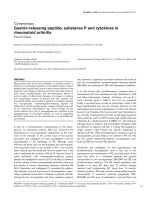
Figure 1 Checklist for Lung Ultrasound reports. Maximum mark per field was previously decided considering the number of parameters
requested.
Tutino et al. Scandinavian Journal of Trauma, Resuscitation and Emergency Medicine 2010, 18:44
/>Page 2 of 4
element of the template provided for reporting. A “ 0”
was given for any incomplete information or any miss-
ing field. Otherwise, a “1” was assigned if the parameter
wasconsideredsufficient(Figure1).Thesumofall
fields, from 0 to 24, was used to evaluate the internal
ICU learning curve trend.
Results
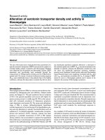
During the study period (April 2008-April 2009), a total
of 637 LUSs were performed, and the marks per month
(median) are shown in Figure 2. Multiple LUS per
patient were possible either for clinical investigation, for
devices positioning, or clinical follow-up.
Significant differences regarding quality standards of
LUS reporting between the first and the last month
were noticed, with a constant positive trend. The worst
and insufficient average vote was f ound in the first
month, when the bedside LUS implementation had just
started. To achieve sufficiency (median mark > = 15), 7
months were necessary, afterwards the standard
remained high. Once data colle ction was completed,
twelve LUS reports were randomly checked with the
same met hod in order to confirm the marks trend,
achieving a median result of 23.
The most common omissions i n LUS reporting con-
cerned three of the six considered echographic fields.
The description of pleural line, B-lines and pneu-
mothorax was generally adequate, whereas incomplete
reporting was commo n for diaphragm motility and lung
consolidations.
Diaphragm motility was often not evaluated with miss-
ing information about the quantification of the excursion.
Concerning consolidations and atelectasis, a precise
definition of their extensions and anatomical localization
was often lacking, compromising an adequat e follow-up
of the lesions.
Also bronchograms were incompletely described,
therefore the diagnosis of the nature of the consolida-
tion was often impossible. Finally, concerning pleural
effusion evaluation, the statement whether it was deter-
mined in supine or lateral position, was often l acking.
Nevertheless, using Balik’ s formula, the estimation of
pleural effusion was in good relation with the effective
drained volume (volume of effusion in millilitres equals
the distance b etween lung and posterior chest wall in
centimetres multiplied by 20) [5].
Discussion
Inourexperiencewehaveshownthattheaccuracyof
LUS description improves over time by using a preset
reportin g module. In this descriptive study, the lack of a
control group does not permit to evaluate the strength
of association between electronic sheet introduction and
LUS quality improvement. Moreover, in our clinical
practice LUS has been widely improved over time, mov-
ing from a procedure-related tool (mere wide to pleural
Figure 2 Monthly median of marks achieved during the study period.
Tutino et al. Scandinavian Journal of Trauma, Resuscitation and Emergency Medicine 2010, 18:44
/>Page 3 of 4
effusion draina ge) to a wider and more frequent clinical
examination method. Therefore, operators skills in LUS
execution, naturally improved as they ga ined experience.
The process of acquiring competency in ultrasound
examination was already described by Schlager and co-
workers in a study evaluating goal-directed ultrasound
in emergency department, where that accuracy
improved with gradually growing experience [6]. Kendall
and Shimp demonstrated that in focused bedside ultra-
sound exam (abdominal right upper quadrant), the sen-
sitivity of the exam was 100% after 25 exams performed
[7]. Although gaining competency in a skill over time is
a well recognized process, our study was aimed to inves-
tigate the quality of the reporting method, rather than to
assess the learning curve of LUS examination. We
believe that a complete LUS reporting should consider a
multitude of parameters and its clinical utility correlates
to accuracy of this diagnostic tool.
Considering the completeness of the reporting , with
the introduction of the standardized report sheet, we
report an increasing quality of the examinations during
the study period, as a prompt for operators to consider
all the parameters required for a complete LUS
reporting.
In the same way, the standardize sheet induced opera-
tors to obtain all the required images necessary for a
complete evaluation of the chest, therefore an adequate
follow-up was possible comparing images taken from
exams performed in sequence. Lack of proper images
easily result in missing pathology or mistaking artefacts
also in other fields of ultrasonography [8].
Although the scoring method we adopted is arbitrary
and far from being validated, it can be regarded as a
useful method to compare LUS examinations, an ever-
growing exam with a strong inter-operator variability.
Conclusions
The use of a standard report scheme for LUS can help
intensivists to improve completeness and accuracy level
of the examination reporting and it permits to follow
the clinical course of chest pathology in ICU patients.
Author details
1
Postgraduate School of Anaesthesia and Intensive Care, Faculty of Medicine,
University of Florence, Italy.
2
Anesthesia and Intensive Care Unit of
Emergency Department, Careggi Teaching Hospital, Florence, Italy.
Authors’ contributions
LT wrote the manuscript, participated in the coordination of the study and
took part in the internal teaching programme. GC and SB were the two
seniors involved in report judgement, they also coordinated the teaching
programme. FB coordinated the ICU ultrasound screening and coordinated,
with the help of RC, the electronic data collection of LUS data during the
study.
AP conceived the study, participated in its design and took part in the
educational program. All authors read and approved the final manuscript.
Competing interests
The authors declare that they have no competing interests.
Received: 20 January 2010 Accepted: 12 August 2010
Published: 12 August 2010
References
1. Arbelot C, Ferrari F, Bouhemad B, Rouby JJ: Lung ultrasound in acuote
respiratory distress syndrome and acute lung injury. Curr Opin Crit Care
2008, 14:70-74.
2. Peris A, Zagli G, Barbani F, Tutino L, Biondi S, di Valvasone S, Batacchi S,
Bonizzoli M, Spina R, Miniati M, Pappagallo S, Giovannini V, Gensini GF: The
value of lung ultrasound monitoring in H1N1 acute respiratory distress
syndrome. Anaesthesia 2009, 65:294-297.
3. Mayo PH, Beaulieu Y, Doelken P, Feller-Kopman D, Harrod C, Kaplan A,
Oropello J, Vieillard-Baron A, Axler O, Lichtenstein D, Maury E, Slama M,
Vignon P: American College of Chest Physicians/La Societe de
Reanimation de Langue Francaise statement on competence in critical
care ultrasonography. Chest 2009, 135:1050-1060.
4. Boddi M, Barbani F, Abbate R, Bonizzoli M, Batacchi S, Lucente E, Chiostri M,
Gensini GF, Peris A: Reduction in deep vein thrombosis incidence in
intensive care after a clinician education program. J Thromb Haemost
2009, 8:121-128.
5. Balik M, Plasil P, Waldauf P, Pazout J, Fric M, Otahal M, Pachl J: Ultrasound
estimation of volume of pleural fluid in mechanically ventilated patients.
Intensive Care Med 2006, 32:318-321.
6. Schlager D, Lazzareschi G, Whitten D, Sanders AB: A prospective study of
ultrasonography in the ED by emergency physicians. Am J Emerg Med
1994, 12:185-189.
7. Kendall JL, Shimp RJ: Performance and interpretation of focused right
upper quadrant ultrasound by emergency physicians. J Emerg Med 2001,
21:7-13.
8. Gaspari RJ, Dickman E, Blehar D: Learning curve of bedside ultrasound of
the gallbladder. J Emerg Med 2009, 37:51-56.
doi:10.1186/1757-7241-18-44
Cite this article as: Tutino et al.: Time needed to achieve completeness
and accuracy in bedside lung ultrasound reporting in Intensive Care
Unit. Scandinavian Journal of Trauma, Resuscitation and Emergency Medicine
2010 18:44.
Submit your next manuscript to BioMed Central
and take full advantage of:
• Convenient online submission
• Thorough peer review
• No space constraints or color figure charges
• Immediate publication on acceptance
• Inclusion in PubMed, CAS, Scopus and Google Scholar
• Research which is freely available for redistribution
Submit your manuscript at
www.biomedcentral.com/submit
Tutino et al. Scandinavian Journal of Trauma, Resuscitation and Emergency Medicine 2010, 18:44
/>Page 4 of 4