Báo cáo y học: " Clinical presentation of a traumatic cervical spine disc rupture in alpine sports: a case report" pdf
Bạn đang xem bản rút gọn của tài liệu. Xem và tải ngay bản đầy đủ của tài liệu tại đây (439.69 KB, 5 trang )
BioMed Central
Page 1 of 5
(page number not for citation purposes)
Scandinavian Journal of Trauma,
Resuscitation and Emergency Medicine
Open Access
Case report
Clinical presentation of a traumatic cervical spine disc rupture in
alpine sports: a case report
Timo M Ecker*
1,2
, Mark Kleinschmidt
†2
, Luca Martinolli
†1
,
Heinz Zimmermann
†1
and Aristomenis K Exadaktylos
†1
Address:
1
Department of Emergency Medicine, University of Bern, Inselspital Bern, 3010 Bern, Switzerland and
2
Department of Orthopaedic
Surgery, University of Bern, Inselspital Bern, 3010 Bern, Switzerland
Email: Timo M Ecker* - ; Mark Kleinschmidt - ; Luca Martinolli - ;
Heinz Zimmermann - ; Aristomenis K Exadaktylos -
* Corresponding author †Equal contributors
Abstract
Isolated non-skeletal injuries of the cervical spine are rare and frequently missed. Different
evaluation algorithms for C-spine injuries, such as the Canadian C-spine Rule have been proposed,
however with strong emphasis on excluding osseous lesions. Discoligamentary injuries may be
masked by unique clinical situations presenting to the emergency physician. We report on the case
of a 28-year-old patient being admitted to our emergency department after a snowboarding
accident, with an assumed hyperflexion injury of the cervical spine. During the initial clinical
encounter the only clinical finding the patient demonstrated, was a burning sensation in the palms
bilaterally. No neck pain could be elicited and the patient was not intoxicated and did not have
distracting injuries. Since the patient described a fall prevention attempt with both arms, a
peripheral nerve contusion was considered as a differential diagnosis. However, a high level of
suspicion and the use of sophisticated imaging (MRI and CT) of the cervical spine, ultimately led to
the diagnosis of a traumatic disc rupture at the C5/6 level. The patient was subsequently treated
with a ventral microdiscectomy with cage interposition and ventral plate stabilization at the C5/C6
level and could be discharged home with clearly improving symptoms and without further
complications.
This case underlines how clinical presentation and extent of injury can differ and it furthermore
points out, that injuries contracted during alpine snow sports need to be considered high velocity
injuries, thus putting the patient at risk for cervical spine trauma. In these patients, especially when
presenting with an unclear neurologic pattern, the emergency doctor needs to be alert and may
have to interpret rigid guidelines according to the situation. The importance of correctly using CT
and MRI according to both – standardized protocols and the patient's clinical presentation – is
crucial for exclusion of C-spine trauma.
Background
Isolated non-skeletal injuries of the cervical spine are rare
and among the most commonly missed injuries – with
serious implications for the patient and physician[1]. In a
cohort of 14,755 C-spine injuries in a level I trauma cen-
tre, Demetriades et al. showed that only 3.8% of the
Published: 12 November 2008
Scandinavian Journal of Trauma, Resuscitation and Emergency Medicine 2008, 16:14 doi:10.1186/1757-7241-16-14
Received: 21 September 2008
Accepted: 12 November 2008
This article is available from: />© 2008 Ecker et al; licensee BioMed Central Ltd.
This is an Open Access article distributed under the terms of the Creative Commons Attribution License ( />),
which permits unrestricted use, distribution, and reproduction in any medium, provided the original work is properly cited.
Scandinavian Journal of Trauma, Resuscitation and Emergency Medicine 2008, 16:14 />Page 2 of 5
(page number not for citation purposes)
patients suffered from an isolated spinal chord injury
without concomitant fracture or subluxation, of which
only 45.5% were diagnosed as a spinal chord injury ini-
tially[1]. Specific trauma mechanisms and collateral inju-
ries that are associated with a high incidence of skeletal C-
spine injuries have been described [2]. Different algo-
rithms for the initial assessment of these patients have
been proposed, such as the NEXUS low risk criteria, or the
Canadian C-spine rule[3,4]. In our institution we employ
the Canadian C-spine rule as a guideline for the applica-
tion of CT scans in trauma patients, since a study by Stiell
et al. has proven the Canadian C-spine rule to be superior
over the NEXUS criteria, especially in alert trauma
patients[5]. Additional radiographic examinations, such
as MRI, are important adjuncts in order to detect soft tis-
sue injuries. However, despite rigid recommendations,
emergency physicians might be challenged by situations
that are rather unusual and cannot be assessed with the
help of standardized scores or algorithms alone, but may
require an individualized approach.
This case report shows the discrepancy between patient
appearance and the extent of injury and at the same time
reflects the difficulty in decision making when algorithms
and guidelines are challenged by an unusual clinical pres-
entation.
Case report
We report the case of a 28-year-old female snowboarder
who suffered from a fall during a descent on a maintained
skiing slope. The exact mechanism of injury was not
reported, but a hyperflexion injury of the C-spine was
assumed. No loss of consciousness was reported. Initially
the patient started to hyperventilate and was calmed by
the layperson that provided the initial support. With the
arrival of the emergency physician on site, the patient had
a Glasgow Coma Scale (GCS) of 15 with stable hemody-
namics and was subsequently transferred to our emer-
gency department by helicopter. Upon arrival, the patient
was immobilized on a vacuum mattress; the C-spine was
stabilized with a Stifneck. Her GCS was 15 and primary
surveys ABCDE including log roll revealed no pathologic
findings. With stable vital signs, a secondary survey was
performed. Since the patient was fully alert without any
distracting injuries, and did not complain of any neck
pain, the Stifneck was opened. Careful examination of the
C-spine revealed no pain on palpation of the Proc. spi-
nosi. She could actively turn her head to more than 45
degrees bilaterally and lift the head in a supine position
without eliciting any neck pain. During the secondary sur-
vey we performed a complete neurologic exam according
to the ASIA criteria. Motor function was graded according
to the muscle strength scale with a score from 0 to 5 and
there were no pathologic findings. The deep tendon
reflexes of the upper and lower extremities bilaterally were
normal. The sensory examination including light touch,
vibration and pinprick, revealed a painful paraesthesia
bilaterally over the palms. Applied to dermatomes the
appropriate neurologic level was C6 and below. However,
we did not find a complete affection of the dermatome
representing the C6 level and neither of the dermatomes
below this level. In the absence of cervical pain, and
motor dysfunction, the underlying cause was not clear. As
a differential diagnoses to C-spine trauma, tingling and
paraesthesia as a consequence of the reported hyperventi-
lation, and a peripheral nerve contusion was considered.
The latter was taken into account, since she had attempted
to prevent her fall with both arms extended. Subse-
quently, in order to safely exclude a non-skeletal injury of
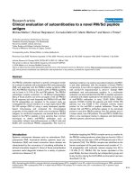
the spinal cord, we decided to perform an MRI. The
images showed a traumatic subligamentous rupture of the
intervertebral disc between C5 and C6 with ventral mye-
locompression (figure 1). The dorsal longitudinal liga-
ment was intact. There was no sign of paravertebral
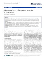
haematoma. Consecutively, an additional CT scan was
performed. The scan revealed a small teardrop fracture of
the ventral base plate of C5 in the paramedian line to the
left (Figure 2). The overall alignment was correct and there
was no sign of myelocompression from osseous struc-
tures, nor lesions of the posterior column or the facet
joints. We initiated treatment with a 30 mg/kg bolus injec-
tion of Methylprednisolone and a maintenance dose of
5,4 g/kg body weight and hour. The patient was trans-
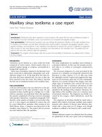
ferred to the intermediate care unit and had surgery the
next day. She underwent ventral microdiscectomy with
cage interposition and ventral plate stabilization at the
C5/C6 level (Figure 3). Postoperatively, the paraesthesia
resolved immediately. At the time of discharge three days
later, there was some residual burning and tingling, but
subjective improvement of the clinical symptoms. The
patient was discharged home without further complica-
tions.
Discussion and conclusion
This case reflects several important issues. First, it con-
firms the findings of Franz et al.[6], who proposed that
injury patterns of modern alpinists have shifted from inju-
ries of the extremities to a higher incidence of spinal inju-
ries. Due to the technical advances of hardware, as well as
altered and more radical slope designs, snowboarding and
skiing injuries have to be clearly considered high velocity
accidents. Thus, as a consequence, the importance of algo-
rithms such as the Canadian C-spine Rule has become
apparent. Stiell et al.[4] have shown the associated risk
between certain trauma mechanisms and the increased
incidence of spinal injuries and were able to formulate
important recommendations for application of CT diag-
nostics in such patients. We apply the Canadian C-spine
Rule as a gold standard in our emergency department,
since other algorithms such as the NEXUS criteria have
Scandinavian Journal of Trauma, Resuscitation and Emergency Medicine 2008, 16:14 />Page 3 of 5
(page number not for citation purposes)
shown to be less sensitive in the detection of injury in the
alert trauma patient [5]. Especially these patients however,
who are not obtunded but might have distracting injuries
or might be under the influence of sedatives or pain kill-
ers, need to be evaluated according to a reliable algorithm.
Second, this case reflects the discussion in the current lit-
erature on clinical and radiographic C-spine evaluation. It
seems clear that obtunded patients should be evaluated
according to the C-spine protocol with an initial CT scan.
Beyond this, it remains questionable which adjunct exam-
inations should be performed. It is evident that conven-
tional radiography is unreliable and not adequate for
diagnosis of C-spine injuries, especially for evaluation of
the cervico-thoracal junction [7]. Computed tomography
has been shown to be the gold standard for diagnosing
skeletal injury [8,9]. Stelfox et al. proved that discontinu-
ation of C-spine immobilization after a negative CT scan
is permitted and does not lead to further complica-
tions[8]. However, several authors are still discussing the
An MRI was obtained in the emergency department for detection of disco-ligamentous injuriesFigure 1
An MRI was obtained in the emergency department for detection of disco-ligamentous injuries. This figure
shows T2 weighted transversal and sagittal MRI images. The scan revealed a traumatic extradural rupture of the intervertebral
disc between C5 and C6 with ventral myelocompression but without disruption of the dorsal longitudinal ligament
The CT scan shows the small teardrop fracture at the ventral base plate of C5Figure 2
The CT scan shows the small teardrop fracture at the ventral base plate of C5. The ruptured disc cannot be clearly
identified.
Scandinavian Journal of Trauma, Resuscitation and Emergency Medicine 2008, 16:14 />Page 4 of 5
(page number not for citation purposes)
importance of MRI as an adjunct. Due to its superiority in
detecting disco-ligamentous injuries[3], it can be used as
an adjunct examination, especially when suspecting soft
tissue trauma [10-13]. The importance of MRI as an
adjunct becomes apparent in our case.
In the light of different available imaging methods, this
case also shows how clinical presentation and extent of
injury may not be clearly associated and how deceptive
the situation may appear to the emergency physician. The
clinical presentation in this case was rather unusual. A
patient with a cord injury typically has pain at the site of
the spinal injury. This may not always be a reliable feature
to exclude traumatic spinal cord injury (TSCI), since
patients with TSCI often have associated brain and sys-
temic injuries (eg, hemothorax, extremity fractures, intra-
abdominal injury) that may limit the patient's ability to
report localized pain. These also complicate the initial
evaluation and management of patients with TSCI, and
affect prognosis[14]. In this case however, we encoun-
tered a patient who was fully communicative and did not
have any distracting injury. The only apparent finding was
the persisting paraesthesia. The clinical presentation led
us to a hesitant use of a CT scan, even though a protocol
like the Canadian C-Spine rule recommends so. The indi-
cation to perform a primary MRI scan instead of a CT scan
was deemed appropriate in this situation, since osseous
lesions of the cervical spine were not assumed. The dis-
crepancy between clinical presentation and MRI finding
was impressive. Without the MRI and in the absence of a
clinically suspicious spine, the differential diagnosis of a
peripheral nerve injury might have been pursued further
and the actual injury might have been missed.
Patients after high velocity accidents with suspected cervi-
cal spine injuries need to be evaluated according to strict
protocols. The gold standard is the Canadian C-spine
Rule. Whereas computed tomography is the gold standard
for detections of skeletal injury, MRI as an adjunct is
important to exclude soft tissue trauma, especially in
symptomatic patients with an unsuspicious CT scan but
an unclear neurologic pattern. Sometimes the clinical sit-
uation may encourage the physician to improvise and
interpret guidelines to make an individual decision
regarding the best imaging method to reveal the patient's
pathology.
Abbreviations
C-spine: Cervical Spine; MRI: Magnet Resonance Tomog-
raphy; CT: Computed Tomography; TSCI: Traumatic Spi-
nal Chord Injury.
Consent
Written informed consent was obtained from the patient
for publication of this case report and any accompanying
images. A copy of the written consent is available for
review by the Editor-in-Chief of this journal.
Competing interests
The authors declare that they have no competing interests.
Authors' contributions
All authors have contributed equally and sufficiently to
the to conception, design and drafting and revision proc-
ess of this manuscript.
After identification of the injury the patient was transferred to the operating roomFigure 3
After identification of the injury the patient was transferred to the operating room. This figure shows the postop-
erative image after discectomy, cage interposition and ventral stabilization. The implants are in correct position.
Publish with BioMed Central and every
scientist can read your work free of charge
"BioMed Central will be the most significant development for
disseminating the results of biomedical research in our lifetime."
Sir Paul Nurse, Cancer Research UK
Your research papers will be:
available free of charge to the entire biomedical community
peer reviewed and published immediately upon acceptance
cited in PubMed and archived on PubMed Central
yours — you keep the copyright
Submit your manuscript here:
/>BioMedcentral
Scandinavian Journal of Trauma, Resuscitation and Emergency Medicine 2008, 16:14 />Page 5 of 5
(page number not for citation purposes)
References
1. Demetriades D, Charalambides K, Chahwan S, Hanpeter D, Alo K,
Velmahos G, Murray J, Asensio J: Nonskeletal cervical spine inju-
ries: epidemiology and diagnostic pitfalls. J Trauma 2000,
48(4):724-7.
2. Blackmore CC, Emerson SS, Mann FA, Koepsell TD: Cervical spine
imaging in patients with trauma: determination of fracture
risk to optimize use. Radiology 1999, 211(3):759-65.
3. Diaz JJ Jr, Aulino JM, Collier B, Roman C, May AK, Miller RS, Guillam-
ondegui O, Morris JA Jr: The early work-up for isolated liga-
mentous injury of the cervical spine: does computed
tomography scan have a role? J Trauma 2005, 59(4):897-903.
4. Stiell IG, Wells GA, Vandemheen KL, Clement CM, Lesiuk H, De
Maio VJ, Laupacis A, Schull M, McKnight RD, Verbeek R, Brison R,
Cass D, Dreyer J, Eisenhauer MA, Greenberg GH, MacPhail I, Morri-
son L, Reardon M, Worthington J: The Canadian C-spine rule for
radiography in alert and stable trauma patients. Jama 2001,
286(15):1841-8.
5. Stiell IG, Clement CM, McKnight RD, Brison R, Schull MJ, Rowe BH,
Worthington JR, Eisenhauer MA, Cass D, Greenberg G, MacPhail I,
Dreyer J, Lee JS, Bandiera G, Reardon M, Holroyd B, Lesiuk H, Wells
GA: The Canadian C-spine rule versus the NEXUS low-risk
criteria in patients with trauma. N Engl J Med 2003,
349(26):2510-8.
6. Franz T, Hasler RM, Benneker L, Zimmermann H, Siebenrock KA,
Exadaktylos AK: Severe spinal injuries in alpine skiing and
snowboarding: a 6-year review of a tertiary trauma centre
for the Bernese Alps ski resorts, Switzerland. Br J Sports Med
2008, 42(1):55-8.
7. Griffen MM, Frykberg ER, Kerwin AJ, Schinco MA, Tepas JJ, Rowe K,
Abboud J: Radiographic clearance of blunt cervical spine
injury: plain radiograph or computed tomography scan? J
Trauma 2003, 55(2):222-6.
8. Stelfox HT, Velmahos GC, Gettings E, Bigatello LM, Schmidt U:
Computed tomography for early and safe discontinuation of
cervical spine immobilization in obtunded multiply injured
patients. J Trauma 2007, 63(3):630-6.
9. Sanchez B, Waxman K, Jones T, Conner S, Chung R, Becerra S: Cer-
vical spine clearance in blunt trauma: evaluation of a com-
puted tomography-based protocol. J Trauma 2005,
59(1):179-83.
10. Stassen NA, Williams VA, Gestring ML, Cheng JD, Bankey PE: Mag-
netic resonance imaging in combination with helical com-
puted tomography provides a safe and efficient method of
cervical spine clearance in the obtunded trauma patient. J
Trauma 2006, 60(1):171-7.
11. Como JJ, Thompson MA, Anderson JS, Shah RR, Claridge JA, Yowler
CJ, Malangoni MA: Is magnetic resonance imaging essential in
clearing the cervical spine in obtunded patients with blunt
trauma? J Trauma 2007, 63(3):544-9.
12. Hogan GJ, Mirvis SE, Shanmuganathan K, Scalea TM: Exclusion of
unstable cervical spine injury in obtunded patients with blunt
trauma: is MR imaging needed when multi-detector row CT
findings are normal? Radiology 2005, 237(1):106-13.
13. Schuster R, Waxman K, Sanchez B, Becerra S, Chung R, Conner S,
Jones T: Magnetic resonance imaging is not needed to clear
cervical spines in blunt trauma patients with normal com-
puted tomographic results and no motor deficits. Arch Surg
2005, 140(8):762-6.
14. Sekhon LH, Fehlings MG: Epidemiology, demographics, and
pathophysiology of acute spinal cord injury. Spine 2001, 26(24
Suppl):S2-12.