Báo cáo y học: " Surgical management of abdominal compartment syndrome; indications and techniques" potx
Bạn đang xem bản rút gọn của tài liệu. Xem và tải ngay bản đầy đủ của tài liệu tại đây (325.43 KB, 5 trang )
BioMed Central
Page 1 of 5
(page number not for citation purposes)
Scandinavian Journal of Trauma,
Resuscitation and Emergency Medicine
Open Access
Review
Surgical management of abdominal compartment syndrome;
indications and techniques
Ari Leppäniemi
Address: Department of Surgery, Helsinki University Hospital, Haartmaninkatu 4, PO Box 340, 00029 HUS, Helsinki, Finland
Email: Ari Leppäniemi -
Abstract
The indications for surgical decompression of abdominal compartment syndrome (ACS) are not
clearly defined, but undoubtedly some patients benefit from it. In patients without recent abdominal
incisions, it can be achieved with full-thickness laparostomy (either midline, or transverse
subcostal) or through a subcutaneous linea alba fasciotomy. In spite of the improvement in
physiological variables and significant decrease in IAP, however, the effects of surgical
decompression on organ function and outcome are less clear. Because of the significant morbidity
associated with surgical decompression and the management of the ensuing open abdomen, more
research is needed to better define the appropriate indications and techniques for surgical
intervention.
Introduction
Sustained increase in intra-abdominal pressure (IAP)
defined as intra-abdominal hypertension (IAH) can with
ensuing onset of organ dysfunction lead to abdominal
compartment syndrome (ACS) [1,2]. If not timely treated,
it can lead to serious organ failures and even death.
Whether the cause is in the abdomen (primary ACS) or
elsewhere (secondary ACS), prevention is of utmost impor-
tance. Fluid resuscitation is a very important cause of IAH
when high volumes are necessary, such as in patients with
severe acute pancreatitis (SAP). In these patients, the exces-
sive use of crystalloids should be avoided.
The first line of treatment is always nonoperative [3].
Nasogastric decompression is useful in patients with gas-
tric dilatation or ileus. Short term use of neuromuscular
blocking agents has been used in appropriate circum-
stances. Removal of excessive fluids with diuretics can be
tried, but extracorporeal techniques, such as continuous
hemodiafiltration, are more effective in rapidly removing
excess fluid. Finally, percutaneous drainage of intraperito-
neal fluid collections is a simple and more effective way to
reduce intra-abdominal volume.
If nonoperative measures fail to relieve ACS, surgical
decompression should be considered. Due to the severe
morbidity associated with all forms of surgical decom-
pression, the indications, timing and technique used
should be carefully evaluated.
Indications for decompression
There is no uniform consensus on the indications for sur-
gical decompression in ACS. In addition to IAP values, the
cause, time-frame and possible need for further laparot-
omy should be considered. As a general rule, when non-
surgical interventions fail to turn around the progressive
deterioration of organ dysfunctions in the presence of ful-
minate ACS, surgical decompression is justified.
Published: 14 April 2009
Scandinavian Journal of Trauma, Resuscitation and Emergency Medicine 2009, 17:17 doi:10.1186/1757-7241-
17-17
Received: 19 February 2009
Accepted: 14 April 2009
This article is available from: />© 2009 Leppäniemi; licensee BioMed Central Ltd.
This is an Open Access article distributed under the terms of the Creative Commons Attribution License ( />),
which permits unrestricted use, distribution, and reproduction in any medium, provided the original work is properly cited.
Scandinavian Journal of Trauma, Resuscitation and Emergency Medicine 2009, 17:17 />Page 2 of 5
(page number not for citation purposes)
Timing of decompression
Clinical experience seems to indicate that early decom-
pression is more effective and associated with lower mor-
tality than delayed decompression performed after several
days after the onset of ACS, but no comparative studies
exist. Obviously, it depends on the time when ACS devel-
ops. Usually ACS is an early phenomenon and prompt
decompression without significant delay is appropriate.
However, if ACS is caused by a later event, such as infec-
tion of the peripancreatic necrosis in SAP, for example,
decompression at that stage is warranted and often com-
bined with necrosectomy. There are no studies on prophy-
lactic surgical decompression, but preventing ACS by
leaving the abdomen open in high risk patients, such as
patients undergoing damage control surgery for trauma or
patients operated for ruptured abdominal aortic aneu-
rysms, is sensible.
Techniques
When selecting the optimal technique for surgical decom-
pression, previous already existing recent abdominal inci-
sions (wounds) are preferable. If a patient has undergone
a midline laparotomy, utilizing the same incision for
decompression is obviously the best choice. In patients
with secondary ACS and particularly those without previ-
ous abdominal incisions, several options exist. The most
commonly used method for surgical decompression is the
midline laparostomy [4]. All layers (skin, fascia, perito-
neum) are divided through a vertical midline incision
extending from the xiphoideum to the pubis with a few
centimeters of fascia left intact at both ends to facilitate
subsequent closure or late reconstruction. Alternatively, a
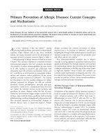
transverse bilaterally extended incision few centimeters
below the costal margins can be used to perform a full-
thickness laparostomy [5] (Fig. 1). A third method utilizes
three short horizontal skin incisions to perform a subcu-
taneous anterior abdominal fasciotomy at the linea alba
(SLAF) with the peritoneum left intact [6].
IAP decrease after full-thickness laparostomy
All techniques have been shown to reduce IAP. The major-
ity of evidence is based on experience from patients with
SAP. In a collective of analysis of 161 patients undergoing
surgical decompression via midline laparostomy, the
mean reported IAP before surgical decompression was
34.6 mmHg and fell to 15.5 mmHg after decompression
(p < 0.001) [4]. The experience with transverse laparos-
tomy is still scarce, but a case report showed the decrease
from 23 mmHg to 10 mmHg [5].
IAP decrease after fasciotomy
The original report of two patients utilizing the SLAF
method showed a decrease of IAP from 30 mmHg to 14
mmHg and 35 mmHg to 23 mmHg, respectively [6].
Another report showed a decrease from 27 mmHg to 11
mmHg [7]. In analyzing the first 10 patients with SAP
undergoing SLAF at the Meilahti hospital in Helsinki, the
mean preoperative IAP was 31 (range 23–45) mmHg and
fell to 20 (10–33) mmHg immediately postoperatively
with a mean decrease of 10 (2–17) mmHg [Leppäniemi A,
unpublished data]. The initial decompressive effect was
deemed sufficient in 7 patients, out of whom 2 developed
recurrent ACS and underwent completion laparostomy.
Selection of decompressive method
Because there are no randomized studies comparing dif-
ferent surgical decompression techniques, the selection of
the technique has to be individualized and based on com-
mon sense and weighing the pros and cons of each tech-
nique. The midline laparostomy is relatively safe, easy to
perform and nearly always effective. Although early com-
plications, such as intestinal fistulas, have been greatly
reduced with careful management and better understand-
ing of the open abdomen, there is a high risk of persistent
open abdomen requiring split-thickness skin grafting and
delayed reconstruction of the abdominal wall (planned
hernia approach). Recently, the use of temporary mesh to
facilitate gradual fascial closure has decreased the planned
hernia rate.
Transverse laparostomy seems to be effective, is little more
time-consuming and could have a higher rate of fascial
closure. In addition, same principles of managing the
open abdomen can be applied without additional equip-
ment. The major disadvantage is the loss of abdominal
and back extensor muscle functions, if fascial closure can
not be achieved. This would require complex reconstruc-
tion procedures including innervated free flaps that not
only restore continuity but also the functional integrity of
the abdomen [8].
Decompressive transverse laparostomyFigure 1
Decompressive transverse laparostomy.
Scandinavian Journal of Trauma, Resuscitation and Emergency Medicine 2009, 17:17 />Page 3 of 5
(page number not for citation purposes)
SLAF is effective in about 50–70%, prevents open abdo-
men and its related complications, but is always associ-
ated with a subsequent hernia. In the acute phase, the cost
effectiveness due to lesser need of nursing care and reop-
eration resources is a significant advantage.
Where to decompress
From a surgical technical point of view, the best place to
perform a decompressive operation is the operation thea-
tre. It provides adequate aseptic conditions, lighting,
equipment and personnel, and is ergonomically better for
the surgeon. However, in urgent situations, decompres-
sion can and sometimes should be performed in the
Intensive Care Unit (ICU). Even under less urgent condi-
tions, a surgical team with proper equipment can go the
ICU and perform the operation there without additional
risk to the patient. The benefit of not requiring the transfer
of a critically ill patient with multiple monitors and ongo-
ing medications is obvious, and it also saves operation
room time.
Effects of surgical decompression on organ
failure and outcome
In a collective review of 250 patients undergoing midline
laparostomy, decompression had an overall positive effect
on hemodynamic, respiratory and renal functions [4].
Central venous pressure and pulmonary artery pressure
decreased, most likely caused by the direct effect of the
decrease in IAP on the thoracic cavity. Cardiac function
improved in the majority of the patients. There was an
improvement in PaO
2
/FIO
2
ratio and decrease in peak air-
way pressure, but the respiratory function remained
severely impaired in most patients. Significant improve-
ment in the urinary output was observed in all but two
studies.
At our institution, among the 26 patients with SAP under-
going surgical decompression for ACS during the past 6
years, mostly a full-thickness midline laparostomy, there
was no significant difference between pre- and postopera-
tive organ dysfunctions score [Leppäniemi, unpublished
data]. The PaO
2
/FIO
2
ratio increased in 50% and
decreased in 50% of the patients. Daily urinary output
increased by >200 ml in 7 patients (27%), and 3 patients
avoided renal replacement therapy. The overall mortality
rate was 46% with preoperative renal failure (p = 0.045),
lower preoperative IAP (p = 0.039) and late (median 7
days) decompression (p = 0.005) being associated with
increased risk of death. It is noteworthy that all 8 patients
undergoing surgical decompression more than 3 days
post-admission died.
It seems that in most cases ACS is an early phenomenon
requiring prompt action. If nonoperative techniques fail
to provide significant decrease in the IAP and improve-
ment in organ functions, surgical decompression should
be performed without additional delay. As institutional
experience in the management of ACS increases, the delay
from onset of the problem to interventions tends to
decrease.
Complications of surgical decompression
Although postoperative bleeding and infection can occur
after any surgical procedure, they are rare after decompres-
sive laparostomy. Recurrent ACS can develop after too
eager closure attempts. However, the major source of mor-
bidity is associated with the management and complica-
tions of the open abdomen.
Management of the open abdomen
In view of the complicated and often fatal outcome of
patients ending up with a persistent open abdomen after
multiple reoperations, often with entero-atmospheric fis-
tulae and persistent infection, the "hostile abdomen" sce-
nario, extreme caution and care should be administered
when managing patients with open abdomen [9].
The ideal cover of the abdominal contents after leaving
the abdomen open should protect the viscera, avoid fistu-
las, be easy to apply and remove, allow easy nursing care,
should not damage the fascia or the skin, be readily avail-
able and inexpensive, and maintain the abdominal
domain. In addition, the preservation of the accessibility
to the abdominal cavity and the feasibility of gradual clo-
sure of the abdominal wall are important.
The easiest method to cover the abdominal viscera after
decompression is a plastic silo (Bogota bag) which is inex-
pensive, readily available and which preserves the intact
fascia when sutured to the skin edges. However, because
the plastic silo or some other form of temporary abdomi-
nal closure allows the fascial edges to retract laterally, the
abdominal cavity looses part of its volume resulting in dif-
ficult fascial closure under significant tension especially, if
the closure is delayed beyond the first week.
The vacuum pack introduced in 1995 utilizes a polyethyl-
ene sheet tucked between the parietal peritoneum and the
bowel, thus preventing the formation of adhesions
between the abdominal wall and the bowel [10]. In 2001
the commercial vacuum-assisted wound management
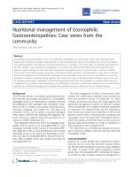
technique was introduced into everyday practise [11]. It
helps the nursing care, but it has not been shown to be
superior to various "self-made" applications used world-
wide [12] (Fig. 2). Nevertheless, even in the management
of the most severe complication of the open abdomen,
the exposed enteric fistula, vacuum-assisted wound man-
agement is able to control the fistula secretion allowing
the wound around it to heal [13,14].
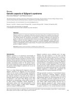
Recently, a technique combining vacuum-dressing and a
temporary mesh has been described [15]. It allows the
Scandinavian Journal of Trauma, Resuscitation and Emergency Medicine 2009, 17:17 />Page 4 of 5
(page number not for citation purposes)
gradual approximation of the fascial edges at every dress-
ing change and eventual removal of the mesh and primary
fascial closure (Fig. 3).
Primary fascial closure
Once the ACS has been treated, the most important aim is
to achieve primary fascial closure as soon as possible with-
out causing recurrent ACS or other complications associ-
ated with premature closure [16]. If the infection source
has been controlled and even if a relaparotomy might be
needed in the near future, every effort should be made to
achieve primary fascial closure during the initial hospital-
ization period and avoid the significant morbidity associ-
ated with leaving the abdomen open for delayed
reconstruction. Gradual fascial closure, often mesh-
assisted, seems to be the best currently available tech-
nique, but other possibilities, such as the components
separation technique at an early stage [17], or fascial clo-
sure with a mesh prosthesis can be considered under
favourable conditions (no infection, enough skin to cover
the prosthesis). However, if primary fascial closure is not
possible, an early decision to resort to the planned hernia
strategy is a good option.
Planned hernia
A planned hernia approach aims at skin coverage with
subsequent delayed abdominal wall reconstruction. The
skin closure is most often achieved with autologous split-
thickness skin grafting over the exposed bowel. Instead of
allowing the bowel surface to granulate before skin graft-
ing, early application of the skin graft over the bowel
seems to enhance the tuning down of the inflammatory
process sustained by the large raw surface, and to make
the subsequent reconstruction process easier.
The conditions favouring a planned hernia strategy
include the inability to reapproximate the retracted
abdominal wall edges, sizeable tissue loss, risk of tertiary
abdominal compartment syndrome, inadequate infection
source control, anterior enteric fistula and poor nutri-
tional status of the patient. One of the factors that must
also be taken into account is the type of the abdominal
wall defect, especially if it is the result of decompressive
laparostomy. A midline laparostomy covered with skin
graft is relatively easily tolerated, whereas one from trans-
verse laparostomy might cause serious disability due to
the loss of abdominal and back extensor muscle func-
tions, if fascial closure can not be achieved [6]. This might
require complex reconstruction procedures to restore not
only continuity but also the functional integrity of the
abdomen [5]. If abdominal decompression has been
achieved with the SLAF technique, the subsequent hernia
can be corrected in a standard fashion utilizing prosthetic
mesh or the components separation technique.
The maturation of the skin graft requires about 9–12
months, after which the grafted skin can be easily
removed from the bowel surface without additional iatro-
genic lesions. Large abdominal wall defects can be recon-
structed with pedicular or microvascular flaps. The most
commonly used is the tensor fascia lata (TFL)-flap [18].
With smaller defects, the components separation tech-
nique or a mesh repair can be also used for late repair pro-
vided that there is enough original skin for skin closure.
Conclusion
IAH is a recently popularized clinical entity that is easily
recognized, when suspected, using intravesicular meas-
urement of IAP. The clinically significant manifestation,
Covering transverse laparostomy with "self-made" negative pressure dressingFigure 2
Covering transverse laparostomy with "self-made"
negative pressure dressing.
Vacuum-dressing with temporary mesh being changedFigure 3
Vacuum-dressing with temporary mesh being
changed.
Publish with BioMed Central and every
scientist can read your work free of charge
"BioMed Central will be the most significant development for
disseminating the results of biomedical research in our lifetime."
Sir Paul Nurse, Cancer Research UK
Your research papers will be:
available free of charge to the entire biomedical community
peer reviewed and published immediately upon acceptance
cited in PubMed and archived on PubMed Central
yours — you keep the copyright
Submit your manuscript here:
/>BioMedcentral
Scandinavian Journal of Trauma, Resuscitation and Emergency Medicine 2009, 17:17 />Page 5 of 5
(page number not for citation purposes)
ACS, requires prompt attempts to reduce IAP starting with
nonoperative measures. If these are insufficient, surgical
decompression is warranted. The choice of the decom-
pressive technique must be individualized. If a full-thick-
ness laparostomy is performed, primary fascial closure
should be attempted during the initial hospitalization
period. If not possible, early resort to the planned hernia
strategy and subsequent abdominal wall reconstruction
are the best options provided that sufficient expertise is
available.
Competing interests
The author declares that they have no competing interests.
Consent section
Written informed consents were obtained from the
patients for publication of this review and accompanying
images. Copies of the written consents are available for
review by the Editor-in-Chief of this journal.
References
1. Malbrain MLNG, Cheatham ML, Kirkpatrick A, Sugrue M, Parr M, de
Waele J, Balogh Z, Leppäniemi A, Olvera C, Ivatury R, D'Amours S,
Wendon J, Hillman K, Johansson K, Kolkman K, Wilmer A: Results
from International Conference of Experts on intra-abdomi-
nal hypertension and abdominal compartment syndrome. I.
Definitions. Intensive Care Med 2006, 32:1722-1732.
2. Cheatham ML, Malbrain MLNG, Kirkpatrick A, Sugrue M, Parr M, De
Waele J, Balogh Z, Leppäniemi A, Olvera C, Ivatury R, D'Amours S,
Wendon J, Hillman K, Wilmer A: Results from International
Conference of Experts on intra-abdominal hypertension and
abdominal compartment syndrome. II. Recommendations.
Intensive Care Med 2007, 33:951-962.
3. De laet I, Malbrain MLNG: ICU management of the patient with
intra-abdominal hypertension: what to do, when and to
whom? Acta Clin Belg Suppl 2007:190-199.
4. De Waele JJ, Hoste EA, Malbrain ML: Decompressive laparotomy
for abdominal compartment syndrome – a critical analysis.
Crit Care 2006, 10:R51.
5. Leppäniemi A, Mentula P, Hienonen P, Kemppainen E: Transverse
laparostomy is feasible and effective in the treatment of
abdominal compartment syndrome in severe acute pancre-
atitis. World J Emerg Surg 2008, 3:6.
6. Leppäniemi A, Hienonen P, Siren J, Kuitunen A, Lindström O, Kemp-
painen E: Treatment of abdominal compartment syndrome
with subcutaneous anterior abdominal fasciotomy in severe
acute pancreatitis. World J Surg 2006, 30:1922-1924.
7. Cheatham M, Fowler J, Pappas P: Subcutaneous linea alba fasci-
otomy: a less morbid treatment for abdominal compart-
ment syndrome. Am Surg 2008, 74:746-749.
8. Pushpakumar SB, Wilhelmi BJ, van-Aalst VC, Banis JC Jr, Barker JH:
Abdominal wall reconstruction in a trauma setting. Eur J
Trauma Emerg Surg 2007, 33:3-13.
9. Leppäniemi A: The hostile abdomen – a systematic approach
to a complex problem. Scand J Surg 2008, 97:218-219.
10. Brock WB, Barker DE, Burns RP: Temporary closure of open
abdominal wounds: the vacuum pack. Am Surg 1995, 61:
30-35.
11. Garner GB, Ware DN, Cocanour CS, Duke JH, McKinley BA, Kozar
RA, Moore FA: Vacuum-assisted wound closure provides early
fascial reapproximation in trauma patients with open abdo-
mens. Am J Surg 2001, 182:630-638.
12. Leppäniemi A: Open abdomen after severe acute pancreatitis.
Eur J Trauma Emerg Surg 2008, 34:17-23.
13. Erdmann D, Drye C, Heller L, Wong MS, Levin SL: Abdominal wall
defects and enterocutaneous fistula treatment with the Vac-
uum Assisted Closure (V.A.C.) system. Plast Reconstr Surg 2001,
108:2066-2068.
14. Becker HP, Willms A, Schwab R: Small bowel fistulas and the
open abdomen. Scand J Surg 2007, 96:263-271.
15. Petersson U, Acosta S, Björck M: Vacuum-assisted wound clo-
sure and mesh-mediated fascial traction – a novel technique
for late closure of the open abdomen. World J Surg 2007,
31:2133-2137.
16. Scott BG, Feanny MA, Hirshberg A: Early definitive closure of the
open abdomen: a quiet revolution. Scand J Surg 2005, 94:9-14.
17. Ramirez OM, Ruas E, Dellon AL: Components separation
method for closure of abdominal-wall defects: and anatomic
and clinical study. Plast Reconstr Surg 1990, 86:519-526.
18. Lyle WG, Gibbs M, Howdieshell TR: The tensor fascia lata free
flap in staged abdominal wall reconstruction after traumatic
evisceration. J Trauma 1999, 46:519-522.