Báo cáo y học: " Rigid fibrescope Bonfils: use in simulated difficult airway by novices" pptx
Bạn đang xem bản rút gọn của tài liệu. Xem và tải ngay bản đầy đủ của tài liệu tại đây (986.87 KB, 7 trang )
BioMed Central
Page 1 of 7
(page number not for citation purposes)
Scandinavian Journal of Trauma,
Resuscitation and Emergency Medicine
Open Access
Original research
Rigid fibrescope Bonfils: use in simulated difficult airway by novices
Tim Piepho
†1
, Rüdiger R Noppens
†1
, Florian Heid
1
, Christian Werner
1
and
Andreas R Thierbach*
2
Address:
1
University Medical Center of the Johannes Gutenberg-University, Department of Anaesthesiology, Langenbeckstr. 1, Mainz, 55131,
Germany and
2
Dept. of Anaesthesia, Intensive Care Medicine, Emergency Medicine and Pain Therapy Klinikum Idar-Oberstein Dr Ottmar-Kohler-
Str. 2 Idar-Oberstein, 55743, Germany
Email: Tim Piepho - ; Rüdiger R Noppens - ; Florian Heid - ;
Christian Werner - ; Andreas R Thierbach* -
* Corresponding author †Equal contributors
Abstract
Background: The Bonfils intubation fibrescope is a promising alternative device for securing the
airway. We examined the success rate of intubation and the ease of use in standardized simulated
difficult airway scenarios by physicians. We compared the Bonfils to a classical laryngoscope with
Macintosh blade.
Methods: 30 physicians untrained in the use of rigid fibrescopes but experienced in airway
management performed endotracheal intubation in an airway manikin (SimMan, Laerdal, Kent, UK)
with three different airway conditions. We evaluated the success rate using the Bonfils (Karl Storz,
Tuttlingen, Germany) or the Macintosh laryngoscope, the time needed for securing the airway, and
subjective rating of both techniques.
Results: In normal airway all intubations were successful using laryngoscope (100%) vs. 82% using
the Bonfils (p < 0.05). In the scenario "tongue oedema" success rate using the Macintosh
laryngoscope was 67% and 83% using the Bonfils. In the scenario "decreased cervical range of
motion with jaw trismus", success rate using the Macintosh laryngoscope was 84% vs. 76%. In
difficult airway scenarios time until airway was secured did not differ between the two devices. Use
of Bonfils was rated "easier" in both difficult airway scenarios.
Conclusion: The Bonfils can be successfully used by physicians unfamiliar with this technique in an
airway manikin. The airway could be secured with at least the same success rate as using a
Macintosh laryngoscope in difficult airway scenarios. Use of the Bonfils did not delay intubation in
the presence of a difficult airway. These results indicate that intensive special training is advised to
use the Bonfils effectively in airway management.
Background
Securing the airway with an endotracheal tube (ET) and
inflated cuff offers several benefits in the treatment of
anaesthetized patients or those in critical condition.
Potential benefits include protection against aspiration,
application of facilitated assisted or controlled mechani-
cal ventilation and positive end expiratory pressure [1].
Despite recent advances in airway management, the laryn-
goscope blade as described by Macintosh (commonly
Published: 22 July 2009
Scandinavian Journal of Trauma, Resuscitation and Emergency Medicine 2009, 17:33 doi:10.1186/1757-7241-17-33
Received: 27 April 2009
Accepted: 22 July 2009
This article is available from: />© 2009 Piepho et al; licensee BioMed Central Ltd.
This is an Open Access article distributed under the terms of the Creative Commons Attribution License ( />),
which permits unrestricted use, distribution, and reproduction in any medium, provided the original work is properly cited.
Scandinavian Journal of Trauma, Resuscitation and Emergency Medicine 2009, 17:33 />Page 2 of 7
(page number not for citation purposes)
referred to as Macintosh blade) remains the so-called
"gold standard" for endotracheal intubation [2].
However, endotracheal intubation is occasionally difficult
or impossible with a consecutive increasing morbidity
and mortality unless oxygenation is immediately estab-
lished [3]. In out-of hospital situations the use of an alter-
native device for endotracheal intubation is required by
emergency physicians in 0.9 to 2.6% of all cases [4-6].
Supraglottic airway devices are generally accepted as alter-
natives in cases of failed endotracheal intubation. How-
ever, endotracheal intubation has additional advantages
and supraglottic airways may fail as well [7-9].
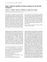
The Bonfils intubation fibrescope (Karl Storz GmbH &
Co. KG, Tuttlingen, Germany; figure 1) is an alternative to
direct laryngoscopy using the Macintosh blade with a
potential benefit especially in difficult airway situations
[10]. The device has a long thin rigid cylindrical body with
a curved tip. A tracheal tube is loaded onto the shaft of the
instrument and is pushed into a locking mechanism. After
lateralized insertion of the Bonfils into the mouth a jaw
thrust maneuver may be used to increase the view into the
retropharyngeal space [11]. The rigid fibrescope is
advanced into the glottic aperture and the tracheal tube is
inserted into the trachea after identifying the vocal cords.
The device is not advanced through the vocal cords at any-
time.
Recent reports demonstrated that the Bonfils is applicable
for tracheal intubation in patients with normal and
expected difficult airway [11,12].
The aim of the study was to compare the success rates of
intubation of physicians inexperienced in the use of a
rigid fibrescope vs. orotracheal intubation performed with
help of Macintosh blade in simulated difficult airway sce-
narios. We hypothesised that physicians inexperienced in
the use of a rigid fibrescope succeed orotracheal intuba-
tion in simulated difficult airway scenarios.
Methods
Thirty physicians with at least one year clinical experience
in anaesthesiology consented to participate in this study.
Prior to the study, each participant completed a question-
naire documenting previous experience with the Macin-
tosh laryngoscope and the Bonfils. All 30 participants had
performed a minimum of 100 endotracheal intubations
each (range 125 to over 2000) at the time of the investiga-
tion using the Macintosh laryngoscope. Only two physi-
cians had prior experience of using the Bonfils in two
cases each. Each physician was given a standardized dem-
onstration of the Bonfils by one of the investigators: After
a theoretically description of the use, the intubation pro-
cedure was practically demonstrated by one of the investi-
gators. The participants did not practice with the
instrument prior to the investigation. The Bonfils was
used without a camera attached and an extra video screen.
View during endotracheal intubation was obtained via the
eyepiece of the instrument.
The sequence in which the participants used the devices
was randomized. All endotracheal intubations were per-
formed using a standard Magill 8.0 mm tracheal tube
(Rüsch, Kernen i.R., Germany) in a Laerdal SimMan man-
ikin (Laerdal, Kent, UK).
The participants were exposed to three different airway
scenarios in a randomized order: 1. normal airway, 2.
tongue oedema and 3. decreased cervical range of motion
with jaw trismus.
The primary endpoints were the success rate of endotra-
cheal intubation and the required time to perform this.
Endotracheal intubation attempt without interposed
mask ventilation should not exceed 30–40 s [1]. There-
fore, failed intubation was defined as an attempt in which
endotracheal intubation was not successful or required
more than 40 s to perform.
Time was measured from insertion of the blade or the
Bonfils until the ET was placed and the first ventilation
with a self-inflating bag was successfully performed.
At the end of each scenario, every participant rated the
ease of use of the devices on a visual analogue scale (from
0 = extremely difficult to 10 = extremely easy).
The Bonfils Intubation Fibrescope (Karl Storz GmbH & Co. KG, Tuttlingen, Germany)Figure 1
The Bonfils Intubation Fibrescope (Karl Storz GmbH
& Co. KG, Tuttlingen, Germany).
Scandinavian Journal of Trauma, Resuscitation and Emergency Medicine 2009, 17:33 />Page 3 of 7
(page number not for citation purposes)
Statistical Analyses
Data are reported as mean ± SEM. Duration of endotra-
cheal intubation was analyzed using one-way analyses of
variance and post hoc Bonferroni test. Nonparametric
data were analyzed by one-way analyses of variance on
ranks (GraphPad Prism version 5.00 for Mac, GraphPad
Software, San Diego California USA). Data for successful
endotracheal intubation were analyzed using the X
2
Test.
Differences were considered statistically significant at an
error probability < 0.05.
Results
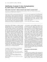
Normal airway
All physicians successfully intubated the trachea using the
Macintosh laryngoscope in the normal airway situation.
In contrast, 82% (25/30) of intubation attempts using the
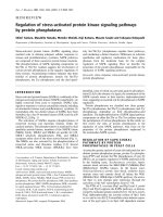
Bonfils succeeded (p < 0.05) (figure 2). The duration of
the intubation attempts using the Bonfils was longer com-
pared to the Macintosh laryngoscope (p < 0.001) (figure
3). The participants rated the use of the Macintosh laryn-
goscope as less difficult in this scenario (p < 0.01) (figure
4).
Tongue oedema
In this scenario, 20/30 attempts (67%) to intubate the tra-
chea using the Macintosh blade were successful. The suc-
cess rate using the Bonfils was 83% (25/30) (p > 0.05;
figure 2). The duration of tracheal intubation was similar
with both devices. In the tongue oedema scenario, physi-
cians ranked the Bonfils easier to use compared to the
Macintosh laryngoscope (p < 0.001).
In comparison to the normal airway scenario, the time
needed for intubating the trachea using the Macintosh
laryngoscope was longer (p < 0.05) (figure 3) and rated
more difficult (p = 0.001). The use of the Bonfils was rated
less difficult (p < 0.05) in this difficult airway scenario
(figure 4).
Decreased cervical range of motion with jaw trismus
No difference was noted in the number of successful
attempts (84% vs. 76%) (figure 2) and their duration (fig-
ure 3) using the Macintosh laryngoscope and the Bonfils,
respectively. The time needed to intubate the trachea
using the laryngoscope was longer compared to intuba-
tion with normal airway (p < 0.05, figure 3). In this sce-
Successful intubation for different airway scenariosFigure 2
Successful intubation for different airway scenarios.
Scandinavian Journal of Trauma, Resuscitation and Emergency Medicine 2009, 17:33 />Page 4 of 7
(page number not for citation purposes)
nario, the Bonfils was rated easier to use compared to the
Macintosh laryngoscope (figure 4; p = 0.001).
Within the groups, the use of the Bonfils was rated easier
(p < 0.05) and the use of the Macintosh laryngoscope
more difficult (p = 0.001, figure 4) in comparison to the
normal airway scenario.
Discussion
The Bonfils rigid fibrescope is a device that is designed to
assist in establishing an airway when direct laryngoscopy
is difficult or fails. Bein described a high success rate after
in-hospital failed direct laryngoscopy in trained users
[13]. But until now, the learning curve of the Bonfils is not
well evaluated. Halligan studied the learning curve of the
Bonfils in two anaesthesiologists. His results suggest that
a performer becomes proficient after 20 to 25 intubations
[14]. According to Byhahn, 10 intubations supervised by
an instructor seem to prove effective [10]. We show that
physicians untrained in the use of the device have poor
rate of successful intubations with first attempt in simu-
lated normal airway situations. However, the success rate
is similar between simulated normal and difficult airway
situations. Our data suggest that the Bonfils might be an
alternative device in the management of difficult intuba-
tion with at least the same rate of successful intubations
than the Macintosh laryngoscope.
Various anatomical or pathologic conditions contribute
to difficult airway management situations. Therefore, we
simulated two potentially difficult airway scenarios. The
simulation of different intubation scenarios has been
widely used for similar studies in the past [15-17].
Although manikin studies have proven a reliable surro-
gate in the clinical context, study settings cannot entirely
simulate the conditions of an emergency airway manage-
ment scenario. The Bonfils requires a minimal retropha-
ryngeal space to allow orientation according to
anatomical landmarks of the fibrescope under direct
vision. In humans it is necessary to create a sufficient ret-
ropharyngeal space to successfully visualize the vocal
cords with the Bonfils fiberscope. This can be achieved
with either a jaw thrust manoeuvre, with a laryngoscope
blade or by lifting the base of the tongue with the index
Time until airway was secured using Bonfils and Macintosh laryngoscope in different airway scenariosFigure 3
Time until airway was secured using Bonfils and Macintosh laryngoscope in different airway scenarios.
Scandinavian Journal of Trauma, Resuscitation and Emergency Medicine 2009, 17:33 />Page 5 of 7
(page number not for citation purposes)
finger of the non-operating hand. Quick and sufficient
endotracheal intubation is almost impossible if a retro-
pharyngeal space is not created using one of these tech-
niques.
A wide variation of results have been described between
different manikins when airway devices have been tested
[18]. While the SimMan's airway is generally considered
very realistic, endotracheal intubation in any kind of man-
ikin in general seems to be easier than in a real patient
[19]. One reason is that tongue of the manikin is rigid and
not as flaccid as in unconscious patients. Therefore a suf-
ficient retropharyngeal space is present and it is not neces-
sary to use a special technique while using the Bonfils in a
manikin setting.
A typical situation which can lead to a difficult endotra-
cheal intubation is the tongue oedema. In this scenario
handling and use of the Bonfils was rated to be easier than
the more common Macintosh laryngoscope. Although all
physicians were skilled in the use of the Macintosh laryn-
goscope the failure rate was – from a patient's point of
view – unacceptably high. Although the failure rate
between both devices was statistically not significant, it is
of clinical relevance. Despite their experience and famili-
arity with the Macintosh laryngoscope, the participants
favoured the Bonfils. Due to the randomized order of the
scenarios a potential learning curve using the Bonfils was
not considered. The advantage of the Bonfils in the tongue
oedema scenario is its small diameter enabling it to be
manoeuvred even in minimal space. In theory a space of
the outer diameter of the laryngeal tube seems to be ade-
quate to perform successful intubation. Furthermore, the
picture from its tip is being transfered to the physician's
eye. Direct visualization of the glottis – a prerequisite
using direct laryngoscopy with a Macintosh laryngoscope
– is not required.
An attached camera with a monitor system allows an eas-
ier and less time consuming endotracheal intubation,
especially in untrained physicians. However, endotra-
cheal intubation was performed in this study by using the
eyepiece of the Bonfils. A major limitation of the video
unit is its portability. Therefore, the use of a video system
in prehospital emergency medicine and emergency intu-
bation can not be suggested for routine use.
The cervical spine has to be immobilized by a rigid collar
until a potential injury has been ruled out in the hospital.
The limited mouth opening and the missing neck exten-
Rating of both techniques in the three scenariosFigure 4
Rating of both techniques in the three scenarios. 10 = extremely easy, 0 = extremely difficult.
Scandinavian Journal of Trauma, Resuscitation and Emergency Medicine 2009, 17:33 />Page 6 of 7
(page number not for citation purposes)
sion is associated with a Cormack and Lehane grade 3 and
4 view [20]. We used a decreased cervical range of motion
with jaw trismus in the manikin to simulate the airway sit-
uation of trauma patients with a rigid collar. No difference
was evident between both devices in respect to the failure
rate and duration of the attempts. However, the partici-
pants of this study favoured the Bonfils. It is likely that the
main reason for this finding is that endotracheal intuba-
tion using the Bonfils can be achieved using less strength
and without any cervical spine movement. This potential
benefit of the Bonfils is in accordance with previously
published manuscripts [21,22]. Several cases of successful
pre-hospital endotracheal intubation using the Bonfils in
patients with an immobilized cervical spine exist [10]. The
small diameter of the Bonfils is in this scenario the advan-
tage, too. It requires only minimal mouth opening to
advance the tip into the pharynx [23].
The use of a flexible intubation fibrescope is a common
device to secure the difficult airway [24]. However, the
insertion of an intubation fibrescope is more difficult and
time consuming in patients with immobilized cervical
spine than in the normal patient and in emergency situa-
tions the endotracheal intubation attempt without inter-
posed mask ventilation should not exceed 30–40 s [1].
Though the failure rate of the Bonfils was less compared
to the Macintosh laryngoscope in the decreased cervical
range of motion with jaw trismus scenario, we do not rec-
ommend using the Bonfils in cases of difficult airway
management without extensive training and clinical rou-
tine. As the success of emergency airway management is
strongly influenced by the level of training and expertise
with the device used, it should be suitable for use in daily
practice.
Conclusion
Our data suggests that in comparison to the Macintosh
laryngoscope the use of the Bonfils in a normal airway is
more difficult and time consuming at least if the physi-
cians are not experienced with the Bonfils device. These
data indicate that physicians must undergo an intensive
training of normal and difficult airway management sce-
narios to improve rate of successfully securing the airway
in these critical situations.
Competing interests
Karl Storz GmbH & Co. KG, Tuttlingen, Germany, pro-
vided the fiberscope used in this study. The Departments
of Anaesthesiology in Mainz and Idar-Oberstein are sup-
ported by Karl Storz GmbH & Co. KG, Tuttlingen, Ger-
many.
Authors' contributions
TP and RRN contributed equally to this article. TP has
made substantial contributions to conception, acquisition
of data and drafting the article. RN has made substantial
contributions to analysis, interpretation of data and in
drafting the article. FH has made substantial contribu-
tions to analysis and interpretation of data. CW has made
substantial contributions to conception and revised the
manuscript critically for important intellectual content.
AT has made substantial contributions to conception,
acquisition of data and revised the manuscript. All
authors read and approved the manuscript.
Acknowledgements
We would like to thank all of the participants, who participated in this
study.
References
1. Thierbach AR: Advanced prehospital airway management
techniques. Eur J Emerg Med 2002, 9:298-302.
2. Macintosh R: A new laryngoscope. Lancet 1943, i:205.
3. Mort TC: The incidence and risk factors for cardiac arrest
during emergency tracheal intubation: a justification for
incorporating the ASA Guidelines in the remote location. J
Clin Anesth 2004, 16:508-516.
4. Adnet F, Jouriles NJ, Le Toumelin P, Hennequin B, Taillandier C,
Rayeh F, Couvreur J, Nougiere B, Nadiras P, Ladka A, Fleury M: Sur-
vey of out-of-hospital emergency intubations in the French
prehospital medical system: a multicenter study. Ann Emerg
Med 1998, 32:454-460.
5. Thierbach A, Piepho T, Wolcke B, Kuster S, Dick W: [Prehospital
emergency airway management procedures. Success rates
and complications]. Anaesthesist 2004, 53:543-550.
6. Orliaguet G, Tartiere S, Lejay M: A prospectivein-field evaluation
of endotracheal intubation byemergency medical services
physicians. J Eur Urgences 1997, 1:27-32.
7. Jolliffe L, Jackson I: Airway management in the outpatient set-
ting: new devices and techniques. Curr Opin Anaesthesiol 2008,
21:719-722.
8. Nolan JP, Soar J: Airway techniques and ventilation strategies.
Curr Opin Crit Care 2008, 14:279-286.
9. Wiese CH, Semmel T, Muller JU, Bahr J, Ocker H, Graf BM: The use
of the laryngeal tube disposable (LT-D) by paramedics dur-
ing out-of-hospital resuscitation-an observational study con-
cerning ERC guidelines 2005. Resuscitation 2009, 80:194-198.
10. Byhahn C, Meininger D, Walcher F, Hofstetter C, Zwissler B: Pre-
hospital emergency endotracheal intubation using the Bon-
fils intubation fiberscope. Eur J Emerg Med 2007, 14:43-46.
11. Halligan M, Charters P: A clinical evaluation of the Bonfils Intu-
bation Fibrescope. Anaesthesia 2003, 58:1087-1091.
12. Bein B, Worthmann F, Scholz J, Brinkmann F, Tonner PH, Steinfath M,
Dorges V: A comparison of the intubating laryngeal mask air-
way and the Bonfils intubation fibrescope in patients with
predicted difficult airways. Anaesthesia 2004, 59:668-674.
13. Bein B, Yan M, Tonner PH, Scholz J, Steinfath M, Dorges V: Tracheal
intubation using the Bonfils intubation fibrescope after failed
direct laryngoscopy. Anaesthesia 2004, 59:1207-1209.
14. Halligan M, Charters P: Learning curve for the Bonfils intuba-
tion fibrescope. Br J Anaesth 2003, 90:826P.
15. Doerges V, Sauer C, Ocker H, Wenzel V, Schmucker P: Airway
management during cardiopulmonary resuscitation – a com-
parative study of bag-valve-mask, laryngeal mask airway and
combitube in a bench model. Resuscitation 1999, 41:63-69.
16. Russi CS, Wilcox CL, House HR: The laryngeal tube device: a
simple and timely adjunct to airway management. Am J Emerg
Med 2007, 25:263-267.
17. Wackett A, Anderson K, Thode H: Bullard laryngoscopy by naive
operators in the cervical spine immobilized patient. J Emerg
Med 2005, 29:253-257.
Publish with BioMed Central and every
scientist can read your work free of charge
"BioMed Central will be the most significant development for
disseminating the results of biomedical research in our lifetime."
Sir Paul Nurse, Cancer Research UK
Your research papers will be:
available free of charge to the entire biomedical community
peer reviewed and published immediately upon acceptance
cited in PubMed and archived on PubMed Central
yours — you keep the copyright
Submit your manuscript here:
/>BioMedcentral
Scandinavian Journal of Trauma, Resuscitation and Emergency Medicine 2009, 17:33 />Page 7 of 7
(page number not for citation purposes)
18. Jordan GM, Silsby J, Bayley G, Cook TM, Difficult Airway Society:
Evaluation of four manikins as simulators for teaching airway
management procedures specified in the Difficult Airway
Society guidelines, and other advanced airway skills. Anaes-
thesia 2007, 62:708-712.
19. Hesselfeldt R, Kristensen MS, Rasmussen LS: Evaluation of the air-
way of the SimMan full-scale patient simulator. Acta Anaesthe-
siol Scand 2005, 49:1339-1345.
20. Heath KJ: The effect of laryngoscopy of different cervical spine
immobilisation techniques. Anaesthesia 1994, 49:843-845.
21. Wahlen BM, Gercek E: Three-dimensional cervical spine move-
ment during intubation using the Macintosh and Bullard
laryngoscopes, the bonfils fibrescope and the intubating
laryngeal mask airway. Eur J Anaesthesiol 2004, 21:907-913.
22. Rudolph C, Schneider JP, Wallenborn J, Schaffranietz L: Movement
of the upper cervical spine during laryngoscopy: a compari-
son of the Bonfils intubation fibrescope and the Macintosh
laryngoscope. Anaesthesia 2005, 60:668-672.
23. Rudolph C, Schlender M: [Clinical experiences with fiber optic
intubation with the Bonfils intubation fiberscope]. Anaesthe-
siol Reanim 1996, 21:127-130.
24. Benumof JL: Management of the difficult adult airway. With
special emphasis on awake tracheal intubation. Anesthesiology
1991, 75:1087-1110.