Báo cáo y học: " Right-sided "trapdoor" incision provides necessary exposure of complex cervicothoracic vascular injury: a case repor" potx
Bạn đang xem bản rút gọn của tài liệu. Xem và tải ngay bản đầy đủ của tài liệu tại đây (1.93 MB, 3 trang )
BioMed Central
Page 1 of 3
(page number not for citation purposes)
Scandinavian Journal of Trauma,
Resuscitation and Emergency Medicine
Open Access
Case report
Right-sided "trapdoor" incision provides necessary exposure of
complex cervicothoracic vascular injury: a case report
Boris Kessel*
1
, Itamar Ashkenazi
2
, Isaak Portnoy
3
, Dan Hebron
4
, Dani Eilam
2
and Ricardo Alfici
2
Address:
1
Trauma Unit, Hillel Yaffe Medical Center, Hadera, Israel,
2
Surgery B Department, Hillel Yaffe Medical Center, Hadera, Israel,
3
Vascular
Surgery Department, Hillel Yaffe Medical Center, Hadera, Israel and
4
Interventional Radiology Unit, Hillel Yaffe Medical Center, Hadera, Israel
Email: Boris Kessel* - ; Itamar Ashkenazi - ; Isaak Portnoy - ;
Dan Hebron - ; Dani Eilam - ; Ricardo Alfici -
* Corresponding author
Abstract
Combined cervicothoracical vascular traumas are very uncommon, mostly resulting from
penetrating injuries. These injuries are accompanied with very high morbidity and mortality rates.
In this manuscript we present a case of hemodinamycally unstable trauma patient whose major
injury was penetrating trauma of both cervical and mediastinal major vessels. The standard surgical
approach of median sternotomy and neck incision was insufficient, and the patient's instability
forced the authors to improvise previously not described right-sided trap-door thoracomy.
Incorporation of such incision in the surgical arsenal may be very effective in selective cases
Introduction
Combined cervicothoracical vascular traumas are uncom-
mon, mostly resulting from high energy penetrating inju-
ries [1,2]. These injuries are diagnostically and
therapeutically challenging even for a very experienced
multidisciplinary trauma team. Patients suffering from
hemodynamic instability are taken immediately to the
operating room. In presence of the right sided vascular
injuries, the common surgical approach is via a median
sternotomy [3]. Combination of anterolateral thoracot-
omy, partial sternotomy and left infra or supraclavicular
incision described as "trap-door" thoracotomy is rarely
performed since it is time-consuming and results in mul-
tiple fractures [4],. We present here a case of hemodi-
namycally unstable trauma patient whose major injury
was penetrating trauma of both cervical and major medi-
astinal vessels. An improvised right sided "trap-door" tho-
racotomy was necessary to achieve vascular control and
reconstruction.
Case description
A twenty eight years white male was admitted to the
trauma resuscitation area following a gunshot assault. On
admission the patient was agitated. Vital signs revealed:
blood pressure of 120/65, heart rate of 110 per minute
and oxygen saturation of 94% on oxygen mask; and respi-
ratory rate of 20 per minute. Physical exam revealed an
entry wound located at the posterior aspect of the right
shoulder. The exit wound was located at the left side of the
neck, posterior to left sternocleidomastoid muscle. A large
right upper chest wall hematoma, extended to the neck
was found which was not pulsating. Pulse on both carotid
arteries was intact. There was no active bleeding from both
entry and exit wounds. The right upper extremity was pale
and swollen, with no palpable pulse. Breath sounds were
Published: 24 September 2009
Scandinavian Journal of Trauma, Resuscitation and Emergency Medicine 2009, 17:46 doi:10.1186/1757-7241-
17-46
Received: 4 July 2009
Accepted: 24 September 2009
This article is available from: />© 2009 Kessel et al; licensee BioMed Central Ltd.
This is an Open Access article distributed under the terms of the Creative Commons Attribution License ( />),
which permits unrestricted use, distribution, and reproduction in any medium, provided the original work is properly cited.
Scandinavian Journal of Trauma, Resuscitation and Emergency Medicine 2009, 17:46 />Page 2 of 3
(page number not for citation purposes)
equal and of good intensity bilaterally. The rest on physi-
cal examination was unremarkable. The patient was
treated with immediately intubation and mechanical ven-
tilation. Intravenous bolus of crystalloids was started.
Portable chest x-ray in the trauma room revealed no pneu-
mothorax.
During the initial stay in the trauma resuscitation area, the
patient became hemodynamically unstable. Despite fluid
administration, he developed tachycardia, up to 136 per
minute, and blood pressure dropped to 80/44. The cervi-
cal hematoma seemed to be increasing in size. Following
this deterioration, the patient was immediately taken to
the operating room.
On surgery, due to clinical impression of injury to the dis-
tal subclavian artery, a right supraclavicular incision was
performed first. Following incision of the platysma and
division of the right sternocleidomastoid muscle, signifi-
cant hemorrhage appeared in the surgical field that was
temporally controlled by direct digit pressure application.
Recognizing this to be hemorrhage possibly arising from
major vessels in Zone I of the neck, a full mid sternotomy
was performed to allow proper exposure and vascular
control. However, even following sternotomy, the athletic
habitus of the patient did not allow delineation and
approach to the major sources of bleeding. The incision
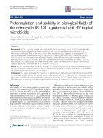
was extended by a right anterolateral thoracotomy (Fig 1),
performed through the third intercostal space. This right-
sided "trapdoor incision" allowed adequate exposure and
proximal control of the mediastinal vessels. Tears of both
the right common carotid artery and the right innominate
artery were found at their confluence. The right jugular
vein was injured as well. Repair of the arterial injury was
achieved by placing a graft patch modified from a collagen
coated knitted polyester vascular prosthesis (Silver Graft,
Datascope, Montvale, USA). The vein was repaired by pri-
mary suture of the tear. At this stage of operation the
patient was hypothermic (34°Celsius) and there was clin-
ical evidence of coagulopathy. We decided not to con-
tinue with the exploration of the distal subclavian artery.
The operation was promptly terminated by packing the
neck and upper mediastinum, followed by temporary clo-
sure of the wounds. Overall, the operation lasted 2 hours
and 47 minutes.
The patient was transferred to the postoperative recovery
unit where he was rewarmed and resuscitated with blood
and fresh frozen plasma. Following 4 hours, he was
hemodynamically stable but still dependent on fluids.
Coagulation tests returned towards normal values. The
right upper extremity was ischemic. At this point, he was
transferred to the angiography suite. We assumed that the
patient had an injury of subclavian artery which was not
dealt with during the initial operation. Via a right groin
approach, neck vessels and selective right subclavian
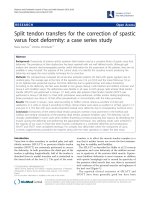
artery angiography was performed." This revealed massive
extravasation from to tear located at the distal part of the
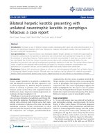
right subclavian artery (Fig 2). Three Fluency covered
stents (Bard Corporate, Murray Hill, NJ) were placed and
no signs of contrast extravasation were demonstrated after
the procedure(Fig 3). Blood flow to right upper extremity
was restored.
The patient was transferred to the Intensive Care Unit.
Twenty four hours later he was reoperated. He underwent
depacking of the neck and upper mediastinum and the
trap-door incision was closed in the usual fashion. At
postoperative day three he was extubated and two days
later he was transferred to the surgical ward. Due to accu-
mulation of pleural blood which would not drain follow-
Displays how the incision was extended by a right anterola-teral thoracotomy, performed through the third intercostal spaceFigure 1
Displays how the incision was extended by a right
anterolateral thoracotomy, performed through the
third intercostal space.
This figure illustrates massive extravasation from to tear located at the distal part of the right subclavian arteryFigure 2
This figure illustrates massive extravasation from to
tear located at the distal part of the right subclavian
artery.
Scandinavian Journal of Trauma, Resuscitation and Emergency Medicine 2009, 17:46 />Page 3 of 3
(page number not for citation purposes)
ing reinsertion of a chest tube, a video-assisted
thoracoscopy was performed at postoperative day ten and
significant amounts of blood clots were evacuated." The
rest of hospital stay was uneventful and the patient was
discharged home after three weeks.
Discussion
Penetrating injuries of the thoracic great vessels are associ-
ated with high morbidity and mortality. Many patients
die on the scene from massive hemorrhage. Mortality is
significant even in patients who survive the initial period
of injury and are alive on their admission to the hospital.
Demetriades et al. report overall mortality of 34.2% in
patients suffering from subclavian and axillary artery inju-
ries [5]. Patients with innominate artery injury usually do
not survive to arrive at the hospital. There are only few
case reports that describe patients who were treated for
combined common carotid and innominate artery inju-
ries [6,7]. In these, the injury was located to the left
hemithorax, unlike our patient in whom these vessels
were injured on the right side.
Management of major vascular injuries in the base of the
neck is complex. If the patient is stable, the first diagnostic
step should be cervical and chest CT-angiography. CT ang-
iography provides the necessary information regarding
the spectrum of vascular, mediastinal and other injuries.
This information is crucial, allowing proper decision mak-
ing concerning the therapeutic plan. In the hemodynami-
cally stable patient, significant vascular injury may be
treated with endovascular stenting.
If the patient is hemodynamically unstable, he/she should
be taken immediately for surgery. The selection of the
incision depends on mediastinal structures that need to
be explored during the surgery. In the case of a clinical sus-
picion for right common carotid artery injury, the oblique
incision along the anterior border of the sternocleidomas-
toid muscle should be performed [8], with extension to
median sternotomy, if proximal control of the injury is
difficult. This surgical exposure usually provides excellent
approach to other injuries of the innominate and com-
mon carotid artery. In our patient, this proved to be insuf-
ficient. The patient instability forced us to improvise a
right "trap-door" thoracomy. Defined by some as being
"obsolete"[5] this incision facilitated to achieve fast con-
trol of bleeding in this patient.
Conclusion
In selective cases median sternotomy does not provide
adequate exposure of the mediastinal great vessels. Incor-
poration of right sided trapdoor thoracotomy may be very
efficient in complex cervicothoracic trauma.
Consent
Written informed consent was obtained from the patient
for publication of this case report and accompanying
images.
Competing interests
The authors declare that they have no competing interests.
Authors' contributions
BK was the case manager and was the main writer to draft
the manuscript. IA and DH helped draft the manuscript
and added significant revisions. IP, DE and RA read the
manuscript and added significant revisions. All authors
discussed the details of the case, implications of the case
and commented on the manuscript at all stages. All
authors read and approved the final manuscript.
References
1. George SM, Croce MA, Fabian TC, Manqiante Ec, Kudsk KA, Voeller GR,
Pate JW: Cervicothoracic arterial injuries: recommendations for
diagnosis and treatment. Worlg J Surg 1991, 15(1):134-139.
2. Johnston RH, Wall MJ, Mattox KL: Innominate artery trauma: a
thirty years experience. J Vasc Surg 1993, 17(1):134-140.
3. Mattox KL, Wall MJ, LeMaire SA: Injury to the thoracic great ves-
sels. 4th edition. Edited by: Mattox KL, Feliciano DV, Moore EE. New
York: McGraw-Hill; 2000:559-582.
4. Boffard K: Manual of Definitive Surgical Trauma Care, Second
Edition. International Association for Trauma Surgery and Intensive Care.
Hodder Arnold. Part 4. Specific Organ Injury, page 90 2007.
5. Demetriades D, Chahwan S, Gomez H, Peng R, Velhamos G, Murray
J, Asensio J, Bongard F: Penetrating injuries to the subclavian
and axillary vessels. J Am Coll Surg 1999, 188(3):290-295.
6. Shin D, Wall M, Mattox K: Combined penetrating injury of the
innominate artery, left common carotid artery, trachea and
esophagus. J Trauma 2000, 49:780-783.
7. Snelleman JA, Tadros T, Lugt A van der, Bogers AJ: Traumatic rup-
ture of the innominate and left common carotid artery: case
report. J Trauma 2002, 52(3):571-572.
8. Feliciano DV: Management of penetrating injuries to carotid
artery. World J Surg 2001, 25:1028-1035.
No signs of contrast extravasation were demonstrated after the procedureFigure 3
No signs of contrast extravasation were demon-
strated after the procedure.