Pacing Options in the Adult Patient with Congenital Heart Disease - part 7 potx
Bạn đang xem bản rút gọn của tài liệu. Xem và tải ngay bản đầy đủ của tài liệu tại đây (418.16 KB, 15 trang )
CHAPTER 19
Ebstein’s anomaly
Ebstein’s anomaly, which represents <1% of all congenital heart diseases,
is of clinical importance to pacemaker physicians because many of the
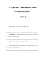
patients, who reach adulthood, develop bradyarrhythmias (Figure 19.1)
[189]. The disorder is characterized by downward displacement of the tri-
cuspid valve orifice so that the cusps, with the exception of the medial
Figure 19.1 Schematic of the apical displacement of the tricuspid valve seen in Ebstein’s
anomaly. This results in a superior “atrialized” portion of the right ventricle, which is
associated with atrial pressure, yet ventricular intracardiac electrogram recordings (broken
circle).
81
82 Chapter 19
two-thirds of the anterior cusp, originate from the right ventricular wall
rather than from the tricuspid annulus [190, 191]. This displacement may
be as low as the junction of the inflow and the outflow portions of the right
ventricle. The displaced tricuspid valve divides the right ventricle into two
parts:
• The atrialized portion which lies between the origin of the normal
tricuspid annulus and the displaced tricuspid orifice.
• The remainder of the true right ventricle, which lies beyond the tricuspid
valve.
The size of the atrialized portion of right ventricle varies greatly and
its walls may be fibrous and paper thin or muscular and well formed.
The tricuspid valve tissue is almost always redundant, wrinkled and the
chordae tendineae poorly developed or absent. The actual valve orifice is
generally smaller than normal and is usually incompetent [190,191].
Cardiac arrhythmias and conduction disturbances occur in 20–25% of
patients with Ebstein’s anomaly [192, 193], and commonly involve pre-
excitation syndromes. Pacemaker therapy is necessary in about 3–4% [194].
The indications for pacemaker therapy include persistent atrial stand-
still [195], atrio-ventricular block which can be de novo [196, 197], post
surgery [198] or following radiofrequency ablation [199]. Due to the mor-
phologic abnormalities in Ebstein’s anomaly, endocardial ventricular lead
placement can be challenging and in those patients who have not had
surgery, there are three pacing options [200].
Pre-valve
The atrialized portion of the right ventricle, which behaves hemodynamic-
ally like the right atrium, has electrophysiologic characteristics of the right
ventricle. This can be used to advantage to position a transvenous ventricu-
lar lead pre-valve in order to pace the right ventricle [201]. By avoiding
crossing the tricuspid valve, lead-induced exacerbation of tricuspid regur-
gitation can be prevented. In its severe form, the muscle in this pre-valve
cul de sac may be poorly formed or absent resulting in a thin aneurysmal
wall and ventricular pacing may therefore require a high voltage or the area
may be electrically silent. If successful, the chest radiograph will show the
ventricular lead tip to be in a postero-medial location (Figure 19.2) and
the ECG reveals right ventricular pacing with a left bundle branch block
configuration and a leftward or superior axis, confirming that the cathode
lies against right ventricular endocardium at the most inferior aspect of
the cardiac silhouette (Figure 19.3).
Ebstein’s anomaly 83
PA RAO
Figure 19.2 Ebstein’s anomaly (pre valve). Two chest cine fluoroscopic views;
Postero-anterior (PA) and right anterior oblique (RAO) showing dual chamber pacing in a
patient with Ebstein’s anomaly. The passive-fixation atrial lead lies in the atrial appendage.
The passive-fixation right ventricular lead lies in the atrialized right ventricular cul de sac
and the RAO view confirms that the tip is against the posterior wall. This pre valve position
was confirmed by echocardiography and pacing thresholds were satisfactory although the
patient was not pacemaker dependent.
DDD AV delay 90 ms
1
11
111
aVR
aVL
aVF
V1
V2
V3
V4
V5
V6
DDD AV delay 90 ms
1
11
111
aVR
aVL
aVF
V1
V2
V3
V4
V5
V6
Figure 19.3 Ebstein’s anomaly (pre valve). Resting 12-lead ECG from the same patient in
Figure 19.2, demonstrating dual chamber pacing. Because of normal atrioventricular
conduction, the atrioventricular delay was programmed to a short 90 ms to force ventricular
pacing. The QRS configuration confirms right ventricular pacing with a superior or
leftward axis.
Post-valve
Although technically difficult, it may be possible, particularly with milder
forms of Ebstein’s anomaly, to pass a transvenous ventricular lead through
84 Chapter 19
PA L Lat
Figure 19.4 Ebstein’s anomaly (post valve). Chest radiographs, postero-anterior (PA) and
left lateral (L Lat), showing dual chamber pacing in a patient with Ebstein’s anomaly. There
is a passive-fixation lead in the right atrial appendage. The ventricular passive-fixation lead
has been positioned at the apex of the true right ventricle beyond the malpositioned
tricuspid valve. In the L Lat view the ventricular lead lies anterior. The lead position was
confirmed by echocardiography.
I aVR V1 V4
II aVL V2 V5
III aVF V3 V6
II
Figure 19.5 Ebstein’s anomaly (post valve). Resting 12-lead ECG from the same patient in
Figure 19.4, demonstrating dual chamber pacing. The pacemaker has been programmed
DOO (to demonstrate consistent pacing). The features are identical to normal pacing from
the apex of the right ventricle; left bundle branch block and left or superior axis.
the displaced tricuspid orifice and then angle the electrode towards the
apex of the right ventricle (Figure 19.4). The ECG would again show a left
bundle branch block appearance with a superior leftwardaxis(Figure 19.5).
When attempting to position a lead post valve, an active-fixation screw-in
Ebstein’s anomaly 85
I aVR V1 V4
II aVL V2 V5
III aVF V3 V6
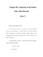
Figure 19.6 Ebstein’s anomaly (cardiac vein). Resting 12-lead ECG demonstrating dual
chamber pacing. The ventricular lead lies in the middle or a low lateral cardiac vein. There is
atrial sensing and ventricular pacing with a right bundle branch block and a normal axis.
This suggests left ventricular pacing from a site above the apex.
lead would be preferable, particularly if the lead can be anchored in the out-
flow tract. An active-fixation lead would also be preferable in the presence
of tricuspid regurgitation. It is likely that the newly developed steerable
catheter will be helpful in placing thin active-fixation leads (Figure 7.5).
Cardiac venous system
When the atrialized right ventricle is unsuitable or it is impossible to tra-
verse the tricuspid orifice, ventricular pacing from the cardiac venous
system may be an option. It may be possible to position a standard tined
lead in one of the cardiac veins. The surface electrocardiogram shows left
ventricular epicardial pacing with a right bundle branch block appear-
ance and the axis will depend on where the lead lies. For instance, if the
lead lies low in the heart such as in the middle or lateral cardiac vein,
then there will be a leftward axis similar to pacing from the cardiac apex
(Figure 19.6). In this position, the lateral chest x-ray helps confirm the car-
diac venous pacing by demonstrating the characteristic posterior position
of the ventricular lead tip (Figure 19.7). Generally leads placed inadvert-
ently in this position provide stable pacing with low thresholds as long
as the lead is gently wedged into a small venous tributary. It may be also
possible to position a lead on the lateral left ventricular epicardial wall
via the upper portion of the coronary sinus similar to the left ventricular
leads of biventricular pacing. Although this would provide physiologic
left ventricular pacing, nevertheless, in pacemaker-dependent patients,
it would not be regarded as safe or desirable, because of the potential
complications discussed earlier.
86 Chapter 19
L Lat
PA
Figure 19.7 Ebstein’s anomaly (cardiac vein). Chest radiographs, postero-anterior (PA) and
left lateral (L Lat), showing dual chamber pacing in a patient with Ebstein’s anomaly (same
patient as in Figure 19.6). There is a passive-fixation lead in the right atrial appendage. The
ventricular passive-fixation lead bypasses the malpositioned tricuspid valve by entering the
coronary sinus and a cardiac vein which is most likely a low lateral cardiac vein (black
arrow). In the L Lat view, the ventricular lead lies posterior which confirms that the lead is
not in the right ventricle (black arrow). In this case the lead position was not confirmed by
transthoracic echocardiography because of poor views. The patient refused a
transesophageal echocardiograph.
Despite the variety of options available for transvenous lead place-
ment, most patients have in the past received ventricular epicardial and
epimyocardial leads [194]. This will be discussed further in the section on
post-operative Ebstein’s anomaly.
SECTION C
Previous corrective or palliative
cardiac surgery
CHAPTER 20
D-Transposition of the great vessels
An infant born with dextro or D-transposition of the great vessels belongs
to one of the cyanotic heart lesion categories and rarely survives the first
year of life without surgical intervention [202]. The condition entails a
normal embryogenic rightward cardiac loop, but failure of the great ves-
sels to completely rotate. Therefore, the aorta arises from a normal right
ventricle and, concomitantly, a pulmonary artery arises from the left vent-
ricle. A venous-arterial circulation exists in parallel rather than in series
with desaturated venous blood recirculating through the systemic circu-
lation without first entering into the pulmonary circulation (Figure 20.1).
Without some type of communication such as a patent ductus arteriosus
or atrial or ventricular septal defects, infants succumb to cyanosis. The
adult pacemaker and ICD implanter would therefore never encounter a
patient with D-transposition of the great vessels who had not undergone
corrective surgery.
Two physiologically related surgeries to correct the cyanosis are per-
formed in the young: the Senning, and the more commonly used Mustard
procedures; redirect venous blood by removal of the atrial septum fol-
lowed by the insertion of an intra-atrial baffle (Figure 20.2). The baffle
creates a physiologic blood flow redistribution in which the desaturated
venous peripheral blood is directed to the mitral valve and left ventricle,
which then exits to the transposed pulmonary artery. Saturated pulmon-
ary venous blood is concomitantly directed to the right atrium, tricuspid
valve, right ventricle and leaves the heart via the aorta. Often the infant may
have first had an atrial septal defect created or a balloon atrial septostomyto
acutely allow for mixing of arterial and venous blood to stabilize acid–base
balance.
The Mustard procedure, which was first reported from Canada in 1964,
uses atrial pericardium or synthetic materials as the intra-atrial baffle [203].
Soon after this atrial switch procedure was first described, there were
a number of reports of early and late atrial arrhythmias and sudden
89
90 Chapter 20
Figure 20.1 Schematic of transposition of the great vessels in the dextro or “D” position
caused by a normal rightward looping of the embryogenic cardiac tube but an arrest of the
normal rotation of the great vessels. As a result, there is atrio-ventricular concordance (atria
and ventricles are normally joined), but ventriculo-arterial discordance (ventricles and great
vessels are not correctly joined). As a consequence, the right-sided ventricle ejects
desaturated venous blood back into the aorta and the left-sided ventricle ejects saturated
pulmonary venous blood back to the pulmonary artery. Unless corrected, the condition is
incompatible with life.
death, which diminished with a change in surgical technique particularly
in regard to cannulation, suturing of the baffle and protection of the
sinus node.
Although the survival rate for the Mustard procedure is generally
regarded as good [204], long-term results continue to show a high incid-
ence of sick sinus syndrome with brady and tachycardias and in particular
junctional, rhythm and atrial tachyarrhythmias [205–208]. At least some of
the sudden deaths were believed to be related to junctional rhythm, atrial
flutter or complete heart block [209–212], particularly during exercise as a
result of ventricular arrhythmias and therefore not prevented by cardiac
pacing [213].
The Senning operation for correction of D-transposition of the great ves-
sels was described before the Mustard procedure [214]. In this operation,
the systemic and pulmonary venous return are rerouted using flaps of
the atrial septum and right atrial free wall. Although no foreign mater-
ial is used, the early results were not particularly good and consequently
very few reports of this operation were published after the introduction of
the Mustard procedure [215, 216]. Because of the extensive atrial surgery
D-Transposition of the great vessels 91
Ventricular lead
Atrial lead
Baffle
Figure 20.2 D-transposition of the great vessels and lead placement. Schematic of the
intra-atrial baffle associated with the Mustard and Senning procedures. After removal of the
inter-atrial septum, the baffle allows desaturated vena caval venous blood to be directed to
the left atrium and ventricle which then permits flow into the pulmonary artery. Saturated
pulmonary venous return is directed to the right atrium, right ventricle and aorta. Atrial and
ventricular pacing leads have been positioned. Both pass behind the baffle at the junction of
the right atrium with the superior vena cava (broken line). The atrial lead must then be
positioned on the roof of the left atrium, whereas the ventricular lead traverses the mitral
valve to the lateral wall of the left ventricle.
required, it is not surprising that sinus node damage was frequent and
that there was a high incidence of atrial arrhythmias and late mortality,
comparable to the Mustard procedure [217].
At the present time, there still exists a significant subset of adult patients
who have undergone a palliative Mustard or Senning procedure. Some
of these patients, who reach adulthood had associated ventricular septal
defects and developed irreversible pulmonary hypertension. The atrial
baffle improves mixing and increases systemic saturations to 70–85%.
However, the ventricular defect remains open. Such patients are typically
treated with aspirin. If pacing is required, addition of a transvenous pacing
system will require coumadin therapy to prevent thromboembolic events.
If ventricular pacing is necessary, the presence of a persistent ventricular
septal defect will limit lead placement. For instance left ventricular septal or
outflow tract pacing, which may be desirable to preserve right ventricular
function, will not be possible.
As the young recipients of either procedure entered adulthood,
more and more required cardiac pacing to alleviate the symptoms of
92 Chapter 20
PA L LatPA L Lat
Figure 20.3 Mustard procedure for D-transposition of the great vessels. Chest radiographs,
postero-anterior (PA) and left lateral (L Lat), showing left atrial pacing in a patient with the
Mustard procedure. In the PA view an active-fixation lead has been passed from the right
subclavicular region to the superior vena cava and the stub of the right atrium. It then
proceeds behind the baffle into the anatomical left atrium, where it is attached to the roof. In
the L Lat view, the lead lies posterior which is very different to the traditional right atrial
appendage position. In this case, intermittent left phrenic nerve stimulation occurred.
bradyarrhythmias and tachyarrhythmias. With the gradual introduction
of the technically demanding but more physiologic, great vessel or arterial
switch procedure originally described by Dr Jatene in the late 1970s [218],
the atrial baffle procedures were slowly abandoned. Because the Jatene or
arterial switch operation does not involve the atria, sinus node dysfunc-
tion does not usually occur. However, the surgery does involve excision
and reimplantation of coronary arteries. Therefore, there is some risk to
sinus or AV nodal arteries and on occasion a pacemaker or ICD may be
required. If so, then it should be regarded as a routine procedure, with nor-
mal ventricular orientation and configuration. However, in those patients
who first underwent a palliative balloon atrial septostomy to stabilize acid-
base balance prior to definitive cardiac repair, an atrial defect patch may
have been used to close the defect. The presence of such material may
complicate atrial lead insertion, especially if Bachmann’s bundle pacing is
anticipated.
To the uninitiated implanter, insertion of a pacemaker or ICD in a
patient with D-transposition of the great vessels corrected with either the
Senning or Mustard procedures, conjures up a frightening scenario. Once
the anatomy is understood, the actual implantation is very straightfor-
ward. However, for a successful outcome, a number of factors must be
considered.
D-Transposition of the great vessels 93
PA L Lat
Figure 20.4 Mustard procedure for transposition of the great vessels. Chest radiographs,
postero-anterior (PA) and left lateral (L Lat), showing dual chamber pacing in a patient with
the Mustard procedure. In the PA view, an active-fixation lead is attached to the roof of the
left atrium. The active-fixation ventricular lead passes behind the baffle and through the
anatomical mitral valve into the body of the left ventricle and is attached to the lateral wall. In
the L Lat view, both leads lie posterior. In this case, both leads caused left phrenic nerve
stimulation with high voltage output pacing.
Provided the pathway is clear, the atrial lead progresses through the
superior vena cava and the stub of the right atrium and proceeds behind
the baffle into the anatomical left atrium (Figures 20.2–20.4). A straight
steroid-eluting screw-in lead with a curved stylet should be used and the
lead positioned as medial (right) as possible to prevent phrenic nerve
stimulation, which unfortunately is a common post implant complica-
tion [209,219]. In this situation, the steerable stylet (Locator
®
Model 4036,
St Jude Medical, Minneapolis, Mn.) is extremely useful (Figure 7.1). The
lead can be very easily placed into the anatomic left atrium using a stand-
ard curved stylet and then positioned medially on the roof, well away from
the phrenic nerve, using the maximum curve of the Locator
®
. Because of
the sharp curve required, the lead may not be adequately secured to the
left atrial wall. Withdrawing the Locator
®
stylet with a modest curve still
present will provide enough stress on the distal end to dislodge the lead if
it isn’t adequately secured (Figures 20.5, 20.6). The recent introduction of
the steerable catheter delivery system (Figure 7.5) has also facilitated lead
implant in the left atrium by permitting a more acute angle to reach the
roof of the left atrium (Figure 20.7).
One of the major issues with pacemaker or ICD implantation is the
presence of venous or baffle stenosis hindering the transvenous path-
way to the left ventricle [220, 221]. Obstruction to the intra-atrial baffle,
94 Chapter 20
LAO
PA
LAO
PA
Figure 20.5 Mustard procedure for transposition of the great vessels. Chest cine
fluoroscopic 40
◦
left anterior oblique (LAO) and postero-anterior (PA) views, showing dual
chamber pacing in a patient with the Mustard procedure. In both views, the upper
active-fixation lead is attached to the roof of the left atrium, but unlike in Figures 20.3 and
20.4, the attachment is arched more medial, well away from the left phrenic nerve. The left
ventricular lead is attached to the body of the chamber and testing revealed no
diaphragmatic stimulation with both leads.
PA RAO
LAO
PA RAO
LAO
Figure 20.6 Mustard procedure for transposition of the great vessels. Chest cine
fluoroscopic 28
◦
left anterior oblique (LAO), postero-anterior (PA) and 28
◦
right anterior
oblique (RAO) views, showing dual chamber pacing in a patient with the Mustard procedure.
In all views, the upper active-fixation lead is attached to the roof of the left atrium, but unlike
in Figures 20.3 and 20.4, the attachment is arched more medial, well away from the left
phrenic nerve. There are two active-fixation leads in the left ventricular chamber. The lower
one is not functioning.
typically at the superior vena cava junction due to patient growth and
angulation, can occur in about 22% of patients, many years after the
Mustard or Senning procedures [222]. Unfortunately, such obstructions
may not be clinically evident as the azygous vein often serves to decom-
press the stenosis by permitting venous runoff to the inferior vena caval
system. The more posterior location of the azygous vein and vena cava
also preclude effective echocardiographic/Doppler evaluations. When
D-Transposition of the great vessels 95
PA
Figure 20.7 Mustard procedure for transposition of the great vessels. Chest cine
fluoroscopic, postero-anterior (PA) view demonstrating the use of the SelectSite
™
catheter
delivery system through the superior vena cava baffle, facilitating lead placement at the
medial location on the roof of the venous left atrium. The acute curvature of the delivery
catheter allows selective lead placement in previously technically difficult to reach locations.
PA
Azygous vein
Baffle obstruction
Figure 20.8 Mustard procedure for transposition of the great vessels. Chest cine
fluoroscopic, postero-anterior (PA) view of a superior vena caval venogram illustrating a
dilated posterior directed azygous vein associated with superior vena cava baffle
obstruction. Venous blood flow eventually communicates with the inferior vena cava.
such a situation is suspected, investigations such as magnetic resonance
imaging, computerized tomographic scans or venograms are required
to delineate the venous anatomy prior to attempted pacemaker implant
(Figure 20.8).