Pacing Options in the Adult Patient with Congenital Heart Disease - part 8 pps
Bạn đang xem bản rút gọn của tài liệu. Xem và tải ngay bản đầy đủ của tài liệu tại đây (351.25 KB, 15 trang )
96 Chapter 20
PA PA
Baffle Obstruction
Stent Placement
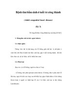
Figure 20.9 Mustard procedure for transposition of the great vessels. Chest cine
fluoroscopic, postero-anterior (PA) views of a superior vena caval venogram. Left: Baffle
obstruction at the junction of the baffle with the right atrium and superior vena cava.
Right: Stent placement with wide opening of the baffle. Patient growth and vena caval-baffle
angulation typically is responsible for such obstructions.
PA
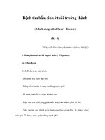
Figure 20.10 Mustard procedure for transposition of the great vessels. Chest cine
fluoroscopic, postero-anterior (PA) view, showing bipolar atrial pacing in a patient with the
Mustard procedure from an era before bipolar leads were available. One lead (cathode) is in
the left atrium and the other (anode) is attached to baffle material as it it was not possible to
attach it to the stump of the right atrium. The anode lead would not pace the atrium, but the
lead combination allowed satisfactory long-term bipolar atrial pacing.
Once identified vascular stents can be effectively used to open the
obstructed baffle regions followed by pacing lead implant (Figure 20.9).
However, due to continued lead-stent neo-intimal interactions follow-
ing implant, vessel restenosis may reoccur necessitating continued close
follow-up care [64]. In a patient with sick sinus syndrome and baffle sten-
osis, it may still be possible to position an atrial lead into a stub of right
atrial appendage close to where it attaches to the baffle (Figure 20.10) [223].
On intraoperative testing, atrial pacing may not be possible or a very high
D-Transposition of the great vessels 97
L Lat
PA
L Lat
PA
L Lat
PA
I II III AVR AVL AVF
V1 V2 V3 V4 V5 V6
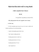
Figure 20.11 Mustard procedure for transposition of the great vessels. Above: Chest
radiographs, postero-anterior (PA) and left lateral (L Lat), showing dual chamber pacing in a
patient with the Mustard procedure. In the PA view, active-fixation leads are attached to the
roof of the left atrium and lateral wall of the left ventricle. In the L Lat view, both leads lie
posterior. Below: Resting 12-lead ECG from the same patient demonstrating dual chamber
pacing with a right bundle branch block indicating left ventricular pacing. There is a right axis
deviation suggesting that the lead is not at the apex of the left ventricle, but rather higher up
in the body of the chamber.
stimulation threshold is recorded. In this situation the lead is actually in
contact with the baffle material and must be repositioned.
For ventricularpacing, thelead mustbe passedto theleft atriumand then
via the anatomical mitral valve to the left ventricle where it is positioned
in the body of the chamber [224]. The major concern once again is phrenic
nerve stimulation which must be tested using 10 volts output from the
pacing system analyser.
The electrocardiograph of dual chamber pacing in patients with the
Mustard or Senning procedure for transposition of the great vessels shows
classical left ventricular pacing with dominant R waves from V1 to V4
(Figure 20.11). The axis is, however, dependent on the position of the lead
in the leftventricle. The higher thelead in the chamber, the more prominent
the R wave in lead III (Figures 20.12, 20.13).
98 Chapter 20
PALAO RAOPALAO RAO
A
B
Figure 20.12 Mustard procedure for transposition of the great vessels. Chest cine
fluoroscopic 40
◦
left anterior oblique (LAO), postero-anterior (PA) and 26
◦
right anterior
oblique (RAO) views, showing dual site left ventricular pacing in a patient with the Mustard
procedure. The upper lead (unipolar) lies just under the mitral valve whereas the lower lead
lies in the body of the chamber.
High LV unipolar
Mid LV bipolar
(a)
(b)
I aVR V1 V4
II aVL V2 V5
III aVF V3 V6
I aVR V1 V4
II aVL V2 V5
III aVF V3 V6
Figure 20.13 Mustard procedure for transposition of the great vessels. Two resting 12-lead
ECGs of the single chamber pacing system shown in Figure 20.12 demonstrating left
ventricular (LV) pacing. The upper ECG shows unipolar pacing from the lead just below the
mitral valve and the lower is bipolar pacing from the mid left ventricle. Unlike right ventricular
pacing, there is little difference in the ECGs from the two sites.
CHAPTER 21
Septal defects including tetralogy
of fallot
It has been well known since the 1950s that surgery for congenital heart
disease and in particular ventricular septal defects may acutely result in
atrioventricular block requiring temporary and occasionally permanent
pacing. This was the result of trauma and edema to the atrioventricular
node, His bundle and more distal conducting pathways. The block prob-
ably resulted from septal sutures [225, 226] and often settled during the
early post operative phase. With increasing knowledge of the aberrant
conduction tissue pathways, particularly with ventricular septal defects,
the incidence of these problems diminished. However, post operatively the
patients frequently demonstrated ECG evidence of conduction tissue dam-
age, which was confirmed with electrophysiology studies [227]. Although
often regarded as benign [228, 229], cases of symptomatic complete heart
block and sudden death were reported years later, similar to the sick sinus
syndrome scenario seen with D-transposition of the great vessels and the
Mustard procedure [230,231].
In patients with asingle surgical repair such as a ventricular septal defect,
the cause could be attributed to the septal sutures, whereas with more com-
plex surgery as with tetralogy of Fallot, the exact identification of the cause
is more complex [225]. For obvious reasons, the more distal the conduc-
tion tissue damage, the less likely the surgery will result in complete heart
block. This fact may also be of significance with the onset of complete heart
block, years later [225]. Of particular importance is that transient complete
heart block at the time of surgery in association with ongoing bifascicular
block, is a more powerful predictor for developing either late complete
heart block or sudden death, than either bifascicular block or transient
heart block alone [225, 232]. Similarly, the progression of a bundle branch
block to trifascicular block also carries a high risk of sudden death [233].
Not all ventricular septal defects require surgery. Thus the implanter
may encounter a patient with a small ventricular septal defect (Maladie de
Roger) in adulthood who requires a ventricular pacing lead (Figure 21.1).
99
100 Chapter 21
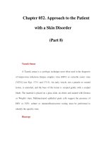
Figure 21.1 Schematic sketch of a ventricular septal defect. The most common site as
shown (broken circle) is the Perimembranous defect (type 2). Other septal defect locations
include Supracristal (type 1), Inflow or AV Canal (type 3) and Muscular (type 4).
As with the atrial septal defect, care must be taken to ensure that the lead is
not inadvertently placed in the left ventricle. Obviously,the same principles
of management as with an atrial septal defect apply (Chapter 16).
Care must also be taken if the ventricular lead is to be positioned in
the right ventricular outflow tract as areas of scarring are to be avoided.
Consideration should be given to device closure prior to positioning the
ventricular lead (Figure 21.2). If the septal defect remains open after lead
insertion anticoagulation with coumadin should be considered.
An important fact to remember when investigating a patient who has
had a ventriculotomy earlier in life is the likely presence of a right bundle
branch block configuration. The presence of certain congenital defects,such
as an atrioventricular septal defect, inherently causes a QRS left axis shift
and AV node conduction delay. An intracardiac electrophysiology study
may also demonstrate prolonged H-V intervals. However, such patients
may not have progressive fascicular conduction disease and may not truly
be candidates for permanent pacing based on those criteria alone. As in
any evaluation, clinical presentation is mandatory.
Not surprisingly, there is also an increased incidence of bradycardia-
tachycardia syndromes following atrial septal defect surgery. This may be
related to the surgery on the septum or the surgical incisions in the free
wall or appendage [234].
Septal defects including tetralogy of fallot 101
PAL Lat
Figure 21.2 Ventricular septal defect device closure. Chest cine fluoroscopic
postero-anterior (PA) and left lateral (L Lat) views, of a star shaped Cardioseal
®
device
(NMT Medical, Boston, MA, USA) (white arrows) inserted into a mid-muscular located
ventricular septal defect. The device size and location may proclude effective septal tract
lead placement.
Since the anatomic pathways to the right atrium and ventricle are normal,
there should be no surprises when implanting a transvenous cardiac pace-
maker in a patient who has had an atrial or ventricular septal defect closed.
However, thepresence of enlarged and potentially scared right sided cham-
bers may make the procedure long, tedious, and more likely to result in
lead dislodgement. Even if the implanting physician’s preferred leads are
tined, it is prudent to choose from the outset, active fixation leads as the tra-
ditional sites may not be suitable. Examples of difficult lead implants due
to interference from ventricular patch material are demonstrated with an
inflow tract attachment in Figure 21.3 and a high outflow tract attachment
in Figure 21.4.
Patients with repaired complete atrioventricular septal defect (endo-
cardial cushion defect) may be particularly difficult cases for ventricular
lead placement. There is often extensive septal synthetic patch material
limiting the sites for successful pacing. The repaired tricuspid valves of
such patients may be regurgitant making lead placement difficult. In such
cases where the patient may become pacemaker-dependent, the “belt and
braces” technique discussed in Chapter 8 should be considered [65].
Another complicating problem may occur with the recent introduction
of interventional devices to close ventricular septal defects. The patients
will not have a sternal or thoracotomy scar and thus the significance of
the closure device may be missed. For those implanters who use the right
ventricular outflow tract there may be significant technical difficulties in
effective septal lead placement (Figure 21.2)
The tetralogy of Fallot, first described by Fallot in 1888, is one
of the most common congenital heart defects causing cyanosis. The
102 Chapter 21
RAOPA
Figure 21.3 Ventricular septal defect repair. Chest cine fluoroscopic postero-anterior (PA)
and right anterior oblique (RAO) views, showing an implanted dual chamber lead system in
a patient following repair of a large ventricular septal defect. The active-fixation right atrial
lead is on the low antero-lateral wall and the active-fixation right ventricular lead is in the
inflow tract of a large right ventricular chamber (black arrow). Attempts to pass the
ventricular lead toward the apex resulted in high stimulation thresholds.
I II
III
V1 V3 V6
PA
L Lat
Figure 21.4 Ventricular septal defect repair. Above: Chest radiographs, postero-anterior
(PA) and left lateral (L Lat), showing an implanted ventricular lead in a patient following
repair of a very large ventricular septal defect and replacement of the mitral valve. The
active-fixation lead has been positioned high in the right ventricular outflow tract. Below:
Resting 6-lead ECG, demonstrating the typical appearance of ventricular pacing from that
site (see Figure 5.2).
Septal defects including tetralogy of fallot 103
Figure 21.5 Schematic sketch of tetralogy of Fallot. The four basic components; right
ventricular outflow obstruction, ventricular septal defect, aorta “overriding” the VSD and right
ventricular hypertrophy are illustrated. Typically the pulmonary obstruction is sub-valvular in
the outflow muscle, although associated pulmonary valve stenosis can occur. In addition to
the VSD closure, surgical repair may involve a trans-annular patch placed across the
pulmonary valve which may enlarge the outflow tract but exacerbate pulmonary valve
insufficiency.
anatomico-pathologic tetralogy consists of an antero-cephalad deviation of
the infundibular right ventricular outflow tract with resultant obstruction,
a ventricular septal defect, the rightward displacement of the aorta over the
ventricular septal defect, and right ventricular hypertrophy (Figure 21.5)
[235]. The addition of an atrial septal defect creates a pentalogy of Fallot. An
atrial septal defect or patent foramen ovale may be present in 82% and a left
superior vena cava draining to the coronary sinus in 11% of patients [236].
Physiologically-related congenital heart conditions include double outlet
right ventricle and pulmonary atresia.
Typically, the patient with tetralogy of Fallot, presenting for pacemaker
or ICD implant, will have had a preliminary systemic to pulmonary
artery anastomosis to improve saturation as an infant (Blalock-Taussig,
Waterston, Potts, Cooley or central graft) followed by the more defin-
itive intracardiac repair later in life. If the subclavian artery was used,
there may be subsequent loss of the pulse on that respective arm. Surgical
repair typically includes patch closure of septal defects with opening of
the narrowed right ventricular infundibulum by placement of a synthetic
or pericardial patch up to or across the pulmonary valve annulus. Most
patients will exhibit at least moderate pulmonary valve insufficiency which
104 Chapter 21
Figure 21.6 Schematic sketch of repair of tetralogy of Fallot. Among patients with nearly
complete outflow tract atresia, surgical repair may involve a valved or non-valved conduit
(Rastelli procedure) to effectively create a jump graft from the right ventricle to pulmonary
artery.
PA
RAO
Figure 21.7 Repair of tetralogy of Fallot. Chest cine fluoroscopic postero-anterior (PA) and
right anterior oblique (RAO) views of a patient following Rastelli repair of a tetralogy of
Fallot. A prior conduit stenosis has been relieved by stent placement. There is a dual
chamber lead system implanted with prolapse of the body of the tined ventricular lead into a
valveless conduit (arrows) as result of free-regurgitant flow. This situation would make future
lead extraction potentially more challenging. An active-fixation atrial lead is in the atrial
appendage.
causes right ventricular dilatation and can complicate ventricular lead
placement. In instances of more severe right ventricular outflow obstruc-
tion or valve atresia, a valve or valveless conduit (Rastelli) is attached from
the right ventricular free wall, extending into the main pulmonary artery
(Figure 21.6).
Septal defects including tetralogy of fallot 105
As expected, following the complicated repair of a tetralogy of Fallot,
the heart will exhibit an extensive amount of ventricular fibrosis which
can complicate pacemaker or ICD implantation. In such patients, the right
ventricular apex, free wall and outflow regions may consist of synthetic
material which will limit effective lead insertion. In addition, free pul-
monary insufficiency with associated blood flow turbulence may cause
excessive lead movement with the potential for excursion into the out-
flow tract (Figure 21.7). Elevated post-implant pacing thresholds may be
anticipated as well as difficulty during lead extraction.
The potential for ventricular arrhythmias and sudden death has been
reported as high as 38% among patients with premature ventricular com-
plexes and elevated ventricular pressures due to recurrent outflow tract
obstruction [237]. Therefore determination of ventricular pressures prior
to pacemaker implantation may help in determining if the patient is a can-
didate for concomitant interventional balloon angioplasty or additional
surgical relief of any residual outflow tract obstruction. Such a patient
being considered for a pacemaker may in fact benefit from an ICD.
CHAPTER 22
Repaired Ebstein’s anomaly
Conduction tissue disease is one of the features of Ebstein’s anomaly, which
usually denotes a more severe form of the syndrome and consequently such
patients may require tricuspid valve reconstructive surgery or replacement
[194]. If at open heart surgery, an annuloplasty has been performed, pacing
or ICD leads can still be inserted into the right ventricle (Figure 22.1) [39].
However, once a mechanical prosthetic valve has been implanted, a trans-
venous lead cannot be positioned in the ventricle via the valve orifice.
Consideration should, therefore, be given to the implantation of a ventricu-
lar lead at the time of the original surgery. As described earlier, the lead
can be positioned in the ventricle, via the atrium and the sewing ring of the
prosthetic tricuspid valve will then cover the lead as it traverses the annu-
lus (Figure 4.3). This lead can be tunneled to the anterior abdominal wall or
subclavicular area and if not used immediately can be capped and buried.
Such a procedure would be potentially very helpful, if there is evidence of
sick sinus syndrome or atrioventricular block which may progress in the
future.
Another situation in Ebstein’s anomaly that may require cardiac pacing
in the future is the propensity for atrial fibrillation. In the presence of a
large atrium resultant from long-standing tricuspid regurgitation, atrial
fibrillation may be present preoperatively or may occur post-operatively.
A pacemaker may be indicated either at surgery or sometime after it for
a slow ventricular response, inappropriate pauses or the need to consider
His bundle ablation.
If a ventricular lead is required in the presence of a mechanical tricuspid
valve prosthesis, an endocardial lead can still be passed via the transvenous
route to the coronary sinus and placed in a cardiac vein on the epicardial
surface [68–74]. However, as discussed in chapter 1, post operatively the
coronary sinus may not drain directly into the right atrium. The surgeon,
in order to protect the conducting system may position the prosthetic tri-
cuspid valve, so that the coronary sinus lies on the ventricular side of the
106
Repaired Ebstein’s anomaly 107
PA
Figure 22.1 Ebstein’s anomaly. Chest cine fluoroscopic postero-anterior (PA) view of a right
ventriculogram demonstrating the relatively narrow effective tricuspid valve orifice
associated with severe insufficiency and dilatation of the right atrium in a postoperative
patient with Ebstein’s anomaly.
valve. The surgical notes should, therefore, be reviewed pre implant and
if necessary attempts made to visualize the ostium of the coronary sinus
before embarking on the pacemaker implantation.
In most cases where the tricuspid valve has been replaced, an epicardial
pacing system will be used. The lead must be attached to the epimyocar-
dial ventricular wall in an area scarred from previous surgery. An open
heart, transatrial approach to position an active-fixation lead in the right
ventricular outflow tract has also been described in an Ebstein’s anomaly
patient with a tricuspid annuloplasty [39].
SECTION D
No venous access to ventricle