Báo cáo y học: " Increased serum soluble Fas after major trauma is associated with delayed neutrophil apoptosis and development of sepsis" pot
Bạn đang xem bản rút gọn của tài liệu. Xem và tải ngay bản đầy đủ của tài liệu tại đây (595.43 KB, 10 trang )
RESEARCH Open Access
Increased serum soluble Fas after major trauma is
associated with delayed neutrophil apoptosis and
development of sepsis
Adnana Paunel-Görgülü, Sascha Flohé, Martin Scholz
*
, Joachim Windolf, Tim Lögters
Abstract
Introduction: Deregulated apoptosis and overshooting neutrophil functions contribute to immune and organ
dysfunction in sepsis and multiple organ failure (MOF). In the present study, we determined the role of soluble Fas
(sFas) in the regulation of posttraumatic neutrophil extrinsic apoptosis and the development of sepsis.
Methods: Forty-seven major trauma patients, 18 with and 29 without sepsis development during the first 10 days
after trauma, were enrolled in this prospective study. Seventeen healthy volunteers served as controls. Blood
samples from severely injured patients were analyzed at day 1, day 5 and day 9 after major trauma. sFas levels,
plasma levels of neutrophil elastase (PMNE) and levels of interleukin (IL)-6 were quantified by enzyme-linked
immunosorbent assay and related to patients’ Sequential Organ Failure Assessment (SOFA) score and Multiple
Organ Dysfunction Score (MODS). Neutrophil apoptosis was determined by propidium iodide staining of
fragmented DNA and flow cytometry. sFas-mediated effects on neutrophil apoptosis were investigated in cells
cultured with agonistic anti-Fas anti bodies in the presence of recombinant sFas, sFas-depleted serum or untreated
serum from septic patients.
Results: Serum levels of sFas in patients who later developed sepsis were significantly increased at day 5 (P < 0.01)
and day 9 (P < 0.05) after trauma compared with patients with uneventful recovery. Apoptosis of patient
neutrophils was significantly decreased during the observation period compared with control cells. Moreover, Fas-
mediated apoptosis of control neutrophils was efficiently inhibited by recombinant sFas and serum from septic
patients. Depletion of sFas from septic patient sera diminished the antiapoptotic effects. In septic patients, sFas
levels were positively correlated with SOFA at day 1 (r = 0.7, P < 0.001), day 5 (r = 0.62, P < 0.01) and day 9 (r =
0.58, P < 0.01) and with PMNE and leukocyte counts (r = 0.49, P < 0.05 for both) as well as MODS at day 5 (r =
0.56, P < 0.01) after trauma.
Conclusions: Increased sFas in patients with sepsis development impairs neutrophil extrinsic apoptosis and shows
a positive correlation with the organ dysfunction scores and PMNE. Therefore, sFas might be a therapeutic target
to prevent posttrauma hyperinflammation and sepsis.
Introduction
Major trauma is frequently associated with activation of
polymorphon uclear neutrophils and systemic inflamma-
tion. Normally, the life span of neutrophils, which con-
stitute an important line of innate host defense, is
limited by apoptosis [1]. During inflammation, neutro-
phils rapidly migrate from the blood into solid tissues to
protect organs from invading bacteria [2]. However, the
life span of these neutrophils is prolonged, resulting in
lung [3], liver [4] and kidney [5] injury. Further, neutro-
phil accumulation in the lung and distant organs repre-
sents a characteristic finding in patients dying of sepsis
[6]. Neutrophils may cause tissue damage by the secre-
tion of reactive oxygen spe cies (ROS) and proteolytic
enzymes, of which neutrophil elastase (PMNE) is the
most abundant [7,8]. There is strong evidence for a
direct correlation between impaired neutrophil apoptosis
and overshooting inflammation [9].
* Correspondence:
Department of Trauma and Hand Surgery, University Hospital Düsseldorf,
Moorenstrasse 5, D-40225 Düsseldorf, Germany
Paunel-Görgülü et al. Critical Care 2011, 15:R20
/>© 2011 Paunel-Görgülü et al.; licensee BioMed Central Ltd. This is an open access article distributed under the terms of the Creative
Commons Attributio n License (http://c reativecommons.org/licenses/by/2.0 ), which permi ts unrestricted use, distribution, and
reproduction in any medium, provided the original work is properly cit ed.
Apoptosis is tightly regulated and might be activated
via membrane-bo und “death” receptors, such as Fas
(extrinsic pathway), or via the mitochondrion (intrinsic
pathway). Fas/Fas ligand (FasL) signaling has emerged as
an important cellular pathway regulating the induction
of apoptosis in a wide variety of tissues as well as acti-
vated immune cells [10,11], thus playing a crucial role
in the resolution of inflammatory responses [9]. The Fas
receptor, also designated as CD95 or Apo-1, is a type I
cell surface glycoprotein which belongs to the tumor
necrosis factor (TNF) receptor superfamily of membrane
receptors and has a broad distribution on various tissues
[12]. The Fas molecule could occur as a cell surface
receptor as well as a soluble protein. The soluble form
of Fas (sFas) is derived either by alternative splicing
from the membrane form or by proteolytic cleavage of
membrane-bound receptors [13,14]. sFas seems to play
an important role as a signaling molecule. It has been
suggested that sFas modifies ligand concentration,
downregulates membrane receptor numbers and specifi-
call y inhibits ligand-receptor associat ion in the extracel-
lular space, thus preventing the induction of apoptosis
in Fas-bearing target cells. Furthermore, expression of
sFas in mice leads to an autoimmune syndrome, and
elevated levels of sFas have been found in some patients
with autoimmune diseases [13]. FasL is a type II integral
membrane protein which is more restricted and tightly
regulated in its expression [12], and the procession by a
matr ix metalloproteinase results in protein cleavage and
release of the extra cellular domain [15]. The biologically
active soluble form of FasL (sFasL) as well as agonistic
anti-Fas antibodies are capable of inducing cytotoxicity,
hepatocyte destruction and mortality in mice through
the interaction with hepatocyte Fas [16,17] and might
contribute to system ic tissue destruction during inflam -
mation [18].
NeutrophilsexpressbothFasanditsendogenous
ligand FasL on their surface, and therefore Fas-FasL
interaction may represent a mechanism of autocrine/
paracrine neutrophil death regulation [19]. Several pre-
vious studies have reported reduced Fas-mediated apop-
tosis in neutrophils obtained from humans with
systemic inflammatory response syndrome (SIRS), burn
injuries or surgical trauma [20,21], without elucidating
the regulatory mechanisms of the disturbed apoptosis.
In the current study, we provide evidence for serum
sFas-mediated inhibition of neutrophil apoptosis and
have determined the prognostic value of sFas in post-
traumatic sepsis.
Materials and methods
Patients
Forty-seven patients were enrolled in this prospective
study. Study approval was obtained from the Ethics
Review Board of the University of Düsseldorf (Düssel-
dorf, Germany). Patient s with blunt or penetrating mul-
tiple injuries who were admitted to our Level I Trauma
Center with an Injury Severity Score (ISS) >16, intensive
care unit (ICU) stay >3 days and ages 18 years and
older were enrolled in this study. Written, informed
consent was obtained from all participants or their legal
representatives if the patients were unconscious. Exclu-
sion criteria were death of the patient on the day of
admission or within the first 2 days on the ICU and
withdrawal of patient consent. In addition, patients with
known preexisting immunological disorders or systemic
immunosuppressive medication were excluded. The
severity of injury was assessed by using the ISS, which is
based on the Abbreviated Injury Scale (AIS) [22], on
admission to the emergency room. SIRS and sepsis were
def ine d using the criteri a outlined in 2005 by the Inte r-
national Sepsis Forum [23]. SIRS was considered to be
present when patients’ conditions fulfilled more than
one SIRS criterion. Patients were determined as septic if
they fulfilled criteria for SIRS and had a proven source
of infection. To evaluate organ dysfunction and/or fail-
ure, the Sequential Organ Failure Assessment (SOFA)
and Multiple Organ Dysfunction (MOD) scores [24]
were determined. Severe sepsis referred to sepsis com-
plicated by organ dysfunction. Organ dysfunction has
been defined using the definition by the SOFA score
with >2 points for at least one system (respiratory, coa-
gulation, liver, c ardiovascular, central nervous or ren al
system). Septic shock was defined as sepsis with acute
persistent circulatory failure unexplained by other causes
(>2 points in SOFA score for the cardiovascular system).
The patients included in this study did not receive
low-dose hydrocortisone therapy as routine adjuvant
treatment for septic shock. Seventeen healthy volunteers
served as the control group.
Blood was collected from healthy volunteers and daily
from patients from the day of admission until day 9.
Heparinized blood was immediately use d after collection
for neutrophil isolation. In parallel, sera and plasma
were harvested by centrifugation and stored at -80°C
until further processing.
Quantification of sFas, sFasL, IL-6 and PMNE by ELISA
sFas (detection limit <47 pg/mL), sFasL (detection limit
<12 pg/mL) (both evaluated by Hoelzel Diagnostika,
Cologne, Germany) and interleukin (IL)-6 (detection
limit <0.70 pg/mL) (evaluated by R&D Systems, Wiesba-
den-Nordenstadt, Germany) were measured in serum
and PMNE (det ection limit 3 ng/mL) (evaluated by
Milenia Biotec, Gießen, Germany) in plasma samples by
using commercially available enzyme-linked immunosor-
bent assay (ELISA) kits according to the manufacturer’s
instructions.
Paunel-Görgülü et al. Critical Care 2011, 15:R20
/>Page 2 of 10
Isolation of human neutrophils
Human neutrophils were isolated by discontinuous den-
sity gradient centrifugation using Percoll medium (Bio-
chrom, Berlin, Germany) as previously described [25].
After hypotonic lysis to remove contaminating erythro-
cytes, cells were suspende d in phosphate-buffered saline
(PBS). Purity and viabili ty were routinely >95% as
assessed by forward and side scatter characteristics of
FACScan (BD Biosciences, Heidelberg, Germany) and
Trypan blue exclusion, respectively.
Immunoprecipitation of sFas from patient serum
The monoclonal anti-Fas antibody clone ZB4 (2 μg;
Millipore, Schwalbach, Germany) was mixed with 40 μL
of Protein G Plus/Protein A-Agarose beads (Calbiochem,
Darmstadt, Germany) and incubate d for 3 hours with
gentle sha king. Then pooled serum from four septic
patients was added and incubated for an additional
17hours at 4°C with gentle shaking. Bound immune com-
plexes were spun down, and the supernatant was stored
at -80°C until use.
Apoptosis assay
To neutralize the apoptotic activity of ag onistic anti-Fas
immunoglobulin (Ig) M antibody (clone CH-11; MBL,
Woburn, MA, USA), antibodies (50 ng/mL) were first
incubated with recombinant human sFas (R&D Systems,
Wiesbaden-Nordenstadt, Germany) for 1 hour and then
added to freshly isolated neutrophil s (1 × 10
6
/mL) from
healthy controls. Cells were further cultured with anti-
Fas antibodies in the presence of sFas for 18 hours in
RPMI 1640 medium containing 2 mM glutamine (Bio-
chrom, Berlin, Germany) and supplemented with 5%
fetal calf serum (FCS) (PAA Laboratories, Coelbe,
Germany), 100 U/mL penicillin and 100 μg/mL strepto-
mycin (Invitrogen, Karlsruhe, Germany) at 37°C in a
humidified atmosphere contain ing 5% CO
2
before being
assessed for apoptosis.
Additionally, pooled patient serum and sera immuno-
precipitated with ZB4 were used to block the activity of
agonistic anti-Fas antibodies (clone CH-11; 200 ng/mL).
After 1 hour of incubation, patient serum (10%) contain-
ing CH-11 antibodies was added to fr eshly isolated con-
trol neutrophils (1 × 10
6
/mL). Cells were further
cultured overnight in RPMI 1640 medium containing
2 mM glutamine (Biochrom, Berlin, Germany) and sup-
plemented with 100 U/mL penicillin and 100 μg/mL
streptomycin (Invitrogen, Karlsruhe, Germany) at 37°C
in a humidified atmosphere containing 5% CO
2
.
Neutrophil apoptosis was measured by flow cytometry
as the percentage of cells with fragmented DNA using
the method described by Nicolleti et al. [26]. Briefly, cell
suspensions of fres hly isolated neutrophils or those
incubated overnight were centrifuged at 450 × g for
5 minutes, and then cells were suspended in 300 μlof
hypotonic fluorochrome solution (50 μg/mL propidium
iodide in 0.1% sodium citrate plus 0.1% Triton X-100).
Cell suspensions were stored in the dark at 4°C for at
least 3 hours before they were analyzed by flow cytome-
try (BD Biosciences, Heidelberg, Germany). A minimum
of 10,000 events were counted per sample. Results are
represented as the percentage of hypodiploid DNA (sub-
G1; percentage apoptosis) corresponding to fragmented
DNA characteristics for apoptotic cells.
Statistical analyses
To evaluate differences between the study groups, a
Kruskal-Wallis test with Dunn’s post hoc test was per-
formed. Correlation between numerical values was eval-
uated by using Spearman’s rank-correlation coefficient
(r). Nonparamet ric receiver operating characteristics
(ROC) curves were generated in which the value for
sensitivity (true positive rate) was plotted against the
false-positive rate (1 - the value of specificity). Analyses
were performed using GraphPad Prism software (version
5; GraphPad Software, San Diego, CA, USA). Compari-
son of ROC curves was performed with MedCalc s oft-
ware (version 11.1.1, MedCalc Software, Mariakerke,
Belgium) using the method described by Delong et al.
[27]. Data were considered to be statistically significant
at P < 0.05.
Results
Demographics and initial blood values outcomes
The 47 patients (31 male, 16 female) enrolled in this
studyhadameanISSof32.9±1.7(range,16to57).
The patients’ mean age was 45.9 ± 2.9 year s (age rang e,
20 to 96 years). Among all patients, 18 (38.3%) devel-
opedsepsiswithin6.1±0.3days(range,4to9days)
after admission. Among the septic patients, nine patients
met the criteria for severe sepsis and four patients met
the criteria for septic shock. The infection site of sepsis
and microbiological pathogens for each patient are given
in Table 1. Five patients died posttraumatically after
30.7 ± 12.3 days (range, 16 to 55 days) as a consequence
of multiple organ failure (MOF). The mean ICU stay
was 18.1 ± 2.6 days (range, 3 to 74 days). The mean age
of the 18 patients (3 female, 15 male) who subsequently
developed sepsis (sepsis group) was 53.5 ± 4.6 (range,
20 to 78 years). The mean ISS in this patient group was
36.7 ± 2.8 (range, 16 to 50). Further patient characteris-
tics as well as injury severity and outcomes are shown
in Table 2.
Levels of sFas and sFasL in patients with or without
sepsis after major trauma
Levels of sFas and sFasL were determined in the serum
of healthy volunteers (control group) and patients within
Paunel-Görgülü et al. Critical Care 2011, 15:R20
/>Page 3 of 10
24 hours after admission (day 1), at day 5 and at day 9
after major trauma (Figure 1). Patients were divided in
two groups: those who subsequently developed sepsis
and those with uneventful recovery after major trauma.
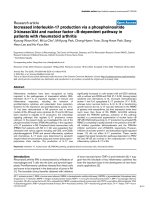
Within the first day after admission, sFas values of
patients who subsequently developed sepsis, but not the
sFas values of those with uneventful outcomes (median,
101.6; interquartile range (IQR), 66.62 to 156.9), were
significantly increased (median, 122.5; IQR, 92.84 to
230.7; P < 0.05) compared with the healthy control
group (median, 70.29; IQR, 42.9 to 93.29) (Figure 1a).
Furthermore, sFas levels in these patients remarkably
increased within the next days and peaked at day 5 after
trauma (median, 230; IQR, 145.2 to 291.2), whereas the
values for pati ents without development of sepsis nor-
malized at this time point (median, 86.46; IQR, 62.95 to
114.2). sFas values in the sepsis group remained
enhanced until day 9 after trauma (median, 187.1; IQR,
80.22 to 297.4) compared with values in the nonsepsis
group at the same time (median, 68.95; IQR, 52.44 to
128.8). Significant intergroup differences were detectable
between patients with sepsis development and healthy
volunteers at day 1 (P < 0.05), day 5 (P < 0.001) and day
9(P < 0.01). Additionally, sFas levels increased signifi-
cantly in sepsis patients at day 5 (P < 0.01) and day 9
(P < 0.05) compared with the nonsepsis patients.
In contrast, for both groups (with or without sepsis),
sFasL values were on an equivalent level compared with
that of healthy cont rols throughout the entire observa-
tion period (P > 0.05) (Figure 1b) and did not show any
intergroup differences.
Prevention of neutrophil apoptosis by recombinant and
serum sFas
It is well established that sFas may bind to membrane-
bound FasL, thus blocking binding of the ligand to the
Fas receptor and preventing apoptosis induction in the
target cell. Therefore, we assumed that elevated serum
levels of sFas may inhibit apoptosis in circulating neu-
trophils and promote prolonged cellular activity. Neu-
trophil apoptosis in both patient groups, tho se who
developed sepsis subsequently and those with an
uneventful recovery, was significantly reduced within the
first day after trauma and c ontinued to be reduced for
theentireperioduntilday9aftertrauma.Thesepsis
patients had a lower rate of neutrophil apoptosis at day
5 (median, 0.94; IQR, 0.6 to 1.67; vs. median, 2.08; IQR
0.64 to 3.36; in the nonsepsis group) and day 9 (median,
Table 1 Infection site of sepsis and microbiological pathogens
Patient Infection site Pathogen Evidence for sepsis, days after trauma
1 Pneumonia Klebsiella pneumoniae 4
2 Pneumonia Klebsiella pneumoniae 5
3 Pneumonia Pseudomonas aeruginosa 8
4 Pneumonia Klebsiella pneumoniae, Pseudomonas aeruginosa 5
5 Pneumonia Klebsiella pneumoniae, Enterococcus faecalis 7
6 Pneumonia Escherichia coli 6
7 Pneumonia Morganella morganii 6
8 Pneumonia Haemophilus influenzae 4
9 Pneumonia Klebsiella pneumoniae 6
10 Peritonitis Enterococcus faecalis 5
11 Pneumonia Escherichia coli 7
12 Pneumonia Pseudomonas aeruginosa 9
13 Pneumonia Staphylococcus aureus 6
14 Pneumonia Staphylococcus aureus 7
15 Pneumonia Klebsiella pneumoniae 7
16 Pneumonia Klebsiella pneumoniae 4
17 Surgical wound infection Enterococcus faecalis 5
18 Pneumonia Enterobacter cloacae 8
Table 2 Demographics, injury severity, and outcome
among subsets of patients
a
Parameter All patients Nonsepsis Sepsis
Number, n 47 29 18
Age, yr (±SEM) 45.9 ± 2.9 41.1 ± 4.4 53.5 ± 4.6
b
ISS (±SEM) 32.9 ± 1.7 30.5 ± 2.0 36.7 ± 2.8
b
ICU, days (±SEM) 18.1 ± 2.6 13.2 ± 2.9 25.9 ± 4.3
b
Sepsis, % (n) 38.3 (18) 0 (0) 100 (18)
Death, % (n) 10.6 (5) 0 (0) 27.8 (5)
Max SOFA day 1 9.2 ± 0.6 8.4 ± 0.9 10.6 ± 0.5
Max SOFA day 5 6.2 ± 0.6 4.4 ± 0.8 9.1 ± 0.7
b
Max SOFA day 9 4 ± 0.6 2.0 ± 0.6 7.1 ± 1.0
b
a
ISS, injury severity score; ICU, intensive care unit length of stay; Max SOFA,
maximal Sequential Organ Failure Assessment score;
b
P < 0.05 between sepsis
and nonsepsis groups.
Paunel-Görgülü et al. Critical Care 2011, 15:R20
/>Page 4 of 10
0.38;IQR,0.28to0.94;vs.median,0.47;IQR0.26to
2.5; in the nonsepsis group), although this difference did
not reach the level of significance (Figure 2a).
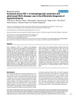
We therefore speculated that sFas prevents the activa-
tion of Fas on trauma neutrophils, leading to strong inhi-
bition of neutrophil extrinsic apoptosis in sepsis. To prove
this hypothesis of sFas-mediated apoptosis inhibition, neu-
trophils from healthy donors were incubated with an ago-
nistic anti-Fas antibody (CH-11) in the presence of serial
dilutions of recombinant human sFas, which has been
shown to inhibit FasL-induced apoptosis of Jurkat cells
[13]. As depicted in Figure 2b, we found that sFas blocks
apoptosis in a concentration-dependent manner.
We further investigated whether sFas in the sera of
patients with sepsis development might also inhibit CH-
11-mediated neutrophil apoptosis. Patient serum con-
tains a broad range of cytokines, especially high levels of
granulocyte macrophage colony- stimulating factor (GM-
CSF), which is known to reduce the neutrophil apopto-
sis rate during inflammation by inhibiting the intrinsic
apoptosis pathway [28]. Because serum containing high
or moderate levels of sFas might also differ in the con-
centrations of the cytokines mentioned above, we pooled
sera from four sepsis patients before immunoprecipita-
tion of sFas by anti-Fas antibodies (ZB4). Then sera
were further used to b lock the proapoptotic activity of
CH-11 monoclonal antibodies. As depicted in Figures 2c
and 2d, neutrophils incubated with agonistic CH-11
antibodies and sera from sepsis patients immunoprecipi-
tated with ZB4 (low sFas levels) displayed a twofold
increased apoptosis rate when compared with cells cul-
tured in the presence of CH-11 antibodies and pooled
serum samples (control; high sFas levels).
Increased levels of PMNE in patients with development
of posttraumatic sepsis
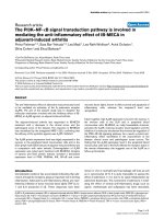
As shown in Figure 3a, leukocyte counts were found to
be significantly increased in septic patients at day 9 after
trauma (median, 12.7; IQR, 9.4 to 17.75) compared with
the number of leukocytes determined in the nonsepsis
group at day 1 (median, 7.7; IQR 6.05 to 9.85; P <0.01)
andday5(median,7;IQR,6.3to10.7;P < 0.05). Neu-
trophil degranulation was further examined by assessing
the levels of PMNE in patients’ plasma (Figur e 3b).
PMNE showed peak levels at day 5 in patients who
developed sepsis (median, 301.4; IQR, 217.5 to 474)
compared with controls (median, 165.9; IQR, 123.1 to
184.4) and in patients with uneventful recovery (median,
162.8; IQR, 111.4 to 268.9; P < 0.05). Interestingly,
PMNE values as well as leukocyte counts were foun d to
correlate with serum sFas concentrations in the sepsis
group at day 5 after trauma (r = 0.49; P < 0.05 for both).
Relation of serum sFas levels with IL-6, SOFA and MOD
scores and its prognostic value in septic patients
IL-6 is a widely accepted inflammatory parameter in
response to major trauma and sepsis. Therefore, IL-6
values in patient serum were determined and correlated
to the sFas val ues. As depicted in Figu re 3c, IL-6 values
of both groups were elevated at day 1 compared with
control values, but decreas ed simultaneously on the fol-
lowing days. Differences were significant at day 5 and
day 9 between the sepsis group (day 5: m edian, 191.7;
IQR, 57.37 to 282.2; day 9: median, 54.94; IQR 29.51 to
191.4) and the nonsepsis group (day 5: median, 41.82;
range, 22.74 to 69.43; P < 0.05; day 9: median, 19; IQR,
4.11 to 26.75; P < 0.05). In all patients, IL-6 showed a
Figure 1 Kinetics of serum soluble Fas (sFas) and soluble Fas ligand (sFasL) after major trauma. (a) sFas levels in patients who developed
sepsis during the first 10 days after trauma (n = 18, dark gray boxes) are significantly elevated when compared to the values determined in
patients with uneventful recovery (n = 29, light gray boxes) and healthy volunteers (n = 17, white box). (b) No alterations in sFasL levels were
observed between different groups. The horizontal line across the boxplots represents the median, and the lower and upper ends of the
boxplots are the 25th and 75th percentiles, respectively. Whiskers indicate the minimum and maximum values, respectively. *P < 0.05, **P < 0.01,
***P < 0.001 vs. control group;
#
P < 0.05,
##
P < 0.01 vs. nonsepsis group.
Paunel-Görgülü et al. Critical Care 2011, 15:R20
/>Page 5 of 10
positive correlation with SOFA score at all time points as
wellaswiththeMODscoreatdays5and9(Table3).
Furthermore, a strong correlation was determ ined
between IL-6 and sFas at day 5 (r = 0.42; P < 0.01) and
day 9 (r =0.4;P < 0.05), but not at d ay 1 after major
trauma. No correlation between sFas and IL-6 values was
found in patients with sepsis development (sepsis group).
To investigate the predictive potential of sFas for the
development of sepsis after major trauma, sFas values
were additionally correlated to SOFA and MOD scores
(Table 3). Elevated sFas concentrations determined in
patients with sepsis development after severe trauma
strongly correlated with patients’ SOFA scores from day
1 until day 9 after trauma. In this patient cohort, sFas
values at day 5 were also significantly correlated to the
MOD score and were positively associated with the
development of multiple organ dysfunction (Table 3).
However, sFas did not correlate with SOFA and MOD
scores of patients with uneventful recovery.
ROC curves
To verify the prognostic potential of sFas in relation to
the established prognostic marker IL-6 for sepsis devel-
opment after major trauma, we established ROC curves
for both parameters at each t ime point. Figure 4 shows
ROCcurvesofsFasandIL-6atday1andday5after
Figure 2 Inhibition of neutrophil extrinsic apoptosis by sFas. (a) Reduced percentage of apoptotic neutrophils isolated from healthy
controls (n = 15, white box), sepsis patients (n = 7, dark gray boxes) and nonsepsis patients (n = 13, light gray boxes) at day 1, day 5 and day 9
after trauma. Boxplots represent the median (heavy line in boxes) and the 25th and 75th percentiles (lower and upper lines of the box,
respectively). Whiskers indicate the minimum and maximum values, respectively. *P < 0.05, **P <0.01, ***P < 0.001 vs. control group.
(b) Neutrophils from healthy controls were incubated with 50 ng/mL anti-Fas antibody (CH-11) in the presence of serial dilutions of recombinant
human soluble Fas (sFas) (range, 0 to 2 μg/mL) for 18 hours. Thereafter cells were lysed in hypotonic solution containing propidium iodide, and
the percentage of apoptotic cells was determined by flow cytometry. Data (means ± SEM) from three independent experiments are presented.
(c) Control neutrophils were incubated with 200 ng/mL agonistic anti-Fas antibodies (clone CH-11) and pooled serum from four sepsis patients
immunoprecipitated by anti-Fas antibodies (clone ZB4) or not. After 18 hours of culture, apoptotic neutrophils with hypodiploid DNA content
were quantified by propidium iodide staining and flow cytometry. Data (means ± SEM) from six independent experiments are depicted.
(d) Representative histogram of CH-11-induced apoptosis in the presence of patient serum. Region M1 describes the percentage of
hypodiploid DNA.
Paunel-Görgülü et al. Critical Care 2011, 15:R20
/>Page 6 of 10
Figure 3 Leukocyte count, pl asma levels of neutrophi l elastase (PMNE) and IL -6 levels after major trauma. (a) Total leukocyte counts
from controls (n = 4, white box), patients with sepsis development (n = 16, dark gray boxes) and patients with uneventful outcome (n = 29,
light gray boxes). (b) PMNE in controls (n = 6, white box), the sepsis group (n = 16, dark gray boxes) and the nonsepsis group (n = 28, light
gray boxes). (c) Serum IL-6 levels in trauma patients with sepsis (n = 18, dark gray boxes) and without sepsis (n = 27, light gray boxes). Boxplots
represent the median (heavy line in boxes) and the 25th and 75th percentiles (lower and upper lines of the box, respectively). Whiskers indicate
the minimum and maximum values, respectively. *P < 0.05, **P < 0.01.
Table 3 Correlations of sFas and IL-6 levels with the
organ dysfunction scoring systems
a
SOFA MODS
Protein Day 1 Day 5 Day 9 Day 1 Day 5 Day 9
sFas
Nonsepsis 0.38
NS
0.25
NS
0.42
NS
0.35
NS
0.25
NS
0.29
NS
Sepsis 0.7
d
0.62
c
0.58
c
0.18
NS
0.56
c
0.13
NS
All 0.54
d
0.56
d
0.61
d
0.28
NS
0.44
c
0.37
b
IL-6
Nonsepsis 0.55
b
0.57
c
0.21
NS
0.08
NS
0.15
NS
0.09
NS
Sepsis 0.46
NS
0.33
NS
0.59
c
0.35
NS
0.25
NS
0.71
c
All 0.35
b
0.54
d
0.57
d
0.2
NS
0.37
b
0.43
c
a
SOFA, Sequential Organ Failure Assessment score; MODS, Multiple Organ
Dysfunction Score; sFas, soluble Fas; IL-6, interleukin-6; NS, not significant;
b
P < 0.05;
c
P < 0.01;
d
P < 0.001.
Figure 4 Receiver operating characteristic (ROC) curves using
sFas and IL-6 as predictors of sepsis. The area under the curve
(AUC) is given for each graph. On day 5, the seven patients who
already had sepsis were excluded from the ROC curve analysis.
Paunel-Görgülü et al. Critical Care 2011, 15:R20
/>Page 7 of 10
trauma. Pairwise comparison of the ROC curves dis-
played no sta tistical difference between the area under
the curve (AUC) for the sFas and IL-6 values at the
depicted time points (day 1, P = 0.694; day 5, P = 0.911).
Discussion
In this study, we have demonstrated that sFas, which
has been found to be significantly elevated in the sera of
trauma patients who subsequently developed sepsis,
inhibits the activation of the Fas pathway and thus
extrinsic apoptosis induction in neutrophils.
Neut rophil apoptosis is reg ulated by the expression of
pro- and antiapoptotic factors and might be initiated by
the activation of TNF family receptors such as Fas by
naturally occurring ligands such as FasL. Many proin-
flammatory cytokines such as GM-CSF, IL-8 and IL-6
are known to prolong neutrophil survival [29]. Recent
studies have shown that proinflammatory mediators
activate both the extracellular signal-regulated kinase
and phosphatidylinositol 3-kinase pathways [30,31] and
might trigger upregulation of antiapoptotic factors such
as Mcl-1 [32], thus promoting intrinsic apoptosis resis-
tance in neutrophils [28].
The Fas/FasL system plays a key role in maintaining
the homeostasis of the immune system. It is widely
accepted that sFas can protect cells against Fas-mediated
apoptosis by binding to FasL, thereby functionally antag-
onizing the Fas-FasL pathway [13]. Evidence has been
reported for a relation between elevated sFas levels and
severe illness [33-37], such as sepsis [36] , malignant dis-
ease [37], autoimmu ne diseases [13] or acute respiratory
distress syndrome [38], or after major surgery [39]. It
has been suggested that sFas decreases neutrophil apop-
tosis in patients postoperatively [39].
In the present study, the sFas levels in patients who
developed sepsis were found to be significantly elevated
at day 1, day 5 and day 9 after major trauma compared
with levels determined in the sera of healthy donors and
at day 5 and day 9 compared with patients with
uneventful recovery. Our in vitro experiments with
recombinant sFas and sera from septic pati ents demon-
strate the abrogation of CH-11-induced neutrophil
apoptosis. We have clearly shown by immunoprecipita-
tion that the antiapoptotic effects of patient serum were
largely mediated by sFas. We therefore postulate that
the antiapoptotic activity of sFas in combination with
the previously reported impaired intrinsic apoptosis
pathway in neutrophils after trauma might be an impor-
tant factor in the ongoing inflammatory injury and pro-
gressive organ dysfunction seen in sepsis patients
[40,41].
Indeed, serum sFas concentrations showed a strong
positive correlation with SOFA and MOD scores, espe-
cially in those patients who developed sepsis.
Additionally, sFas values in patients with septic shock
tended to be higher at day 5 and day 9 after trauma
compa red with the sFas levels in patients suffering from
sepsis and severe sepsis (data not shown). Thus, our
data demonstrate that sFas levels correlate with patient
prognosis and might be used as an additional prognostic
sepsis marker already at day 1 after trauma when sepsis
is clinically not apparent. Moreover, as an interesting
new aspect, we found sFas levels in patients with sepsis
development to be persistently increased even at day 5
and day 9 after trauma, thus showing an association
with the reduced neutrophil apoptosis found at these
time points. Surprisingly, no significant differences in
peripheral circulating leukocyte numbers between both
patients groups could be found. This finding might be
explained by the fact that activated neutrophils become
rapidly recruited to the injured tissue and thus cannot
be further detected in the peripheral circulation.
These data show for the first time the role of sFas as a
predictor for sepsis and the potential link to neutrophil
activity and the pathophysiology of major trauma. How-
ever, the ISSs of the patients in our series ranged
between 16 and 57. This heterogeneity between patients
in terms of injury severity as well as the small number
of patients included may present potential limitations of
the current study.
In contrast to the work of Papathanassoglou et al.
[33], here sFas strongly correlated with IL-6 levels in
serum from trauma patients, except for day 1. Neverthe-
less, no association was found bet ween IL-6 and sFas in
patients with sepsis development. IL-6 levels did not
specifically correlate with SOFA and MOD scores of the
sepsis group, pointing to sFas as a marker for sepsis and
clinical outcome.
The highest s Fas serum concentrations as well as t he
best correl ation with leukocyte counts, PMNE, IL-6 and
MODS were found at day 5 after severe trauma. Inter-
estingly, at this time point, sepsis frequently develops
clinically [8]. Because it is known that sFas may also
influence the adaptive T cell-mediated immunity
[42,43], it may be speculated that sFas might contribute
to T cell anergy and sepsis.
In this study, reduced neutrophil apoptosis has also
been observed in patients who did not develop sepsis.
This finding indicates that sFas-mediated effects on neu-
trophils contribute to the development of organ dys-
function due to prolonged neutrophil hyperactivity, but
not directly to the development of sepsis. Moreover, it is
likel y that sFas might additionally promote a phenotypi-
cal and functi onal change in neutrophils, resulting in an
indirect inhibition of T cell function, which is widely
accepted to be associated with sepsis development
[44,45]. In this context, impairment of T cell prolifera-
tion by soluble CD83 molecules, neutrophil-derived
Paunel-Görgülü et al. Critical Care 2011, 15:R20
/>Page 8 of 10
arginase and ROS has been reported [46-48]. Neverthe-
less, the relationship between neutrophil hyperactivity
and the extensive lympho cyte apoptosis seen in sepsis-
related immunosuppression is currently incompletely
understood and should be elucidated in future studies.
Conclusions
In summary, the present study demonstrates for the first
time a role of serum sFas in the inhibition of neutrophil
extrinsic apoptosis associated with incr eased levels of
PMNE, a marker for systemic inflammation. Our results
show a high correlation between sFas and patients’
SOFA and MOD scores in sepsis and thus provide evi-
dence for the clinical significance of the risk for the
development of sepsis and MOF. Thus, sFas may repre-
sent a feasible target for new therapeutic strategies to
limit neutrophil life span and hyperactivity.
Key messages
• Serum sFas levels have been shown to be signifi-
cantly elevated in patients with sepsis development
after major trauma compared with patients with
uneventful recovery and healthy controls.
• Fas-mediated neutrophil apoptosis was efficiently
inhibited by serum sFas from sepsis patients. Ele-
vated sFas l evels were associate d with increased
levels of PMNE, a marker for neutrophil activity.
• sFas showed a positiv e correlation with SOFA and
MOD scores and sepsis development in severely
injured patients.
• sFas may represent a feasible target for new thera-
peutic strategies to prevent n eutrophil hyperactivity
and sepsis.
Abbreviations
AIS: Abbreviated Injury Scale; ARDS: acute respiratory distress syndrome;
AUC: area under curve; ERK: extracellular signal-regulated kinase; FasL: Fas
ligand; FCS: fetal calf serum; GM-CSF: granulocyte macrophage colony-
stimulating factor; ICU: intensive care unit; IL: interleukin; IQR: interquartile
range; ISS: Injury Severity Score; MOD(S): Multiple Organ Dysfunction (Score);
MOF: multiple organ failure; PI3K: phosphatidylinositol 3-kinase; PBS:
phosphate-buffered saline; PMNE: neutrophil elastase; ROC: receiver
operating characteristics; ROS: reactive oxygen species; SEM: standard error
of the mean; sFas: soluble Fas; sFasL: soluble Fas ligand; SIRS: systemic
inflammatory response syndrome; SOFA: Sequential Organ Failure
Assessment; TNF: tumor necrosis factor.
Acknowledgements
The authors thank Samira Seghrouchni for excellent technical assistance. This
study was supported by a grant from the Forschungskomission of the
Heinrich Heine University Düsseldorf.
Authors’ contributions
AP-G and SF conceived the study, analysed and interpreted data and
drafted the manuscript. Experimental work was performed by AP-G. TL
contributed to the acquisition and analysis of patient data as well as to the
writing of the manuscript. MS and JW critically revised the manuscript for
intellectual content and gave important advice. All authors read and
approved the final manuscript.
Competing interests
The authors declare that they have no competing interests.
Received: 14 July 2010 Revised: 14 December 2010
Accepted: 13 January 2011 Published: 13 January 2011
References
1. Savill JS, Wyllie AH, Henson JE, Walport MJ, Henson PM, Haslett C:
Macrophage phagocytosis of aging neutrophils in inflammation:
programmed cell death in the neutrophil leads to its recognition by
macrophages. J Clin Invest 1989, 83:865-875.
2. Nathan C: Points of control in inflammation. Nature 2002, 420:846-852.
3. Abraham E: Neutrophils and acute lung injury. Crit Care Med 2003, 31:
S195-S199.
4. Ramaiah SK, Jaeschke H: Role of neutrophils in the pathogenesis of acute
inflammatory liver injury. Toxicol Pathol 2007, 35:757-766.
5. Kuligowski M, Kitching A, Hickey M: Leukocyte recruitment to the
inflamed glomerulus: A critical role for platelet-derived P-selectin in the
absence of rolling. J Immunol 2006, 176:6991-6999.
6. Brown KA, Brain SD, Pearson JD, Edgeworth JD, Lewis SM, Treacher DF:
Neutrophils in development of multiple organ failure in sepsis. Lancet
2006, 368:157-169.
7. Donnelly SC, MacGregor I, Zamani A, Gordon MW, Robertson CE,
Steedman DJ, Little K, Haslett C: Plasma elastase levels and the
development of the adult respiratory distress syndrome. Am J Respir Crit
Care Med 1995, 151:1428-1433.
8. Bhatia R, Dent C, Topley N, Pallister I: Neutrophil priming for elastase
release in adult blunt trauma patients. J Trauma 2006, 60:590-596.
9. Savill J: Apoptosis in resolution of inflammation. J Leukoc Biol 1997,
61:375-380.
10. Lynch DH, Ramsdell F, Alderson MR: Fas and FasL in the homeostatic
regulation of immune responses. Immunol Today 1995, 16:569-574.
11. Siegel RM, Chan FK, Chun HJ, Lenardo MJ: The multifaceted role of Fas
signaling in immune cell homeostasis and autoimmunity. Nat Immunol
2000, 1:469-474.
12. Nagata S: Fas ligand-induced apoptosis. Annu Rev Genet 1999, 33:29-55.
13. Cheng J, Zhou T, Liu C, Shapiro JP, Brauer MJ, Kiefer MC, Barr PJ,
Mountz JD: Protection from Fas-mediated apoptosis by a soluble form of
the Fas molecule. Science 1994, 263:1759-1762.
14. Cascino I, Fiucci G, Papoff G, Ruberti G: Three functional soluble forms of
the human apoptosis-inducing Fas molecule are produced by
alternative splicing. J Immunol 1995, 154:2706-2713.
15. Kayagaki N, Kawasaki A, Ebata T, Ohmoto H, Ikeda S, Inoue S, Yoshino K,
Okumura K, Yagita H:
Metalloproteinase-mediated release of human Fas
ligand. J
Exp Med 1995, 182:1777-1783.
16. Song E, Chen J, Ouyang N, Su F, Wang M, Heemann U: Soluble Fas ligand
released by colon adenocarcinoma cells induces host lymphocyte
apoptosis: an active mode of immune evasion in colon cancer. Br J
Cancer 2001, 85:1047-1054.
17. Ogasawara J, Watanabe-Fukunaga R, Adachi M, Matsuzawa A, Kasugai T,
Kitamura Y, Itoh N, Suda T, Nagata S: Lethal effect of the anti-Fas
antibody in mice. Nature 1993, 364:806-809.
18. Matute-Bell o G, Liles WC, Steinberg KP, Kiener PA, Mongovin S, Chi EY,
Jonas M, Martin TR: Soluble Fas ligand induces epithelial cell apoptosis
in humans with a cute lung injury (ARDS). JImmunol1999,
163:2217-2225.
19. Liles WC, Kiener PA, Ledbetter JA, Aruffo A, Klebanoff SJ: Differential
expression of Fas (CD95) and Fas ligand on normal human phagocytes:
implications for the regulation of apoptosis in neutrophils. J Exp Med
1996, 184:429-440.
20. Jimenez MF, Watson RW, Parodo J, Evans D, Foster D, Steinberg M,
Rotstein OD, Marshall JC: Dysregulated expression of neutrophil apoptosis
in the systemic inflammatory response syndrome. Arch Surg 1997,
132:1263-1270.
21. Chitnis D, Dickerson C, Munster AM, Winchurch RA: Inhibition of apoptosis
in polymorphonuclear neutrophils from burn patients. J Leukoc Biol 1996,
59:835-839.
22. Greenspan L, McLellan BA, Greig H: Abbreviated Injury Scale and Injury
Severity Score: a scoring chart. J Trauma 1985, 25:60-64.
23. Calandra T, Cohen J, International Sepsis Forum Definition of Infection in
the ICU Consensus Conference: The international sepsis forum consensus
Paunel-Görgülü et al. Critical Care 2011, 15:R20
/>Page 9 of 10
conference on definitions of infection in the intensive care unit. Crit Care
Med 2005, 33:1538-1548.
24. Marshall JC, Cook DJ, Christou NV, Bernard GR, Sprung CL, Sibbald WJ:
Multiple Organ Dysfunction Score: a reliable descriptor of a complex
clinical outcome. Crit Care Med 1995, 23:1638-1652.
25. Maianski NA, Mul FP, van Buul JD, Roos D, Kuijpers TW: Granulocyte
colony-stimulating factor inhibits the mitochondria-dependent
activation of caspase-3 in neutrophils. Blood 2002, 99:672-679.
26. Nicoletti I, Migliorati G, Pagliacci MC, Grignani F, Riccardi C: A rapid and
simple method for measuring thymocyte apoptosis by propidium iodide
staining and flow cytometry. J Immunol Methods 1991, 139:271-279.
27. DeLong ER, DeLong DM, Clarke-Pearson DL: Comparing the areas under
two or more correlated receiver operating characteristic curves: a
nonparametric approach. Biometrics 1988, 44:837-845.
28. Paunel-Görgülü A, Zörnig M, Lögters T, Altrichter J, Rabenhorst U, Cinatl J,
Windolf J, Scholz M: Mcl-1-mediated Impairment of the intrinsic
apoptosis pathway in circulating neutrophils from critically ill patients
can be overcome by Fas stimulation. J Immunol 2009, 183:6198-6206.
29. Maianski NA, Maianski AN, Kujpers TW, Roos D: Apoptosis of neutrophils.
Acta Haematol 2004, 111:56-66.
30. Tilton B, Andjelkovic M, Didichenko SA, Hemmings BA, Thelen M: G-
Protein-coupled receptors and Fcγ-receptors mediate activation of Akt/
protein kinase B in human phagocytes. J Biol Chem 1997,
272:28096-28101.
31. McLeish KR, Knall C, Ward RA, Gerwins P, Coxon PY, Klein JB, Johnson GL:
Activation of mitogen-activated protein kinase cascades during priming
of human neutrophils by TNF-α and GM-CSF. J Leukoc Biol 1998,
64:537-545.
32. Derouet M, Thomas L, Cross A, Moots RJ, Edwards SW: Granulocyte
macrophage colony-stimulating factor signaling and proteasome
inhibition delay neutrophil apoptosis by increasing the stability of Mcl-1.
J Biol Chem 2004, 279:26915-26921.
33. Papathanassoglou ED, Moynihan JA, Vermillion DL, McDermott MP,
Ackerman MH: Soluble fas levels correlate with multiple organ
dysfunction severity, survival and nitrate levels, but not with cellular
apoptotic markers in critically ill patients. Shock 2000, 14:107-112.
34. Marsik C, Halama T, Cardona F, Wlassits W, Mayr F, Pleiner J, Jilma B:
Regulation of Fas (APO-1, CD95) and Fas ligand expression in leukocytes
during systemic inflammation in humans. Shock 2003, 20:493-496.
35. Torre D, Tambini R, Manfredi M, Mangani V, Livi P, Maldifassi V, Campi P,
Speranza F: Circulating levels of FAS/APO-1 in patients with the systemic
inflammatory response syndrome. Diagn Microbiol Infect Dis 2003,
45:233-236.
36. De Freitas I, Fernandez-Somoza M, Essenfeld-Sekler , Cardier JE: Serum
levels of the apoptosis-associated molecules, tumor necrosis factor-α
/
tumor necrosis factor type-I receptor and Fas/FasL, in sepsis. Chest 2004,
125:2238-2246.
37. Mitani K, Nishioka Y, Yamabe K, Ogawa H, Miki T, Yanagawa H, Sone S:
Soluble Fas in malignant pleural effusion and its expression in lung
cancer cells. Cancer Sci 2003, 94:302-307.
38. Lee KS, Choi YH, Kim YS, Baik SH, Oh YJ, Sheen SS, Park JH, Hwang SC,
Park KJ: Evaluation of bronchoalveolar lavage fluid from ARDS patients
with regard to apoptosis. Respir Med 2008, 102:464-469.
39. Iwase M, Kondo G, Watanabe H, Takaoka S, Uchida M, Ohashi M,
Nagumo M: Regulation of Fas-mediated apoptosis in neutrophils after
surgery-induced acute inflammation. J Surg Res 2006, 134:114-123.
40. Liacos C, Katsaragakis S, Konstadoulakis MM, Messaris EG, Papanicolaou M,
Georgiadis GG, Menenakos E, Vasiliadi-Chioti A, Androulakis G: Apoptosis in
cells of bronchoalveolar lavage: a cellular reaction in patients who die
with sepsis and respiratory failure. Crit Care Med 2001, 29:2310-2317.
41. Fialkow L, Fochesatto Filho L, Bozzetti MC, Milani AR, Rodrigues Filho EM,
Ladniuk RM, Pierozan P, de Moura RM, Prolla JC, Vachon E, Downey GP:
Neutrophil apoptosis: a marker of disease severity in sepsis and sepsis-
induced acute respiratory distress syndrome. Crit Care 2006, 10:R155.
42. Silvestris F, Grinello D, Tucci M, Cafforio P, Dammacco F: Enhancement of T
cell apoptosis correlates with increased serum levels of soluble Fas
(CD95/Apo-1) in active lupus. Lupus 2003, 12:8-14.
43. Ma Y, Ye F, Lv W, Cheng Q, Chen H, Xie X: Correlation between soluble
Fas level and apoptosis of T cells in ovarian carcinoma. Eur J Obstet
Gynaecol Reprod Biol 2008, 138:204-211.
44. Choudhry MA, Ahmad S, Thompson KD, Sayeed MM: T-lymphocyte Ca
2+
signalling and proliferative responses during sepsis. Shock 1994,
1:466-471.
45. Roth G, Moser B, Krenn C, Brunner M, Haisjackl M, Almer G, Gerlitz S,
Wolner E, Boltz-Nitulescu G, Ankersmit HJ: Susceptibility to programmed
cell death in T-lymphocytes from septic patients: a mechanism for
lymphopenia and Th2 predominance. Biochem Biophys Res Commun 2003,
308:840-846.
46. Dudziak D, Nimmerjahn F, Bornkamm GW, Laux G: Alternative splicing
generates putative soluble CD83 proteins that inhibit T cell proliferation.
J Immunol 2005, 174:6672-6676.
47. Munder M, Schneider H, Luckner C, Giese T, Langhans CD, Fuentes JM,
Kropf P, Mueller I, Kolb A, Modolell M, Ho AD: Suppression of T-cell
functions by human granulocyte arginase. Blood 2006, 108:1627-1634.
48. Kusmartsev S, Su Z, Heiser A, Dannull J, Eruslanov E, Kübler H, Yancey D,
Dahm P, Vieweg J: Reversal of myeloid cell-mediated
immunosuppression in patients with metastatic renal cell carcinoma.
Clin Cancer Res 2008, 14:8270-8278.
doi:10.1186/cc9965
Cite this article as: Paunel-Görgülü et al.: Increased serum soluble Fas
after major trauma is associated with delayed neutrophil apoptosis and
development of sepsis. Critical Care 2011 15:R20.
Submit your next manuscript to BioMed Central
and take full advantage of:
• Convenient online submission
• Thorough peer review
• No space constraints or color figure charges
• Immediate publication on acceptance
• Inclusion in PubMed, CAS, Scopus and Google Scholar
• Research which is freely available for redistribution
Submit your manuscript at
www.biomedcentral.com/submit
Paunel-Görgülü et al. Critical Care 2011, 15:R20
/>Page 10 of 10