Báo cáo y học: " Can ‘permissive’ hypercapnia modulate the severity of sepsis-induced ALI/ARDS" doc
Bạn đang xem bản rút gọn của tài liệu. Xem và tải ngay bản đầy đủ của tài liệu tại đây (323.52 KB, 9 trang )
Introduction
Ventilatory strategies that reduce lung stretch by reduc-
ing tidal and minute ventilation, which results in a
‘permissive’ hypercapnic acidosis, improve outcome in
patients with acute lung injury/acute respiratory distress
syndrome (ALI/ARDS) [1,2]. Reassuringly, evidence from
clinical studies attests to the safety and lack of detri-
mental eff ects of hypercapnic acidosis [2]. Of parti cu lar
importance, a secondary analysis of data from the
ARDSnet tidal volume study [1] demonstrated that the
presence of hypercapnic acidosis at the time of randomi-
zation was associated with improved patient survival in
patients who received high tidal volume ventilation [3].
ese fi ndings have resulted in a shift in paradigms
regarding hypercapnia – from avoidance to tolerance –
with hypercapnia increasingly permitted in order to
realize the benefi ts of low lung stretch. Conse quently, low
tidal and minute volume ventilation and the accom-
panying `permissive’ hypercapnia are now the standard
of care for patients with ALI/ARDS, and are increasingly
used in the ventilatory management of a diverse range of
diseases leading to acute severe respira tory failure, inclu-
d ing asthma and chronic obstructive pulmonary disease.
e infl ammatory response plays a central role in the
pathogenesis of injury and in the repair process in ALI/
ARDS [4]. Infl ammation is a highly conserved process in
evolution, which is essential for survival. It functions to
resolve the injurious process, facilitate repair, and return
the host to a state of homeostasis. e ideal infl ammatory
process is rapid, causes focused destruction of pathogens,
yet is specifi c and ultimately self-limiting [5]. In contrast,
when the infl ammatory response is dysregulated or persis-
tent, this can lead to excessive host damage, and
contribute to the pathogenesis of lung and systemic organ
injury, leading to multiple organ failure and death. e
potential for hypercapnia and/or its associated acidosis to
potently inhibit the immune response is increasingly
recognized [6,7]. Where the host immune response is a
major contri bu tor to injury, such as in non-septic ALI/
ARDS, these eff ects would be expected to result in
potential benefi t. is has been demonstrated clearly in
relevant pre-clinical ALI/ARDS models, where hyper-
capnic acidosis has been demonstrated to attenuate ALI
induced by free radicals [8], pulmonary [9] and systemic
ischemia-reperfusion [10], pulmonary endo toxin instilla-
tion [11], and excessive lung stretch [12]. e protective
eff ects of hypercapnic acidosis in these models appear
due, at least in part, to its anti-infl ammatory eff ects.
e eff ects of hypercapnia in sepsis-induced lung
injury, where a robust immune response to microbial
infec tion is central to bacterial clearance and recovery, is
less clear. Of concern, severe sepsis-induced organ
failure, whether pulmonary or systemic in origin, is the
leading cause of death in critically ill adults and children
[13]. Sepsis-induced ARDS is associated with the highest
mortality rates. Evidence suggests that approximately
40% of patients with severe sepsis develop ARDS [13].
Furthermore, infection frequently complicates critical
illness due to other causes, with an infection prevalence
of over 44% reported in this population [14]. ese issues
underline the importance of understanding the eff ects of
hypercapnia on the immune response, and the implica-
tions of these eff ects in the setting of sepsis.
Hypercapnia and the innate immune response
Function of the innate immune response
e immune system can be viewed as having two inter-
connected branches, namely the innate and adaptive
immune responses [5]. e innate immune system is an
ancient, highly conserved response, being present in
Can ‘permissive’ hypercapnia modulate the
severity of sepsis-induced ALI/ARDS?
Gerard Curley, Mairead Hayes, and John G La ey*
This article is one of eleven reviews selected from the Annual Update in Intensive Care and Emergency Medicine 2011 (Springer Verlag) and
co-published as a series in Critical Care. Other articles in the series can be found online at Further
information about the Annual Update in Intensive Care and Emergency Medicine is available from />REVIEW
*Correspondence: john.la
Department of Anestheisa, Clinical Sciences Institute, National University, Galway,
Ireland
Curley et al. Critical Care 2011, 15:212
/>© 2011 Springer-Verlag Berlin Heidelberg.
This work is subject to copyright. All rights are reserved, whether the whole or part of the material is concerned, speci cally the rights of
translation, reprinting, reuse of illustrations, recitation, broadcasting, reproduction on micro lm or in any other way, and storage in data
banks. Duplication of this publication or parts thereof is permitted only under the provisions of the German Copyright Law of September9,
1965, in its current version, and permission for use must always be obtained from Springer-Verlag. Violations are liable for prosecution
under the German Copyright Law.
some form in all metazoan organisms. is response is
activated by components of the wall of invading micro-
organisms, such as lipopolysaccharide (LPS) or peptido-
glycan, following the binding of these pathogen-asso-
ciated molecular patterns to pattern recognition recep-
tors, such as the Toll-like receptors (TLRs) on tissue
macrophages. e innate immune response is also activa-
ted by endogenous `danger’ signals, such as mitochon-
drial components [15], providing an elegant explanation
for why non-septic insults can also lead to organ injury
and dysfunction. An infl ammatory cascade is then
initiated, involving cytokine signaling activation of
phago cytes that kill bacteria, as is activation of the (later)
adaptive immune response.
Activation of the innate immune response
Hypercapnic acidosis has been demonstrated to inhibit
multiple components of the host innate immune res-
ponses. Activation of the innate immune response
initiates a conserved signaling cascade that culminates in
the activation of transcription factors, such as nuclear
factor kappa-B (NF-κB) [5]. ese transcription factors
drive the expression of multiple genes that activate and
regulate the pro-infl ammatory and repair processes.
Increasing evidence suggests that hypercapnic acidosis
directly inhibits the activation of NF-κB [16]. Intriguingly,
this eff ect of hypercapnic acidosis may be a property of
the CO
2
rather than its associated acidosis [17–19]. If
confi rmed, this fi nding suggests the presence of a
molecular CO
2
sensor in mammalian cells. is mecha-
nism of action of hypercapnic acidosis has been demon-
strated to underlie some of the anti-infl ammatory eff ects
of hypercapnia [16], and to be a key mechanism by which
hypercapnia – whether buff ered or not – reduces
pulmonary epithelial wound healing [18].
Coordination of the innate immune response
Hypercapnic acidosis also interferes with coordination of
the innate immune response by reducing cytokine signal-
ing between immune eff ector cells. Hypercapnic acidosis
reduces neutrophil [20] and macrophage [21] production
of pro-infl ammatory cytokines such as tumor necrosis
factor (TNF)-α, interleukin (IL)-1β, IL-8 and IL-6. Hyper-
capnic acidosis reduced endotoxin stimulated macro-
phage release of TNFα and IL-1β in vitro [21]. Peritoneal
macrophages incubated under hypercapnic conditions
demonstrated a prolonged reduction in endotoxin-
stimulated TNF-α and IL-1β release [22]. In contrast, a
recent study reported rapid onset and rapid reversibility
of IL-6 inhibition by hypercapnia in mature macrophage
stimulated with LPS [19]. e mechanism underlying
hypercapnic acidosis-mediated inhibition of cytokine
and chemokine production appears to be mediated at
least in part via inhibition of activation of NF-κB.
The cellular innate immune response
Neutrophils and macrophages are important eff ectors of
the innate immune response in the setting of bacterial
infection. Neutrophils rapidly migrate from the blood-
stream to areas of infection, and rapidly phagocytose
invading microorganisms. Tissue macrophages and their
blood borne monocyte counterparts are activated by
bacterial products such as endotoxin, and coordinate the
activation of the adaptive immune response in the setting
of infection by presenting foreign antigen to lymphocytes
and secreting chemokines. Both monocytes and
macrophages phagocytose and kill pathogens by similar
mechanisms but at a slower rate than neutrophils.
Hypercapnic acidosis may impact on the cellular
immune response via both direct and indirect mecha-
nisms. Hypercapnic acidosis inhibits neutrophil expres-
sion of the chemokines, selectins and intercellular
adhesion molecules [16,20], which facilitate neutrophil
binding to the endothelium and migration out of the
vascular system. e potential for hypercapnic acidosis
to inhibit neutrophil chemotaxis and migration to the
site of injury has been confi rmed in vivo, where hyper-
capnic acidosis inhibits pulmonary neutrophil infi ltration
in response to endotoxin instillation [11]. Hypercapnic
acidosis directly impairs neutrophil phagocytosis in vitro
[23]. is inhibitory eff ect appears to be a function of the
acidosis per se, with buff ering restoring neutrophil
phagocytosis [24]. Hypercapnic acidosis also inhibits
phagocytosis of opsonized polystyrene beads by human
alveolar macrophages, although the levels of CO
2
utilized
to demonstrate this eff ect were well beyond the range
encountered clinically [19].
Neutrophils and macrophages kill ingested bacteria by
producing free radicals such as superoxide, hydrogen
peroxide, and hypochlorous acid, and releasing these into
the phagosome. is is a pH-dependent process, with
free radical production decreased at low pH [25]. Hyper-
capnic acidosis inhibits the generation of oxidants such
as superoxide by unstimulated neutrophils and by
neutrophils stimulated with opsonized Escherichia coli or
with phorbol esters [20]. In contrast, hypocapnic alkalosis
stimulates neutrophil oxidant generation [20]. Inhibition
of the intracellular pH changes with acetazolamide
attenuated these eff ects. More recently, hypercapnic
acidosis has been demonstrated to reduce oxidative
reactions in the endotoxin injured lung by a mechanism
involving inhibition of myeloperoxidase-dependent
oxida tion [26]. e potential for hypercapnic acidosis to
reduce free radical formation, while benefi cial where host
oxidative injury is a major component of the injury
process, may be disadvantageous in sepsis, where free
radicals are necessary to cause bacterial injury and death.
Neutrophil apoptosis following phagocytic activity
generally occurs within 48 hours of release into the
Curley et al. Critical Care 2011, 15:212
/>Page 2 of 9
circulation. Conversely, neutrophil death via necrosis
causes release of intracellular contents, including harmful
enzymes, which can cause tissue destruction. Neutrophils
appear to have an increased probability of dying by
necrosis following intracellular acidifi cation during
phagocytosis [27]. Hypercapnic acidosis may, therefore,
increase the probability of neutrophil cell death occurring
via necrosis rather than apoptosis.
Hypercapnia and the adaptive immune response
e adaptive immune system is activated by the innate
response following activation of pattern recognition
receptors that detect molecular signatures from micro-
bial pathogens. Specifi c major histocompatibility complex
molecules on T and B lymphocytes also bind microbial
components. ese activation events lead to the genera-
tion of T and B lymphocyte-mediated immune responses
over a period of several days.
Much of the focus to date regarding the eff ects of
hypercapnic acidosis on immune response to injury and/
or infection has been on the innate immune response.
Less is known about the eff ects of hypercapnic acidosis
on adaptive or acquired immunity. However, important
clues as to the potential for hypercapnic acidosis to
modulate the adaptive response come from the cancer
literature. e tumor microenvironment is characterized
by poor vascularization, resulting in tissue hypoxia and
acidosis. In a situation analogous to sepsis, acidosis in
this setting may hamper the host immune response to
tumor cells, potentially leading to increased tumor
growth and spread. e cytotoxic activity of human
lympho kine activated killer cells and natural killer cells is
diminished at acidic pH [28]. Metabolic acidosis reduces
lysis of various tumor cell lines by cytotoxic T-lympho-
cytes [29]. In contrast, the motility of IL-2-stimulated
lymphocytes appears to be stimulated in the presence of
an acidifi ed extracellular matrix and severe extracellular
acidosis (pH 6.5) also appears to enhance the antigen
presenting capacity of dendritic cells [30]. e net eff ect
of these contrasting actions of metabolic acidosis on the
adaptive immune response is unclear. However, the
demonstration that hypercapnic acidosis enhanced
systemic tumor spread in a murine model [31] raises
clear concerns regarding the potential for hypercapnic
acidosis to suppress cell-mediated immunity.
Hypercapnia and acidosis modulate bacterial
proliferation
Carbon dioxide has broadly similar eff ects within the
various families of microorganisms, but the sensitivity to
CO
2
varies across the families, e.g., yeasts are quite
resistant to the inhibitory eff ects of CO
2
, Gram-positive
organisms are somewhat less resistant, and Gram-
negative organisms are the most vulnerable [32]. Optimal
anaerobic E. coli growth occurs at a CO
2
tension (PCO
2
)
of 0.05 atmospheres, which is similar to the PCO
2
in the
gut. e aerobic growth rate of E. coli was not inhibited
by a PCO
2
of 0.2 atmospheres but was inhibited at partial
pressures above 0.6 atmospheres [33]. It is important to
remember that these levels are extremely high in the
context of human physiology.
Of concern, however, is the demonstration by Pugin et
al. that more clinically relevant degrees of metabolic
acidosis can directly enhance bacterial proliferation in
vitro [34]. Cultured lung epithelial cells exposed to cyclic
stretch similar to that seen with mechanical ventilation
produced a lactic acidosis that markedly enhanced the
growth of E. coli [34]. is was a direct eff ect of hydrogen
ions, as direct acidifi cation of the culture medium to a
pH of 7.2 with hydrochloric acid enhanced E. coli growth.
In contrast, alkalinizing the pH of conditioned media
from stretched lung cells abolished the enhancement of
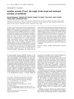
E. coli growth. A range of Gram-positive and Gram-
negative bacteria (including E. coli, Proteus mirabilis,
Serratia rubidaea, Klebsiella pneumoniae, Enterococcus
faecalis, and Pseudomonas aeruginosa) isolated from
patients with ventilator-associated pneumonia (VAP),
grew better in acidifi ed media (Fig.1). Interestingly, this
eff ect was not seen with a methicillin resistant Staphylo-
coccus aureus (MRSA) strain, which appeared to grow
best at an alkaline pH [34].
e eff ects of hypercapnic acidosis on bacterial
proliferation at levels encountered in the context of
permissive hypercapnia are unclear. e net eff ect is
likely to be a combination of the eff ects of the acidosis
and of the hypercapnia. Nevertheless, the demonstration
that clinically relevant levels of metabolic acidosis
enhance bacterial growth is of concern.
Implications for hypercapnia in sepsis
Immunocompetence is essential to an eff ective host
response to microbial infection. Hypercapnia and/or
acidosis may modulate the interaction between host and
bacterial pathogen via several mechanisms, resulting in a
broad based suppression of the infl ammatory response.
Hypercapnia, acidosis and the host response
e initial host response to invading pathogens is domi-
nated by neutrophil activation, migration to the infective
site, and phagocytosis and killing of bacteria. Compart-
mentalized release of neutrophil proteolytic enzymes and
myeloperoxidase-dependent oxygen radicals results in
eff ective pathogen destruction. However, excessive
release of these potent mediators into the extracellular
space results in damage to host tissue and worsening ALI.
Consistent with this is the fi nding that recovery of
neutrophil count in neutropenic patients worsens the
severity of ALI [35]. Hypercapnic acidosis may reduce
Curley et al. Critical Care 2011, 15:212
/>Page 3 of 9
the potential for damage to host tissue during the
response to infection, by reducing lung neutrophil
recruit ment [10], adherence [16], intracellular pH
regulation [12], oxidant generation [8], and phagocytosis
[23]. ese mechanisms are considered to underlie some
of the protective eff ects of hypercapnic acidosis in non-
sepsis induced ALI [7]. However, these eff ects of hyper-
capnic acidosis may be detrimental in sepsis, given the
central role of neutrophil mediated phagocytosis of
microbial pathogens and activation of the cytokine
cascade to the host response to infection. In this context,
defects in neutrophil function are associated with
increased sepsis severity and worse outcome [36].
Early versus late bacterial infection
e eff ects of this hypercapnic acidosis-induced immune
modulation may vary depending upon the stage of the
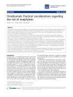
infective process. e anti-infl ammatory properties of
hypercapnic acidosis may reduce the intensity of the
initial host response to infection, thus attenuating tissue
damage (Fig.2). However, the mechanisms whereby bac-
teria mediate tissue injury are complex and not limited to
the contribution from an excessive host response. In late
or prolonged pneumonia, in which tissue injury from
direct bacterial spread and invasion makes a signifi cant
contribution, hypercapnic acidosis might impair
bactericidal host responses. In the absence of eff ective
antibiotic therapy, this may lead to enhanced bacterial
spread and replication leading to more severe tissue
destruction and lung and systemic organ injury (Fig.2).
Impact on repair following injury
Hypercapnic acidosis has been demonstrated to retard
the repair process following lung cell and tissue injury.
Hypercapnia slowed resealing of stretch-induced cell
membrane injuries [37] and inhibited the repair of
pulmo nary epithelial wounds [18] by a mechanism
involv ing inhibition of the NF-κB pathway. ese fi ndings
raise the potential that hypercapnic acidosis could lead to
increased bacterial translocation through defects in the
pulmonary epithelium, while also delaying the recovery
process following a septic insult.
Recent studies in relevant preclinical models have
signifi cantly advanced our understanding of the eff ects of
hypercapnic acidosis in both pulmonary and systemic
sepsis-induced ALI/ARDS. ese studies reveal the
importance of severity, site, and stage of the infective
process, the need for antibiotic therapy, and the utility of
buff ering the hypercapnic acidosis in this setting.
Hypercapnia in pulmonary sepsis
Early lung infection
e eff ect of hypercapnic acidosis on pneumonia-induced
ALI appears to depend on the stage and severity of the
infection. In an acute severe bacterial pneumonia-
induced lung injury, hypercapnic acidosis improved
physio logical indices of injury [38]. Intriguingly, these
protective eff ects were mediated by a mechanism inde-
pen dent of neutrophil function. In contrast, hypercapnic
acidosis did not alter the magnitude of lung injury in a
less severe acute bacterial pneumonia [39]. Importantly
these in vivo studies showed no increase in bacterial
count in animals exposed to hypercapnic acidosis, a
reassuring fi nding given concerns regarding retardation
of the host bactericidal response and potential bacterial
proliferation.
In the clinical setting, many critically ill patients will
have established infection at the time of presentation.
Figure 1. Bacterial pathogens proliferate more rapidly in the
setting of metabolic acidosis. All bacterial strains tested, except for
a methicillin-resistant S. aureus, had a marked growth advantage at
moderately acidic pH levels (7.2–7.6) relevant to the clinical setting.
Gram-negative bacteria are represented by dark blue bars while
Gram-positive bacteria are represented by light blue bars. From [34]
with permission.
Curley et al. Critical Care 2011, 15:212
/>Page 4 of 9
us animal models of established bacterial pneumonia,
in which hypercapnic acidosis was introduced several
hours following induction of infection with E. coli, more
closely resemble the clinical setting. In an established
pneumonia model, hypercapnic acidosis induced after
the development of a signifi cant pneumonia-induced
lung injury reduced physiological indices of lung injury
[40]. Of importance, these protective eff ects of hyper cap-
nic acidosis were enhanced in the presence of appropriate
antibiotic therapy [40]. Again, reassuringly, lung bacterial
loads were similar in the hypercapnic acidosis and
normo capnia groups [40].
Prolonged lung infection
In an animal model of prolonged untreated pneumonia,
sustained hypercapnic acidosis worsened histological and
physiological indices of lung injury, including compliance,
arterial oxygenation, alveolar wall swelling and neutrophil
infi ltration [23]. Of particular concern to the clinical
setting, hypercapnic acidosis was associated with a higher
lung bacterial count. e mechanism underlying this eff ect
appeared to be inhibition of neutrophil function, as
evidenced by impaired phagocytotic ability in neutrophils
isolated from hypercapnic rats [23]. Of importance to the
clinical context, the use of appropriate antibiotic therapy
abolished these deleterious eff ects of hypercapnia,
reducing lung damage and lung bacterial load to levels
comparable to those seen with normocapnia.
ese fi ndings have been confi rmed and considerably
expanded in a recent study of hypercapnia in the fruit fl y
[41]. Helenius et al., in a series of elegant in vivo studies,
found that prolonged hypercapnia decreased expression
of specifi c anti-microbial peptides in Drosophilia
melano gaster [41]. Hypercapnia decreased bacterial
resis tance in adult fl ies exposed to pathogens as
evidenced by increased bacterial loads and increased
mortality in fl ies inoculated with E. faecalis, A. tume-
faciens, or S. aureus [41]. e previously demon strated
suppressive eff ects of hypercapnic acidosis on the NF-κB
pathway appeared to underlie the decreased resistance to
infection [41]. ese fi ndings raise signifi cant concerns
regarding the safety of hypercapnia in the setting of
prolonged pneumonia, particularly in the absence of
eff ective antibiotic therapy.
Figure 2. Potential mechanisms underlying the e ects of hypercapnic acidosis in sepsis. Panel A represents early sepsis, in which
hypercapnic acidosis may reduce the host in ammatory response and decrease the contribution of bacterial toxin mediated injury to tissue injury
and damage. This might result in an overall decrease in lung injury. Panel B represents late or prolonged bacterial sepsis, where a hypercapnic
acidosis-mediated decrease in the host response to bacterial infection might result in unopposed bacterial proliferation, thereby increasing direct
bacterial tissue invasion and injury, and worsening lung injury. ALI: acute lung injury; HCA: hypercapnic acidosis.
Curley et al. Critical Care 2011, 15:212
/>Page 5 of 9
Hypercapnia in systemic sepsis
A growing body of evidence attests to a benefi cial role of
hypercapnia in the setting of systemic sepsis. Improve-
ments in hemodynamic parameters and lung injury have
been demonstrated in evolving, established, and pro-
longed systemic sepsis in animal models. is is in
contrast to the detrimental eff ects of hypercapnic
acidosis seen in prolonged pulmonary sepsis, suggesting
that the eff ects of hypercapnic acidosis depend not only
on the stage of the infective process, but also on the site
of the primary infection.
Early systemic sepsis
Hypercapnic acidosis reduces the severity of early septic
shock and lung injury induced by systemic sepsis. In a
rodent model of peritoneal sepsis induced by cecal
ligation and puncture, hypercapnic acidosis slowed the
development of hypotension, preserved central venous
oxygen saturation, and attenuated the rise in serum
lactate compared to control conditions, in the fi rst
3hours post injury [42]. e severity of early lung injury
was reduced as evidenced by a decrease in the alveolar-
arterial oxygen gradient, and reduced lung permeability,
compared to normocapnia. Alveolar neutrophil concen-
tration was reduced by hypercapnic acidosis but IL-6 and
TNF-α were unchanged [42]. Of importance, there were
no diff erences in bacterial loads in the lung, blood, or
peritoneum in the hypercapnia group.
Prolonged systemic sepsis
Using an ovine model of fecal peritonitis, Wang et al
compared the eff ects of hypercapnic acidosis with those
of dobutamine [43]. Over an 18-hour study period,
hyper capnic acidosis resulted in improved hemo-
dynamics of a magnitude comparable to that of dobu-
tamine. Compared with normocapnia, both hypercapnic
acidosis and dobutamine raised cardiac index and
systemic oxygen delivery and reduced lactate levels. In
addition, hyper capnic acidosis attenuated indices of lung
injury, including lung edema, alveolar-arterial oxygen
partial pressure diff erence and shunt fraction. Hyper-
capnic acidosis did not decrease survival time compared
to normo capnia in this setting [43]. In a more prolonged
systemic sepsis model, Costello et al. demonstrated that
sustained hypercapnic acidosis reduced histological
indices of lung injury compared with normocapnia in
rodents following cecal ligation and puncture [42].
Reassuringly there was no evidence of an increased
bacterial load in the lung, blood, or peritoneum of
animals exposed to hypercapnia.
Intraperitoneal hypercapnia
Direct intra-abdominal administration of CO
2
– by
means of a pneumoperitoneum – reduces the severity of
abdominal sepsis-induced lung and systemic organ
injury. Insuffl ation of CO
2
into the peritoneal cavity prior
to laparotomy for endotoxin contamination increased
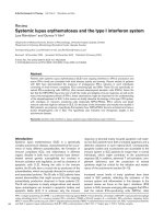
animal survival [44]. Most recently, CO
2
pneumo peri-
toneum has been demonstrated to increase survival in
mice with polymicrobial peritonitis induced by cecal
ligation and puncture (Fig. 3) [31]. ese protective
eff ects of intraperitoneal carbon dioxide insuffl ation
appear be due to the immunomodulatory eff ects of
hyper capnic acidosis, which include an IL-10 mediated
downregulation of TNF-α [44]. Importantly, these eff ects
appear to be mediated by the localized peritoneal
acidosis, rather than by any systemic eff ect.
Bu ering hypercapnic acidosis in sepsis
e immunomodulatory eff ects of hypercapnic acidosis
in sepsis may occur as a function of either hypercapnia or
acidosis. As discussed, evidence suggests that hyper-
capnic acidosis exerts certain eff ects via its associated
acidosis [24], while other eff ects appear be a function of
the hypercapnia per se [17]. Buff ered hypercapnia, i.e.,
hypercapnia in the presence of normal pH, may be seen
in ALI/ARDS patients as a renal compensatory measure,
or as a result of the administration of bicarbonate, a
common clinical practice in the ICU, and one that was
Figure 3. Insu ation of CO
2
into the peritoneal cavity improves
survival following cecal ligation and puncture-induced systemic
sepsis. Animals were rst subjected to cecal ligation and puncture.
Four hours later, animals underwent a laparotomy and induction of
a CO
2
pneumoperitoneum (laparotomy + CO
2
), laparotomy alone, or
no laparotomy; survival was determined over the following 8 days.
Modi ed from [31] with permission.
Curley et al. Critical Care 2011, 15:212
/>Page 6 of 9
permitted in the ARDSnet tidal volume study [1]. Aside
from well established concerns regarding the use of
sodium bicarbonate, there is evidence from animal models
of lung and systemic sepsis that the anti-infl am matory and
protective eff ects of hypercapnic acidosis are lost with
buff ering. is has signifi cant implications in clinical
scenarios where the buff ering of hypercapnia resulting
from protective ventilator strategies is considered.
Pulmonary sepsis
In rodent models of acute pneumonia induced by intra-
tracheal E. coli and by endotoxin, buff ered hypercapnia
worsened lung injury [24]. Compared with normocapnic
controls, buff ered hypercapnia increased multiple indices
of lung injury including arterial oxygenation, lung compli-
ance, pro-infl ammatory pulmonary cytokine concen-
trations, and measurements of structural lung damage. In
these experiments, buff ered hypercapnia was established
in the animals by exposure to hypercapnic conditions until
renal buff ering to normal pH had occurred, thus avoiding
the confounding eff ects of exogenous acid or alkali
administration. is contrasts with the protective eff ects
of hypercapnic acidosis in similar models [11,38]. Of note,
buff ered hypercapnia did not reduce the phagocytic
capacity of neutrophils, and did not increase lung bacterial
load in these studies [24].
Systemic sepsis
In a study designed to assess the contribution of acidosis
versus hypercapnia to the eff ects of hypercapnic acidosis
on the lung and hemodynamic profi le in systemic sepsis,
Higgins et al. exposed rats to environmental hypercapnia
until renal buff ering had restored pH to the normal range
[45]. Both buff ered hypercapnia and hypercapnic acidosis
reduced the severity of early shock and attenuated the
increase in serum lactate compared with normocapnia.
In contrast, buff ered hypercapnia did not attenuate
physiologic or histologic indices of lung injury in these
studies [45]. Reassuringly, there was no evidence to
suggest that buff ered hypercapnia worsened the degree
of lung injury compared to normocapnia, and buff ered
hypercapnia did not increase the bacterial load in the
lungs or the bloodstream [45].
Hypercapnia and sepsis: where are we now?
e generally benefi cial eff ects of hypercapnic acidosis in
the setting of experimental non-septic infl ammatory
injury contrast with a more complex spectrum of eff ects
in the setting of live bacterial infection. Hypercapnia and/
or acidosis exert diverse – and potentially confl icting –
eff ects on the innate and adaptive immune responses.
Overall, hypercapnic acidosis appears to suppress the
immune response, although the net eff ect of its multiple
actions appears to vary depending on the site of infection
and also on whether the acidosis produced by the
hypercapnia is buff ered or not. Hypercapnic acidosis
appears to protect the lung from injury induced by
evolving or more established lung and systemic bacterial
sepsis in relevant pre-clinical models. In contrast, the
eff ects of hypercapnic acidosis in prolonged untreated
bacterial sepsis appear to diff er depending on the source
of the infection, with the immunosuppressive eff ects of
hypercapnic acidosis worsening lung injury in the setting
of prolonged pneumonia. is deleterious eff ect is
abrogated by eff ective antibiotic therapy. In contrast,
hyper capnic acidosis reduced lung damage caused by
pro longed systemic sepsis, again highlighting the poten-
tial importance of the source of infection. Finally, buff er-
ing of the acidosis induced by hypercapnia does not
confer signifi cant benefi t in the setting of lung or
systemic sepsis, and may actually worsen lung injury in
the setting of pneumonia.
Taken together, recent experimental fi ndings in
relevant pre-clinical models provide some reassurance
regarding the safety of hypercapnia in sepsis, particularly
in early pneumonia, and in the setting of abdominal
sepsis. However, in the setting of prolonged pneumonia,
the immunosuppressive eff ects of hypercapnia remain a
concern. While the use of ventilation strategies resulting
in hypercapnia is clearly justifi ed in patient with ALI/
ARDS, care is warranted in the setting of sepsis. e
fi nding that deleterious eff ects of hypercapnia in the
setting of prolonged pneumonia are abrogated by appro-
priate antibiotic therapy is of importance.
Clinicians should carefully consider the use of early
empiric antibiotic therapy in hypercapnic ALI/ARDS
patients in whom sepsis is suspected or confi rmed.
However, concerns persist, particularly where antibiotic
cover may be suboptimal, or the bacteria are more
resistant to antibiotic therapy. e fi ndings that hyper-
capnia may increase septic lung injury in the setting of
prolonged pneumonia is also of relevance to other patient
groups, such as patients with infective exacerbations of
chronic obstructive airways disease.
Conclusion
Hypercapnia is an integral component of protective lung
ventilatory strategies in patients with severe respiratory
failure. e potential for hypercapnia to modulate the
immune response, and the mechanisms underlying these
eff ects are increasingly well understood. e fi ndings that
hypercapnic acidosis is protective in systemic sepsis, and
in the earlier phases of pneumonia-induced sepsis,
provide reassurance regarding the safety of hypercapnia
in the clinical setting. However, the potential for hyper-
capnic acidosis to worsen injury in the setting of pro-
longed lung sepsis must be recognized. Additional
studies are needed to further elucidate the mechanisms
Curley et al. Critical Care 2011, 15:212
/>Page 7 of 9
underlying the eff ects of hypercapnia and acidosis in the
setting of sepsis-induced lung injury.
Acknowledgement
This work was supported by funding from the Health Research Board, Dublin,
Ireland (Grant No: RP/2008/193), and the European Research Council, Brussels,
Belgium, under the Framework 7 Programme (Grant No: ERC-2007-StG
207777).
Competing interests
The authors declare that they have no competing interests.
List of abbreviations used
ALI: acute lung injury; ARDS: acute respiratory distress syndrome; IL:
interleukin; LPS: lipopolysaccharide; MRSA: methicillin resistant Staphylococcus
aureus; NF-κB: nuclear factor kappa-B; TLR: toll-like receptor; TNF: tumor
necrosis factor; VAP: ventilator-associated pneumonia.
Published: 22 March 2011
References
1. The ARDS Network: Ventilation with lower tidal volumes as compared with
traditional tidal volumes for acute lung injury and the acute respiratory
distress syndrome. N Engl J Med 2000, 342:1301–1308.
2. Hickling KG, Walsh J, Henderson S, Jackson R: Low mortality rate in adult
respiratory distress syndrome using low-volume, pressure-limited
ventilation with permissive hypercapnia: a prospective study. Crit Care Med
1994, 22:1568–1578.
3. Kregenow DA, Rubenfeld GD, Hudson LD, Swenson ER: Hypercapnic acidosis
and mortality in acute lung injury. Crit Care Med 2006, 34:1–7.
4. Ware LB, Matthay MA: The acute respiratory distress syndrome. N Engl J Med
2000, 342:1334–1349.
5. Barton GM: A calculated response: control of in ammation by the innate
immune system. J Clin Invest 2008, 118:413–420.
6. La ey JG, Kavanagh BP: Carbon dioxide and the critically ill–too little of a
good thing? Lancet 1999, 354:1283–1286.
7. La ey JG, O’Croinin D, McLoughlin P, Kavanagh BP: Permissive hypercapnia–
role in protective lung ventilatory strategies. Intensive Care Med 2004,
30:347–356.
8. Shibata K, Cregg N, Engelberts D, Takeuchi A, Fedorko L, Kavanagh BP:
Hypercapnic acidosis may attenuate acute lung injury by inhibition of
endogenous xanthine oxidase. Am J Respir Crit Care Med 1998,
158:1578–1584.
9. La ey JG, Jankov RP, Engelberts D, et al.: E ects of therapeutic hypercapnia
on mesenteric ischemia-reperfusion injury. Am J Respir Crit Care Med 2003,
168:1383–1390.
10. La ey JG, Tanaka M, Engelberts D, et al.: Therapeutic hypercapnia reduces
pulmonary and systemic injury following in vivo lung reperfusion. Am J
Respir Crit Care Med 2000, 162:2287–2294.
11. La ey JG, Honan D, Hopkins N, Hyvelin JM, Boylan JF, McLoughlin P:
Hypercapnic acidosis attenuates endotoxin-induced acute lung injury. Am
J Respir Crit Care Med 2004, 169:46–56.
12. Sinclair SE, Kregenow DA, Lamm WJ, Starr IR, Chi EY, Hlastala MP: Hypercapnic
acidosis is protective in an in vivo model of ventilator-induced lung injury.
Am J Respir Crit Care Med 2002,
166: 403–408.
13. Hudson LD, Milberg JA, Anardi D, Maunder RJ: Clinical risks for development
of the acute respiratory distress syndrome. Am J Respir Crit Care Med 1995,
51:293–301.
14. Vincent JL, Bihari DJ, Suter PM, et al.: The prevalence of nosocomial infection
in intensive care units in Europe. Results of the European Prevalence of
Infection in Intensive Care (EPIC) Study. EPIC International Advisory
Committee. JAMA 1995, 274:639–644.
15. Zhang Q, Raoof M, Chen Y, et al.: Circulating mitochondrial DAMPs cause
in ammatory responses to injury. Nature 2010, 464:104–107.
16. Takeshita K, Suzuki Y, Nishio K, et al.: Hypercapnic acidosis attenuates
endotoxin-induced nuclear factor-kappa B activation. Am J Respir Cell Mol
Biol 2003, 29:124–132.
17. Cummins EP, Oliver KM, Lenihan CR, et al.: NF-kB links CO2 sensing to innate
immunity and in ammation in mammalian cells. J Immunol 2010,
185:4439–44.
18. O’Toole D, Hassett P, Contreras M, et al.: Hypercapnic acidosis attenuates
pulmonary epithelial wound repair by an NF-kappaB dependent
mechanism. Thorax 2009, 64:976–982.
19. Wang N, Gates KL, Trejo H, et al.: Elevated CO2 selectively inhibits
interleukin-6 and tumor necrosis factor expression and decreases
phagocytosis in the macrophage. Faseb J 2010, 24:2178–2190.
20. Coakley RJ, Taggart C, Greene C, McElvaney NG, O’Neill SJ: Ambient pCO2
modulates intracellular pH, intracellular oxidant generation, and
interleukin-8 secretion in human neutrophils. J Leukoc Biol 2002,
71:603–610.
21. West MA, Baker J, Bellingham J: Kinetics of decreased LPS-stimulated
cytokine release by macrophages exposed to CO2. J Surg Res 1996,
63:269–274.
22. Gupta A, Watson DI: E ect of laparoscopy on immune function. Br J Surg
2001, 88:1296–1306.
23. O’Croinin DF, Nichol AD, Hopkins N, et al.: Sustained hypercapnic acidosis
during pulmonary infection increases bacterial load and worsens lung
injury. Crit Care Med 2008, 36:2128–2135.
24. Nichol AD, O’Cronin DF, Howell K, et al.: Infection-induced lung injury is
worsened after renal bu ering of hypercapnic acidosis. Crit Care Med 2009,
37:2953–2961.
25. Swallow CJ, Grinstein S, Sudsbury RA, Rotstein OD: Relative roles of Na+/H+
exchange and vacuolar-type H+ ATPases in regulating cytoplasmic pH
and function in murine peritoneal macrophages. J Cell Physiol 1993,
157:453–460.
26. Nichol AD, O’Cronin DF, Naughton F, Hopkins N, Boylan J, McLoughlin P:
Hypercapnic acidosis reduces oxidative reactions in endotoxin-induced
lung injury. Anesthesiology 2010, 113:116–125.
27. Coakley RJ, Taggart C, McElvaney NG, O’Neill SJ: Cytosolic pH and the
in ammatory microenvironment modulate cell death in human
neutrophils after phagocytosis. Blood 2002, 100:3383–3391.
28. Severin T, Muller B, Giese G, et al.: pH-dependent LAK cell cytotoxicity.
Tumour Biol 1994, 15:304–310.
29. Redegeld F, Filippini A, Sitkovsky M: Comparative studies of the cytotoxic T
lymphocyte-mediated cytotoxicity and of extracellular ATP-induced cell
lysis. Di erent requirements in extracellular Mg2+ and pH. J Immunol 1991,
147:3638–3645.
30. Vermeulen M, Giordano M, Trevani AS, et al.: Acidosis improves uptake of
antigens and MHC class I-restricted presentation by dendritic cells.
JImmunol 2004, 172:3196–3204.
31. Metzelder M, Kuebler JF, Shimotakahara A, Chang DH, Vieten G, Ure BL: CO2
pneumoperitoneum increases survival in mice with polymicrobial
peritonitis. Eur J Pediatr Surg 2008, 18:171–175.
32. Dixon NM, Kell DB: The inhibition by CO2 of the growth and metabolism of
micro-organisms. J Appl Bacteriol 1989, 67:109–136.
33. Mori H, Kobayashi T, Shimizu S: E ect of carbon dioxide on growth of
microorganisms in fed-batch cultures. J Ferment Technol 1983, 61:211–213.
34. Pugin J, Dunn-Siegrist I, Dufour J, Tissieres P, Charles PE, Comte R: Cyclic
stretch of human lung cells induces an acidi cation and promotes
bacterial growth. Am J Respir Cell Mol Biol 2008, 38:
362–370.
35. Azoulay E, Darmon M, Delclaux C, et al.: Deterioration of previous acute
lung injury during neutropenia recovery. Crit Care Med 2002, 30: 781–786.
36. Alves-Filho JC, de Freitas A, Spiller F, Souto FO, Cunha FQ: The role of
neutrophils in severe sepsis. Shock 2008, 30 (Suppl 1): 3–9.
37. Doerr CH, Gajic O, Berrios JC, et al.: Hypercapnic acidosis impairs plasma
membrane wound resealing in ventilator-injured lungs. Am J Respir Crit
Care Med 2005, 171:1371–1377.
38. Ni Chonghaile M, Higgins BD, Costello JF, La ey JG: Hypercapnic acidosis
attenuates severe acute bacterial pneumonia-induced lung injury by a
neutrophil-independent mechanism. Crit Care Med 2008, 36:3135–3144.
39. O’Croinin DF, Hopkins NO, Moore MM, Boylan JF, McLoughlin P, La ey JG:
Hypercapnic acidosis does not modulate the severity of bacterial
pneumonia-induced lung injury. Crit Care Med 2005, 33:2606–2612.
40. Chonghaile MN, Higgins BD, Costello J, La ey JG: Hypercapnic acidosis
attenuates lung injury induced by established bacterial pneumonia.
Anesthesiology 2008, 109:837–848.
41. Helenius IT, Krupinski T, Turnbull DW, et al.: Elevated CO2 suppresses speci c
Drosophila innate immune responses and resistance to bacterial infection.
Proceedings of the National Academy of Sciences of the United States of America
2009, 106:18710–18715.
42. Costello J, Higgins B, Contreras M, et al.: Hypercapnic acidosis attenuates
Curley et al. Critical Care 2011, 15:212
/>Page 8 of 9
shock and lung injury in early and prolonged systemic sepsis. Crit Care Med
2009, 37:2412–2420.
43. Wang Z, Su F, Bruhn A, Yang X, Vincent JL: Acute hypercapnia improves
indices of tissue oxygenation more than dobutamine in septic shock. Am J
Respir Crit Care Med 2008, 177:178–183.
44. Fuentes JM, Hanly EJ, Aurora AR, et al.: CO2 abdominal insu ation
pretreatment increases survival after a lipopolysaccharide-contaminated
laparotomy. J Gastrointest Surg 2006, 10:32–38.
45. Higgins BD, Costello J, Contreras M, Hassett P, D OT, La ey JG: Di erential
E ects of Bu ered Hypercapnia versus Hypercapnic Acidosis on Shock
and Lung Injury Induced by Systemic Sepsis. Anesthesiology 2009,
111:1317–1326.
doi:10.1186/cc9994
Cite this article as: Curley G, et al.: Can ‘permissive’ hypercapnia modulate
the severity of sepsis-induced ALI/ARDS?. Critical Care 2011, 15:212.
Curley et al. Critical Care 2011, 15:212
/>Page 9 of 9