Báo cáo y học: "Endothelial Real-time ultrasound-guided percutaneous dilatational tracheostomy: a feasibility study" doc
Bạn đang xem bản rút gọn của tài liệu. Xem và tải ngay bản đầy đủ của tài liệu tại đây (2.83 MB, 10 trang )
Real-time ultrasound-guided percutaneous
dilatational tracheostomy: a feasibility study
Rajajee et al.
Rajajee et al. Critical Care 2011, 15:R67
(22 February 2011)
RESEARCH Open Access
Real-time ultrasound-guided percutaneous
dilatational tracheostomy: a feasibility study
Venkatakrishna Rajajee
1*
, Jeffrey J Fletcher
1
, Lauryn R Rochlen
2
, Teresa L Jacobs
1
Abstract
Introduction: Ultrasound (US) performed prior to percutaneous tracheostomy (PT) may be useful in avoiding injury
to pretracheal vascular structures and in avoiding high placement of the tube. Bedside real-time US guidance with
visualization of needle path is routinely utilized for other procedures such as central venous catheterization, and
may enhance the safety and accuracy of PT without causing airway occlusion or hypercarbia. Our objective was to
demonstrate that PT performed under real-time US guidance with visualization of needle path during tracheal
puncture is feasible, including in patients with features that increase the technical difficulty of PT.
Methods: Mechanically ventilated patients with acute brain injury requiring tracheostomy underwent US guided
PT. The orotracheal tube was withdrawn using direct laryngoscopy. The trachea was punctured under real-time US
guidance (with visualization of the needle path) while using the acoustic shadows of the cricoid and the tracheal
rings to identify the level of puncture. After guidewire passage the site and level of entry was verified using the
bronchoscope, which was then withdrawn. Following dilatation and tube placement, placement in the airway was
confirmed using auscultation and the “lung sliding” sign on US. Bronchoscopy and chest X-ray were then
performed to identify any complications.
Results: Thirteen patients successfully underwent US guided PT. Three patients were morbidly obese, two were in
cervical spine precautions and one had a previous tracheostomy. In all 13 patients bronchoscopy confirmed that
guidewire entry was through the anterior wall and between the first and fifth tracheal rings. There was no case of
tube misplacement, pneumothorax, posterior wall injury, significant bleeding or other complication during the
procedure.
Conclusions: Percutaneous tracheostomy performed under real-time ultrasound guidance is feasible and appears
accurate and safe, including in patients with morbid obesity and cervical spine precautions. Larger studies are
required to further define the safety and relative benefits of this technique.
Trial registration: UMIN Clinical Trials Registry, UMIN000005023.
Introduction
Percutaneous tracheostomy (PT) is now a commonly
performed bedside procedure in the Intensiv e Care Unit
(ICU). Several studies have demonstrated that PT is a
safe and cost-effective alternative to open, surgical tra-
cheostomy [1-3]. Bronchoscopic guidance during PT
maybeusefulinavoidinginjury to surrounding struc-
tures, high placement of the tube, injury to the posterior
tracheal wall and in confirming endotracheal placement
[4,5]. The use of bronchoscopy, however, requires the
availability of specialized equipment, staff and specific
expertise. In patients with acute brain injury, acute ele-
vations in intracranial pressure may occur during the
performance of bronchoscopy [6].
Preliminary reportssuggestthatsonographicdelinea-
tion of anatomy prior to tracheal puncture during PT
may help prevent bleeding from pretracheal vascular
structures and placement of the tracheal tube above the
first tracheal ring [7-9]. The use of real-time ultrasono-
graphy, with actual visualization of the needle path up
to the anterior tracheal wall should further decrease the
risk of puncture above the first tracheal ring as well as
the risk of injury to surrounding structures and the
* Correspondence:
1
Departments of Neurosurgery and Neurology, University of Michigan Health
System, 3552 Taubman Health Care Center, 1500 E. Medical Center Dr., SPC
5338, Ann Arbor, MI 48109-533 8, USA
Full list of author information is available at the end of the article
Rajajee et al. Critical Care 2011, 15:R67
/>© 2011 Rajajee et al.; licensee BioMed Central Ltd. This is an open access article distributed under the terms of the Creative Commons
Attribution License ( which permits unrestricted use, distribution, and reproduction in
any medium, provided the original work is properly cited.
posterior tracheal wall. While the use of real-time sono-
graphic imaging with visualization of the needle path is
routin ely used for other bedside procedures, such as the
insertion of central venous catheters [10,11], real-time
sonographic guidance of the needle path during PT has
not yet been described in the literature. Real-time gui-
dance during PT may be particularly useful when factors
that increase the technical difficulty of the procedure
(morbid obesity, difficult anatomy, cervical spine precau-
tions) are present. Ultrasound imaging may permit accu-
rate delineation of the position of the tracheal rings
prior to puncture in these patients despite the absence
of clearly palpable tracheal anatomy (in patien ts with
morbid obesity) and without extending the neck (in
patients with cervical spine precautions). Our objective
was to demonstrate that PT performe d under real-time
US guidance with visualization of needle path during
tracheal puncture is feasible, i ncluding in patients with
features that increase the technical difficulty of PT.
Materials and methods
Approval was obtained from the Institutional Review
Board of the University of Michigan for this st udy. Con-
secutive patients in the neuro-intensive care unit of the
University of Michigan scheduled to undergo bedside
tracheostomy between May and November 2010 were
prospectively enrolled to undergo ultrasound guided
percutaneous tracheostomy (US-PT) based on consent
and investigator availability. Consent was obtained from
next of kin. Initial sonographic examination of anatomy
was performed after consent was obtained. Following
consent, criteria for performing PT under standard
bronchoscopic guidance rather than US-PT were: 1) the
inability to clearly visualize the first tracheal ring above
the sternal notch on ultrasound and 2) the inability to
obtain at least a Cormack-Lehane Grade 2b view (view
of the arytenoids) on direct laryngoscopic examination.
All US-PTs were performed by asingleintensivist(VR)
with six years’ experience performing PT and three
years’ experience with the use of point-of-care u ltra-
sound for evaluation of anatomy prior to PT.
Timing of and indications for tracheostomy
The decision to perform tracheostomy was made in
accordance with the usual practice at our institution.
The number of days on mechanical ventilation prior to
PT, and the indication for tracheostomy were recorded.
Cervical spine precautions, sub-optimal anatomy to pal-
pation, obesity (Body Mass Index, BMI, ≥30 kg/m
2
)and
previous tracheostomy were not considered to be auto-
matic contra-indications to PT, in accordance with pre-
viously published studies that have demonstrated the
safety of this technique in these groups of patient s
[12-14].
Percutaneous tracheostomy technique
Tracheal and pre-tracheal anatomy was examined using
palpation as well as the ultras ound (Figures 1a-c and 2)
after enrollment for US-PT. The ultrasound was used to
confirm that the first tracheal ring was clearly visible
above the sternal notch with the neck in the anticipated
position for the tracheostomy (extension for most
patients, neutral position for patients with cervical spine
precautions). For morbidly obese patients the ultrasound
was used to estimate the thickness of soft tissue between
the skin and the trachea at the level of the second tra-
cheal ring (Figure 3), as well as the internal diameter of
the trachea itself at that level, with the head i n the neu-
tral position, in order to assess the need for an
extended-length tracheostomy tube and the most appro-
priate size of tracheostomy tube. The use of skin to tra-
chea sonographic measurements to determine
appropriate tracheostomy tube length has been pre-
viously described [15].
A propofol infusion was used for all patients for the
duration of the procedure (bot h tracheostomy and sub-
sequen t bronchoscopy), titrated to deep sedation (Rich-
mond Agitation Sedation Score of -5) prior to
administration of the neuromuscular blockade. Fentanyl
and vecuronium were administered to all patients prior
to commencement of the procedure. Following induc-
tion, the endotracheal tube was withdrawn under direct
laryngoscopic vision until the cuff was positioned imme-
diately inferior to the vocal cords. Standard Macintosh
and Miller laryngoscope blades of the appropriate size
were used. The Cook C iaglia Blue Rhino
®
G2 (Cook
Medical Inc., Bloomington, IN, USA) single stage dilator
percutaneous tracheostomy kit w as used. Continuous
monitoring of heart rate, blood pressure and pulse oxi-
metry was performed. Intracranial pressure (ICP) was
monitored in patients with external ventricular drains
(Bactiseal
®
catheters, Codman & Shurtleff Inc., Rayn-
ham , MA, USA) or intraparenchymal probes (Codman
®
MicroSensor, Codman & Shurtleff Inc., Raynham, MA,
USA) in place. All ICP elevations to >25 mmHg as well
as the peak ICP during the procedure were recorded,
along with the stage of the procedure during which ele-
vations and peak ICP were noticed.
A Sonosite M-Turbo
®
(SonoSite Inc., Bothell, WA,
USA) point-of-care ultrasound machine was used, with
a 10 to 5 MHz l inear array pro be and a sterile sheath.
The mode of imaging was set to maximal resolution and
depth of imaging adjusted to keep the trachea just
within the screen. Transverse/axial (rather than longitu-
dinal/sagittal) real-time imaging of the trachea was per-
formed to permit clear visualization of the needle path
up to the midline of the anterior wall of the trachea. On
axial imaging, the airway in the neck is immediately
apparent in the midline with mixed hyper-echogenecity
Rajajee et al. Critical Care 2011, 15:R67
/>Page 2 of 9
within the air-filled lumen . The cricoid cartilage (Figure
1a) was identified using its relatively larger acoustic sha-
dow within the anterior wall of the larynx caudal to the
cricothyroid membrane and the tracheal rings identified
by their relatively thin acoustic shadows within the
anterior wall of the trachea (Figure 1b). The thyroid
gland and isthmus were delineated (Figures 1c and 2).
The point of tracheal puncture was selected using the
following criteria on sonographic imaging: below the
first tracheal ring but above the fifth tracheal ring and
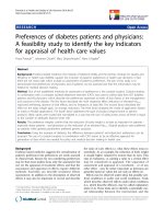
Figure 1 Axial images of trachea and pretracheal structures on ultrasound. Asterisk: Tracheal lumen. (a) Arrow- acoustic shadow of cricoid
cartilage. (b) Arrow- acoustic shadow of first tracheal ring. (c) Arrow: Anterior tracheal wall between first and second tracheal rings. Arrowhead-
Thyroid isthmus.
Figure 2 Axial image of trachea and surrounding st ructures with depiction o f pre-tracheal veins using color duplex imaging. Tr,
Tracheal lumen; Th, lobes of thyroid; I, thyroid isthmus; Arrowheads, pre-tracheal veins.
Rajajee et al. Critical Care 2011, 15:R67
/>Page 3 of 9
no vascular structure (Figure 2) in the path of the nee-
dle. Ideally, the space between the second and third
rings or the third and fourth tracheal rings was selected;
however, the precise inter-tracheal ring space was con-
sidered less i mportant than passage below the first and
above the fifth tracheal rings. Puncture through the
thyroid isthmus was permitt ed. The 15 G needle was
introduced perpendicularly to the skin and the needle
path was determined by the distinct acoustic shadow
ahead of the needle followed by the displacement of tis-
sue layers seen with needle passage (Figure 4a-d). Inden-
tation of the anterior tracheal wall by the needle was
then sometimes visible. A dvancement of the needle was
halted when the needle was seen to reach and then just
pass the anterior wall, with a palpable change in resis-
tance as the lumen was entered. The goal was to punc-
ture the anterior quadrant of the trachea, as close as
possible to the midline, as is our practice with standard
bronchoscopic guidance. Endotracheal position of the
tip was confirmed by the aspiration of air into a saline-
fille d syringe. The needle was then angled slightly caud-
ally to prevent retrograde passage of the guidewire. The
guidewire was then introduced and the needle removed.
The bronchoscope was then passed through the endo-
tracheal tube, the exact point of guidewire entry
recorded and the trachea visualized for any sign of
injury or posterior wall puncture. The bronchoscope
was then removed. A 2 cm horizontal incision was
made at the point of guidewire entry and blunt dissec-
tion was carried out. The 14Fr dilator was then used to
create the initial stoma, followed by the single-stage
“Rhino Horn” dilator over the guidewire and guiding
catheter. The appropriate-sized tracheostomy tube fitted
over an appropriate loading tube was then passed
through the stoma and secured. Endotracheal placement
of the tube was confirmed immediately using ausculta-
tion, verification of appropriate breath delivery on the
ventilator and the presence of the sonographic “lung-
sliding” bilaterally, as previously described. The lung
sliding sign is the visible “sliding” of the visceral pleura
on the parietal pleura on ultrasound imaging through
the intercostal space along with a characteristic appear-
ance on M-mode [16,17]. When seen bilaterally with
each delivered breath through the tracheal tube, this
sign denotes bilateral lung expansion.
The bronchoscope was then re-introduced through
the tracheostomy tube as well as the oro-tracheal tube
to look for a ny complications, such as airway injury,
tube misplacement or tracheal ring fracture. A ch est X-
ray was obtained on all patients to look for further
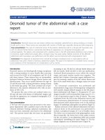
Figure 3 Measurement of skin to anterior tracheal wall thickness at the level of the second tracheal ring. Measured distance is 1.23 cm.
Rajajee et al. Critical Care 2011, 15:R67
/>Page 4 of 9
complications, such as pneumothorax or pneumo-med-
iastinum. Bronchoscopy was performed using Olympus
BF-1T30, BF-1T40 and BF-P40 fiber-optic broncho-
scopes with an Olympus Evis Exera 2 video system
(Olympus America, Center Valley, PA, USA).
Results
A total of 13 patients underwent US-PT. Sonographic
delineation of anatomy was possible in all enrolled sub-
jects and no patients required conversion to standard
bronchoscopic PT. There were nine women and four
men, with a mean age of 46 years (standard deviation 15
years, range 20 t o 68 years). The median BMI was 28.4
kg/m
2
(95% central range: 19.3 to 62.5 kg/m
2
). Diagnoses
were: aneurysmal subarachnoid hemorrhage (SAH, n =
4), severe traumatic brain injury (TBI, n = 2), ischemic
stroke (n = 2 ), intracerebral hemorrhage (n =2),severe
sepsis (n = 1), hepatic encephalopathy with chronic
obstructive pulmonary disease (COPD) (n = 1) and stiff-
person syndrome (n = 1). Tracheostomy was performed a
mean of four days (SD: 3 days, range 0 to 12 days) follow-
ing initiation of mechanical ventilation. Two patients
required tracheostomy because of the need for prolonged
mechanical ventilation. The indication for tracheostomy
in the other 11 patients was poor mental status with an
inability to cough effectively and clear secretions.
Two of 13 patients (including one with BMI 36 kg/
m
2
) were in cervical spine precautions. One patient
(with BMI 33 kg/m
2
) had had a previous tracheostomy.
Six of 13 patients were obese (BMI ≥30 kg/m
2
), while
threeweremorbidlyobese(BMI≥40 kg/m
2
). One
patient with extreme obesity had a BMI of 65.9 kg/m
2
.
Four patients, including all three patients with BMI >40
kg/m
2
and one patient in cervical spine precautions had
anatomy that could not be adequately defined by
palpation.
Ultrasound findings
Tracheal anatomy could be adequately defined in all
patients on ultrasound and tracheal puncture achieved
with a single advance of the needle in all patients. Ade-
quate sonographic delineation of anatomy with the lin-
ear probe was possible in all enrolled patients,
regardless of BMI. The first tracheal ring was visualized
above the sternal notch in all patients. Two patients
were found to have midline pretracheal veins, presumed
to be inferior thyroid veins, in the planned path of
puncture, requiring a change in the site of puncture.
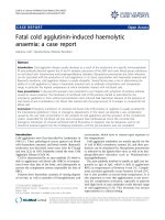
Theneedlepathcouldbedefinedusingtheacoustic
shadow ahead of the needle followed by displacement of
tissue in all patients (Figure 4a-d). In 4 of 13 (31%)
patients, actual indentation of the anterior tracheal wall
Figure 4 Acoustic shadow (Arrow) and displacement of tissue depicting the path of the needle during tracheal puncture.
Rajajee et al. Critical Care 2011, 15:R67
/>Page 5 of 9
during tracheal puncture could be seen. In these
patients, a subsequent straightening of the anterior wal l
was seen once the anterior wall had been breached.
Skin to trachea measurements in the three morbidly
obese (BMI >40 kg/m
2
) patients were 1.23 cm (BMI 42
kg/m
2
, internal tracheal diameter, ITD, 1.34 cm), 1.4 cm
(BMI 43 kg/m
2
, ITD 1.51 cm) and 2.97 cm (BMI 65.9
kg/m
2
, ITD 1.44 cm). Accordingly, the first two mor-
bidly obese patients had standard length Shiley
®
(Covi-
dien-Nellcor, Boulder, CO, USA) size 8.0 tracheostomy
tubes placed while the patient with BMI 66 kg/m
2
had
an extended proxi mal length Tracheo soft
®
size 7.0 tube
(Covidien-Nellcor, Boulder, CO, USA) placed
successfully.
Bronchoscopic findings
Guidewire placement was through the anterior quadrant
and was judged adequate in all patients on broncho-
scopy. Guidewire entry was between the third and
fourth tracheal rings in seven patients, second and third
rings in three patients, fourth and fifth rings in two
patients and first and second rings in one patient. Both
patients with guidewire entry between the fourth and
fifth tracheal rings had pretracheal vascular structures
that were specifically avoided. No complications were
found on bronchoscopy, including no clearly visible tra-
cheal ring fractures and no posterior wall injury/
puncture.
Monitoring of physiological parameters
Therewerenoepisodesofhypoxia(pulseoximetry
<90%) or significant hemodynamic instability during the
performance of P T. Seven of 13 patients had ICP moni-
tored during the procedure (2 with TBI, 2 with SAH
and 1 with ischemic stroke). Of note, all but two of
these patients (both of whom had undergone decom-
pressive craniectomy) demonstrated transient (recorded
as lasting for less than five minutes each time) eleva-
tions of ICP to >25 mmHg. The average maximum ICP
seen during the procedure was 29 mmHg (SD: 9
mmHg, range 15 to 39 mm Hg). Of note, the maximum
recorded ICP during the procedure was always during
bronchoscopy, although a smaller increase in ICP, for
much shorter duration (recorded as bein g less than one
minute in each instance), was also noted during direct
laryngosc opy and passage of the single stage dilator and
the tracheostomy tube. Retention of the bronchoscope
in the airway was limited to no more than five minutes
at a time, to limit ICP elevation.
Complications and follow-up
No complications were seen on bronchoscopy or chest
X-ray. The tube was seen to be in good position within
the trachea in all patients on bronchoscopy and chest
X-ray, with the tip positioned within the thoracic cav ity
and at least 2 cm above the trachea. Follow-up was
available for an average period of four months (SD:
three months, range one week to seven months) follow-
ing tracheostomy. Three patients had died, all from
withdrawal of care, of causes unrelated to tracheos tomy
(two for failure to demonstrate any neurological recov-
ery and one for failure to wean from mechanical ventila-
tion with multiple medical co-morbidities). Five patients
had undergone successful decanulation of the tracheal
tube, a mean of 17.6 days (SD: 4.5 days, range 12 to 24
days) from tracheostomy. One female patient on
mechanical ventilation with BMI 33 kg/m
2
,adequately
palpable pre-procedure neck anatomy and a standard
length Shiley
®
6.0 tube suffered dislodgment of the tra-
cheostomy tube seven days after tracheostomy during a
period of severe agitation with hea d shaking and devel-
oped acute respiratory distress while the tube was dis-
lodged. The tube was emergently replaced through the
stoma and the patient had no permanent injury from
the accidental decanulation. The tube was subsequently
empirically changed to an extended proximal length size
6.0 tube to decrease the risk of future dislodgement. No
other complications, minor or major, were observed in
any patient during the available period of follow-up.
Discussion
The purpose of our study was to demonstrate the feasi-
bility of performing percutaneous tracheostomy under
real-time ultrasound guidance with actual visualization
of the needle path and to assess the accuracy of this
technique in placement of the guidewire b elow the first
tracheal ring. Tracheostomy was typically performed
early (mean four days after initiation of mechanical ven-
tilation). Our practice is to perform early tracheostomy,
within one week of intubation, for patients with acute
brain injury, who, in the judgment of the treating clini-
cian, are likely to require mechanical ventilation, or a
definitive airway (because of poor mental status and the
inability to cough or handle secretions) for more than
two to three weeks. Our rationale for performing early
tracheostomy, the benefits of which are a subject of
debate, is a potential reduction in the number of ventila-
torandICUdays[18,19]aswellasimprovementin
patient comfort and reduced need for sedation [20].
The use of real-time sonography with visualization of
the needle path for central venous catheter placement is
now widespread and may decrease the rate of complica-
tions [10,11]. We believe that this technique, which has
not previously been described in the literature with PT,
has many potential advantages over other techniques of
PT. The first is the ability to consistently place the tra-
cheostomy tube below the first tracheal ring. Pla cement
of the tracheal tube above the first tracheal ring may
Rajajee et al. Critical Care 2011, 15:R67
/>Page 6 of 9
increase the risk of late sub-glottic cicatrization and ste-
nosis [21-23]. In one study of patients who underwent
autopsy following PT, 5 of 15 patients had the tracheal
tube placed above the first tracheal ring when the tube
was placed blindly v s zero of 11 patients when PT was
performed with ultrasound guidance [8]. In this study,
however, demonstration of the trachea on ultrasound
was in sagittal section to determine the appropriate level
of puncture. Actual visualization of the needle path and,
therefore, the actual level of puncture was not possible.
Real-time imaging of the need le path allows visual con-
firmation that the anterior wall has been passed, at
which point the needle is advanced no further and pos-
terior wall injury is avoided. Although a special metal
stopper was used in the aforementioned study to avoid
posterior wall injury, it is custom-made and not widely
available. A further strength of our study is that all
guidewire and final tracheal tube positions were imme-
diately verified with bronchsocopy, unlike previous stu-
dies with ultrasound which either did not use real-time
guidance or were able to confirm tube position only i n
select patients who underwent autopsy.
In this limited feasibility study, the presence of morbid
obesity, sub-optimal palpable neck anatomy, previous
tracheostomy or cervical spine precautions did not
appear to be a barrier to the performanc e of US-PT.
Prior studies have shown that PT should not be auto-
matically contraindicated in these groups of patients
[12-14]. About half the patients in our series had one of
these factors: morbid obesity in three (including one
patient with BMI 65.9 kg/m
2
), cervical spine precautions
in two and previous tracheostomy in one. We believe
that our technique of real-time guidance will further
enhance the safety and ease of performance of PT in
these sub-groups. In our series, these factors appeared
to present no increased difficulty for the performance of
ultrasound guided puncture, as long as the first tracheal
ring was clearly visible above the sternal notch. Particu-
larly useful may be the ability to measure the pretra-
cheal soft tissue thickness in the morbidly obese and the
consequent ability to assess the need for extended-
length tracheostomy tube placement, as has been
described earlier [15]. The patient in our study who suf-
fered a late dislodgement while severely agitated had not
had these measurements performed as she was not mor-
bidly obese and appeared to have well-palpable anatomy
prior to the procedure. It is possible that routine mea-
surements of pretracheal thickness, even in patients
with normal palpab le anatomy, may help better select
the optimal tube for placement and decrease the rate of
subsequent tracheostomy dislodgement [15].
Another advantage of US-PT is the ability to avoid
vascular structures anterior to the trachea. Prior stud ies
have demonstrated a potential role for pre-procedure
ultrasound imaging in transverse section to identify vas-
cular structures and reducing the ri sk of bleeding [7,24].
In one study, bleeding from injury to vascular structures
which would have likely been identified had ultrasound
been used was considered significant i n 24 of 497 (5%)
PTs performed without pre-procedure US evaluation,
with 6 of 24 patients requiring conversion to surgical
tracheostomy [25]. Pre-procedure ultrasound resulted in
a change in the planned site of tracheal puncture in 24%
of patients in another study [26]. These studies did not
use real-time guidance. In our study, 2 of 10 patients
(20%) had the planned site of puncture moved (both
caudally) to avoid vascular structures. The use of real-
time imaging may be preferab le for avoiding vascular
structures compared to pre-proc edure imaging alone,
since avoidance of a vascular structure such as an infer-
ior thyroid vein cannot be taken for granted without
actual visualization of the needle path.
US-PT also does not have some of the disadvantages
of bronchoscopy. This may be particularly relevant to
the group of patients in whom this study was performed
- patients with acute brain injury. Our study confirms a
previously reported observation that bronchoscopy is
associated with a pre dictable, if transient, increase in
intracranial pressure, probably caused by hypoventilation
and hypercarbia [6]. This may be particularly true when
a policy of performing early, rather than late, tracheost-
omy is used [27], as is the practice in several neuro-
ICUs including ours. Although PT has been demon-
strated to be safe, overall, in patients requiring ICP
monitoring [28], the use of real-time ultrasound gui-
dance minimizes hypercarbia and the consequent eleva-
tion of ICP compared to puncture under continuous
bronchoscopic monitoring and, therefore, may be pre-
ferable for patients at significant risk of developing
intracranial hypertension and ICP plateau waves.
The ability to perform US-PT without bronchoscopy
is limited, however, by the need to safely retract the oro-
tracheal tube to a position high enough to permit tra-
cheal puncture while avoiding accidental extubation. We
used direct laryngoscopy for this purpose, to demon-
strate one potential method of safely performing US-PT
without bronchoscopy. An adequate laryngoscopic grade
of view was, therefore, a pre-requisite. For patients with
an inadequate laryngoscopic view of the glottis, or
operators who do not routinely perform direct laryngo-
scopy, other non-bronchoscopic options for retraction
of the orotracheal tube may exist. For patients with
poor laryngoscopic views with standard blades, use of a
video laryngoscope may provide a superior view [29].
One study described using Doppler ultrasound over the
trachea to determine the correct position of the orotra-
cheal tube [30], a technique which is, in our ane cdotal
experience, less reliable than direct laryngoscopy or
Rajajee et al. Critical Care 2011, 15:R67
/>Page 7 of 9
bronchoscopy. Laryngeal mask airways have been used
successfully instead of orotracheal tubes during PT
[31,32], although the relative safety of this technique is
debatabl e [33]. In view of the other advantages, detailed
above, it is possible that r eal-time ultrasound guidance
of the needle path will find a role as a routine adjunct,
rather than alternative, to standard bronchoscopy-
guided PT.
Our study is limited in being only a prelim inary
demonstration of the feasibility of using real-time ultra-
sound guidance for tracheal puncture during PT. Also,
long term follow-up was not available to detect the inci-
dence of late tracheal stenosis. Larger, randomized stu-
dies are required to better define the relative advantages
of this technique, appropriate candidates and the safety
of US-PT performed without bronchoscopic confirma-
tion of guidewire and cannula placement. We believe
our study lays the foundation for future clinical trials.
Conclusions
Percutaneous tracheostomy can be performed safely
using real-time sonographic visualization of the needle
path to ensure avoidance of vascular structur es and pla-
cement of the tracheostomy tube below the first tracheal
ring, including in patients with morbid obesity and cer-
vical spine precautions.
Key messages
• Percutaneous tracheostomy can be performed
using real-time ultrasound guidance for visualization
of the needle path during tracheal puncture.
• Real- time ultrasound guidance during percutanous
tracheostomy can be used to guide placement of the
tracheal tube below the first tracheal ring and to
avoid vascular structures.
• Real-time ultrasound guidance can facilitate percu-
tanous tracheostomy in patient s with morbid obesity
and cervical spine precautions.
Abbreviations
BMI: body mass index; COPD: chronic obstructive pulmonary disease; ICP:
intracranial pressure; ICU: intensive care unit; ITD: internal tracheal diameter;
PT: percutaneous tracheostomy; SAH: subarachnoid hemorrhage; SD:
standard deviation; TBI: traumatic brain injury; US: ultrasound; US-PT:
ultrasound guided percutaneous tracheostomy.
Acknowledgements
Institutional Review Board approval: University of Michigan, Ann Arbor, MI,
USA.
Author details
1
Departments of Neurosurgery and Neurology, University of Michigan Health
System, 3552 Taubman Health Care Center, 1500 E. Medical Center Dr., SPC
5338, Ann Arbor, MI 48109-533 8, USA.
2
Department of Anesthesiology,
University of Michigan Health System, University Hospital, 1500 E. Medical
Center Drive, Room 1H247, Ann Arbor, MI 48109-5048, USA.
Authors’ contributions
VR conceived of the study, participated in its design and coordination, and
drafted the manuscript. JFF, LRR and TLJ participated in the design and
coordination of the study, and helped to draft the manuscript. All authors
read and approved the final manuscript.
Competing interests
The authors declare that the y have no competing interests.
Received: 13 December 2010 Revised: 18 January 2011
Accepted: 22 February 2011 Published: 22 February 2011
References
1. Heikkinen M, Aarnio P, Hannukainen J: Percutaneous dilational
tracheostomy or conventional surgical tracheostomy? Crit Care Med 2000,
28:1399-1402.
2. Freeman BD, Isabella K, Cobb JP, Boyle WA, Schmieg RE Jr, Kolleff MH,
Lin N, Saak T, Thompson EC, Buchman TG: A prospective, randomized
study comparing percutaneous with surgical tracheostomy in critically ill
patients. Crit Care Med 2001, 29:926-930.
3. Delaney A, Bagshaw SM, Nalos M: Percutaneous dilatational tracheostomy
versus surgical tracheostomy in critically ill patients: a systematic review
and meta-analysis. Crit Care 2006, 10:R55.
4. Marelli D, Paul A, Manolidis S, Walsh G, Odim JN, Burdon TA, Shennib H,
Vestweber KH, Fleiszer DM, Mulder DS: Endoscopic guided percutaneous
tracheostomy:early results of a consecutive trial. J Trauma 1990,
30:433-435.
5. Barba CA, Angood PB, Kauder DR, Latenser B, Martin K, McGonigal MD,
Phillips GR, Rotondo MF, Schwab CW: Bronchoscopic guidance makes
percutaneous tracheostomy a safe, cost-effective, and easy-to-teach
procedure. Surgery 1995, 118:879-883.
6. Reilly PM, Anderson HL III, Sing RF, Schwab CW, Bartlett RH: Occult
hypercarbia. An unrecognized phenomenon during percutaneous
endoscopic tracheostomy. Chest 1995, 107:1760-1763.
7. Hartfield A, Bodenham A: Portable ultrasonic scanning of the anterior
neck before percutaneous dilatational tracheostomy. Anaesthesia 1999,
54:660-663.
8. Sustić A, Kovac D, Zgaljardić Z, Zupan Z, Krstulović B: Ultrasound-guided
percutaneous dilatational tracheostomy: a safe method to avoid cranial
misplacement of the tracheostomy tube. Intensive Care Med 2000,
26:1379-1381.
9. Sustić A: Role of ultrasound in the airway management of critically ill
patients. Crit Care Med 2007, 35:S173-S177, Review.
10. Slama M, Novara A, Safavian A, Ossart M, Safar M, Fagon JY: Improvement
of internal jugular vein cannulation using an ultrasound-guided
technique. Intensive Care Med 1997, 23:916-919.
11. Karakitsos D, Labropoulos N, De Groot E, Patrianakos AP, Kouraklis G,
Poularas J, Samonis G, Tsoutsos DA, Konstadoulakis MM, Karabinis A: Real-
time ultrasound-guided catheterisation of the internal jugular vein: a
prospective comparison with the landmark technique in critical care
patients. Crit Care 2006, 10:R162.
12. Sustic A, Krstulovic B, Eskinja N, Zelic M, Ledic D, Turina D: Surgical
tracheostomy versus percutaneous dilational tracheostomy in patients
with anterior cervical spine fixation: preliminary report. Spine 2002,
27:1942-1945.
13. Mansharamani NG, Koziel H, Garland R, LoCicero J III, Critchlow J, Ernst A:
Safety of bedside percutaneous percutaneous dilatational tracheostomy
in obese patients in the ICU. Chest
2000, 117:1426-1429.
14.
Meyer M, Critchlow J, Mansharamani N, Angel LF, Garland R, Ernst A:
Repeat bedside percutaneous dilational tracheostomy is a safe
procedure. Crit Care Med 2002, 30:986-988.
15. Szeto C, Kost K, Hanley JA, Roy A, Christou N: A simple method to predict
pretracheal tissue thickness to prevent accidental decannulation in the
obese. Otolaryngol Head Neck Surg 2010, 143:223-229.
16. Lichtenstein D, Menu Y: A bedside ultrasound sign ruling out
pneumothorax in the critically ill: lung sliding. Chest 1995, 108:1345-1348.
17. Chun R, Kirkpatrick AW, Sirois M, Sargasyn AE, Melton S, Hamilton DR,
Dulchavsky S: Where’s the tube? Evaluation of hand-held ultrasound in
confirming endotracheal tube placement. Prehospital Disaster Med 2004,
19:366-369.
Rajajee et al. Critical Care 2011, 15:R67
/>Page 8 of 9
18. Terragni PP, Antonelli M, Fumagalli R, Faggiano C, Berardino M,
Pallavicini FB, Miletto A, Mangione S, Sinardi AU, Pastorelli M, Vivaldi N,
Pasetto A, Della Rocca G, Urbino R, Filippini C, Pagano E, Evangelista A,
Ciccone G, Mascia L, Ranieri VM: Early vs late tracheotomy for prevention
of pneumonia in mechanically ventilated adult ICU patients: a
randomized controlled trial. JAMA 2010, 303:1483-1489.
19. Griffiths J, Barber VS, Morgan L, Young JD: Systematic review and meta-
analysis of studies of the timing of tracheostomy in adult patients
undergoing artificial ventilation. BMJ 2005, 330:1243.
20. Nieszkowska A, Combes A, Luyt CE, Ksibi H, Trouillet JL, Gibert C, Chastre J:
Impact of tracheotomy on sedative administration, sedation level, and
comfort of mechanically ventilated intensive care unit patients. Crit Care
Med 2005, 33:2527-2533.
21. Walz MK, Schmidt U: Tracheal lesion caused by percutaneous dilatational
tracheostomy–a clinico-pathological study. Intensive Care Med 1999,
25:102-105.
22. McFarlane C, Denholm SW, Sudlow CL, Moralee SJ, Grant IS, Lee A:
Laryngotracheal stenosis: a serious complication of percutaneous
tracheostomy. Anaesthesia 1994, 49:38-40.
23. Van Heurn LW, Theunissen PH, Ramsay G, Brink PR: Pathologic changes of
the trachea after percutaneous dilatational tracheostomy. Chest 1996,
109:1466-1469.
24. Flint AC, Midde R, Rao VA, Lasman TE, Ho PT: Bedside ultrasound
screening for pretracheal vascular structures may minimize the risks of
percutaneous dilatational tracheostomy. Neurocrit Care 2009, 11:372-376.
25. Muhammad JK, Major E, Wood A, Patton DW: Percutaneous dilatational
tracheostomy: haemorrhagic complications and the vascular anatomy of
the anterior neck. A review based on 497 cases. Int J Oral Maxillofac Surg
2000, 29:217-222.
26. Kollig E, Heydenreich U, Roetman B, Hopf F, Muhr G: Ultrasound and
bronchoscopic controlled percutaneous tracheostomy on trauma ICU.
Injury 2000, 31:663-668.
27. Kocaeli H, Korfali E, Taşkapilioğlu O, Ozcan T: Analysis of intracranial
pressure changes during early versus late percutaneous tracheostomy in
a neuro-intensive care unit. Acta Neurochir (Wien) 2008, 150:1263-1267.
28. Milanchi S, Magner D, Wilson MT, Mirocha J, Margulies DR: Percutaneous
tracheostomy in neurosurgical patients with intracranial pressure
monitoring is safe. J Trauma 2008, 65:73-79.
29. Mort TC: Tracheal tube exchange: feasibility of continuous glottic
viewing with advanced laryngoscopy assistance. Anesth Analg 2009,
108:1228-1231.
30. Reilly PM, Sing RF, Giberson FA, Anderson HL, Rotondo MF, Tinkoff GH,
Schwab CW: Hypercarbia during tracheostomy: a comparison of
percutaneous endoscopic, percutaneous Doppler, and standard surgical
tracheostomy. Intensive Care Med
1997, 23:859-864.
31. Dosemeci L, Yilmaz M, Gurpinar F, Ramazanoglu A: The use of the
laryngeal mask airway as an alternative to the endotracheal tube during
percutaneous dilatational tracheostomy. Intensive Care Med 2002,
28:63-67.
32. Sustić A, Zupan Z, Antoncić I: Ultrasound-guided percutaneous
dilatational tracheostomy with laryngeal mask airway control in a
morbidly obese patient. J Clin Anesth 2004, 16:121-123.
33. Ambesh SP, Sinha PK, Tripathi M, Matreja P: Laryngeal mask airway vs
endotracheal tube to facilitate bedside percutaneous tracheostomy in
critically ill patients: a prospective comparative study. J Postgrad Med
2002, 48:11-15.
doi:10.1186/cc10047
Cite this article as: Rajajee et al.: Real-time ultrasound-guided
percutaneous dilatational tracheostomy: a feasibility study. Critical Care
2011 15:R67.
Submit your next manuscript to BioMed Central
and take full advantage of:
• Convenient online submission
• Thorough peer review
• No space constraints or color figure charges
• Immediate publication on acceptance
• Inclusion in PubMed, CAS, Scopus and Google Scholar
• Research which is freely available for redistribution
Submit your manuscript at
www.biomedcentral.com/submit
Rajajee et al. Critical Care 2011, 15:R67
/>Page 9 of 9