Báo cáo y học: "Virus-associated hemophagocytic syndrome as a major contributor to death in patients with 2009 influenza A (H1N1) infection" potx
Bạn đang xem bản rút gọn của tài liệu. Xem và tải ngay bản đầy đủ của tài liệu tại đây (787.58 KB, 8 trang )
RESEARCH Open Access
Virus-associated hemophagocytic syndrome as a
major contributor to death in patients with 2009
influenza A (H1N1) infection
Gernot Beutel
1*
, Olaf Wiesner
2
, Matthias Eder
1
, Carsten Hafer
3
, Andrea S Schneider
4
, Jan T Kielstein
3
,
Christian Kühn
5
, Albert Heim
6
, Tina Ganzenmüller
6
, Hans-Heinrich Kreipe
7
, Axel Haverich
5
, Andreas Tecklenburg
8
,
Arnold Ganser
1
, Tobias Welte
2
and Marius M Hoeper
2
Abstract
Introduction: Virus-associated hemophagocytic syndrome (VAHS) is a severe complication of various viral
infections often resulting in multiorgan failure and death. The pur pose of this study was to describe baseline
characteristics, development of VAHS, related treatments and associated mortality rate of consecutive critically ill
patients with confirmed 2009 influenza A (H1N1) infection and respiratory failure.
Methods: We conducted a prospective observational study of 25 critically ill patients with 2009 influenza A (H1N1)
infection at a single-center intensive care unit in Germany between 5 October 2009 and 4 January 2010.
Demographic data, comorbidities, diagnosis of VAHS, illness progression, treatments and survival data were
collected. The primary outcome measure was the development of VAHS and related mortality. Secondary outcome
variables included duration of mechanical ventilation, support of extracorporeal membrane oxygenation and
duration of viral shedding.
Results: VAHS developed in 9 (36%) of 25 critically ill patients with confirmed 2009 influenza A (H1N1) infection,
and 8 (89%) of them died. In contrast, the mortality rate in the remaining 16 patients without VAHS was 25% (P =
0.004 for the survival difference in patients with or without VAHS by log-rank analysis). The patients were relatively
young (median age, 45 years; interquartile range (IQR), 35 to 56 years of age); however, 18 patients (72%)
presented with one or more risk factors for a severe course of illness. All 25 patients received mechanical
ventilation for severe acute respiratory distress syndrome and refractory hypoxemia, with a median duration of
mechanical ventilation of 19 days (IQR, 13 to 26 days). An additional 17 patients (68%) required extracorporeal
membrane oxygenation for a median of 10 days (IQR, 6 to 19 days).
Conclusions: The findings of this study raise the possibility that VAHS may be a frequent complication of severe
2009 influenza A (H1N1) infection and represents an important contributor to multiorgan failure and death.
Introduction
In the spring of 2009, novel human influenza A (H1N1)
(A/H1N1/2009) infection began spreading from Mexico
around the globe, causing a worldwide pandemic [1-3].
Contrary to initial fears, most patients experienced a
mild clinical course. Some patients did become critically
ill with respiratory failure, however, requiring intensive
care and ventilator support. Mortality rates were high in
these patients, especially in those who developed multi-
organ failure [4-6].
The mechanisms leading to multiorgan failure and
death in patients with influenza infection are not well
understood. Septicemia is a leading cause of seasonal
influenza, mainly due to secondary infection by other
microorganisms, principally Gram-positive or Gram-
negative bacteria. The first reports of fatal A/H1N1/
2009 infections, howev er, only described septicemi a
occasionally [7,8]. Other pathomechanisms may also
* Correspondence:
1
Departments of Hematology, Hemostasis, Oncology, and Stem Cell
Transplantation, Hannover Medical School, Carl-Neuberg-Strasse 1, D-30625
Hannover, Germany
Full list of author information is available at the end of the article
Beutel et al. Critical Care 2011, 15:R80
/>© 2011 Beut el et al .; licensee BioMed Central Ltd. This is an open access article distributed under the terms of the Creative Commons
Attribution License (http://crea tivecommons.org/licenses/by/2.0), which pe rmits unrestricted use, distribution, and re prod uction in
any medium, provide d the origin al work is properly cited.
contribute to severe multiorgan failure, with several
reports suggesting that patients with severe influenza
infection may develop a virus-associated hemophagocy-
tic syndrome (VAHS) [8-10].
VAHS may present as an aggressiv e, life-threateni ng
disease, with previous reports implicating its role in fatal
cases of season al (H3N2) influenza as well as avian
(H5N1) influenza virus [8,9,11]. Analogously to heredi-
tary hemophag ocytic lymphohistiocytosis (HLH ), VAHS
is associated with massive cytokine release ("cytokine
storm”), elevated plasma levels of soluble interleukin 2
receptor (sIL-2R) and other inflammatory mediators and
the accumulation of activated T-lymphocytes and
macrophages in various organs, frequently resulting in
multiorgan failure and death [12-16].
In Germany, peak infection rates for A/H1N1/2009
occurred between October 2009 and December 2009,
that is, in the first winter season after the initial out-
break in Mexico. In our tertiary care center, the first cri-
tically ill patient with A/H1N1/2009 infection and
respiratory failure was admitted on 5 October 2009.
This patient required mechanical ventilation and extra-
corporeal membrane oxygenation (ECMO) f or 2 weeks.
The patient’s lung function eventually recovered, and
the patient was successfully weaned from ECMO sup-
port but subsequently died as a result of progressive
multiorgan failure 25 days after hospital admission.
There was no evidence of secondary septic complica-
tions, and VAHS was identified as the most likely cause
of multiorgan failure. Therefore, we systematically and
prospectively assessed al l further patients with A/H1N1/
2009 admitted to our intensive care unit (ICU) for the
development of VAHS.
This report describes a series of 25 consecutive criti-
cally ill patients with severe A/H1N1/2009 infection in
whom VAHS was found to be a leading contributor to
death.
Materials and methods
Study design and patient eligibility
Between 5 October 2009 and 4 Janu ary 2010, we pro-
spectively studied 25 adult patients (22 Caucasian, 2
Turkish and 1 Arabian) with confirmed severe A/H1N1/
2009 infection admitted to the medical ICU at Hann-
over Medical School, Hannover, Germany. All critically
ill patients were defined as those requiring invasive
mechanical ventilation, having a fraction of inspired
oxygen level greater than 60% or receiving intravenous
infusion of vasopressor or inotropic medication. Addi-
tional venovenous ECMO support was necessary in 17
patients. In each c ase, the diagnosis of A/H1N1/2009
infection was confirmed by real-time reverse transcrip-
tase polymerase chain reaction (RT-PCR) assay [17].
Data collection
Data collection included patient demographics as well as
the presence of the number of predefined comorbidities.
Presumed infectious organisms from upper and lower
respiratory tract specimens were identified by perform-
ing A/H1N1/2009 RT-PCR assays within 48 hours of
admission. Further viral, microbiological and fungal sur-
veillance included twice-weekly nasopharyngeal swabs,
bronchial lavage, and twice-w eekly analysis of blood and
urine cultures. In addition to daily routine laboratory
analysis, which included C-reactive protein (CRP), pro-
calcitonin, and la ctate dehydrogenase (LDH) levels,
thrice-weekly measurements of serum ferritin and sIL-
2R levels as well as weekly measurements to detect tri-
glyceridemia and hypofibrinogenemia were performed.
VAHS was suspected when patients developed fever,
cytopenia affecting at least two lineages, hepatitis or
splenomegaly and/or when serum levels of sIL-2R and
ferritin were increased. The presence of two or more of
these features triggered the performance of bone mar-
row aspiration and biopsy. The diagnosis of VAHS was
made according to established HLH diagnostic criteria if
three of four major criteria (fever, cytopenia, hepatitis or
splenomegaly) and at least one minor criterion (evidence
of hemophagocytosis in bone marrow samples or
increase in serum level of sIL-2R or ferritin, respectively)
were present [18].
We further obtained information regarding the total
duration of hospitalization, mechanical ve ntilation and
ECMO support, as well as the duration and use of anti-
viral, antibiotic and antifungal treatments. Severity of ill-
ness was assessed using the Acute Physiology and
Chronic Health Evaluation II [19] and Sepsis-related
Organ Failure Assessment scores [20]. Sev erity of illness
before the commencement of ECMO was assessed on
the basis of ventilatory parameters, arterial blood gas
values and chest radiograph findings.
The primary outcome measure was the development
of VAHS an d VAHS-related mortality. Secondary out-
come variables included the duration of mechanical ven-
tilation, ECMO support and t he duration of viral
shedding.
Standard treatments
Antiviral treatment consisted of oral oseltamivir at doses
of 75 to 150 mg twice daily and/or intr avenous zana mi-
vir at a dose of 600 mg twice daily (individually pro-
vided on a compassionate use basis by GlaxoSmithKline,
Munich, Germany) [21]. The standard therapeutic
course for each compound lasted 5 days. If ongoing
viral shedding was present, additional treatment courses
were administered until A/H1N1/2009 infection was no
longer detectable by RT-PCR assay. Early corticosteroid
Beutel et al. Critical Care 2011, 15:R80
/>Page 2 of 8
treatment was not routinely used in this patient
population.
Patients with VAHS were intended to be treated
according to the recommendations of the Histiocyte
Society with a modified HLH-94 protocol which con-
sisted of intravenous etoposide (100 to 150 mg/m
2
once
weekly) and intravenous dexamethasone (8 mg/m
2
once
daily) [22-24].
Our diagnostic and therapeutic approach was
approved by the local institutional review board (Ethics
Committee of the Hannover Medical School, Reference
953-2011). In agreement with local regulations,
informed consent was waived, as all patients were trea-
ted according to the standards of care in our center.
Statistical analysis
Descriptive analysis was performed using medians and
interquartile ranges (IQR). All statistical parameters
were tested for normal distribut ion using the Shapiro-
Wilk test of normality. Discrete variables were compared
using Pearson’s c
2
test or Fisher’sexacttest.Fornor-
mally distributed data, co ntinuous variables of patients
with and without VAHS were an alyzed using the Welch
two-sample t-test. Otherwise, the Wilcoxon rank-sum
test was used. Probability of survival was determined on
the basis of survival curves using t he Kaplan-Meier
method. Differences between groups were calculated
using a stratified log-rank test (Fleming-Harrington G r
family). Hazard ratios for the development of VAHS as
a time-dependent variable were evaluated by using a
Cox proportional regression model. Last survival status
for all patients was assessed on 31 March 2010. Two-
sided P values <0.05 were considere d statistically signifi-
cant differences. R-Project software version 2.10.1 for
Linux was used for statistical computation.
Results
Characteristics of patients
Between 5 October 2009 and 4 January 4 2010, 25 adult
patients (22 Caucasian, 2 Turkish and 1 Arabian) ful-
filled the study’ s eligibility criteria. All patients were
admitted with severe respiratory failure requiring inva-
sive mechanical ventilation (n = 25, 100%) and venove-
nous ECMO support (n = 17, 68%). The median age
was45years(IQR,35to56years),and16patients
(64%) were men. Seven of these patients (28%) had no
preexisting medical conditions, whereas 18 patients
(72%) presented with one or more risk factors, including
obesity (n = 10), cardiovascular disease (n =8),chronic
pulmonary disease (n = 4), chronic ren al insufficiency (n
= 4), immunosuppressive therapy after organ transplan-
tation (n = 3), diabetes mellitus (n = 3), liver diseas e (n
= 2), malignant lymphoma (n = 2) and pregnancy (n =
2) (Table 1). In all patients, A/H1N 1/2009 infection was
identified by RT-PCR assay, whereas seasonal subtypes
of influenza A were not detectable.
Severity of illness
The median durations of mechanical ventilation and
ECMO support wer e 19 days (IQR, 3 to 26 days) and 10
days (IQR, 6 to 19 days), respectively. Before ECMO
commencement, patients had a median respiratory rate
of 24 breaths/minute (IQR, 20 to 26/breaths/minute), a
median positive end-expi ratory pressure of 18 cmH
2
O
(IQR, 15 to 20 cmH
2
O) and a median peak airway pres-
sure of 34 cm H
2
O (IQR, 31 to 36 cm H
2
O). The med-
ian partial pressure of oxygen in arterial blood (PaO
2
)
level was 66 mmHg (IQR, 56 to 85 mmHg), with a
PaO
2
/fractionofinspiredoxygenratioof85mmHg
(IQR, 59 to 138 mmHg). In the course of critical illness,
21 patients (84%) r eceived vasopressor or inotrope ther-
apy and 14 patients (56%) received renal replacement
therapy.
Antiviral treatment and virus shedding
Oseltamivir was used as antiviral treatment in 24
patients (96%) for a median of 7 days (IQR, 4 to 10
days), and zanamivir was used as antiviral therapy in 15
patients (60%) for a median of 7 days (IQR, 5 to 12
days). The median duration of viral shedding from dis-
ease onset to the last positive A/H1N1/2009 infection
RT-PCR assay was 19 days (IQR, 14 to 26 days). In
patients without VAHS, the median v iral shedding t ime
was 15 days (IQR, 12 to 22 days) as opposed to a med-
ian of 21 days (IQR, 14 to 26 days) ( P = 0.13) in patients
with VAHS.
Occurrence of VAHS
Nine patients (36%) fulfilled the diagnostic criteria f or
VAHS. The median time from the onset of symptoms
to the diagnosis of VAHS was 23 days (IQR, 15 to 29
days), and the median time from admission to the ICU
to the diagnosis of VAHS was 16 days (IQR, 11 to 25
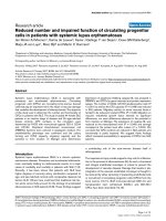
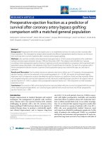
days). Within the first 16 days after symptom onset,
the predicted hazard ratio revealed a 12-fold increase
(log hazard ratio, 2.5) for the development of VAHS
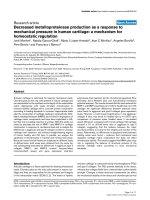
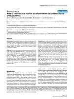
(Figure 1). When VAHS was diagnosed, patients
demonstrated cytopenia affecting at least two lineages,
hepatitis or splenomegaly with a bone marrow speci-
men demonstrating characteristic features of hemopha-
gocytosis (Figure 2). At the same time, serum analysis
revealed markedly elevated levels of ferritin, sIL-2R,
LDH and CRP (Table 1). However, there was no evi-
dence of uncontrolled bacterial infection in any of
these patients on the basis of repeated sterile cultures
from the tracheobronchial tree, blood and urine.
Over the course of time, patients who presented with
VAHS developed multiorgan dysfunction with hepatitis
Beutel et al. Critical Care 2011, 15:R80
/>Page 3 of 8
(n = 9, 100%), renal f ailure (n = 8, 89%), pancytopenia
(n = 8, 89%) and lactic acidosis (n = 7, 78%).
VAHS-directed therapy and mortality
Treatment of VAHS was started in six of the nine
patients with VAHS (n = 4 with etoposide and dexa-
methasone and n = 2 with steroids only). Three patients
were moribund when VAHS was diagnosed and were no
longer considered candidates for treatment with etopo-
side and dexamethasone. Despite VAHS-directed ther-
apy, five of the six patients who were treated died as a
result of uncontrolled disease progress leading to multi-
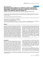
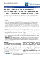
organ failure. Overall, eight (89%) of the nine pa tients
withconfirmedVAHSdiedcomparedto4(25%)of16
patients without VAHS (Figure 3). This difference was
statistically significant (P = 0.004). Overall, 12 patients
(48%) died, all as a result of multiorgan failure.
Discussion
The present case series confirms previous postmortem
analyses that A/H1N1/2009 infection can cause severe
and fatal infections in humans, even in the absence of
risk factors [25]. More importantly, our data show that
VAHS should be taken into consideration as a major
pathogenetic mechanism contributing to multiorgan fail-
ure and death in patients with severe viral infections.
Summary of study findings
The occurrence of VAHS in approximately one-th ird (9
of 25, 36%) of our patients was unexpected. Of the nine
patients diagnosed with VAHS, eight (89%) failed to sur-
vive. By c omparison, only 4 (25%) of the remaining 16
patients without VAHS died, suggesting that VAHS
development either cont ributes greatly to or is itself
causative of death in this patient population. On the
basis of our initial experience, we prospectively screened
all patients admitted to our ICU with A/H1N1/2009
infection for the development of VAHS, and it is t here-
fore unlikely that we missed cases. VAHS was not an
initial feature of A/H1N1/2009 infection but developed
a median of 23 days (IQR, 15 to 29 days) after symptom
onset and a median of 16 days (IQR , 11 to 25 days)
after ICU admission. The duration of viral shedding
tended to be longer in patients with VAHS than in
Table 1 Baseline demographic and clinical characteristics of critically ill patients with H1N1 infection
a
Characteristics All patients
(n = 25)
Patients with VAHS
(n =9)
Patients without VAHS
(n = 16)
P
value
Median age, yr (IQR) 45 (35 to 56) 53 (39 to 56) 38 (32 to 52) 0.32
Sex, F/M 9/16 2/7 7/9 0.52
Any comorbidity, n (%)
b
18 (72%) 5 (56%) 13 (81%) 0.36
Obesity, n (%)
c
10 (40%) 3 (33%) 7 (44%) 0.67
Median APACHE II score at admission (IQR) 21 (19 to 30) 28 (23 to 32) 21 (18 to 23) 0.29
Median SOFA score at admission (IQR) 11 (10 to 13) 13 (11 to 16) 11 (9 to 12) 0.22
Median duration of mechanical ventilation, days
(IQR)
19 (13 to 26) 25 (17 to 26) 18 (11 to 25) 0.69
Patients on ECMO support, n (%) 17 (68%) 9 (100%) 8 (50%) 0.02
e
Median duration of ECMO support, days (IQR) 10 (6 to 19) 10 (4 to 19) 11 (8 to 20) 0.90
Median duration of viral shedding, days (IQR) 19 (14 to 26) 21 (14 to 26) 15 (12 to 22) 0.13
Patients treated with oseltamivir, n (%)
d
24 (96%) 9 (100%) 15 (94%) 0.44
Median duration of oseltamivir treatment, days (IQR) 7 (4 to 10) 10 (5 to 12) 7 (4 to 10) 0.32
Patients treated with intravenous zanamivir, n (%)
d
15 (60%) 6 (67%) 9 (56%) 0.61
Median duration of zanamivir treatment, days (IQR) 7 (5 to 12) 6 (5 to 7) 10 (5 to 13) 0.32
Median peak CRP level, mg/l (IQR) 313 (271 to 344) 337 (324 to 345) 302 (241 to 315) 0.03
f
Median peak LDH level, U/l (IQR) 1,175 (703 to 3,744) 3,819 (1,096 to 9,403) 933 (674 to 1,729) 0.03
g
Median peak serum sIL-2R level, kU/l (IQR) 2,289 (1,416 to
5,793)
8,188 (5,120 to 10,650) 1,433 (1,092 to 1,904) 0.001
f
Median peak serum ferritin level, μg/l (IQR) 1,067 (835 to 5,986) 7,576 (4,708 to 68,070) 861 (487 to 1,060) <0.001
g
Patients requiring renal replacement therapy, n (%) 14 (56%) 8 (89%) 6 (38%) 0.03
e
Mortality, n (%) 12/25 (48%) 8/9 (89%) 4/16 (25%) 0.004
h
a
VAHS, virus-associated hemophagocytic syndrome; IQR, interquartile range; APACHE II, Acute Physiology and Chronic Health Evaluation II; SOFA, ; ECMO,
extracorporeal membrane oxygenation; CRP, C-reactive protein; LDH, lactate dehydrogenase; sIL-2R, soluble interleukin-2 receptor;
b
comorbidities were obesity,
cardiovascular or chronic pulmonary disease, chronic renal insufficiency, immunosuppressive therapy after organ transplantation, diabetes mellitus, liver disease,
malignant lymphoma and pregnancy (see Materials and methods for details);
c
obesity was defined as body mass index >30 kg/m
2
;
d
patients received sequential
therapy, that is, antiviral therapy was started with oseltamivir but was switched to intravenous zanamivir in patients with persistent viral shedding; one patient
received intravenous zanamivir as initial therapy;
e
difference between VAHS and non-VAHS was significant based on Fisher’s exact test for count data;
f
difference
between VAHS and non-VAHS was significant based on the Welch two-sample t-test;
g
difference between VAHS and non-VAHS was significant based on the
Wilcoxon rank-sum test;
h
difference between VAHS and non-VAHS was significant based on log-rank analysis.
Beutel et al. Critical Care 2011, 15:R80
/>Page 4 of 8
those who did not develo p VAHS. Although this differ-
ence is not statistically significant, it supports our
hypothesis that prolonged clearance of influenza A (A/
H1N1/2009) virus infection may lead to the develop-
ment of an initial pulmonary hemophagocytosis followed
by secondary systemic manifestation. Notably, in all
patients who developed VAHS, A/H1N1/2009 infection
was still detectable by RT -PCR assay when the syn-
drome was diagnosed, suggesting that persistent A/
H1N1/2009 infection might have been a trigger of
VAHS in our patient population.
Study strengths and limitations
To date, VAHS has mostly been reported in postmor-
tem analyses of patients infected with A/H1N1/2009
[25-27], raising the question whether this syndrome was
disproportionately prevalent in our series or whether it
was underdiagnosed in oth ers. In the ICU setting, the
clinical pattern of VAHS often mimics septicemia, and
thus patients with VAHS may easily be misdiagnosed
with septic multiorgan failure.
The mortality rate in our series, however, appears
higher than those reported in other series of patients
with severe A/H1N1/2009 infection [5,28]. In contrast to
the practice at some other centers, we did not routinely
administer early corticosteroid therapy, as this approach
is not supported by robust data [29-32]. We cannot
exclude the possibility that our strategy of avoiding corti-
costeroids in the early phase of A/H1N1/2009 infection
may have contributed to the high incidence of VAHS and
the rather poor outcomes in our cohort of patients.
The use of ECMO may also have been a risk factor for
the development of VAHS. All patients in our series
who developed VAHS had received ECMO support for
some time during the course of their illness, and eight
of nine were still receiving ECMO therapy when VAHS
was diagnosed. VAHS did not occur in patients without
Figure 2 Bone marrow smears showing large hist iocytes with vacuolated cytoplasm phagocytic granulocytes (a) and containing
nucleated red blood cells (erythrophagocytosis (b)) (Wright-Giemsa stain; original magnification, ×600).
Figure 1 Predicted hazard ratio for the development of virus-
associated hemophagocytic syndrome (VAHS) revealed a 12-
fold increase (log-hazard ratio, 2.5) within the first 16 days
after symptom onset.
Beutel et al. Critical Care 2011, 15:R80
/>Page 5 of 8
ECMO support. Although the pathogenesis of VAHS is
incompletely understood, there is ample evidence that
extensive cytokine activation is a key factor [33]. It is
conceivable that the use of ECMO could have been a
trigger or an amplifier of cytokine activation [34]. These
aspects should be further studied, especially as ECMO
has been widely used in patients with severe A/H1N1/
2009 infection in ICUs around the globe.
It is generally recommended that patients with VAHS
be treated with dexamethasone and etoposide according
to a modified HLH-94 protocol, although the efficacy of
thisregimenislesswellestablishedinVAHSthanin
hereditary hemophagocytic lymphohistiocytosis
[22,23,35]. Althou gh antiviral defense might be ham-
pered by the use of dexamethasone or etoposide ther-
apy, there is evidence that early treatment may improve
survival in patients with VAHS associated with Epstein-
Barr virus infection or influenza A (H5N1) virus infec-
tion, respectively [36-38]. The potential mechanism of
action is b elieved to be modulation of the (hyper)acti-
vated inflammatory response [9]. In our case series, the
development of VAHS was associated with rapid clinical
deterioration and the development of multiorgan failure.
Treatment of VAHS with etoposide a nd/or corticoster-
oids did not prevent a fatal outcome in t he majority of
our patients. In co nsidering the refractory course exhib-
ited in patients receiving treatment, it remains im possi-
ble to determine whether this r eflects an overall lack of
treatment efficacy, an unknown harmful effect stemming
from the treatment or the result of late treatment
initiation, that is, when patients had already developed
terminal multiorgan failure.
Conclusions
In summary, our findings raise t he possibility that
VAHS may be a frequent complication of severe A/
H1N1/2009 infection and represents an important con-
tributor to multiorgan failure and death in these
patients. Therefore, physicians’ awareness and timely
diagnosis of VAHS is crucial for early and successful
treatment. These observations are p reliminary, but may
nevertheless have important implications for future
management of patients with A/H1N1/2009 infection as
well as other severe viral infections.
Key messages
• Severe A/H1N1/2009 infection is frequently asso-
ciated with VAHS.
• VAHS represents an important contributor of mul-
tiorgan failure and death.
• Physicians’ awareness of and regular screening
using VAHS diagnostic criteria are crucial for the
timely diagnosis of VAHS.
• Early treatment of VAHS with corticostero ids and/
or etoposide may improve patient outcomes.
Abbreviations
APACHE II: Acute Physiology and Chronic Health Evaluation II; CRP: C-
reactive protein; ECMO: extracorporeal membrane oxygenation; HLH:
hereditary hemophagocytic lymphohistiocytosis; ICU: intensive care unit; IQR:
interquartile range; LDH: lactate dehydrogenase; PCT: procalcitonin; RT-PCR:
reverse transcriptase polymerase chain reaction; SIL-2R: soluble interleukin 2
receptor; SOFA: Sepsis-related Organ Failure Assessment; VAHS: virus-
associated hemophagocytic syndrome.
Acknowledgements
The authors thank the nurses and physicians of the medical intensive care
unit for their excellent patient care.
Author details
1
Departments of Hematology, Hemostasis, Oncology, and Stem Cell
Transplantation, Hannover Medical School, Carl-Neuberg-Strasse 1, D-30625
Hannover, Germany.
2
Department of Respiratory Medicine, Hannover
Medical School, Carl-Neuberg-Strasse 1, D-30625 Hannover, Germany.
3
Department of Nephrology, Hannover Medical School, Carl-Neuberg-Strasse
1, D-30625 Hannover, Germany.
4
Department of Gastroenterology,
Hepatology and Endocrinology, Hannover Medical School, Carl-Neuberg-
Strasse 1, D-30625 Hannover, Germany.
5
Department of Cardio-, Thoracic-,
Transplantation and Vascular Surgery, Hannover Medical School, Carl-
Neuberg-Strasse 1, D-30625 Hannover, Germany.
6
Institute for Virology,
Hannover Medical School, Carl-Neuberg-Strasse 1, D-30 625 Hannover,
Germany.
7
Institute for Pathology, Hannover Medical School, Carl-Neuberg-
Strasse 1, D-30625 Hannover, Germany.
8
Hospital Administration, Hannover
Medical School, Carl-Neuberg-Strasse 1, D-30625 Hannover, Germany.
Authors’ contributions
GBE, MED, AGA, MHO and JKI designed the research. GBE, MED, TGA, CHA,
AHE, MHO, JKI, CKU, HKR, OWI and ASC performed the research. MHO, ATE
and TWE contributed new drugs. GBE, TGA and OWI collected data. GBE,
MED, AGA, MHO, TGA, CHA, JKI, CKU, ASC and TWE analyzed and interpreted
data. GBE performed statistical analysis. GBE, MED, AGA, TGA, CHA, AHE,
Figure 3 Kaplan-Meier curve show ing estimated survival rates
of patients with 2009 influenza A (H1N1) infection with or
without virus-associated hemophagocytic syndrome (VAHS). P
= 0.004 by log-rank analysis.
Beutel et al. Critical Care 2011, 15:R80
/>Page 6 of 8
MHO, JKI, HKR, ASC and TWE wrote and/or critically revised the manuscript.
All authors read and approved the final manuscript.
Competing interests
The authors declare that they have no competing interests.
Received: 30 November 2010 Revised: 8 February 2011
Accepted: 2 March 2011 Published: 2 March 2011
References
1. Novel Swine-Origin Influenza A (H1N1) Virus Investigation Team,
Dawood FS, Jain S, Finelli L, Shaw MW, Lindstrom S, Garten RJ,
Gubareva LV, Xu X, Bridges CB, Uyeki TM: Emergence of a novel swine-
origin influenza A (H1N1) virus in humans. N Engl J Med 2009,
360:2605-2615.
2. Zarocostas J: World Health Organization declares A (H1N1) influenza
pandemic. BMJ 2009, 338:b2425.
3. Centers for Disease Control and Prevention (CDC): Update: novel influenza
A (H1N1) virus infections-worldwide, May 6, 2009. MMWR Morb Mortal
Wkly Rep 2009, 58:453-458.
4. Chowell G, Bertozzi SM, Colchero MA, Lopez-Gatell H, Alpuche-Aranda C,
Hernandez M, Miller MA: Severe respiratory disease concurrent with the
circulation of H1N1 influenza. N Engl J Med 2009, 361:674-679.
5. Kumar A, Zarychanski R, Pinto R, Cook DJ, Marshall J, Lacroix J, Stelfox T,
Bagshaw S, Choong K, Lamontagne F, Turgeon AF, Lapinsky S, Ahern SP,
Smith O, Siddiqui F, Jouvet P, Khwaja K, McIntyre L, Menon K, Hutchison J,
Hornstein D, Joffe A, Lauzier F, Singh J, Karachi T, Wiebe K, Olafson K,
Ramsey C, Sharma S, Dodek P: Critically ill patients with 2009 influenza A
(H1N1) infection in Canada. JAMA 2009, 302:1872-1879.
6. Perez-Padilla R, de la Rosa-Zamboni D, Ponce de Leon S, Hernandez M,
Quiñones-Falconi F, Bautista E, Ramirez-Venegas A, Rojas-Serrano J,
Ormsby CE, Corrales A, Higuera A, Mondragon E, Cordova-Villalobos JA,
INER Working Group on Influenza: Pneumonia and respiratory failure from
swine-origin influenza A (H1N1) in Mexico. N Engl J Med 2009,
361:680-689.
7. Louie JK, Acosta M, Winter K, Jean C, Gavali S, Schechter R, Vugia D,
Harriman K, Matyas B, Glaser CA, Samuel MC, Rosenberg J, Talarico J,
Hatch D, California Pandemic (H1N1) Working Group: Factors associated
with death or hospitalization due to pandemic 2009 influenza A (H1N1)
infection in California. JAMA 2009, 302:1896-1902.
8. To KF, Chan PK, Chan KF, Lee WK, Lam WY, Wong KF, Tang NL, Tsang DN,
Sung RY, Buckley TA, Tam JS, Cheng AF: Pathology of fatal human
infection associated with avian influenza A H5N1 virus. J Med Virol 2001,
63:242-246.
9. Henter JI, Chow CB, Leung CW, Lau YL: Cytotoxic therapy for severe avian
influenza A (H5N1) infection. Lancet 2006, 367:870-873.
10. Henter JI, Palmkvist-Kaijser K, Holzgraefe B, Bryceson YT, Palmer K: Cytotoxic
therapy for severe swine flu A/H1N1. Lancet 2010, 376:2116.
11. Potter MN, Foot AB, Oakhill A: Influenza A and the virus associated
haemophagocytic syndrome: cluster of three cases in children with
acute leukaemia. J Clin Pathol 1991, 44:297-299.
12. Henter JI, Elinder G, Soder O, Hansson M, Andersson B, Andersson U:
Hypercytokinemia in familial hemophagocytic lymphohistiocytosis. Blood
1991, 78:2918-2922.
13. Osugi Y, Hara J, Tagawa S, Takai K, Hosoi G, Matsuda Y, Ohta H, Fujisaki H,
Kobayashi M, Sakata N, Kawa-Ha K, Okada S, Tawa A: Cytokine production
regulating Th1 and Th2 cytokines in hemophagocytic
lymphohistiocytosis. Blood 1997, 89:4100-4103.
14. Arico M, Danesino C, Pende D, Moretta L: Pathogenesis of
haemophagocytic lymphohistiocytosis.
Br J Haematol 2001, 114:761-769.
15.
Komp
DM, McNamara J, Buckley P: Elevated soluble interleukin-2 receptor
in childhood hemophagocytic histiocytic syndromes. Blood 1989,
73:2128-2132.
16. Tang YM, Xu XJ, Song H, Yang SL, Shi SW, Wei J, Pan BH, Zhao FY, Liao C,
Luo CF: Early diagnostic and prognostic significance of a specific Th1/
Th2 cytokine pattern in children with haemophagocytic syndrome. Br J
Haematol 2008, 143:84-91.
17. Panning M, Eickmann M, Landt O, Monazahian M, Olschläger S,
Baumgarte S, Reischl U, Wenzel JJ, Niller HH, Günther S, Hollmann B,
Huzly D, Drexler JF, Helmer A, Becker S, Matz B, Eis-Hübinger A, Drosten C:
Detection of influenza A(H1N1)v virus by real-time RT-PCR. Euro Surveill
2009, 14:19329.
18. Filipovich AH: Hemophagocytic lymphohistiocytosis (HLH) and related
disorders. Hematology Am Soc Hematol Educ Program 2009, 127-131.
19. Knaus WA, Draper EA, Wagner DP, Zimmerman JE: APACHE II: a severity of
disease classification system. Crit Care Med 1985, 13:818-829.
20. Vincent JL, Moreno R, Takala J, Willatts S, De Mendonça A, Bruining H,
Reinhart CK, Suter PM, Thijs LG: The SOFA (Sepsis-related Organ Failure
Assessment) score to describe organ dysfunction/failure. On behalf of
the Working Group on Sepsis-Related Problems of the European Society
of Intensive Care Medicine. Intensive Care Med 1996, 22:707-710.
21. Kidd IM, Down J, Nastouli E, Shulman R, Grant PR, Howell DCJ, Singer M:
H1N1 pneumonitis treated with intravenous zanamivir. Lancet 2009,
374:1036-1036.
22. Henter JI, Samuelsson-Horne A, Aricò M, Egeler RM, Elinder G, Filipovich AH,
Gadner H, Imashuku S, Komp D, Ladisch S, Webb D, Janka G, Histocyte
Society: Treatment of hemophagocytic lymphohistiocytosis with HLH-94
immunochemotherapy and bone marrow transplantation. Blood 2002,
100:2367-2373.
23. Janka GE, Schneider EM: Modern management of children with
haemophagocytic lymphohistiocytosis. Br J Haematol 2004, 124:4-14.
24. Imashuku S, Teramura T, Tauchi H, Ishida Y, Otoh Y, Sawada M, Tanaka H,
Watanabe A, Tabata Y, Morimoto A, Hibi S, Henter JI: Longitudinal follow-
up of patients with Epstein-Barr virus-associated hemophagocytic
lymphohistiocytosis. Haematologica 2004, 89:183-188.
25. Harms PW, Schmidt LA, Smith LB, Newton DW, Pletneva MA, Walters LL,
Tomlins SA, Fisher-Hubbard A, Napolitano LM, Park PK, Blaivas M, Fantone J,
Myers JL, Jentzen JM: Autopsy findings in eight patients with fatal H1N1
influenza. Am J Clin Pathol 2010, 134:27-35.
26. Soto-Abraham MV, Soriano-Rosas J, Diaz-Quinonez A, Silva-Pereyra J,
Vazquez-Hernandez P, Torres-Lopez O, Roldan A, Cruz-Gordillo A, Alonso-
Viveros P, Navarro-Reynoso F: Pathological changes associated with the
2009 H1N1 virus. N Engl J Med 2009, 361:2001-2003.
27. Zheng Y, Yang Y, Zhao W, Wang H: Novel swine-origin influenza A (H1N1)
virus-associated hemophagocytic syndrome: a first case report. Am J
Trop Med Hyg 2010, 82
:743-745.
28.
ANZIC
Influenza Investigators, Webb SA, Pettilä V, Seppelt I, Bellomo R,
Bailey M, Cooper DJ, Cretikos M, Davies AR, Finfer S, Harrigan PW, Hart GK,
Howe B, Iredell JR, McArthur C, Mitchell I, Morrison S, Nichol AD,
Paterson DL, Peake S, Richards B, Stephens D, Turner A, Yung M: Critical
care services and 2009 H1N1 influenza in Australia and New Zealand. N
Engl J Med 2009, 361:1925-1934.
29. Falagas ME, Vouloumanou EK, Baskouta E, Rafailidis PI, Polyzos K, Rello J:
Treatment options for 2009 H1N1 influenza: evaluation of the published
evidence. Int J Antimicrob Agents 2010, 35:421-430.
30. Hui DS, Lee N, Chan PK: Clinical management of pandemic 2009
influenza A(H1N1) infection. Chest 2010, 137:916-925.
31. Quispe-Laime AM, Bracco JD, Barberio PA, Campagne CG, Rolfo VE,
Umberger R, Meduri GU: H1N1 influenza A virus-associated acute lung
injury: response to combination oseltamivir and prolonged
corticosteroid treatment. Intensive Care Med 2010, 36:33-41.
32. Confalonieri M, Cifaldi R, Dreas L, Viviani M, Biolo M, Gabrielli M:
Methylprednisolone infusion for life-threatening H1N1-virus infection.
Ther Adv Respir Dis 2010, 4:233-237.
33. Woo PC, Tung ET, Chan KH, Lau CC, Lau SK, Yuen KY: Cytokine profiles
induced by the novel swine-origin influenza A/H1N1 virus: implications
for treatment strategies. J Infect Dis 2010, 201:346-353.
34. McILwain RB, Timpa JG, Kurundkar AR, Holt DW, Kelly DR, Hartman YE,
Neel ML, Karnatak RK, Schelonka RL, Anantharamaiah GM, Killingsworth CR,
Maheshwari A: Plasma concentrations of inflammatory cytokines rise
rapidly during ECMO-related SIRS due to the release of preformed
stores in the intestine. Lab Invest 2010, 90:128-139.
35. Henter JI, Horne A, Aricó M, Egeler RM, Filipovich AH, Imashuku S,
Ladisch S, McClain K, Webb D, Winiarski J, Janka G: HLH-2004: diagnostic
and therapeutic guidelines for hemophagocytic lymphohistiocytosis.
Pediatr Blood Cancer 2007, 48:124-131.
36. Imashuku S, Hibi S, Ohara T, Iwai A, Sako M, Kato M, Arakawa H,
Sotomatsu M, Kataoka S, Asami K, Hasegawa D, Kosaka Y, Sano K,
Igarashi N, Maruhashi K, Ichimi R, Kawasaki H, Maeda N, Tanizawa A, Arai K,
Abe T, Hisakawa H, Miyashita H, Henter JI: Effective control of Epstein-Barr
Beutel et al. Critical Care 2011, 15:R80
/>Page 7 of 8
virus-related hemophagocytic lymphohistiocytosis with
immunochemotherapy. Blood 1999, 93:1869-1874.
37. Imashuku S, Kuriyama K, Teramura T, Ishii E, Kinugawa N, Kato M, Sako M,
Hibi S: Requirement for etoposide in the treatment of Epstein-Barr virus-
associated hemophagocytic lymphohistiocytosis. J Clin Oncol 2001,
19:2665-2673.
38. Imashuku S, Kuriyama K, Sakai R, Nakao Y, Masuda S, Yasuda N, Kawano F,
Yakushijin K, Miyagawa A, Nakao T, Teramura T, Tabata Y, Morimoto A,
Hibi S: Treatment of Epstein-Barr virus-associated hemophagocytic
lymphohistiocytosis (EBV-HLH) in young adults: a report from the HLH
Study Center. Med Pediatr Oncol 2003, 41:103-109.
doi:10.1186/cc10073
Cite this article as: Beutel et al.: Virus-associated hemophagocytic
syndrome as a major contributor to death in patients with 2009
influenza A (H1N1) infection. Critical Care 2011 15 :R80.
Submit your next manuscript to BioMed Central
and take full advantage of:
• Convenient online submission
• Thorough peer review
• No space constraints or color figure charges
• Immediate publication on acceptance
• Inclusion in PubMed, CAS, Scopus and Google Scholar
• Research which is freely available for redistribution
Submit your manuscript at
www.biomedcentral.com/submit
Beutel et al. Critical Care 2011, 15:R80
/>Page 8 of 8