Báo cáo y học: "The integrins are a superfamily of cell adhesion receptors that bind to extracellular matrix ligands, cell-surface ligands, and soluble ligands. They are transmembrane α" ppsx
Bạn đang xem bản rút gọn của tài liệu. Xem và tải ngay bản đầy đủ của tài liệu tại đây (254.23 KB, 9 trang )
Genome Biology 2007, 8:215
Protein family review
The integrins
Yoshikazu Takada*, Xiaojing Ye* and Scott Simon
†
Addresses: *Department of Dermatology, University of California Davis School of Medicine, Second Avenue, Sacramento, CA 95817, USA.
†
Department of Biomedical Engineering, College of Engineering, University of California, East Health Sciences Drive, Davis, CA 95616, USA.
Correspondence: Yoshikazu Takada. Email:
Summary
The integrins are a superfamily of cell adhesion receptors that bind to extracellular matrix ligands,
cell-surface ligands, and soluble ligands. They are transmembrane αβ heterodimers and at least 18
α and eight β subunits are known in humans, generating 24 heterodimers. Members of this family
have been found in mammals, chicken and zebrafish, as well as lower eukaryotes, including
sponges, the nematode Caenorhabditis elegans (two α and one β subunits, generating two integrins)
and the fruitfly Drosophila melanogaster (five α and one β, generating five integrins). The α and β
subunits have distinct domain structures, with extracellular domains from each subunit contri-
buting to the ligand-binding site of the heterodimer. The sequence arginine-glycine-aspartic acid
(RGD) was identified as a general integrin-binding motif, but individual integrins are also specific
for particular protein ligands. Immunologically important integrin ligands are the intercellular
adhesion molecules (ICAMs), immunoglobulin superfamily members present on inflamed endo-
thelium and antigen-presenting cells. On ligand binding, integrins transduce signals into the cell
interior; they can also receive intracellular signals that regulate their ligand-binding affinity. Here
we provide a brief overview that concentrates mostly on the organization, structure and function of
mammalian integrins, which have been more extensively studied than integrins in other organisms.
Published: 1 June 2007
Genome Biology 2007, 8:215 (doi:10.1186/gb-2007-8-5-215)
The electronic version of this article is the complete one and can be
found online at />© 2007 BioMed Central Ltd
Gene organization and evolutionary history
The integrins are a superfamily of cell adhesion receptors that
recognize mainly extracellular matrix ligands and cell-surface
ligands, although some soluble ligands have been identified [1].
They are transmembrane αβ heterodimers, and at least 18 α
and eight β subunits are known in humans [2] (Figure 1; lists of
the integrin subunits present in mouse, chicken, zebrafish,
Caenorhabditis elegans, Xenopus laevis and Drosophila
melanogaster are given in Additional data file 1). Integrin α
and β subunits are totally distinct, with no detectable homology
between them; sequence identity among α subunits is about
30% and among β subunits 45%, indicating that both the α and
the β gene families evolved by gene duplication (Figure 2). In
humans, genes for both α and β subunits are located on various
chromosomes. However, genes for integrins expressed in
leukocytes (subunits αL, αM, αD, and αX) are clustered at
16p11, while for those expressed in platelets and endothelial
cells, the αIIb and β3 genes are at 17q21.32, and the α6, α4, and
αV cluster at 2q31 (Table 1). Some integrin 〈 subunits (〈1, 〈2,
〈10, 〈11, 〈M, 〈L, 〈D, and 〈X) contain a so-called I (insertion or
interaction), or A, domain, while others do not. The I-domain
integrin α subunits are closely related to each other (Figure 2a).
Also closely related to each other are the family of non-I-
domain α subunits that recognize the RGD motif (αV, α8, α5,
and αIIb) and the family of laminin-binding α subunits (α3,
α6, and α7). Studies on integrin genes from lower and higher
eukaryotes clearly indicate that integrin genes (both α and β
)
derived from a common ancestral gene by gene duplications. A
genomic analysis among 24 invertebrate and vertebrate species
revealed that the α and β integrin structure, along with the
inserted α I domain, has been highly conserved during the
evolution of vertebrates [3].
Characteristic structural features
The crystal structures of human integrins αVβ3 [4,5] and
αIIbβ3 [6] show that the extracellular portion of an integrin
heterodimer consists of multiple domains (Figure 3a). The
headpiece of αVβ3, which contains the ligand-binding site,
consists of the β-propeller domain and the plexin-sema-
phorin-integrin (PSI) domain of the αV subunit, and the β
I-like, or βA, domain and the hybrid domain of the β sub-
unit. The β-propeller domain contains seven repeats of
about 60 amino acids each that fold into a seven-bladed
β-propeller structure similar to the β subunit of a hetero-
trimeric G protein. I domains contain a metal-ion-dependent
adhesive site (MIDAS) and I-like domains contain a
structurally similar metal-binding motif. The RGD-binding
site is located at the interface between the β-propeller
domain and the β I-like domain and amino-acid residues
from the two domains interact directly with the RGD peptide
of a ligand [5]. Mutagenesis studies have identified many
other amino-acid residues that are critical for ligand binding
[7,8]. These residues are discontinuous in the primary
structure but are exposed on the surface of the headpiece
and generate the ligand-binding surface. The crystal
structure of a complete integrin-ligand complex has not yet
been published, but by comparing the crystal structures of
RGD-bound and unbound forms it has been found that the
disulfide-linked loop structure in the β I-like domain under-
goes conformational changes (a movement of 1.5 Å towards
the RGD peptide), and the α helix 7 in the β I-like domain
moves downward on ligand binding [4,5]. Also, the hybrid
domain swings outward upon integrin activation. In the
I-domain integrins, the I domain can be present in either an
open (active) or a closed (inactive) conformation. These are
major conformational changes that affect ligand binding in
the headpiece.
215.2 Genome Biology 2007, Volume 8, Issue 5, Article 215 Takada et al. />Genome Biology 2007, 8:215
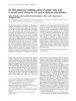
Figure 2
Phylogenetic trees of integrin subunits. Trees for (a) integrin α and (b)
integrin β subunits are adapted from [58] and [59], respectively.
Drosophila βPS
Human β7
Sea urchin βG
C. elegans βPat3
Human β2
Human β1
Human β6
Human β8
Human β3
Human β5
Human β4
Sponge βPo1
Drosophila Fly βν
Coral βCn1
Crayfish β
(a)
(b)
αM
αX
αL
αE
α1
α2
α10
α11
Ascidian αHr1
I domain
α4
α9
αV
α8
α5
αIIb
C. elegans αF54F2.1
Drosophila αPS2
Sea urchin αSU2
Sea urchin αP
Ascidian αHR2
α6
α7
α3
Drosophila αPS1
C. elegans αF54G8.3
Sponge α
Drosophila αPS3
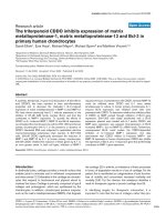
Figure 1
The members of the human integrin superfamily and how they combine
to form heterodimeric integrins. At least 18 α subunits and eight
β subunits have been identified in humans, which are able to generate
24 different integrins. Integrin subunits that bind to each other to form a
heterodimer are connected by solid lines. Each integrin has distinct
ligand-binding specificity and tissue and cell distribution.
α
4
α
5
α
6
α
7
α
8
α
9
α
10
α
11
α
V
β
3
α
IIb
β
4
α
X
β
2
α
L
α
M
α
D
β
7
α
E
α
1
β
1
α
2
α
3
β
5
β
6
β
8
The cytoplasmic tails of human integrin subunits are less
than 75 amino acids long (the β4 tail is an exception at a
length of approximately 1,000 amino acids, which includes
four fibronectin type III repeats). There is striking
homology among the β-subunit cytoplasmic tails, but the
α-subunit tails are highly divergent except for a conserved
GFFKR motif next to the transmembrane region, which is
important for association with the β tail. A large number of
cytoskeletal and signaling proteins have been reported to
bind to β cytoplasmic tails and some have been found to
interact with specific α tails. Most integrin β tails contain
one or two NPxY/F motifs (where x is any amino acid) that
are part of a canonical recognition sequence for
phosphotyrosine-binding (PTB) domains, which are
protein modules present in a wide variety of signaling and
cytoskeletal proteins. Phosphorylation of the tyrosine (Y)
in the NPxY/F motif may represent a mode of regulating
integrin interactions with other proteins at the cytoplasmic
face of the plasma membrane. The integrin tails recruit
several proteins, such as talin, that bind actin filaments,
and thus form a connection to the cytoskeleton, a
connection that is essential for most, if not all,
Genome Biology 2007, Volume 8, Issue 5, Article 215 Takada et al. 215.3
Genome Biology 2007, 8:215
Table 1
Human integrin subunits
Protein
Gene Protein Location accession
symbol name Synonyms Gene accession number (chromosome) number
ITGA1 α1 CD49a NM_181501 5q11.2 P56199
ITGA2 α2 CD49b, α2 subunit of very late antigen 2 (VLA-2) NM_002203 5q23-q31 P17301
ITGA2B αIIb GTA, CD41, GP2B, HPA3, CD41b, GPIIb NM_000419 17q21.32 P08514
ITGA3 α3 CD49c, α3 subunit of VLA-3 NM_002204, NM_005501 17q21.33 P26006
ITGA4 α4 CD49d, α4 subunit of VLA-4 NM_000885 2q31.3 AAB25486
ITGA5 α5 CD49e, fibronectin receptor alpha NM_002205 12q11-q13 P08648
ITGA6 α6 CD49f, ITGA6B NM_000210 2q31.1 P23229
ITGA7 α7 NM_002206 12q13 Q86W93
ITGA8 α8 NM_003638 10p13 P53708
ITGA9 α9 NM_002207 3p21.3 Q13797
ITGA10 α10 NM_003637 1q21 O75578
ITGA11 α11 NM_001004439, NM_012211 15q23 Q9UKX5
ITGAD αD NM_005353 16p11.2 Q13349
ITGAE αE CD103, human mucosal lymphocyte antigen 1α NM_002208 17p13 P38570
ITGAL αL CD11a (p180), lymphocyte function-associated NM_002209 16p11.2 P20701
antigen 1 (LFA-1) α subunit
ITGAM αM Mac-1, CD11b, complement receptor 3 (CR3) subunit J03925, NM_000632, 16p11.2 P11215
ITGAV αV CD51, MSK8, vitronectin receptor α (VNRα) NM_002210 2q31-q32 P06756
ITGAX αX CD11c, CR4 subunit NM_000887 16p11.2 P20702
ITGB1 β1 Fibronectin receptor β, CD29, MDF2, MSK12 BC020057 10p11.2 P05556
ITGB2 β2 Leukocyte cell adhesion molecule, CD18, CR3 subunit, NM_000211 21q22.3 P05107
CR4 subunit
ITGB3 β3 CD61; GP3A; GPIIIa, platelet glycoprotein IIIa NM_000212 17q21.32 P05106
ITGB4 β4 CD104 NM_000213 17q25 P16144
ITGB5 β5 NM_002213 3q21.2 P18084
ITGB6 β6 NM_000888 2q24.2 P18564
ITGB7 β7 NM_000889 12q13.13 P26010
ITGB8 β8 NM_002214 7p15.3 P26012
integrin-mediated functions. The structural basis for talin’s
unique ability to activate integrins through PTBs has been
defined [9]. Structural data on integrins are mostly derived
from mouse and human and the structural basis for the
activation of integrins through their cytoplasmic domains
in other species is not yet known.
Localization and function
Integrins function as traction receptors that can both
transmit and detect changes in mechanical force acting on
the extracellular matrix. In mammals, some integrins are
limited to certain cell types or tissues: αIIbβ3 to platelets;
α6β4 to keratinocytes; αEβ7 to T cells, dendritic cells and
mast cells in mucosal tissues; α4β1 to leukocytes; α4β7 to a
subset of memory T cells; and the β2 integrins to
leukocytes. Other integrins are widely distributed, such as
αVβ3, which is expressed on endothelium. The RGD
sequence in fibronectin was originally identified as an
integrin-binding motif [10] and this and related sequences
in extracellular matrix molecules do act as integrin-binding
motifs in vivo. However, integrins also recognize many
non-RGD sequences in their ligands, such as the tripeptide
LDV in the immunoglobulin superfamily member vascular
cell adhesion molecule 1 (VCAM-1), which is expressed on
inflamed endothelium and is bound by α4β1. This pattern
of integrin recognition and activation appears to be
conserved among most mammals studied.
In regard to ligand specificity, the mammalian integrins
can be broadly grouped into laminin-binding integrins
(α1β1, α2β1, α3β1, α6β1, α7β1, and α6β4), collagen-binding
integrins (α1β1, α2β1, α3β1, α10β1, and α11β1), leukocyte
integrins (αLβ2, αMβ2, αXβ2, and α
Dβ2), and RGD-
recognizing integrins (α5β1, αVβ1, αVβ3, αVβ5, αVβ6,
αVβ8, and αIIbβ3). Individual integrins have unique
ligand specificities (Table 2). They are further defined by
those α subunits that can contain the I domain: these are
α1, α2, α10, α11, αL, αM, αX, αD, and αE. Non-I-domain
subunits are α3, α4, α5, α6, α7, α8, α9, αV, and αIIb. In I-
domain integrins, the I domains play a central role in
ligand binding and intercellular adhesion. Integrin binding
among invertebrate species is less well studied; RGD
sequences have been found in species as diverse as sea
urchins and amoebae, however, and integrins and their
biochemical functions are likely to be highly conserved in
metazoa, due to the essential nature of their function.
Upon binding an extracellular ligand, integrins generate an
intracellular signal and, conversely, their functioning can
be regulated by signals from within the cell [1]. They serve
as transmembrane links between extracellular contacts
(other cells or the extracellular matrix) and the actin
microfilaments of the cytoskeleton, whose behavior
integrins also regulate and modulate. Many different
proteins on the cytoplasmic side of the membrane, such as
talin, vinculin, and ERM (ezrin, radixin, moesin)
actin-binding proteins, act as linker proteins to connect the
cytoplasmic domains of integrins to the cytoskeleton,
resulting in complex interactions [1]. Extracellular ligation
of integrins triggers a large variety of signal transduction
events that modulate cell behaviors such as adhesion,
proliferation, survival or apoptosis, shape, polarity,
motility, haptotaxis, gene expression, and differentiation,
mostly through effects on the cytoskeleton.
The deletion of individual genes by gene knockout in mice
shows that particular integrins play a critical role in
development (the β1 integrins), vasculogenesis (αV inte-
grins), lymphangiogenesis (α9β1), thrombus formation
(αIIbβ3), the integrity of the skin (α6β4), and immune
responses (the β2 integrins) (Table 3). Knockout of the
gene for β3 enhanced tumorigenesis and angiogenesis
[11,12], enhanced wound healing [13], and enhanced
inflammation and atherosclerosis [14], suggesting that
αVβ3 normally suppresses these processes.
215.4 Genome Biology 2007, Volume 8, Issue 5, Article 215 Takada et al. />Genome Biology 2007, 8:215
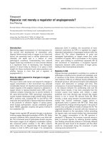
Figure 3
The extracellular region of a human integrin. (a) The crystal structure
represents a net form of integrin αVβ3 with no bound RGD peptide
(Protein Data Bank (PDB) code 1JV2) [3,4]. See PBD code 1L5G for the
RGD-bound form. (b) The I (inserted or interactive) domain is present in
seven human α subunits between β-propeller repeats 2 and 3, and is
involved in ligand binding. An I-like domain is present in all human integrin
β subunits along with four EGF-like repeats. Both the I and I-like domains
have a Rossmann fold.
l domain
l-domain-like structure
α
β
1
234567
(b)
EGF-like repeats
Membrane
(a)
Inside-out signaling regulates integrin affinity
The affinity of individual integrins for their ligands in
mammals is tightly regulated by their heterodimeric
structure and by cytoplasmic signals from within the cell
(inside-out signaling). Integrins can be activated
intracellularly by signals from G-protein-coupled receptors
that lead to phosphorylation of the cytoplasmic domain of
the β subunit. The association of the α and β cytoplasmic
tails appears to be required to maintain an integrin in the
inactive state; the association is disrupted by treatment
with agonists such as chemokines that are known to cause
integrin activation and which signal via G-protein-coupled
receptors [15] (Figure 4).
The cytoskeletal adaptor protein talin has been proposed to
play a role in regulating integrin affinity. Binding of the
talin head region to the integrin β cytoplasmic tail causes
dissociation of the α and β tails and induces a confor-
mational change in the extracellular region that increases
its affinity for its ligand [15]. Two models have been
proposed for this change in affinity. In both, the inactive
integrin is in a bent conformation, with the headpiece
facing the membrane. In the ‘deadbolt model’ the bent
conformation is maintained in an activated integrin, but
piston-like movements of the transmembrane regions
cause sliding of the extracellular stalks of the α and β
subunits, which disrupts the interaction between the head-
piece and the β stalk just beyond the membrane (the
deadbolt) [16]. In the ‘switchblade model’, dissociation of
the α and β cytoplasmic and transmembrane regions leads
to dislocation of an epidermal growth factor (EGF)-like
repeat in the β stalk, which causes the head region to
extend outwards in a switchblade-like movement [17]. In
both models, these proposed events correlate within
seconds with integrin ‘activation’, leading to
conformational changes in the ligand-binding pocket of the
headpiece that increase its affinity for ligand.
The affinity directly regulates the nature of the ligand
binding and appears to tune the degree and kinetics of cell
adhesion. In leukocytes, for instance, αLβ2 in an
intermediate-affinity state will interact with its ligand on
endothelium to help decelerate the leukocytes, which roll
slowly along the vessel wall but do not arrest (Figure 4a).
Conversion of αLβ2 to the high-affinity state by
intracellular signals from other receptors mediates their
complete arrest (Figure 4b) and signals cell polarization
and leukocyte movement across the post-capillary venule
wall into the inflamed tissue [18].
Outside-in signaling relays signals from the
extracellular environment
It has been proposed that on binding extracellular ligands,
mammalian integrins cluster in the membrane and
transduce signals to the interior of the cell (outside-in
signaling; Figure 4b). Extracellular ligand binding induces
conformational changes, including the outward swing of
the hybrid domain, separation of the α and β ‘leg’ domains
(Figure 3b), and separation of the transmembrane
domains, that lead to the interaction of the cytoplasmic
tails with intracellular signaling molecules [16]. These
include enzymes (for example, the focal adhesion kinase/c-
Src, and the small GTPases Ras and Rho) and adaptors (for
example, Cas/Crk and paxillin) that assemble within
dynamic adhesion structures, including focal adhesions
that bind cells to the extracellular matrix and podosomes
(small foot-like extensions of plasma membrane) [15,19].
In this manner, the affinity of an integrin and its valence in
binding ligands such as intercellular adhesion molecule-1
(ICAM-1) regulate the extent of outside-in signaling at the
site of focal adhesive contacts (Figure 4). These contacts
are active sites that transduce information such as the
density of extracellular ligand or the magnitude and
direction of extracellular forces on the cell. Integrins can
also be activated from the outside by the binding of
divalent cations to the metal-ion-binding sites in the I and
I-like domains in the α and β subunits, respectively.
Binding of RGD-containing peptides or related compounds
to a site in the headpiece of the integrin heterodimers has
been shown in crystal structures of αVβ3 [4,5] and αIIbβ3
[6]. The binding site is composed of the β-propeller
domain of the α subunit and the I-like domain of the β
subunit. The original crystal structure of integrin αVβ3
revealed a bent conformation of the head region associated
with low affinity for ligand [4,5]. It was therefore proposed
that the bent form does not bind ligand or carry out
outside-in signaling and that activated integrins have an
extended form (see the switchblade model described
above). Interestingly, it has been shown that the bent form
of αVβ3 can still bind to fibronectin [20] (see the deadbolt
model described above). Several intermediate forms of
integrin conformation have been postulated that confer
ligand-binding affinities and a different activation and cell
adhesion status from either the bent or the extended
forms [21].
The medical potential of antagonists to human
integrins
The interaction of integrins with their ligands is a major target
for the development of therapeutic drugs. A humanized anti-
β3 antibody (abciximab) that blocks the binding of platelet
integrin αIIbβ3 to fibrinogen has been used in the clinic to
prevent thrombosis [22]. A humanized anti-α4 antibody
(natalizumab) that can block the α4β1-VCAM interaction or
the α4β7-mucosal addressin cell adhesion molecule
(MAdCAM) interaction on mucosal endothelium has been
tested in clinical trials. Natalizumab blocks leukocyte
trafficking across the blood-brain barrier and thereby
moderates inflammation in multiple sclerosis [23]. Anti-α4
antibody is also effective in clinical trials in ameliorating
inflammatory bowel diseases, for example, Crohn’s disease.
Genome Biology 2007, Volume 8, Issue 5, Article 215 Takada et al. 215.5
Genome Biology 2007, 8:215
Many RGD-based low-molecular-weight integrin
antagonists have been developed and some of them have
been approved as therapeutics (for example, eptifibatide and
tirofiban as inhibitors of αIIbβ3 to reduce platelet
aggregation and the formation of blood clots) [24]. As more
becomes known about the relationship between integrin
three-dimensional structure and how this regulates affinity for
ligand and signaling into the cell, antagonists can be designed
that stabilize a specific conformation, thereby promoting or
blocking specific intercellular adhesion functions.
Frontiers
Integrins are transmembrane molecules that are essential
for both embryonic development and immunological function
by binding to a wide variety of ligands, including extra-
cellular matrix molecules and members of the immuno-
globulin superfamily. Their capacity to specifically recognize
particular amino-acid motifs and regulate binding affinity to
them lies in their heterodimeric structure. This molecular
design incorporates a remarkable ability to direct confor-
mational changes initiated at the cytoplasmic domain, and
also to signal extracellular ligand binding back to the inside
of the cell. Much of our current knowledge of the myriad of
functions attributed to ligand binding of a particular αβ pair
comes from gene knockout studies in mouse or from rare
hereditary disorders in humans. Only a handful of crystal
structures of integrins bound to their ligands have been
solved. From these data it appears that small variations in
the particular structure or charge of a ligand (that is, down
215.6 Genome Biology 2007, Volume 8, Issue 5, Article 215 Takada et al. />Genome Biology 2007, 8:215
Table 2
Ligand-binding specificities of human integrins
Integrins Ligands
α1β1 Laminin, collagen
α2β1 Laminin, collagen, thrombospondin, E-cadherin, tenascin
α3β1 Laminin, thrombospondin, uPAR
α4β1 Thrombospondin, MAdCAM-1, VCAM-1, fibronectin, osteopontin, ADAM, ICAM-4
α5β1 Fibronectin, osteopontin, fibrillin, thrombospondin, ADAM, COMP, L1
α6β1 Laminin, thrombospondin, ADAM, Cyr61
α7β1 Laminin
α8β1 Tenascin, fibronectin, osteopontin, vitronectin, LAP-TGF-β, nephronectin,
α9β1 Tenascin, VCAM-1, osteopontin, uPAR, plasmin, angiostatin, ADAM [25], VEGF-C, VEGF-D [26]
α10β1 Laminin, collagen
α11β1 Collagen
αVβ1 LAP-TGF-β, fibronectin, osteopontin, L1
αLβ2 ICAM, ICAM-4
αMβ2 ICAM, iC3b, factor X, fibrinogen, ICAM-4, heparin
αXβ2 ICAM, iC3b, fibrinogen, ICAM-4, heparin, collagen [27]
αDβ2 ICAM, VCAM-1, fibrinogen, fibronectin, vitronectin, Cyr61, plasminogen
αIIbβ3 Fibrinogen, thrombospondin, , fibronectin, vitronectin, vWF, Cyr61, ICAM-4, L1, CD40 ligand [28]
αVβ3 Fibrinogen, vitronectin, vWF, thrombospondin, fibrillin, tenascin, PECAM-1, fibronectin, osteopontin, BSP, MFG-E8, ADAM-15, COMP,
Cyr61, ICAM-4, MMP, FGF-2 [29], uPA [30], uPAR [31], L1, angiostatin [32], plasmin [33], cardiotoxin [34], LAP-TGF-β, Del-1
α6β4 Laminin
αVβ5 Osteopontin, BSP, vitronectin, CCN3 [35], LAP-TGF-β
αVβ6 LAP-TGF-β, fibronectin, osteopontin, ADAM
α4β7 MAdCAM-1, VCAM-1, fibronectin, osteopontin
α
Eβ7 E-cadherin
αVβ8 LAP-TGF-β
References are included for recently discovered ligands only. Abbreviations: ADAM, a disintegrin and metalloprotease; BSP, bone sialic protein; CCN3,
an extracellular matrix protein; COMP, cartilage oligomeric matrix protein; Cyr61, cysteine-rich protein 61; L1, CD171; LAP-TGF-β, TGF-β latency-
associated peptide; iC3b, inactivated complement component 3; PECAM-1, platelet and endothelial cell adhesion molecule 1; uPA, urokinase; uPAR,
urokinase receptor; VEGF, vascular endothelial growth factor; vWF, von Willebrand Factor.
to single atoms) can strongly influence the binding affinity
and the capacity of the integrin to maintain a conformation
that signals back into the cell. This implies that ligand
binding can influence allosteric changes in the integrin,
which in turn dictate how the integrin reports on the
environment in which the cell finds itself. Thus, integrins
serve as both sensors of their molecular surroundings and
effectors that conduct motile forces exerted by the cell’s
cytoskeleton and from the dynamic environment (that is,
shear forces within blood vessels). We are just beginning to
understand the structural and chemical basis of this sensor-
effector system. A particularly exciting development is the
discovery of small molecules that bind tightly to the ligand-
binding pocket or to other domains and allosterically
stabilize integrin conformations that promote or antagonize
binding. For instance, small molecules have been discovered
that can allosterically tune conformations of α
L
β
2
that favor
low, intermediate, and high-affinity binding. In this manner,
it is possible to steer the adhesive response of a leukocyte in
a blood vessel to promote tethering and rolling, firm arrest,
or no binding at all. It may be possible to apply such small
molecules as therapeutics either to promote leukocyte
recruitment at sites of infection or to block their accumu-
lation in chronic inflammatory diseases such as in
rheumatoid arthritis and psoriasis. As more knowledge
accumulates relating amino-acid sequence to common
structural motifs associated with the allosteric control of
ligand recognition and outside-in signaling to the cytoplasm,
it will become possible to design small molecules that target
these critical domains.
Additional data files
Additional data are available online with this article:
Additional data file 1 contains tables of the integrin subunits
present in the mouse, chicken, zebrafish, nematodes,
Xenopus laevis, and D. melanogaster.
Genome Biology 2007, Volume 8, Issue 5, Article 215 Takada et al. 215.7
Genome Biology 2007, 8:215
Table 3
Phenotypes of deletions of integrin subunits in the mouse
Integrin subunit Viability Fertility Phenotype Reference
α1 + + Defects in bone healing and reduced tumor angiogenesis [36]
α2 + + Reduced branching morphogenesis and platelet adhesion [37]
α3 Perinatal lethal + Kidney, lung, and skin defects [38]
α4 Embryonic lethal – Placental and heart defects [39]
α5 Embryonic lethal – Mesodermal and vascular defects [40]
α6 Perinatal lethal + Epidermal detachment, defect in neurogenesis [41]
α7 + + Muscular dystrophy [42]
α8 Perinatal lethal + Kidney defect [43]
α9 Perinatal lethal + Chylothorax (defect in lymphatic drainage) [44]
αv Embryonic and perinatal lethal + Cerebral hemorrhage [45]
αM + + Neutrophil adhesion and degranulation [46]
αL + + Neutrophil emigration [47]
αD + + Reduced T-cell response and T-cell phenotypic changes [48]
αE + + Skin inflammation [49]
β1 Embryonic lethal – Fails to gastrulate [45,50]
β2 + + Leukocyte adhesion deficiency [51]
β3 + + Platelet defect [52]
β4 Perinatal lethal + Epidermal detachment [53]
β5 + + Accelerated age-related blindness [54]
β6 + + Inflammation in skin and lungs [55]
β7 + + Gut-associated lymphocyte defects [56]
β8 Embryonic and perinatal lethal + Cerebral hemorrhage [57]
References
1. Hynes RO: Integrins: bidirectional, allosteric signaling
machines. Cell 2002, 110:673-687.
2. Shimaoka M, Springer TA: Therapeutic antagonists and confor-
mational regulation of integrin function. Nat Rev Drug Discov
2003, 2:703-716.
3. Huhtala M, Heino J, Casciari D, de Luise A, Johnson MS: Integrin
evolution: insights from ascidian and teleost fish genomes.
Matrix Biol 2005, 24:83-95.
4. Xiong JP, Stehle T, Diefenbach B, Zhang R, Dunker R, Scott DL,
Andrzej J, Goodman SL, Arnaout MA: Crystal structure of the
extracellular segment of integrin
αα
v
ββ
3. Science 2001, 294:339-
345.
5. Xiong JP, Stehle T, Zhang R, Joachimiak A, Frech M, Goodman SL,
Arnaout MA: Crystal structure of the extracellular segment
of integrin alpha Vbeta3 in complex with an Arg-Gly-Asp
ligand. Science 2002, 296:151-155.
6. Xiao T, Takagi J, Coller BS, Wang JH, Springer TA: Structural basis
for allostery in integrins and binding to fibrinogen-mimetic
therapeutics. Nature 2004, 432:59-67.
7. Puzon-McLaughlin W, Takada Y: Critical residues for ligand
binding in an I domain-like structure of the integrin beta1
subunit. J Biol Chem 1996, 271:20438-20443.
8. Kamata T, Tieu KK, Irie A, Springer TA, Takada Y: Amino acid
residues in the alpha IIb subunit that are critical for ligand
binding to integrin alpha IIbbeta 3 are clustered in the beta-
propeller model. J Biol Chem 2001, 276:44275-44283.
9. Wegener KL, Partridge AW, Han J, Pickford AR, Liddington RC,
Ginsberg MH, Campbell ID: Structural basis of integrin activa-
tion by talin. Cell 2007, 128:171-182.
10. Pierschbacher MD, Ruoslahti E: Cell attachment activity of
fibronectin can be duplicated by small synthetic fragments
of the molecule. Nature 1984, 309:30-33.
11. Taverna D, Moher H, Crowley D, Borsig L, Varki A, Hynes RO:
Increased primary tumor growth in mice null for beta3- or
beta3/beta5-integrins or selectins. Proc Natl Acad Sci USA 2004,
101:763-768.
12. Taverna D, Crowley D, Connolly M, Bronson RT, Hynes RO: A
direct test of potential roles for beta3 and beta5 integrins in
growth and metastasis of murine mammary carcinomas.
Cancer Res 2005, 65:10324-10329.
13. Reynolds LE, Conti FJ, Lucas M, Grose R, Robinson S, Stone M, Saun-
ders G, Dickson C, Hynes RO, Lacy-Hulbert A, et al.: Accelerated
re-epithelialization in beta3-integrin-deficient- mice is asso-
ciated with enhanced TGF-beta1 signaling. Nat Med 2005,
11:
167-174.
14. Weng S, Zemany L, Standley KN, Novack DV, La Regina M, Bernal-
Mizrachi C, Coleman T, Semenkovich CF: Beta3 integrin deficiency
promotes atherosclerosis and pulmonary inflammation in
high-fat-fed, hyperlipidemic mice. Proc Natl Acad Sci USA 2003,
100:6730-6735.
15. Ginsberg MH, Partridge A, Shattil SJ: Integrin regulation. Curr Opin
Cell Biol 2005, 17:509-516.
16. Arnaout MA, Mahalingam B, Xiong JP: Integrin structure,
allostery, and bidirectional signaling. Annu Rev Cell Dev Biol
2005, 21:381-410.
17. Luo BH, Carman CV, Springer TA: Structural basis of integrin
regulation and signaling. Annu Rev Immunol 2007, 25:619-647.
18. Green CE, Schaff UY, Sarantos MR, Lum AF, Staunton DE, Simon SI:
Dynamic shifts in LFA-1 affinity regulate neutrophil rolling,
arrest, and transmigration on inflamed endothelium. Blood
2006, 107:2101-2111.
19. Shattil SJ: Integrins and Src: dynamic duo of adhesion signal-
ing. Trends Cell Biol 2005, 15:399-403.
20. Adair BD, Xiong JP, Maddock C, Goodman SL, Arnaout MA, Yeager
M: Three-dimensional EM structure of the ectodomain of
integrin {alpha}V{beta}3 in a complex with fibronectin. J Cell
Biol 2005, 168:1109-1118.
215.8 Genome Biology 2007, Volume 8, Issue 5, Article 215 Takada et al. />Genome Biology 2007, 8:215
Figure 4
Leukocyte recruitment to the endothelial surface. (a) Binding of glycoprotein selectin ligands (yellow and purple) on the leukocyte to selectins (blue) on
the endothelial surface, and weak binding of low-affinity leukocyte integrins (green) to ICAMs (pale yellow) on the endothelium facilitates cell tethering
and rolling. This binding, together with signals from chemokines (pink), generates inside-out signals (yellow arrows) that shift the bound integrins to a
high-affinity ligand-binding state. (b) Leukocyte arrest is mediated by clusters of high-affinity integrins (red) binding to ICAMs on the endothelial cells.
These focal clusters can themselves signal outside-in to affect functions such as cell polarization and migration.
*
*
*
*
*
*
*
Inside-out signaling Outside-in signaling
Chemokines
Integin
Selectin
ICAM
Chemokine
receptor
(a) Leukocyte rolling Leukocyte arrest(b)
Glycoprotein
selectin ligand
21. Sarantos MR, Raychaudhuri S, Lum AF, Staunton DE, Simon SI:
Leukocyte function-associated antigen 1-mediated adhesion
stability is dynamically regulated through affinity and valency
during bond formation with intercellular adhesion mole-
cule-1. J Biol Chem 2005, 280:28290-28298.
22. Gabriel HM, Oliveira EI: Role of abciximab in the treatment of
coronary artery disease. Expert Opin Biol Ther 2006, 6:935-942.
23. O’Connor P: Natalizumab and the role of alpha 4-integrin
antagonism in the treatment of multiple sclerosis. Expert
Opin Biol Ther 2007, 7:123-136.
24. Meyer A, Auernheimer J, Modlinger A, Kessler H: Targeting RGD
recognizing integrins: drug development, biomaterial
research, tumor imaging and targeting. Curr Pharm Des 2006,
12:2723-2747.
25. Eto K, Huet C, Tarui T, Kupriyanov S, Liu HZ, Puzon-McLaughlin W,
Zhang XP, Sheppard D, Engvall E, Takada Y: Functional classifica-
tion of ADAMs based on a conserved motif for binding to
integrin alpha 9beta 1; implications for sperm-egg binding
and other cell interactions. J Biol Chem 2002, 277:17804-17810.
26. Vlahakis NE, Young BA, Atakilit A, Sheppard D: The lymphangio-
genic vascular endothelial growth factors VEGF-C and -D
are ligands for the integrin {alpha}9{beta}1. J Biol Chem 2005,
280:4544-4552.
27. Garnotel R, Rittie L, Poitevin S, Monboisse J-C, Nguyen P, Potron G,
Maquart F-X, Randoux A, Gillery P: Human blood monocytes
interact with type I collagen through {alpha}x{beta}2 inte-
grin (CD11c-CD18, gp150-95). J Immunol 2000, 164:5928-5934.
28. Andre P, Prasad KSS, Denis CV, He M, Papalia JM, Hynes RO, Phillips
DR, Wagner DD: CD40L stabilizes arterial thrombi by a
[beta]3 integrin-dependent mechanism. Nat Med 2002, 8:247-
252.
29. Rusnati M, Tanghetti E, Dell’Era P, Gualandris A, Presta M: alphav-
beta3 integrin mediates the cell-adhesive capacity and bio-
logical activity of basic fibroblast growth factor (FGF-2) in
cultured endothelial cells. Mol Biol Cell 1997, 8:2449-2461.
30. Tarui T, Akakura N, Majumdar M, Andronicos N, Takagi J, Mazar AP,
Bdeir K, Kuo A, Yarovoi SV, Cines DB, et al.: Direct interaction of
the kringle domain of urokinase-type plasminogen activator
(uPA) and integrin alpha v beta 3 induces signal transduc-
tion and enhances plasminogen activation. Thromb Haemost
2006, 95:524-534.
31. Tarui T, Mazar AP, Cines DB, Takada Y: Urokinase-type plas-
minogen activator receptor (CD87) is a ligand for integrins
and mediates cell-cell interaction. J Biol Chem 2001, 276:3983-
3990.
32. Tarui T, Miles LA, Takada Y: Specific interaction of angiostatin
with integrin {alpha}v{beta}3 in endothelial cells. J Biol Chem
2001, 276:39562-39568.
33. Tarui T, Majumdar M, Miles LA, Ruf W, Takada Y: Plasmin-
induced migration of endothelial cells: A potential target for
the anti-angiogenic action of angiostatin. J Biol Chem 2002,
277:33564-33570.
34. Wu PL, Lee SC, Chuang CC, Mori S, Akakura N, Wu WG, Takada Y:
Non-cytotoxic cobra cardiotoxin A5 binds to integrin alpha
vbeta 3 and inhibits bone resorption: Identification of car-
diotoxins as non-RGD integrin-binding proteins of the Ly-6
family. J Biol Chem 2006, 281:7937-7945.
35. Lin CG, Chen C-C, Leu S-J, Grzeszkiewicz TM, Lau LF: Integrin-
dependent functions of the angiogenic inducer NOV
(CCN3): implication in wound healing. J Biol Chem 2005, 280:
8229-8237.
36. Gardner H, Kreidberg J, Koteliansky V, Jaenisch R: Deletion of
integrin alpha 1 by homologous recombination permits
normal murine development but gives rise to a specific
deficit in cell adhesion. Dev Biol 1996, 175:301-313.
37. Chen J, Diacovo TG, Grenache DG, Santoro SA, Zutter MM: The
alpha(2) integrin subunit-deficient mouse: a multifaceted
phenotype including defects of branching morphogenesis
and hemostasis. Am J Pathol 2002, 161:337-344.
38. Kreidberg JA, Donovan MJ, Goldstein SL, Rennke H, Shepherd K,
Jones RC, Jaenisch R: Alpha 3 beta 1 integrin has a crucial role
in kidney and lung organogenesis. Development 1996, 122:3537-
3547.
39. Yang JT, Rayburn H, Hynes RO: Cell adhesion events mediated
by alpha 4 integrins are essential in placental and cardiac
development. Development 1995, 121:549-560.
40. Yang JT, Rayburn H, Hynes RO: Embryonic mesodermal defects
in alpha 5 integrin-deficient mice. Development 1993, 119:1093-
1105.
41. Georges-Labouesse E, Messaddeq N, Yehia G, Cadalbert L, Dierich
A, Le Meur M: Absence of integrin alpha 6 leads to epider-
molysis bullosa and neonatal death in mice. Nat Genet 1996,
13:370-373.
42. Mayer U, Saher G, Fassler R, Bornemann A, Echtermeyer F, von der
Mark H, Miosge N, Poschl E, von der Mark K: Absence of integrin
alpha 7 causes a novel form of muscular dystrophy. Nat Genet
1997, 17:318-323.
43. Muller U, Wang D, Denda S, Meneses JJ, Pedersen RA, Reichardt LF:
Integrin alpha8beta1 is critically important for epithelial-
mesenchymal interactions during kidney morphogenesis.
Cell 1997, 88:603-613.
44. Huang XZ, Wu JF, Ferrando R, Lee JH, Wang YL, Farese RV Jr, Shep-
pard D: Fatal bilateral chylothorax in mice lacking the inte-
grin alpha9beta1. Mol Cell Biol 2000, 20:5208-5215.
45. Bader BL, Rayburn H, Crowley D, Hynes RO: Extensive vasculo-
genesis, angiogenesis, and organogenesis precede lethality
in mice lacking all alpha v integrins. Cell 1998, 95:507-519.
46. Lu H, Smith CW, Perrard J, Bullard D, Tang L, Shappell SB, Entman
ML, Beaudet AL, Ballantyne CM: LFA-1 is sufficient in mediating
neutrophil emigration in Mac-1-deficient mice. J Clin Invest
1997, 99:1340-1350.
47. Ding ZM, Babensee JE, Simon SI, Lu H, Perrard JL, Bullard DC, Dai
XY, Bromley SK, Dustin ML, Entman ML, et al.: Relative contribu-
tion of LFA-1 and Mac-1 to neutrophil adhesion and migra-
tion. J Immunol 1999, 163:5029-5038.
48. Wu YM, Robinson DR, Kung HJ: Signal pathways in up-regula-
tion of chemokines by tyrosine kinase MER/NYK in prostate
cancer cells. Cancer Res 2004, 64:7311-7320.
49. Schon MP, Schon M, Warren HB, Donohue JP, Parker CM: Cuta-
neous inflammatory disorder in integrin alphaE (CD103)-
deficient mice. J Immunol 2000, 165:6583-6589.
50. Stephens LE, Sutherland AE, Klimanskaya IV, Andrieux A, Meneses J,
Pedersen RA, Damsky CH: Deletion of beta 1 integrins in mice
results in inner cell mass failure and peri-implantation
lethality. Genes Dev 1995, 9:1883-1895.
51. Wilson RW, Ballantyne CM, Smith CW, Montgomery C, Bradley A,
O’Brien WE, Beaudet AL: Gene targeting yields a CD18-
mutant mouse for study of inflammation. J Immunol 1993, 151:
1571-1578.
52. Hodivala-Dilke KM, McHugh KP, Tsakiris DA, Rayburn H, Crowley
D, Ullman-Cullere M, Ross FP, Coller BS, Teitelbaum S, Hynes RO:
Beta3-integrin-deficient mice are a model for Glanzmann
thrombasthenia showing placental defects and reduced sur-
vival. J Clin Invest 1999, 103:229-238.
53. van der Neut R, Krimpenfort P, Calafat J, Niessen CM, Sonnenberg
A: Epithelial detachment due to absence of hemidesmo-
somes in integrin beta 4 null mice. Nat Genet 1996, 13:366-369.
54. Huang X, Griffiths M, Wu J, Farese RV Jr, Sheppard D: Normal
development, wound healing, and adenovirus susceptibility
in beta5-deficient mice. Mol Cell Biol 2000, 20:755-759.
55. Huang XZ, Wu JF, Cass D, Erle DJ, Corry D, Young SG, Farese RV,
Jr., Sheppard D: Inactivation of the integrin beta 6 subunit
gene reveals a role of epithelial integrins in regulating
inflammation in the lung and skin. J Cell Biol 1996, 133:921-928.
56. Wagner N, Lohler J, Kunkel EJ, Ley K, Leung E, Krissansen G, Rajew-
sky K, Muller W: Critical role for beta7 integrins in formation
of the gut-associated lymphoid tissue. Nature 1996, 382:366-
370.
57. Zhu J, Motejlek K, Wang D, Zang K, Schmidt A, Reichardt LF: beta8
integrins are required for vascular morphogenesis in mouse
embryos. Development 2002, 129:2891-2903.
58. Miyazawa S, Azumi K, Nonaka M: Cloning and characterization
of integrin alpha subunits from the solitary ascidian, Halo-
cynthia roretzi. J Immunol 2001, 166:1710-1715.
59. Brower DL, Brower SM, Hayward DC, Ball EE: Molecular evolu-
tion of integrins: genes encoding integrin beta subunits from
a coral and a sponge. Proc Natl Acad Sci USA 1997, 94:9182-9187.
Genome Biology 2007, Volume 8, Issue 5, Article 215 Takada et al. 215.9
Genome Biology 2007, 8:215