Báo cáo y học: "The impact of chromatin modifiers on the timing of locus replication in mouse embryonic stem cells" pot
Bạn đang xem bản rút gọn của tài liệu. Xem và tải ngay bản đầy đủ của tài liệu tại đây (828.17 KB, 13 trang )
Genome Biology 2007, 8:R169
comment reviews reports deposited research refereed research interactions information
Open Access
2007Jørgensenet al.Volume 8, Issue 8, Article R169
Research
The impact of chromatin modifiers on the timing of locus
replication in mouse embryonic stem cells
Helle F Jørgensen
*
, Véronique Azuara
*†
, Shannon Amoils
*
,
Mikhail Spivakov
*
, Anna Terry
*
, Tatyana Nesterova
‡
, Bradley S Cobb
*
,
Bernard Ramsahoye
§
, Matthias Merkenschlager
*
and Amanda G Fisher
*
Addresses:
*
Lymphocyte Development Group, MRC Clinical Sciences Centre, Imperial College School of Medicine, London W12 0NN, UK.
‡
Developmental Epigenetics, MRC Clinical Sciences Centre, Imperial College School of Medicine, London W12 0NN, UK.
§
Developmental
Epigenetics, University of Edinburgh, Western General Hospital, Edinburgh EH4 2XR, UK.
†
Current address: Institute of Reproductive and
Developmental Biology, Imperial College School of Medicine, London W12 0NN, UK.
Correspondence: Helle F Jørgensen. Email: Amanda G Fisher. Email:
© 2007 Jørgensen et al.; licensee BioMed Central Ltd.
This is an open access article distributed under the terms of the Creative Commons Attribution License ( which
permits unrestricted use, distribution, and reproduction in any medium, provided the original work is properly cited.
Regulation of replication timing in ES cells<p>A panel of mutant embryonic stem (ES) cell lines lacking important chromatin modifiers was used to dissect the relationship between chromatin structure and replication timing, revealing the importance of several chromatin modifiers for maintaining correct replication of satellite sequences in pluripotent ES cells.</p>
Abstract
Background: The time of locus replication during S-phase is tightly regulated and correlates with
chromatin state. Embryonic stem (ES) cells have an unusual chromatin profile where many
developmental regulator genes that are not yet expressed are marked by both active and repressive
histone modifications. This poised or bivalent state is also characterized by locus replication in early
S-phase in ES cells, while replication timing is delayed in cells with restricted developmental options.
Results: Here we used a panel of mutant mouse ES cell lines lacking important chromatin modifiers
to dissect the relationship between chromatin structure and replication timing. We show that
temporal control of satellite DNA replication is sensitive to loss of a variety of chromatin modifiers,
including Mll, Eed, Dnmt1, Suv39h1/h2 and Dicer. The replication times of many single copy loci,
including a 5 Mb contiguous region surrounding the Rex1 gene, were retained in chromatin modifier
mutant ES cells, although a subset of loci were affected.
Conclusion: This analysis demonstrates the importance of chromatin modifiers for maintaining
correct replication of satellite sequences in pluripotent ES cells and highlights the sensitivity of
some single copy loci to the influence of chromatin modifiers. Abundant histone acetylation is
shown to correlate well with early replication. Surprisingly, loss of DNA methylation or histone
methylation was tolerated by many loci, suggesting that these modifications may be less influential
for the timing of euchromatin replication.
Background
DNA labeling experiments have shown that replication pat-
terns are faithfully inherited through multiple cell divisions
[1]. Individual genes replicate at similar times in each cell of a
given type but locus replication timing often differs between
cell types. In embryonic stem (ES) cells, the timing of DNA
Published: 17 August 2007
Genome Biology 2007, 8:R169 (doi:10.1186/gb-2007-8-8-r169)
Received: 6 March 2007
Revised: 26 June 2007
Accepted: 17 August 2007
The electronic version of this article is the complete one and can be
found online at />R169.2 Genome Biology 2007, Volume 8, Issue 8, Article R169 Jørgensen et al. />Genome Biology 2007, 8:R169
replication of several genes is altered in response to differen-
tiation [2,3], which reflects changes in both gene expression
and the decline in developmental potential that accompanies
lineage commitment [4]. More generally, replication timing is
influenced by both chromosome context [5,6] and underlying
nucleotide composition [3]. Genome-wide and single gene
analyses have shown that early replication timing correlates
with transcriptional activity (reviewed in [7]) as well as with
chromatin accessibility, or permissivity [8], and is often asso-
ciated with enrichment of acetylated histones [9-11]. The
exact relationship between chromatin structure and time of
locus replication in S-phase remains unresolved.
Chromatin structure depends on both the action of sequence-
specific DNA binding proteins and epigenetic features such as
post-translational modifications of histones, the extent of
DNA methylation and nuclear location (reviewed in [12]).
Proteins capable of changing these parameters, chromatin
modifiers, are important for establishing and maintaining
particular chromatin configurations. For example, enzymes
that methylate Lys4 on histone H3 or acetylate histone H3 or
H4 are thought to be important for retaining accessibility
whereas histone deacetylases (HDACs) and histone methyl
transferases (HMTases) that target histone H3 Lys9, Lys27
and histone H4 Lys20 are important for the formation of
repressive chromatin. Other factors, including DNA methyl-
transferases, methyl-DNA binding proteins, polycomb
repressor complexes (PRCs), nucleosome remodeling com-
plexes and Dicer-dependent short interfering RNA (siRNA),
also induce or stabilize repressed chromatin states.
Recently, we showed that many genes encoding key develop-
mental regulators replicate early in ES cells, despite being
inactive at this stage [4]. Importantly, the promoters of these
genes displayed an unusual chromatin profile, being enriched
for both marks of active (H3K9ac, H3K4me2/3) and repres-
sive (H3K27me3) chromatin [4,13]. This bivalent structure is
interpreted as representing a 'poised' yet non-expressed state,
in which H3K27 methylation is key to ensure repression
[4,14,15]. Upon differentiation, many lineage inappropriate
genes switch from early to late replication [2,3], suggesting
that early replication of lineage specifiers in undifferentiated
ES cells is actively maintained. Here, a genetic approach was
used to analyze the impact of different chromatin modifiers
on the replication timing profile of mouse ES cells. We show
that, while early replication in ES cells correlates with peaks
of increased histone acetylation, the replication times of
many, but not all, single copy genes was preserved, even in
mutant cells where polycomb group (PcG)-, H3K9me- or CpG
methylation-mediated repression was abrogated. This con-
clusion is based on analysis of multiple individual genes and
extended chromosome walking. The replication timing of
repetitive DNA was consistently altered in many mutant ES
cell lines and we demonstrate that DNA methylation is partic-
ularly important for the temporal regulation of pericentric
DNA duplication in ES cells.
Results and discussion
Replication timing of many genes is unchanged in
mutant ES cells
Mutation of chromatin modifiers in vivo often results in
embryonic lethality and impaired development (Table 1).
Despite this, murine ES cell lines lacking individual modifiers
have been established and, in many cases, shown to retain
multi-lineage potential. Using a panel of mutant ES cell lines
(described in detail in Table 1) we examined whether a lack of
specific histone methyltransferases, DNA methyltrans-
ferases, the NuRD nucleosome remodeling complex or Dicer
activity was sufficient to alter the temporal profile of locus
replication in ES cells. All ES cell lines examined displayed ES
cell morphology, expressed markers that are characteristic of
murine ES cells (such as Oct4, alkaline phosphatase and
SSEA-1) and had cell cycle profiles that were comparable with
wild-type ES cells (supplementary Table 1 in Additional data
file 1, and supplementary Figure 1 in Additional data file 2).
The replication timing profiles of Oct4, Esg1, Nkx2.9 and
Mash1 for four independently derived wild-type (white bars)
and eight mutant ES cell lines that lack Mll, Eed, Dnmt 1,
Dnmt 3a/3b, Mbd3, G9a, Suv39 h1/h2 or Dicer (gray bars)
are shown in Figure 1a. Histograms indicate the abundance of
newly synthesized DNA corresponding to each locus in sam-
ples prepared from sequential stages of the cell cycle (G1-S,
S1, S2, S3, S4 and G2/M) for all the wild-type and mutant ES
cell lines. Oct4, which replicates early in S-phase in all cell
types analyzed, showed only minor differences between wild-
type and mutant cell lines (upper panel). Similarly, there was
little variation in the early replication of Esg1 and late replica-
tion of Mash1 in wild-type and mutant ES cells, even though
these loci are capable of switching replication timing upon
differentiation; Esg1 has been shown to shift to late replica-
tion upon neural induction while Mash1 shifts to become
early replicating [2,16]. Nkx2.9, a neural specific gene that
replicates in mid S-phase in undifferentiated ES cells showed
some variation between wild-type and mutant cells. This
analysis was extended to include a wider set of candidate loci
that also have been shown to be permissive for changes in
replication timing [2,4,17]. Figure 1b summarizes the data for
14 genes (shown in supplementary Figure 2 in Additional data
file 2), in which replication timing is color-coded according to
peak abundance in G1-S and/or S1 (early, green), S2 (mid-
early, lime), S2 and S3 (middle, yellow), S3 (mid-late,
orange), S4 and/or G2/M (late, red). Early replication of
Nanog, Zfp57, Oct4, Esg1, Sox2 and Rex1 was unaffected in
mutant ES cells lacking either a chromatin activator (Mll) or
repressive chromatin modifiers (Eed, Dnmt1, Dnmt3a/3b,
Mbd3, G9a, Suv39h1/h2 and Dicer) compared to wild-type
cells (OS25 and WT). The replication of several middle- and
late-replicating genes was also unchanged in chromatin mod-
ifier mutant ES cells, although three loci (Mage a2, Ebf,
Sox3
), in addition to Nkx2.9, showed some changes in repli-
cation patterns in the mutant lines. Sox3 replicated earlier in
ES cells that lacked Dnmt1, Dnmt 3a/3b or Dicer but slightly
Genome Biology 2007, Volume 8, Issue 8, Article R169 Jørgensen et al. R169.3
comment reviews reports refereed researchdeposited research interactions information
Genome Biology 2007, 8:R169
later in Eed-deficient ES cells. Mage a2 was sensitive to loss
of G9a (supplementary Figure 3 in Additional data file 2).
This gene is transcriptionally regulated by G9a [18] (supple-
mentary Figure 2 in Additional data file 2), and belongs to the
Mage genes that are DNA methylated in adult somatic tissues
[19]. Replication of Ebf, a gene that replicates earlier in pro-
and pre-B cells than in ES cells [17], showed slight shifts in
replication in G9a, Suv39 h1/2 and Dicer-deficient ES cells.
From a total of 14 loci analyzed in Figure 1b, four showed a
temporal shift in one or more mutant ES cell lines. We ana-
lyzed the sequence context of the genes (supplementary Table
2 in Additional data file 1) but neither GC content nor Line
density was obviously different between genes that change
replication timing or those that remain unchanged in the
mutant cells. Bivalent genes were represented among loci that
showed shifts in response to loss of chromatin modifiers
(such as Nkx2.9 and Msx1) as well as those that did not
change their timing of replication (such as Math1 and Sox1;
supplementary Figure 2 in Additional data file 2). Some rep-
lication timing changes were in the predicted direction (that
is, an advance upon loss of a repressive chromatin modifier)
whereas others were counter-intuitive, which we cannot
explain. Importantly, however, we did not observe consistent
shifts towards earlier or later replication in response to
removal of a specific chromatin modifier. These results sug-
gest that while some loci may be more sensitive to chromatin
changes than others, none of the chromatin modifiers studied
here is capable of overt de-regulation of the temporal order of
gene replication in ES cells. This was true even for cells lack-
ing Eed (and hence devoid of methylated H3K27), a factor
previously shown to be important for transcriptional repres-
sion and chromatin bivalency in ES cells [15]. Thus, our data
do not support a model where methylation of specific histone
residues or CpG dinucleotides confers replication at a certain
time in S-phase. To explore this further, we also analyzed a
large contiguous region surrounding Rex1 (Figure 1c), a gene
Table 1
Characteristics of chromatin modifiers and mutant ES cells
Name Protein function ES cell lines Phenotype of KO/DKO mice Phenotype of KO/DKO ES cells Reference
Mll HMTase: tri-
methylation of H3K4
KO: High 6 Embryonic lethal (E11.5-14.5)
Homeotic transformations
Mis-regulation of Hox gene
expression
Mis-regultion of Hox genes
Failure of in vitro differentiation
to hematopoietic pre-cursors
[42-44]
Eed Subunit of PRC2
Cofactor for Ezh2
(H3K27 HMTase)
KO: B1.3, G8.1 Embryonic lethal (E6.5)
Failure to maintain inactive X in
trophoblast derivatives
Loss of H3K27me2/3
Reduced H3K27me1
Contribute to all tissues of
chimeras
[4,45-47]
Dnmt1 Maintenance DNA
methyl transferase
KO: c/c Embryonic lethal (E11.5) Reduced DNA methylation level
Reduced differentiation
[36,48,49]
Dnmt 3a/3b De novo DNA methyl
transferase
DKO: clone 10 (early
passage)
Embryonic lethal (E11.5) Lack de novo DNA methylation
activity DNA methylation levels
slightly reduced in early passage
(severly reduced in late passage
cells)
Retains differentiation potential
at early passages
[27,37,50]
Mbd3 Subunit of NuRD
(nucleosome
remodeling and
HDAC complex)
KO: Fix2 Embryonic lethal (implantation) Loss of the NuRD (nucleosome
remodeling and HDAC)
complex
Severe differentiation block
[38,51]
G9a HMTase: H3K9me
H3K9me2
euchromatic
WT: Col4
KO: 2-3
Tg: 15-3
Embryonic lethal (E12.5) Reduced H3K9me2
Increased H3K4me2, H3K9ac
Reduced H3K9 methylation in
euchromatin
[18,25]
Suv39 h1/h2 H3K9me3 (hetero-
chromatic)
WT: wt26
DKO: DN57, DN72
Increased prenatal lethality
Growth retarded
B-cell lymphomas
Male sterility
Chromosome instablility in
fibroblasts
Reduced H3K9me3 level;
Reduced H3K9me3 at
pericentric heterochromatin
Increased H3K27me3 at
pericentric heterochromatin
Decreased CpG methylation of
satellite repeats
Increased transcription of major/
minor satellite
[25,35,52,53]
Dicer RNase, essential for
siRNA/miRNA
pathway in mammals
WT: D3
KO: D3-S5, D3-S6
Embryonic lethal (E7.5) Increased transcription of
repeats Slow growth
[30,54,55]
miRNA, microRNA.
R169.4 Genome Biology 2007, Volume 8, Issue 8, Article R169 Jørgensen et al. />Genome Biology 2007, 8:R169
Figure 1 (see legend on next page)
OS25 (WT) L E ME L E E L
Mll KO L E ME L E ME L
Eed KO L E ME L E ME L
Dnmt 1 KO L E ME L E E L
Dnmt 3a/3b DKO L E ME L E E L
Mbd3 KO L E E L E E ML
G9a WT L E ME L E ME L
G9a KO L E ME L E ME L
Suv39 h1/h2 WT L E ME L E E ML
Suv39 h1/h2 DKO L E ME L E E L
Dicer WT L E E L E ML L
Dicer KO L E E L E ML L
Msr1
Slc7a2
Frg1
Loc2
Adam26
Loc4
Rex1
Esg1
Nanog
Oct4
Zfp57
Nkx2.9
Sox2
Rex1
Mage a2
Mash1
Ebf
Sox3
β-Globin
Myf5
NeuroD1
`
`
Esg1
Oct4
Nkx2.9
Mash1
Locus abundance (percentage of total)
(a)
(b)
(d)(c)
G1
S1
S2
S3 S4
G2
Cell cycle fraction
G1-S S1 S2 S3 S4 G2/M
Cell cycle fraction
Locus abundance (percentage of total)
Early
(E)
Mid-Early
(ME)
Middle
(M)
Mid-Late
(ML)
Late
(L)
WT Mutant
0.5 Mb
OS25 (WT) EEEEEEMMLMLMLMLLLL
Mll KO EEEEEEMMLMLMLMLLLL
Eed KO EEEEEEMLMLMLMLLLMLL
Dnmt 1 KO EEEEEEMMLMLMLMMLMLL
Dnmt 3a/3b DKO EEEEEEMMEMLMLMEMLMLL
Mbd3 KO EEEEEEMEMLMLMLMLLN.D.L
G9a WT EEEEEEMMLMLMLMLMLN.D.L
G9a KO EEEEEEMLMELMLMMLMLL
Suv39 h1/h2 WT EEEEEEMMMLMLMLMLMLL
Suv39 h1/h2 DKO EEEEEEMMMMLMLMLN.D.L
Dicer WT EEEEEEMMLMMLMLMLMLL
Dicer KO EEEEEEMLMLMLMLMEMLMLL
Genome Biology 2007, Volume 8, Issue 8, Article R169 Jørgensen et al. R169.5
comment reviews reports refereed researchdeposited research interactions information
Genome Biology 2007, 8:R169
that is expressed and early replicating in ES cells, but
switches to late replication in differentiated cells and con-
comitantly looses histone acetylation as the gene is silenced
[2]. Chromosome walking has previously identified two
domains within this 5 Mb region that replicate early in ES
cells and switch to late replication upon differentiation
(marked by red lines in Figure 1c) [2] (P Perry and VA, unpub-
lished). Analysis of this entire region in each of the chromatin
modifier mutants showed that the boundaries of early and
late replication were retained.
An explanation for why the replication times of several loci
are unchanged in mutant ES cells might be that other modifi-
cations compensate for this loss - for example, increased DNA
methylation might compensate for loss of Eed-mediated
repression. To address this possibility we knocked-down Eed
(using short hairpin RNA) in ES cells that already lacked
Dnmt1 (supplementary Figure 4a in Additional data file 2)
but were unable to detect additional changes in the replica-
tion profiles of early (Oct4, Rex1), middle (Nkx2.9) or later
replicating loci (Sox3, Mash1,
β
-Globin) (supplementary Fig-
ure 4b in Additional data file 2). Collectively, these data sug-
gest that only a minority of loci (5/23; supplementary Figure
2 in Additional data file 2) change their replication timing in
response to severe reduction of DNA methylation (Dnmt 1
knock out (KO)), methylation of H3K27 (Eed KO), euchro-
matic H3K9 methylation (G9a KO) or NuRD activity (Mbd3
KO), despite being sensitive to changes that occur during nor-
mal differentiation [2,4,16].
Histone acetylation and replication timing in ES cells
To assess whether histone acetylation levels are indicative of
early replicating regions in ES cells, as has been suggested for
other cell types [9,11], we compared the abundance of histone
acetylation at the candidate loci using the chromatin-immu-
noprecipitation (ChIP) assay. Replication timing domains are
very large (0.2-2 Mb) compared to promoter regions that are
conventionally analyzed by ChIP. We therefore applied cus-
tom-made tiling arrays to examine approximately 200 kb
regions surrounding the loci for enrichment of acetyl-H3K9.
Early replicating loci, such as Sox2, Nanog and Rex1, con-
tained numerous peaks of acetylation (eight- to ten-fold
enrichment relative to H3; Figure 2a and Additonal data file
3). Loci that replicated in the second half of S-phase showed
much fewer peaks and the enrichment was less pronounced
(one- to two-fold). Basal histone acetylation levels were, how-
ever, relatively constant across each of the regions analyzed,
irrespective of whether they replicated early or late.
To assess whether enhanced histone acetylation was suffi-
cient to determine early replication, we treated ES cells for
24-48 h with doses of the HDAC inhibitor Trichostatin A
(TSA), which raised the global levels of histone acetylation in
nuclei (as judged by immunofluorescence; data not shown)
without compromising cell viability, proliferation or mor-
phology. TSA treatment of wild-type OS25 ES cells did not
affect the replication timing of any of the loci tested, including
the region surrounding Rex1 (Figure 2b). Similar treatment
has been reported to advance replication of the cystic fibrosis
transmembrane conductance (CFTR) gene in cell lines [20].
The failure of TSA treatment to impact on replication of these
genes in ES cells suggests that either temporal shifts are
highly gene specific or that HDAC inhibition by TSA treat-
ment merely increases histone acetylation at sites that are
already acetylated and early replicating in ES cells. Consistent
with the latter explanation, TSA treatment was recently
shown to increase histone acetylation and expression of genes
such as Hox B1 and Brachyury that replicate early in ES cells
(L Mazzarella and HFJ, unpublished) [21].
Altered replication of satellite sequences in ES cells
lacking specific chromatin modifiers
Next we assessed the replication of three different murine
repeat sequences. X141 is a complex X-linked repeat that is
constitutively late replicating and heterochromatic [6,22].
Minor and major satellites are simple direct repeats located
around the centromeres of mouse chromosomes that, in wild-
type ES cells, replicate in mid-early and mid-late stages of S-
phase, respectively (Figure 3a). In mutant ES cells, late repli-
cation of X141 was retained but the timing of both minor and
major satellites was altered. Minor satellite replication was
selectively delayed in ES cells lacking Mll, which catalyses
methylation of H3K4, an activating histone mark. The repli-
cation of both satellite sequences was delayed in Eed deficient
ES cells, which lack repressive H3K27me3 (Figure 3a).
Retarded replication of the major satellite was also seen in
cells lacking the Suv39h1/h2 HMTases compared with
matched wild-type controls. In contrast, major satellite repli-
cation was advanced in Dnmt1 KO and G9a KO ES cells. Inter-
estingly, a comparison of matched mutant and wild-type ES
cells showed advanced replication of major satellite
sequences in the absence of Dicer, consistent with the pro-
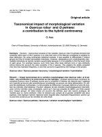
Replication timing of many genes is unchanged in ES mutantsFigure 1 (see previous page)
Replication timing of many genes is unchanged in ES mutants. (a) Replication timing analysis of Oct4, Esg1, Nkx2.9 and Mash1 in wild-type (white bars;
OS25, G9a WT, Suv39 h1/h2 WT, Dicer WT) and mutant (gray bars; Mll KO, Eed KO, Dnmt 1 KO, Dnmt3a/3b DKO, Mbd3 KO, G9a KO, Suv39 h1/h2
DKO and Dicer KO) ES cells. The histograms show the relative locus replication within each cell cycle fraction as measured by qPCR for all the wild-type
and mutant ES cells analyzed. The mean values and standard error of at least two independent experiments are shown. (b,c) Summary of replication
timing of candidate genes (b) and loci surrounding the Rex1 gene (c). In (c), positions of genes are indicated by black boxes and the two regions changing
replication timing upon ES cell differentiation are indicated by red bars. (d) Replication timing categories and color code. Early replication (E) is defined by
peak abundance in the G1-S and/or S1 fractions, mid-early (ME) by peak replication in S2, middle (M) in S2 and S3, mid-late (ML) in S3 and late (L)
replicating loci have peak abundance in S4 and/or G2.
R169.6 Genome Biology 2007, Volume 8, Issue 8, Article R169 Jørgensen et al. />Genome Biology 2007, 8:R169
posed role of siRNA in silencing repetitive elements [23]. In a
recent study, an advance in the replication of the major
satellite in Suv39h1/h2 double knockout (DKO) relative to
wild-type fibroblasts was reported [24], although the authors
noted that this advance was, in fact, not statistically signifi-
cant. The apparent discrepancy between their observation
and ours could be the result of intrinsic differences in the
mutant cell lines used, or reflect secondary adaptations to loss
of chromatin components. In this regard, compensatory
chromatin modifications have been previously described,
including an increase in H3K27me3 levels in Suv39h1/h2
deficient ES cells [25].
Minor and major satellites both carry DNA methylation and
share some histone marks [26], but their chromatin structure
is remarkably dissimilar. Major satellite DNA replicates in
mid to late S-phase in ES and somatic cells and is character-
ized by hypoacetylation, trimethylation of H3K9 and H4K20
and DNA methylation [25,27,28]. The minor satellite con-
tains the centromeric H3 variant CenpA, lacks appreciable
amounts of the repressive H3K9me3 and does not bind HP1
[28,29]. Instead, this repeat carries the permissive H3K4me2
mark [28] and it replicates in the first half of S-phase (Figure
3). We show that the replication timing of major and minor
satellites responds very differently to mutation of Mll, Dnmt1,
Suv39h1/h2, Dicer and G9a, supporting the view that the two
repeats are regulated differently.
Earlier replication of the major satellite in Dicer KO cells
might reflect increased repeat RNA accumulation, as has
been reported in some cells upon loss of Dicer [23,30],
prompting us to measure transcript levels of the repeats in
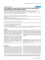
Histone acetylation and replication timing in ES cellsFigure 2
Histone acetylation and replication timing in ES cells. (a) The level of acetyl-H3K9 relative to total H3 is shown for each probe in >200 kb regions
surrounding the candidate loci (values represent log2{acetylH3K9/H3}). The peaks of histone acetylation were identified using the hidden Markov model
and are marked in blue. The location of candidate genes are shown (black box) relative to other genes (white box) within each region. Arrows show the
position of the primers used for the replication timing analysis and the size bars (black) represent 25 kb. Raw data are available in Additional data file 3. (b)
The replication timing of candidate loci in untreated ES cells (white bars) and after incubation with 10 nM (black bars) or 20 nM (gray bars) TSA is shown
as histograms. The mean values and standard error from at least two (two to three) independent experiments are shown.
Esg1
Nanog
Oct4
Zfp57
Nkx2.9
Sox2
Rex1
Mage a2
Mash1
Ebf
Sox3
b-Globin
Myf5
NeuroD1
2G4S3S2S1S1G2G4S3S2S1S1G
OS25
OS25 + 10 nM TSA
OS25 + 20 nM TSA
(b)
Cell cycle fraction Cell cycle fraction
(a)
G1 S1 S2 S3 S4 G2
Cell cycle fraction
Slc7a2
Loc2
Frg1
Msr1
Adam26
Loc4
Locus abundance (percentage of total)
Nanog
Nkx2.9
Sox2
Rex1
Mash1
Sox3
Myf5
Histone acetylation level [Log(acetylH3K9/H3)]
3
1
3
1
3
1
3
1
3
1
3
1
3
1
Locus abundance (percentage of total)
Locus abundance (percentage of total)
Replication timing profilesHistone acetylation (ChIP)
Genome Biology 2007, Volume 8, Issue 8, Article R169 Jørgensen et al. R169.7
comment reviews reports refereed researchdeposited research interactions information
Genome Biology 2007, 8:R169
each of the mutant cell lines (Table 2). Despite variation
among different lines of wild-type ES cells (major 0.7-4,
minor 0.1-5), a significant increase in major satellite tran-
script levels was seen in Dicer KO cells (17 compared with 4 in
matched wild-type cells). Increased minor and major satellite
transcripts were also seen in Eed deficient ES cells but not in
other mutant lines that also change satellite replication tim-
ing (for example, Dnmt 1 KO). These data suggest that while
chromatin modifiers can influence satellite transcript levels,
precocious replication is not an invariable consequence of
satellite transcription.
To determine whether satellite sequences are particularly
sensitive to loss of chromatin modifiers or if this is a general
repeat-associated feature, we analyzed long interspersed
nuclear elements (LINEs) and short interspersed nuclear ele-
ments (SINEs), which are found as single copies interspersed
with genes and other unique sequences at many locations in
the genome, as well as the tandemly repeated rDNA array. In
wild-type ES cells, SINE B1 replicates early whereas LINE 1
elements show replication in all fractions of the S-phase (Fig-
ure 4), consistent with the known genomic distribution of
these repeats; SINEs are primarily associated with gene rich
regions (which replicate early), whereas LINEs are enriched
in AT-rich, gene poor regions (which often replicate late, but
can change replication timing depending on the cell type [3]).
The rDNA sequence, which in fibroblasts comprises an early
replicating active and a late replicating silent fraction [31],
replicated synchronous very early in S-phase in wild-type ES
cells (Figure 4), possibly reflecting a high demand for biosyn-
thesis in these rapidly dividing cells. Analysis of these three
repeat sequences in the mutant ES cell lines revealed only
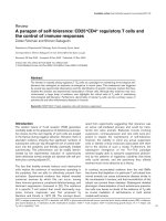
Satellite replication in ES cells is altered by mutation of chromatin modifiersFigure 3
Satellite replication in ES cells is altered by mutation of chromatin modifiers. (a) Summary of replication timing of repeat sequences in mutant ES cell lines.
Top, ideogram of the acrocentric mouse chromosome X, showing the position of minor satellite (Minor sat), major satellite (Major sat) and the X-linked
X141 repeat. (b) Examples show replication timing of repeats in wild type (WT, white bars (OS25 for Mll, Eed and Dnmt1; matched wild-type lines for
G9a, Suv39 h1/h2 and Dicer)) compared to ES cells mutant for the indicated chromatin modifier (black bars). The mean values and standard error of at
least two (two to five) independent experiments are shown.
Minor sat
G1 S1 S2 S3 S4 G2
Cell cycle fraction
Locus abundance (percentage of total)
X141
Major sat
Mll
G9a
Suv39
h1/h2
Dicer
Minor sat Major sat
G1 S1 S2 S3 S4 G2
Dnmt 1
Eed
WT Mutant
OS25 (WT) ME ML L
Mll KO M ML L
Eed KO ML L L
Dnmt 1 KO ME ME L
Dnmt 3a/3b DKO ME ML L
Mbd3 KO ME ML L
G9a WT ME ML L
G9a KO M ME ML
Suv39 h1/h2 WT ME ML L
Suv39 h1/h2 DKO ME L L
Dicer WT ME L L
Dicer KO M ML L
(b)(a)
R169.8 Genome Biology 2007, Volume 8, Issue 8, Article R169 Jørgensen et al. />Genome Biology 2007, 8:R169
very small changes with respect to wild-type cells. Replication
of rDNA was extended in Eed deficient cells and slightly
delayed in ES cells lacking Dicer.
DNA methylation selectively affects major satellite
replication timing
Our data show that loss of Dnmt1 in ES cells (which causes
genome-wide loss of CpG methylation; Table 1) results in
early replication of the pericentric major satellite sequence
without widespread changes in the replication timing of
euchromatic loci or other repeat elements (Figures 3 and 4).
To verify that reduction in DNA methylation is sufficient to
precipitate this advance in major satellite replication, we
experimentally demethylated wild-type ES cells. Exposure for
three days to the Dnmt inhibitor 5-azacytidine reduced DNA
methylation (from 0.88 in untreated to 0.21 in 5-azacytidine-
treated cells, compared to 0.11 in the Dnmt1 KO ES cell line;
Figure 5b) and caused an advanced replication of the major
satellite (Figure 5a). The replication timing of the minor sat-
ellite as well as single copy genes (
α
-Globin, Mash1 and Myf5)
was unaffected in treated cells. Collectively, these results sug-
gest that DNA methylation per se is important for maintain-
ing the correct temporal replication of the major satellite.
A role for DNA methylation in replication of heterochromatic
foci has been previously observed in fibroblasts and during
development [32]. Here we show that DNA methylation is
important in maintaining late replication specifically of major
satellite repeats in undifferentiated ES cells. As DNA methyl-
ation of the major satellite is also reduced in Suv39h1/h2
DKO ES cells (Table 1), it is perhaps surprising that these
mutant cells have delayed major satellite replication (Figure
Table 2
Relative transcript levels* of repeats in ES cell lines
Minor satellite Major satellite X141
WT (OS25) 0.4 ± 0.1 1.8 ± 0.5 1.8 ± 0.3
Eed KO 7.2 ± 6.5
†
18.5 ± 17.0
†
1.8 ± 0.2
Dnmt1 KO 0.3 ± 0.1 2.1 ± 0.0 2.0 ± 1.3
Dnmt3a/3b DKO 0.1 ± 0.0 5.7 ± 2.4 1.5 ± 0.9
G9a WT 0.1 ± 0.0 0.7 ± 0.1 0.5 ± 0.7
G9a KO 0.4 ± 0.4 1.0 ± 0.2 ND
Suv39 h1/h2 WT 4.9 ± 0.8 2.5 ± 1. 6 1.1 ± 0.3
Suv39 h1/h2 DKO 4.0 ± 1.3 2.2 ± 0.7 2.1 ± 0.5
Dicer WT 0.2 ± 0.0 4.1 ± 0.0 2.7 ± 0.0
Dicer KO 0.9 ± 0.6 17.4 ± 5.5 4.1 ± 1.2
C2C12 dif 100 100 100
*Levels of repeat sequence RNA were normalized to a house keeping gene (Ube) and expressed as a percentage of the levels detected in
differentiated mouse myocytes derived from C2C12 (C2C12 dif), samples in which high levels of major and minor satellite transcripts have
previously been detected [56]. Controls without reverse transcriptase were analyzed in parallel to dismiss contamination with genomic DNA.
†
Four independent samples showed variable but increased transcript levels: major satellite, 8.2%, 8.7%, 13.4%, and 43.8% of C2C12; minor satellite,
2.6%, 7.1%, 11.8%, and 13.8% of C2C12. ND, none detected.
Replication timing of repetitive elements in wild-type and mutant ES cell linesFigure 4
Replication timing of repetitive elements in wild-type and mutant ES cell
lines. The replication timing was determined for retrotransposons (LINE
and SINE B1) and rDNA repeats in wild-type OS25 ES cells and in mutant
ES cells lacking Eed, Dnmt 1, Dnmt 3a/3b, G9a or Dicer. The mean values
and standard error of at least two independent experiments are shown.
WT (OS25)
Dnmt 3a/3b
DKO
G9a KO
Dnmt 1 KO
Dicer KO
Eed KO
rDNASineB Line
Locus abundance (percentage of total)
G1 S1 S2 S3 S4 G2 G1 S1 S2 S3 S4 G2 G1 S1 S2 S3 S4 G2
Cell cycle fraction
Genome Biology 2007, Volume 8, Issue 8, Article R169 Jørgensen et al. R169.9
comment reviews reports refereed researchdeposited research interactions information
Genome Biology 2007, 8:R169
3). It is possible that other chromatin modifications
compensate for the loss of H3K9me3 to ensure heterochro-
matin formation in Suv39h1/h2 deficient cells, an idea that is
consistent with enhanced H3K27me3 at pericentric regions in
these cells [25].
Conclusion
We show that the timing of mouse satellite replication is
altered in ES cells lacking specific repressive chromatin mod-
ifiers. In particular, replication was advanced by mutation of
Dnmt1, G9a or Dicer, consistent with their repressive nature.
Earlier replication of major satellite was also induced by 5-
azacytidine treatment, demonstrating the importance of DNA
methylation for correct timing of this sequence. The sensitiv-
ity of satellite repeats to chromatin modifiers may be a reflec-
tion of their complexity and size. Genome-wide studies have
shown that replication timing of non-repetitive sequences is
constant over large 0.2-2 Mb regions [7], which often include
multiple loci that are regulated by different mechanisms.
Repetitive regions, on the other hand, have a more uniform
chromatin structure, which may make them more vulnerable
to loss of specific chromatin modifiers. Consistent with this
idea, the major and minor satellites comprise simple direct
repeats with high copy numbers (50-200,000) [26] whereas
the stable X141 is part of a much more complex repetitive
region and is represented only 80-90 times in the mouse
genome [22]. Interestingly, the size of the late replicating
fraction of the tandemly repeated rDNA array in fibroblasts
was shown to depend on NoRC, an ATP-dependant chroma-
tin remodeling complex [33].
Of the single copy genes examined in the study, we show that
the replication timing of some loci are more sensitive to the
loss of individual chromatin modifiers than others. Overall,
the apparent stability of gene replication profiles in mutant
ES cell lines suggests that for many single copy loci, replica-
tion timing is not primarily controlled by methylation of spe-
cific histone residues or DNA methylation, but, in agreement
with previous studies [4,8,9,11], histone acetylation is shown
to be a good predictor of replication timing. These data are
consistent with a mechanistic link between early origin firing
and acetylation in mammalian cells, as has been previously
demonstrated in yeast [10].
Materials and methods
ES cell culture and drug treatment
ES cells used in this study were wild-type OS25 [34], G9a
knock out (KO) clone 2-3, G9a wild-type clone col4 (G9a WT),
G9a transgene rescue clone 15-3 (G9a tg) [18], Suv39 h1/h2
double KO (DKO) clones DN57/DN72 and wild-type litter-
mate clone wt26 (Suv39 h1/h2 WT) [35], Eed KO clones B1.3/
G8.1 [4], Dnmt1 c/c (Dnmt1 KO) [36], Dnmt3a/Dnmt3b DKO
Major satellite replication is specifically advanced by 5-azacytidine treatmentFigure 5
Major satellite replication is specifically advanced by 5-azacytidine treatment. (a) Replication timing of 5-azacytidine treated (black bars) and untreated ES
cells (white bars). The mean values and standard error of at two independent experiments are shown. (b) Demethylation index (HpyCh4IV digested to
undigested genomic DNA) of the major satellite in untreated (OS25), 5-azacytidine-treated and Dnmt1 KO ES cells. Sox2 (no HpyCh4IV site) serves as an
internal control. Diagrams show the positions of primers and HpyChIV site. The standard deviation is shown in brackets.
OS25 + 5AzaC OS25 - 5AzaC
Major
sat
Minor
sat
X141
α-Globin
Mash1
Myf5
Methylation Index
(digested/undigested)
Major sat
Control
(Sox2)
HpyCh4IV
OS25
-5AzaC
OS25
+5AzaC
Dnmt1 KO
0.88
(0.05)
0.21
(0.05)
0.11
(0.05)
0.99
(0.10)
1.13
(0.06)
1.13
(0.08)
(a)
G1 S1 S2 S3 S4 G2 G1 S1 S2 S3 S4 G2
Cell cycle fraction
Locus abundance (percentage of total)
(b)
R169.10 Genome Biology 2007, Volume 8, Issue 8, Article R169 Jørgensen et al. />Genome Biology 2007, 8:R169
clone 10 (Dnmt3a/3b DKO) [27,37], Mbd3 KO clone Fix2
[38], Dicer KO clones D3-S5/D3-S6 and Dicer wild-type
clone D3 (described below). ES cells were derived from Dicer
flox/flox blastocysts [39] and transfected with the CRE-ER
transgene to produce the Dicer flox/flox ES clone D3 (Dicer
WT). The Dicer KO clones (D3-S5, D3-S6) were established
after tamoxifen treatment (800 nM; Sigma, Poole, UK) of the
D3 clone. Deletion of both alleles was confirmed by
genotyping.
The ES cell lines were maintained in the undifferentiated
state by culturing on gelatinized plates in KO-DMEM (Invit-
rogen, Carlsbad, CA, USA) supplemented with leukemia
inhibitory factor (LIF), 10% ES-tested fetal calf serum
(GlobePharm, Surrey, UK), L-glutamine, 2-mercaptoethanol,
non-essential amino acids and antibiotics. For Eed KO and
Dicer cells (WT and KO), a feeder layer of mitotic inactivated
fibroblasts was used and the medium was additionally sup-
plemented with 5% knockout serum replacement (KSR, Inv-
itrogen). OS25 cells were grown on gelatinized plates in
Glasgow-MEM (Invitrogen) supplemented with LIF, FCS-
gold (PAA, Yeovil, UK), L-glutamine, 2-mercaptoethanol,
non-essential amino acids, sodium pyruvate, sodium bicarbo-
nate and antibiotics. All ES cell lines examined in this study
were Oct4 positive as determined by immunofluorescence
(>92 %, data not shown). Undifferentiated ES cells (OS25)
were treated with 10 nM (Sigma) for 48 h, 20 nM TSA for 24
h or 15 μM 5-azacytidine (Sigma) for 72 h.
Replication timing assay
The protocol described by Azuara et al. [6] was used. Briefly,
asynchronous cell populations were pulse labeled with bro-
modeoxyuridine (BrdU; 30 minutes), fixed in 70% ethanol,
stained with propidium iodide and fractionated according to
DNA content by fluorescence assisted cell sorting (FACS). For
ES cells grown on a feeder layer, the feeder cells were
removed by differential attachment; less than 1% fibroblasts
remained after 20-25 minutes plating in non-gelatinized
plates. Pre-plating of feeder-dependent ES cells in this way
may result in a slight delay in the apparent time of replication
for genes that normally replicate very early in S-phase. Six cell
cycle fractions were collected, G1-S, G2 and four fractions
covering S-phase, S1-S4, where S1 corresponds to early S-
phase and S4 to late S-phase. An equal amount of BrdU
labeled Drosophila DNA was added to each fraction to control
for equal recovery. After isolation of total genomic DNA, the
DNA was sheared by sonication, denatured and newly repli-
cated, BrdU-labeled DNA was immunoprecipitated using
anti-BrdU antibody (BD, Franklin Lakes, NJ, USA). After
purification, quantitative real-time PCR (qPCR) was
employed to determine the relative quantity of specific loci in
each fraction. The sequences of primers for qPCR analysis are
given in Table 3. Locus replication was categorized based on
the peak fraction(s) as early (peak in G1 or S1), middle-early
(peak in S2), middle (peak in S2 and S3), middle-late (peak in
S3) or late (peak in S4 or G2).
Note regarding replication timing of repeated sequences
As mentioned above, we assessed the proportion of a specific
DNA sequence within newly replicated DNA for each cell
cycle fraction relative to the total from all six fractions. This
means that for single copy genes, a change in one allele will
give a shift for 50% of the signal whereas for a multi-copy
locus, only a small fraction (1% for a sequence repeated 100
times) will shift. Changes in single copy loci are, therefore,
much more readily detected than changes in repeated
sequences. Variability in locus replication among multi-copy
loci would be predicted to result in a spread-out signal
detected across multiple cell cycle fractions.
ChIP
Exponentially growing wild-type ES cells (OS25) were para-
formaldehyde (1%) fixed for 10 minutes at room temperature,
lysed and chromatin immuno-precipitated essentially as
described [40]. Briefly, chromatin (50 μg) was pre-cleared 2
h at 4°C, incubated with antibodies (2 μl IgG (Z0259, DAKO,
Copenhagen, Denmark); 2 μl anti-H3 (ab-1791-100, binds H3
independent of modification state, Abcam, Cambridge, UK),
5 μl anti-H3K9me2, 10 μl anti-H3K9me3 or 5 μl anti-H3K9ac
(07-441, 07-442, 07-352, Upstate/Millipore, Billerica, MA,
USA) at 4°C over night (ON) and the immune-complexes col-
lected by adding protein-A sepharose (Sigma) (incubated 2 h
at 4°C). Unbound chromatin was removed by washing 4× in
ChIP wash buffer (0.1% SDS, 1% Triton X-100, 2 mM EDTA,
150 mM NaCl, 20 mM Tris.Cl pH 8.1 and protease inhibitors)
and 1× in high salt ChIP wash buffer (0.1% SDS, 1% Triton X-
100, 2 mM EDTA, 150 mM NaCl, 20 mM Tris.Cl pH 8.1 and
protease inhibitors) after which 250 μl elution buffer was
added (1% SDS, 0.1 M NaHCO
3
, 100 μg/ml RNaseA, 500 μg/
ml Proteinase K). After incubating at 37°C for 2 h and at 65°C
ON, DNA was purified using a Gel purification kit (Qiagen,
Crawley, UK), using 2 × 40 μl of 10 mM Tris.Cl pH 8 for
elution. ChIP samples were analyzed by qPCR (sequences of
primers are given in Table 3) or microarray hybridization.
Microarray analysis
Input and ChIP samples were amplified by LM-PCR as
advised by Nimblegen, Reykjavik, Iceland. Labeling and
hybridization was done by Nimblegen using a custom
designed 50mer tiling array (100 bp average resolution) cov-
ering a region from 100 kb upstream to 100 kb downstream
of the analyzed genes. Normalized and scaled Chip: input
ratios for anti-H3K9ac and anti-H3 ChIP hybridizations were
produced by Nimblegen. The log2(H3K9ac/H3) ratios were
calculated from these data and plotted against the chromo-
somal position of the probes. Blue lines in Figure 2 indicate
peaks in the dataset detected by a hidden Markov model-
based algorithm using TileMap [41]. Gaps in the profile arise
from repetitive regions in the genome that are not repre-
sented on the array.
Genome Biology 2007, Volume 8, Issue 8, Article R169 Jørgensen et al. R169.11
comment reviews reports refereed researchdeposited research interactions information
Genome Biology 2007, 8:R169
Analysis of transcript levels
RNA was isolated from 3-5 × 10
6
cells using the RNAeasy mini
prep kit (Qiagen) with on-column DNase treatment. To
remove residual genomic DNA, RNA (1.2 μg) was treated with
RNAfree (Ambion, Austin, TX, USA) for 50 minutes before
reverse transcription using SuperScript III (Invitrogen) and
random primers in 20 μl reactions according to the manufac-
turer's instructions.
qPCR analysis
The sequence of primer pairs used in this study is given in
Table 3. All primer pairs were tested for efficiency (>1.95) and
linearity (R
2
> 0.99). Reactions (30 μl) were set up using a
Qiagen SYBR green kit with the appropriate template (2 μl
(corresponding to 200 cell equivalents) for replication tim-
ing, 1.5 μl of 1:5 diluted cDNA for gene expression, 2% of
eluted DNA for ChIP, 2 μl (corresponding to 1.7 ng) genomic
Table 3
Primer sequences
Locus Forward Reverse T
ann
Replication timing/ChIP
Gbe GGTGCAGATCATCCCCTTGA TTACCCGACGGCGAAAG 60
α-Globin CCACAAGCTGCGTGTGGAT ATGCCGCCTGCCAGGT 60
Oct4 GGGTGAGAAGGCGAAGTCTGAA GTGAGCCGTCTTTCCACCAGG 55
Nanog CCCTCTGAGTTTGACCGGTGA CAAGCTAGGATGTTAGGTCTCCCTG 60
Esg1 AAAGACGAACACAGAGTCAAACACC CACCTGCTCGATGTGAGACATTC 60
Zfp57 TGCAAGATAAGAACGAGGAGCAGGAG CCTTTGCGGCTTTGTGGATTTGTG 60
Rex1 TTTGCGGGAATCCAGCAGT CGTCCCATCGCCACTCTAGAC 55
Sox2 CCATCCACCCTTATGTATCCAAG CGAAGGAAGTGGGTAAACAGCAC 55
Nkx2.9 AAGTGCGAGGCGCTCG TGGCACCTTCCGGACTTG 60
Sox3 TGCCCAGATGGCTTCCTATT ACCCGGACATTCTCCGCT 60
Mage a2 TTGGTGGACAGGGAAGCTAGGGGA CGCTCCAGAACAAAATGGCGCAGA 60
Ebf AGATCTGGTTGAAGCCCTGTATGG CATGTCACATCTCAGATCCTGTGTTCT 60
Mash1 CCAGGCTGGAGCAAGGGA CGGTTGGCTTCGGGAGC 55
β
-Globin GGTGAACTTTACTGCTGAGGAAAAG TCACCACCAACCTCTTCAACAT 60
Myf5 GGAGATCCGTGCGTTAAGAATCC CGGTAGCAAGACATTAAAGTTCCGTA 55
Math1 CCCTCACTCAGGTCGCCTG CGTGCGAGGAGCCAATCA 55
Sox1 ACAAGAGGAGGCAGCGAACC TCGCAGGTGGAAAGTTTCTCC 55
Msx1 ACAGAAAGAAATAGCACAGACCATAAGA TTCTACCAAGTTCCAGAGGGACTTT 55
Frg1 AAGGAGCCTATATCCATGCACTGGAC GCCTCCCTGCCATTGCTTGT 60
Slc7a2 GACAAGGAACAGGGCGAGAAG CTTTCCTCATCCTGGGCTTGAGTA 60
Msr1 GCCACCAATGCCCTAGAATTTC GGCAGGCTCTCACTAGGAAGC 60
Loc2 ACTAGCAACTGGACATAAGAGTACACTACC ATTACATATGGTGTCTGGAAGCCAG 60
Adam 26 CCTTGAACAACGCCCTTTTGTG GCAAGCTCCCAAAACAGGTGT 60
Loc4 TAAGGTAGGCAGTGAGAGACATCCA GGTGTAAGAAGGTTAGAACTAA 60
X141 GGGTCATAAAACGCTTTTCCAGGAA TAGCACTGGAGATCAGATTGACGCCT 60
Minor satellite TGATATACACTGTTCTACAAATCCCGTTTC ATCAATGAGTTACAATGAGAAACATGGAAA 55
Major satellite GACGACTTGAAAAATGACGAAATC CATATTCCAGGTCCTTCAGTGTGC 55
rDNA CCTGTGAATTCTCTGAACTC CCTAAACTGCTGACAGGGTG 60
IAP TTGATAGTTGTGTTTTAAGTGGTAAATAAA AAAACACCACAAACCAAAATCTTCTAC 60
LINE 1 TTTGGGACACAATGAAAGCA CTGCCGTCTACTCCTCTTGG 60
SINE B1 GTGGCGCACGCCTTTAATC GACAGGGTTTCTCTGTGTAG 60
Gene expression
Mage a2 GAAGATCTCAGGAGTGTCAGGACTG TCAGCCATTATGACTGTCCTAGGTAA 60
Ubc AGGAGGCTGATGAAGAGCTTGA TGGTTTGAATGGATACTCTGCTGGA 60
Oct4 CCCAAGGTGATCCTCTTCTGCTT GAGAAGGTGGAACCAACTCCCG 60
CpG methylation assay
Major sat GACGACTTGAAAAATGACGAAATC CATATTCCAGGTCCTTCAGTGTGC 55
Sox2 TGGACTGCGAACTGGAGAAGG CGCCCGGAGTCTAGCTCTAAATATT 60
IAP, intracisternal A particle. T
ann
, annealing temperature.
R169.12 Genome Biology 2007, Volume 8, Issue 8, Article R169 Jørgensen et al. />Genome Biology 2007, 8:R169
DNA for analysis of DNA methylation) and analyzed on
Chromo4™ Real-Time PCR Detector (Bio-Rad, Hercules, CA,
USA) with Opticon Monitor™ software.
DNA methylation assay
Triplicate reactions of genomic DNA with or without the
methylation sensitive restriction enzyme HpyCh4IV (New
England Biolabs, Beverly, USA) were incubated for 3 h and
the extent of digestion analyzed by qPCR. The primers for the
major satellite span a HpyCh4IV site. The Sox2 primers,
which do not span a HpyCh4IV site, were used to control for
equal DNA content.
Additional data files
The following additional data are available with the online
version of this paper. Additional data file 1 contains supple-
mentary materials and methods, legends to supplementary
Figures 1-4, and supplementary Tables 1 and 2.
Supplementary Table 1 contains the percentages of cells pos-
itive for ES cell markers and Supplementary Table 2 contains
coordinates, GC content and Line density of the genes ana-
lysed in Figure 1b. Additional data file 2 contains supplemen-
tary Figures 1-4. Supplementary Figure 1 shows alkaline
phosphatase staining and cell cycle profiles of the mutant ES
cell lines analysed. Supplementary Figure 2 contains
replication timing profiles of all loci analysed in each of the
mutant ES cell lines analysed. Supplementary Figure 3 shows
analysis of the Mage a2 gene. Supplementary Figure 4 shows
analysis of short hairpin RNA mediated knockdown of Eed in
Dnmt1 mutant ES cells. Additional data file 3 is an archive
containing microarray data for the histone acetylation
analysis.
Additional data file 1Supplementary materials and methods, legends to supplementary Figures 1-4, and supplementary Tables 1 and 2Supplementary Table 1 contains the percentages of cells positive for ES cell markers and supplementary Table 2 contains coordinates, GC content and line density of the genes analyzed in Figure 1b.Click here for fileAdditional data file 2Supplementary Figures 1-4Supplementary Figure 1 shows alkaline phosphatase staining and cell cycle profiles of the mutant ES cell lines analyzed. Supplemen-tary Figure 2 contains replication timing individual profiles of all genes in each of the mutant ES cell lines analyzed. Supplementary Figure 3 shows analysis of the Mage a2 gene. Supplementary Fig-ure 4 shows analysis of short hairpin RNA mediated knockdown of Eed in Dnmt1 mutant ES cells.Click here for fileAdditional data file 3Microarray data for the histone acetylation analysisMicroarray data for the histone acetylation analysis.Click here for file
Abbreviations
BrdU, bromodeoxyuridine; ChIP, chromatin immunoprecip-
itation; DKO, double knock out; Dnmt, DNA methyl
transferase; ES, embryonic stem; HDAC, histone deacetylase;
HMTase, histone methyl transferase; KO, knock out; LINE,
long interspersed nuclear element; PRC, polycomb repressor
complex; qPCR, quantitative real-time PCR; SINE, short
interspersed nuclear element; siRNA, short interfering RNA;
TSA, trichostatin A.
Authors' contributions
HFJ, VA and AGF designed the study described in this report.
HFJ performed the experiments, analysed the data, and was
responsible for writing the initial versions of the manuscript.
SA performed cell culture experiments. MS and AT were
involved in performing the microarray analysis. MS analysed
the microarray data. TN, BSC and BR contributed material/
reagents/analysis tools. MM and AGF supervised and over-
saw the completion of the studies as well as the writing of the
manuscript. All authors read and approved the final version
of the manuscript.
Acknowledgements
We thank Brian Hendrich, Thomas Jenuwein, Yoichi Shinkai and En Li for
kind gifts of mutant ES cell lines and Rosalind John for advice on culturing
Eed KO cells. Pascale Perry and Luca Mazzarella are thanked for sharing
unpublished information. Eric O'Connor and Eugene Ng are acknowledged
for FACS sorting and Ingrid Devonish for administrative help. This study
was supported by the MRC, the EU Epigenome Network of Excellence, the
Parkinson's Disease Society (VA) and HEROIC, High-throughput Epigenetic
Regulatory Organisation in Chromatin, an Integrated Project funded by the
European Union (LSHG-CT-2005-018883).
References
1. Jackson DA, Pombo A: Replicon clusters are stable units of
chromosome structure: evidence that nuclear organization
contributes to the efficient activation and propagation of S
phase in human cells. J Cell Biol 1998, 140:1285-1295.
2. Perry P, Sauer S, Billon N, Richardson WD, Spivakov M, Warnes G,
Livesey FJ, Merkenschlager M, Fisher AG, Azuara V: A dynamic
switch in the replication timing of key regulator genes in
embryonic stem cells upon neural induction. Cell Cycle 2004,
3:1645-1650.
3. Hiratani I, Leskovar A, Gilbert DM: Differentiation-induced repli-
cation-timing changes are restricted to AT-rich/long inter-
spersed nuclear element (LINE)-rich isochores. Proc Natl Acad
Sci USA 2004, 101:16861-16866.
4. Azuara V, Perry P, Sauer S, Spivakov M, Jorgensen HF, John RM, Gouti
M, Casanova M, Warnes G, Merkenschlager M, et al.: Chromatin
signatures of pluripotent cell lines. Nat Cell Biol 2006, 8:532-538.
5. Ofir R, Wong ACC, McDermid HE, Skorecki KL, Selig S: Position
effect of human telomeric repeats on replication timing. Proc
Natl Acad Sci USA 1999, 96:11434-11439.
6. Azuara V, Brown KE, Williams RR, Webb N, Dillon N, Festenstein R,
Buckle V, Merkenschlager M, Fisher AG: Heritable gene silencing
in lymphocytes delays chromatid resolution without affect-
ing the timing of DNA replication. Nat Cell Biol 2003, 5:668-674.
7. Donaldson AD: Shaping time: chromatin structure and the
DNA replication programme. Trends Genet 2005, 21:444-449.
8. Lin CM, Fu H, Martinovsky M, Bouhassira E, Aladjem MI: Dynamic
alterations of replication timing in mammalian cells. Curr Biol
2003, 13:1019-1028.
9. Schubeler D, Lorincz MC, Cimbora DM, Telling A, Feng YQ, Bouhas-
sira EE, Groudine M: Genomic targeting of methylated DNA:
influence of methylation on transcription, replication, chro-
matin structure, and histone acetylation. Mol Cell Biol 2000,
20:9103-9112.
10. Vogelauer M, Rubbi L, Lucas I, Brewer BJ, Grunstein M: Histone
acetylation regulates the time of replication origin firing.
Mol
Cell 2002, 10:1223-1233.
11. Zhang J, Xu F, Hashimshony T, Keshet I, Cedar H: Establishment of
transcriptional competence in early and late S phase. Nature
2002, 420:198-202.
12. Ai Leen L, Dorothy EP, Beth AS: Control of gene expression and
assembly of chromosomal subdomains by chromatin regula-
tors with antagonistic functions. Chromosoma 2005,
114:242-251.
13. Bernstein BE, Mikkelsen TS, Xie X, Kamal M, Huebert DJ, Cuff J, Fry
B, Meissner A, Wernig M, Plath K, et al.: A bivalent chromatin
structure marks key developmental genes in embryonic
stem cells. Cell 2006, 125:315-326.
14. Boyer LA, Plath K, Zeitlinger J, Brambrink T, Medeiros LA, Lee TI,
Levine SS, Wernig M, Tajonar A, Ray MK, et al.: Polycomb com-
plexes repress developmental regulators in murine embry-
onic stem cells. Nature 2006, 441:349-353.
15. Jorgensen HF, Giadrossi S, Casanova M, Endoh M, Koseki H, Brock-
dorff N, Fisher AG: Stem cells primed for action: polycomb
repressive complexes restrain the expression of lineage-spe-
cific regulators in embryonic stem cells. Cell Cycle 2006,
5:1411-1414.
16. Williams RR, Azuara V, Perry P, Sauer S, Dvorkina M, Jorgensen H,
Roix J, McQueen P, Misteli T, Merkenschlager M, et al.: Neural
Genome Biology 2007, Volume 8, Issue 8, Article R169 Jørgensen et al. R169.13
comment reviews reports refereed researchdeposited research interactions information
Genome Biology 2007, 8:R169
induction promotes large-scale chromatin reorganisation of
the Mash1 locus. J Cell Sci 2006, 119:132-140.
17. Roessler S, Gyory I, Imhof S, Spivakov M, Williams RR, Busslinger M,
Fisher AG, Grosschedl R: Distinct promoters mediate the reg-
ulation of Ebf1 gene expression by interleukin-7 and Pax5.
Mol Cell Biol 2007, 27:579-594.
18. Tachibana M, Sugimoto K, Nozaki M, Ueda J, Ohta T, Ohki M, Fukuda
M, Takeda N, Niida H, Kato H, et al.: G9a histone methyltrans-
ferase plays a dominant role in euchromatic histone H3
lysine 9 methylation and is essential for early embryogenesis.
Genes Dev 2002, 16:1779-1791.
19. Sigalotti L, Coral S, Nardi G, Spessotto A, Cortini E, Cattarossi I, Col-
izzi F, Altomonte M, Maio M: Promoter methylation controls
the expression of MAGE2, 3 and 4 genes in human cutaneous
melanoma. J Immunother 2002, 25:16-26.
20. Englmann A, Clarke LA, Christan S, Amaral MD, Schindelhauer D,
Zink D: The replication timing of CFTR and adjacent genes.
Chromosome Res 2005, 13:183-194.
21. McCool KW, Xu X, Singer DB, Murdoch FE, Fritsch MK: The role
of histone acetylation in regulating early gene expression
patterns during early embryonic stem cell differentiation. J
Biol Chem 2007, 282:6696-6706.
22. Nasir J, Fisher EM, Brockdorff N, Disteche CM, Lyon MF, Brown SD:
Unusual molecular characteristics of a repeat sequence
island within a Giemsa-positive band on the mouse X
chromosome. Proc Natl Acad Sci USA 1990, 87:399-403.
23. Lippman Z, Martienssen R: The role of RNA interference in het-
erochromatic silencing. Nature 2004, 431:364-370.
24. Wu R, Singh PB, Gilbert DM: Uncoupling global and fine-tuning
replication timing determinants for mouse pericentric
heterochromatin. J Cell Biol 2006, 174:185-194.
25. Peters AH, Kubicek S, Mechtler K, O'Sullivan RJ, Derijck AA, Perez-
Burgos L, Kohlmaier A, Opravil S, Tachibana M, Shinkai Y, et al.: Par-
titioning and plasticity of repressive histone methylation
states in mammalian chromatin. Mol Cell 2003,
12:1577-1589.
26. Martens JH, O'Sullivan RJ, Braunschweig U, Opravil S, Radolf M, Stein-
lein P, Jenuwein T: The profile of repeat-associated histone
lysine methylation states in the mouse epigenome. EMBO J
2005, 24:800-812.
27. Okano M, Bell DW, Haber DA, Li E: DNA methyltransferases
Dnmt3a and Dnmt3b are essential for de novo methylation
and mammalian development. Cell 1999, 99:247-257.
28. Sullivan BA, Karpen GH: Centromeric chromatin exhibits a
histone modification pattern that is distinct from both
euchromatin and heterochromatin. Nat Struct Mol Biol 2004,
11:1076-1083.
29. Guenatri M, Bailly D, Maison C, Almouzni G: Mouse centric and
pericentric satellite repeats form distinct functional
heterochromatin. J Cell Biol 2004, 166:493-505.
30. Kanellopoulou C, Muljo SA, Kung AL, Ganesan S, Drapkin R, Jenuwein
T, Livingston DM, Rajewsky K: Dicer-deficient mouse embryonic
stem cells are defective in differentiation and centromeric
silencing. Genes Dev 2005, 19:489-501.
31. Berger C, Horlebein A, Gogel E, Grummt F: Temporal order of
replication of mouse ribosomal RNA genes during the cell
cycle. Chromosoma 1997, 106:479-484.
32. Selig S, Ariel M, Goitein R, Marcus M, Cedar H: Regulation of
mouse satellite DNA replication time. EMBO J 1988,
7:419-426.
33. Li J, Santoro R, Koberna K, Grummt I: The chromatin remodeling
complex NoRC controls replication timing of rRNA genes.
EMBO J 2005, 24:120-127.
34. Billon N, Jolicoeur C, Ying QL, Smith A, Raff M: Normal timing of
oligodendrocyte development from genetically engineered,
lineage-selectable mouse ES cells. J Cell Sci 2002,
115:3657-3665.
35. Peters AH, O'Carroll D, Scherthan H, Mechtler K, Sauer S, Schofer
C, Weipoltshammer K, Pagani M, Lachner M, Kohlmaier A, et al.: Loss
of the Suv39h histone methyltransferases impairs mamma-
lian heterochromatin and genome stability. Cell 2001,
107:323-337.
36. Li E, Bestor TH, Jaenisch R:
Targeted mutation of the DNA
methyltransferase gene results in embryonic lethality. Cell
1992, 69:915-926.
37. Jackson M, Krassowska A, Gilbert N, Chevassut T, Forrester L, Ansell
J, Ramsahoye B: Severe global DNA hypomethylation blocks
differentiation and induces histone hyperacetylation in
embryonic stem cells. Mol Cell Biol 2004, 24:8862-8871.
38. Kaji K, Caballero IM, MacLeod R, Nichols J, Wilson VA, Hendrich B:
The NuRD component Mbd3 is required for pluripotency of
embryonic stem cells. Nat Cell Biol 2006, 8:285-292.
39. Cobb BS, Nesterova TB, Thompson E, Hertweck A, O'Connor E,
Godwin J, Wilson CB, Brockdorff N, Fisher AG, Smale ST, et al.: T
cell lineage choice and differentiation in the absence of the
RNase III enzyme Dicer. J Exp Med 2005, 201:1367-1373.
40. Baxter J, Sauer S, Peters A, John R, Williams R, Caparros ML, Arney
K, Otte A, Jenuwein T, Merkenschlager M, et al.: Histone
hypomethylation is an indicator of epigenetic plasticity in
quiescent lymphocytes. EMBO J 2004, 23:4462-4472.
41. Ji H, Wong WH: TileMap: create chromosomal map of tiling
array hybridizations. Bioinformatics 2005, 21:3629-3636.
42. Ayton P, Sneddon SF, Palmer DB, Rosewell IR, Owen MJ, Young B,
Presley R, Subramanian V: Truncation of the Mll gene in exon 5
by gene targeting leads to early preimplantation lethality of
homozygous embryos. Genesis 2001, 30:201-212.
43. Yagi H, Deguchi K, Aono A, Tani Y, Kishimoto T, Komori T: Growth
disturbance in fetal liver hematopoiesis of Mll-mutant mice.
Blood 1998, 92:108-117.
44. Yu BD, Hess JL, Horning SE, Brown GA, Korsmeyer SJ: Altered Hox
expression and segmental identity in Mll-mutant mice.
Nature 1995, 378:505-508.
45. Montgomery ND, Yee D, Chen A, Kalantry S, Chamberlain SJ, Otte
AP, Magnuson T: The murine polycomb group protein Eed is
required for global histone H3 lysine-27 methylation. Curr Biol
2005, 15:942-947.
46. Faust C, Lawson KA, Schork NJ, Thiel B, Magnuson T: The Poly-
comb-group gene eed is required for normal morphogenetic
movements during gastrulation in the mouse embryo. Devel-
opment 1998, 125:4495-4506.
47. Wang J, Mager J, Chen Y, Schneider E, Cross JC, Nagy A, Magnuson
T: Imprinted X inactivation maintained by a mouse Poly-
comb group gene. Nat Genet 2001, 28:371-375.
48. Lei H, Oh SP, Okano M, Juttermann R, Goss KA, Jaenisch R, Li E: De
novo DNA cytosine methyltransferase activities in mouse
embryonic stem cells. Development 1996, 122:3195-3205.
49. Tucker KL, Talbot D, Lee MA, Leonhardt H, Jaenisch R: Comple-
mentation of methylation deficiency in embryonic stem cells
by a DNA methyltransferase minigene. Proc Natl Acad Sci USA
1996, 93:12920-12925.
50. Chen T, Ueda Y, Dodge JE, Wang Z, Li E: Establishment and main-
tenance of genomic methylation patterns in mouse embry-
onic stem cells by Dnmt3a and Dnmt3b. Mol Cell Biol 2003,
23:5594-5605.
51. Hendrich B, Guy J, Ramsahoye B, Wilson VA, Bird A: Closely
related proteins MBD2 and MBD3 play distinctive but inter-
acting roles in mouse development. Genes Dev 2001,
15:710-723.
52. Lehnertz B, Ueda Y, Derijck AA, Braunschweig U, Perez-Burgos L,
Kubicek S, Chen T, Li E, Jenuwein T, Peters AH: Suv39h-mediated
histone H3 lysine 9 methylation directs DNA methylation to
major satellite repeats at pericentric heterochromatin. Curr
Biol 2003, 13:1192-1200.
53. Peters AH, Mermoud JE, O'Carroll D, Pagani M, Schweizer D, Brock-
dorff N, Jenuwein T: Histone H3 lysine 9 methylation is an epi-
genetic imprint of facultative heterochromatin. Nat Genet
2002, 30:77-80.
54. Bernstein E, Kim SY, Carmell MA, Murchison EP, Alcorn H, Li MZ,
Mills AA, Elledge SJ, Anderson KV, Hannon GJ: Dicer is essential
for mouse development. Nat Genet 2003, 35:215-217.
55. Murchison EP, Partridge JF, Tam OH, Cheloufi S, Hannon GJ: Char-
acterization of Dicer-deficient murine embryonic stem cells.
Proc Natl Acad Sci USA 2005, 102:12135-12140.
56. Terranova R, Sauer S, Merkenschlager M, Fisher AG: The
reorganisation of constitutive heterochromatin in differenti-
ating muscle requires HDAC activity. Exp Cell Res 2005,
310:344-356.
57. Tachibana M, Sugimoto K, Nozaki M, Ueda J, Ohta T, Ohki M, Fukuda
M, Takeda N, Niida H, Kato H, et al.: G9a histone methyltrans-
ferase plays a dominant role in euchromatic histone H3
lysine 9 methylation and is essential for early embryogenesis.
Genes Dev 2002, 16:1779-1791.