Báo cáo y học: "The temporal program of peripheral blood gene expression in the response of nonhuman primates to Ebola hemorrhagic fever" ppsx
Bạn đang xem bản rút gọn của tài liệu. Xem và tải ngay bản đầy đủ của tài liệu tại đây (1.25 MB, 14 trang )
Genome Biology 2007, 8:R174
comment reviews reports deposited research refereed research interactions information
Open Access
2007Rubinset al.Volume 8, Issue 8, Article R174
Research
The temporal program of peripheral blood gene expression in the
response of nonhuman primates to Ebola hemorrhagic fever
Kathleen H Rubins
*†‡
, Lisa E Hensley
§
, Victoria Wahl-Jensen
§
,
Kathleen M Daddario DiCaprio
§
, Howard A Young
¶
, Douglas S Reed
§
,
Peter B Jahrling
§
, Patrick O Brown
†¥
, David A Relman
*#**
and
Thomas W Geisbert
§
Addresses:
*
Department of Microbiology and Immunology, 299 Campus Dr., Stanford University School of Medicine, Stanford, California
94305, USA.
†
Department of Biochemistry, 279 Campus Dr., Stanford University School of Medicine, Stanford, California 94305, USA.
‡
Whitehead Institute for Biomedical Research, Nine Cambridge Center, Cambridge, Massachusetts 02142, USA.
§
US Army Medical Research
Institute of Infectious Diseases, 1425 Porter St., Fort Detrick, Maryland 21702-5011, USA.
¶
National Cancer Institute - Frederick, 1050 Boyles
St., Frederick, Maryland 21702, USA.
¥
Howard Hughes Medical Institute, 279 Campus Dr., Stanford University School of Medicine, Stanford,
California 94305, USA.
#
Department of Medicine, 300 Pasteur Dr., Stanford University School of Medicine, Stanford, California 94305, USA.
**
Veterans Affairs Palo Alto Health Care System, 3801 Miranda Ave., Palo Alto, California 94304, USA.
Correspondence: Kathleen H Rubins. Email:
© 2007 Rubins et al; licensee BioMed Central Ltd.
This is an open access article distributed under the terms of the Creative Commons Attribution License ( which
permits unrestricted use, distribution, and reproduction in any medium, provided the original work is properly cited.
Primate transcriptional response to Ebola<p>Primate blood cells were analysed for changes in global gene expression patterns at several time points following infection with Ebola virus, providing insights into potential mechanisms of viral pathogenesis and host defense.</p>
Abstract
Background: Infection with Ebola virus (EBOV) causes a fulminant and often fatal hemorrhagic
fever. In order to improve our understanding of EBOV pathogenesis and EBOV-host interactions,
we examined the molecular features of EBOV infection in vivo.
Results: Using high-density cDNA microarrays, we analyzed genome-wide host expression
patterns in sequential blood samples from nonhuman primates infected with EBOV. The temporal
program of gene expression was strikingly similar between animals. Of particular interest were
features of the data that reflect the interferon response, cytokine signaling, and apoptosis.
Transcript levels for tumor necrosis factor-α converting enzyme (TACE)/α-disintegrin and
metalloproteinase (ADAM)-17 increased during days 4 to 6 after infection. In addition, the serum
concentration of cleaved Ebola glycoprotein (GP
2 delta
) was elevated in late-stage EBOV infected
animals. Of note, we were able to detect changes in gene expression of more than 300 genes before
symptoms appeared.
Conclusion: These results provide the first genome-wide ex vivo analysis of the host response to
systemic filovirus infection and disease. These data may elucidate mechanisms of viral pathogenesis
and host defense, and may suggest targets for diagnostic and therapeutic development.
Published: 28 August 2007
Genome Biology 2007, 8:R174 (doi:10.1186/gb-2007-8-8-r174)
Received: 12 February 2007
Revised: 4 May 2007
Accepted: 28 August 2007
The electronic version of this article is the complete one and can be
found online at />R174.2 Genome Biology 2007, Volume 8, Issue 8, Article R174 Rubins et al. />Genome Biology 2007, 8:R174
Background
Ebola virus causes severe and often lethal hemorrhagic fever
in humans and nonhuman primates. Ebola virus (EBOV) is
one of two genera that comprise the family Filoviridae. The
EBOV genus consists of four distinct species: Ivory Coast
Ebola virus, Reston Ebola virus, Sudan Ebola virus, and Zaire
Ebola virus (ZEBOV) [1]. Sudan Ebola virus and ZEBOV have
been associated with human disease outbreaks in Central
Africa, with case fatality rates averaging about 50% for Sudan
Ebola virus and ranging from 75% to 90% for ZEBOV [2].
Although Reston Ebola virus is highly lethal in nonhuman
primates [3,4], the few data available suggest that it is non-
pathogenic in humans [5]. The pathogenic potential of Ivory
Coast Ebola virus is unclear because there has only been a sin-
gle confirmed nonfatal human case [6] and a second sus-
pected nonfatal case [7]. In addition to natural outbreaks,
EBOV is an important concern as a potential biologic threat
agent of deliberate use because these viruses have low infec-
tious doses and clear potential for dissemination by aerosol
route [8]. Currently, there are no approved preventive vac-
cines or postexposure treatments for EBOV hemorrhagic
fever, but recent advances have led to the development of sev-
eral candidate therapeutics and vaccines for EBOV [9-11].
The mechanisms of EBOV pathogenesis are only partially
understood, but dysregulation of normal host immune
responses (including destruction of lymphocytes [2] and
increases in levels of circulating proinflammatory cytokines
[12]) is thought to play a major role. Several animal models of
EBOV hemorrhagic fever have been developed, notably a
cynomolgus macaque (Macaca fascicularis) model [13,14],
which closely resembles human infection [2,15]. ZEBOV
infection in cynomolgus macaques results in uniform
lethality at days 6 to 7 after infection [16-19].
The majority of studies conducted in nonhuman primates
have focused on end-point examination when animals are in
the final stages of disease, and have restricted their analyses
to small numbers of cytokines or mRNA transcripts. cDNA
microarrays have been used by our group to study mecha-
nisms of viral pathogenesis in a nonhuman primate model of
an agent, albeit unrelated, that also causes overwhelming,
systemic infection [20,21]. In order to understand better the
early events in EBOV pathogenesis, we examined global
changes in gene transcript abundance, using cDNA microar-
rays, in sequential blood samples from 21 cynomolgus
macaques over the entire time course of ZEBOV infection.
Results
Dataset overview
We characterized the host gene expression program in
peripheral blood mononuclear cells (PBMCs) of cynomolgus
macaques during a temporal survey of ZEBOV infection. The
dataset from these experiments comprises about 3.2 million
measurements of transcript abundance in a total of 65 blood
samples from 21 animals using 85 DNA microarrays. Addi-
tional data file 1 shows animal numbers corresponding to
blood samples. Samples are arranged in the table order
(namely, days 0 to 6 after infection), from right to left, in all
figures. The bleed schedule is provided in Additional data file
2. Figure 1 provides an overview of the temporal changes in
gene expression patterns in PBMCs. The gene expression pro-
gram exhibits surprisingly consistent patterns of temporal
regulation among all animals sampled, with very few changes
with respect to baseline evident at days 1 and 2 after infection,
followed by dramatic and widespread changes at days 4 to 6
after infection. During this latter phase there were changes of
at least threefold in the relative abundance of transcripts for
more than 3,760 elements (1,832 unique named genes; Fig-
ure 1 and Additional data file 3). The average pair-wise corre-
lation of the expression profiles of these 3,760 elements
(1,832 named genes) between different animals at days 4, 5,
and 6 after infection was 0.85, demonstrating the consistency
of host response in this model. In comparison, using the same
criteria the average pair-wise correlation of the transcript
abundance patterns between animals in a cynomolgus
macaque model of smallpox infection was 0.55 over 2,387 ele-
ments for the same time frame [20].
Cytokine response and innate immune activation
A significant increase in cytokine and chemokine transcripts
was observed at days 4 to 6 after infection (Figure 2a). Tran-
scripts encoding the proinflammatory cytokines IL-1β, IL-6,
IL-8, and tumor necrosis factor (TNF)-α were markedly
increased in late-stage animals (average fold increase at day 5
after infection: IL-1β, 3.9; IL-6, 4.3; IL-8, 11.3; and TNF-α,
5.2; Figure 2b). In addition, several chemokines (macrophage
inflammatory protein [MIP]-1α, MIP-1β, growth related
oncogene-α, growth related oncogene-β, monocyte chemoat-
tractant protein [MCP]-1, MCP-2, MCP-3, and MCP-4) exhib-
ited increased transcript levels at days 4 to 6 after infection in
all animals (Figure 2a). Transcripts for several other
cytokines (IL-2, IL-4, IL-10, and IL-12) were detected on the
array, but their levels did not change significantly during the
course of infection. We measured levels of soluble cytokines
by ELISA. All measured cytokines for which we also have gene
expression data are shown in Figure 2b. IL-6 and MCP-1
showed marked increases by day 4 after infection; and MIP-
1α and MIP-1β exhibited moderate increases on day 4, coin-
ciding with gene expression data. By day 5 these four
cytokines were elevated, and there was also an increase in
TNF-α and IL-18 in serum. The ELISA data closely parallel
the microarray mRNA expression data.
We previously identified a set of genes representing the TNF-
α/nuclear factor-κB (NF-κB) B regulon as a prominent fea-
ture of the PBMC response to bacterial lipopolysaccharide
[22]. We extracted these genes from the ZEBOV dataset and
saw marked induction in transcripts regulated by TNF-α/NF-
κB (Figure 3).
Genome Biology 2007, Volume 8, Issue 8, Article R174 Rubins et al. R174.3
comment reviews reports refereed researchdeposited research interactions information
Genome Biology 2007, 8:R174
Apoptosis
Lymphocyte apoptosis in the lymph node and spleen has pre-
viously been identified as a hallmark of ZEBOV infection and
a potential contributor to pathogenesis [23-25]. In order to
determine whether we could also detect evidence of apoptosis
in circulating PBMCs we examined the dataset for genes with
Gene Ontology (GO) annotation for involvement in apoptosis
(pro-apoptotic or anti-apoptotic). Transcripts of a set of genes
that play a role in regulating apoptosis increased on days 4 to
6 after infection (Figure 4a). These genes included Bcl-2 fam-
ily members and interacting proteins: BCL2-antagonist of cell
death, BH3 interacting domain death agonist, BCL2-like 1
(BCL2L1/BCL-X), BCL2-related protein A1, TNF superfamily
member 10 (also known as TNF related apoptosis inducing
ligand [TRAIL]), caspase-5, caspase-8, FADD (Fas-associ-
ated death domain protein)-like apoptosis regulator, caspase
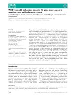
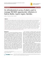
Overview of gene expression in peripheral blood mononuclear cells from Ebola infected macaquesFigure 1
Overview of gene expression in peripheral blood mononuclear cells from Ebola infected macaques. A total of 3,670 elements (1,832 named genes)
exhibited a threefold change or greater in mRNA abundance from at least three different arrays. The data for these 3,670 elements were hierarchically
clustered [67]. Data from individual elements or genes are represented as a single row, and samples from individual monkeys at different days after
infection are shown as columns. Red and green colors denote expression levels greater or less, respectively, than baseline values (average of two to three
samples taken at day -1 and day -6 before inoculation). The intensity of the color reflects the magnitude of the change from baseline.
Day 0
Day 1
Day 2
Day 3
Day 4
Day 5
Day 6
• TRAIL receptor 3
•CD68
• IL-18 binding protein
• IL-2 receptor
• IL-15 receptor
IFN cluster:
STAT1, GBP1, IRF7
IFITM1, GBP1, ISG20
MX1, IFIT1/2, OAS2
•CD59
• MCP-1
•IL1α
• IL1 receptor
• Bcl6
•CFLAR
•NFκB
•TLR1
•TLR4
• Grancalcin
•IFNγ receptor
•TOSO
•TRAIL
• Bcl2A1
•CD14
• Factor VIII
•IL-6
•TNFα
•NFκBIA
•TNFα induced
•RelB
•IL-8
•MIP1α/MIP1β
Increased transcript abundance
Decreased transcript abundance
•Granzyme K
•Granzyme A
•TGFβ-stimulated
• Integrin β5
•CD9
• Integrin α2
• Immunoglobulin κ
• Immunoglobulin μ
•CD40
•CD8
•CD20
• Killer-cell lectin
like receptor
• T cell receptor α
•CD3
•CD1c
• IL-2 receptor
•CD19
•LCK
• MHC class II
• Ribosomal proteins
• T cell receptor β
•CD2
•CD5
•CD6
•CD69
• CD79a/b
•CD86
• CXCR4
•CD74
R174.4 Genome Biology 2007, Volume 8, Issue 8, Article R174 Rubins et al. />Genome Biology 2007, 8:R174
1 apoptosis-related cysteine peptidase/IL-1β convertase, IL-
1β, and IL-1α. TRAIL transcript abundance increased as
much as 35-fold above background at day 5 in some animals,
with average expression being 19.4-fold above baseline (Fig-
ure 4b). We confirmed induction of several of these tran-
scripts (BCL-X, BCL2-related protein A1, and BCL2-
antagonist/killer 1) by RNAse protection assay (Figure 4b).
Interferon response
The earliest major transcriptional response apparent in all
animals by day 2 or 3 was an increase in transcript levels of a
large set of interferon (IFN) regulated genes (Figure 1),
including the following: myxovirus resistance protein (MX)1
and MX2, IFN-γ inducible protein-10, 2'-5' oligoadenylate
synthetase-1, -2, and -3, guanylate binding protein-1 and -2,
signal transducer and activators of transcription (STAT)-1,
double-stranded DNA activated protein kinase, and IFN-γ
receptors 1 and 2. This response increased even further on
day 4 and remained high throughout the time course of infec-
tion. We extracted the set of IFN regulated transcripts using
previously published lists of known IFN-α, IFN-β, and IFN-γ
induced genes [20,26,27] and arranged the gene expression
data for these genes using a self-organzing map (Figure 5a).
MX1 expression in circulating cells was confirmed by immu-
nohistochemistry (Figure 5b).
Fibrin deposition and dissolution
Several transcripts related to the process of fibrin dissolution,
including those for urokinase plasminogen activator (uPA)
and uPA receptor, as well as the plasminogen activator inhib-
itor type 1 of the plasminogen-cleaving serine proteases,
increased during days 4 to 6 after infection (Figure 6a,c,d).
Expression of transcripts encoding uPA and uPA receptor
rapidly increased from baseline on day 4 after infection and
peaked on day 5 after infection (average fold above back-
ground: uPA, 9.5; uPA receptor, 14.1). uPA protein expression
was confirmed by ELISA, and followed a similar trend as gene
expression, but it continued to increase at day 6 after infec-
tion (Figure 6b).
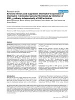
Cytokine gene expressionFigure 2
Cytokine gene expression. (a) A list of all cytokines and chemokines (as defined by Gene Ontology annotation) was used to extract gene expression data.
Genes with at least a 2.5-fold change from baseline in at least three arrays are displayed. (b) Transcript levels of cytokine mRNA in peripheral blood
mononuclear cells and ELISAs for detection of soluble cytokines in the serum. IL, interleukin; MCP, monocyte chemoattractant protein; MIP, macrophage
inflammatory protein; TNF, tumor necrosis factor.
GROβ
β
MIP-1
α
MIP-1
β
MIP-3
α
M-CSF
IL-1
α
IL-1
β
IL-6
IL-8
MCP-1
GRO
α
MCP-2
MCP-3
MCP-4
MIP-3
TNF
α
IP-10
Day 0
Day 1
Day 2
Day 3
Day 4
Day 5
Day 6
(a)
IL-18
IL-18
0 1 2 3
4
5 6
-2
1
2
3
4
5
6
-2000
0
2,000
4,000
6,000
8,000
10,000
Day post infection
Fold change from baseline
pg/mL
IL-6
0 1 2 3
4
5 6
-3
-2
1
2
3
4
5
6
7
-250
0
250
500
750
1,000
Day post infection
Fold change from baseline
pg/mL
MCP-1
0 1 2 3 4 5 6
1
10
20
30
40
50
60
70
80
0
1,000
2,000
3,000
4,000
5,000
6,000
Day post infection
Fold change from baseline
pg/mL
MIP-1
α
0 1 2 3 4 5 6
-2
1
2
3
4
5
6
7
8
9
-2000
0
2,000
4,000
6,000
8,000
10,000
Day post infection
Fold change from baseline
pg/mL
MIP-1
β
0 1 2 3 4 5 6
1
3
5
7
9
-200
0
200
400
600
800
1,000
Day post infection
Fold change from baseline
pg/mL
TNF
α
0 1 2 3 4 5 6
1
3
5
7
9
-10
0
10
20
30
40
50
60
Day post infection
Fold change from baseline
pg/mL
Gene expression
ELISA
(b)
Genome Biology 2007, Volume 8, Issue 8, Article R174 Rubins et al. R174.5
comment reviews reports refereed researchdeposited research interactions information
Genome Biology 2007, 8:R174
Proteolytic cleavage of the Ebola virus glycoprotein
We noted an increase in TNF-α converting enzyme/α-disin-
tegrin and metalloproteinase (ADAM)-17 at days 4 to 6 after
infection, peaking at an average 3.1-fold increase above base-
line at day 5 after infection. Dolnik and coworkers [28] dem-
onstrated that ADAM-17 is responsible for shedding of the
EBOV glycoprotein (GP) ectodomain from cell surfaces in
vitro [28]. We also detected the cleaved ectodomain of GP,
GP
2Δ
, in sera from terminal (day 7 after infection) ZEBOV
infected animals (Figure 7c), which was present at higher
concentrations than the positive control of cell culture super-
natant from ZEBOV infected Vero cells (Figure 7c).
Pre-symptomatic transcriptional response in
peripheral blood mononuclear cells
In order to determine whether we could detect gene expres-
sion changes before clinical symptoms appeared, we analyzed
the complete dataset for genes that exhibited significant
changes before day 3 after infection. The expression levels of
317 elements (202 unique named genes) either increased or
decreased by at least twofold, in at least three animals, at day
1 or 2 after infection (Figure 8). IL-1β, which was highly
induced at later stages of infection (Figure 2), was initially
repressed on the first day after infection. Genes that were
induced during the first 2 days after infection included early
stress response genes (early growth response, Fos, Jun) and
IFN responsive genes (MX1 and 2, STAT-1, IFN-γ inducible
protein-10, guanylate binding protein-1 and -2). Animals had
no detectable clinical illness at days 1 and 2, were feeding nor-
mally, had normal physical activity patterns on days 1 and 2,
and normal results for all measured laboratory values (com-
plete blood count, differential, chemistries, ELISA, and tem-
perature). Levels of plasma viremia were undetectable until
day 3 after infection (Figure 8b). In addition, there were only
mild symptoms at day 3 after infection; three out of ten ani-
mals sampled had elevated temperature, and three out of 15
had early signs of rash (very mild) and a slight increase in D-
dimers.
Changes in cell component mixtures of peripheral
blood mononuclear cells
In samples of whole blood or PBMCs, variations in the indi-
vidual cell subtypes (lymphocytes, monocytes) that comprise
the mixed cell population can lead to observed differences in
gene expression responses. An increase or decrease in one cell
type changes the overall proportion of that cell type's unique
transcripts in the total pool of RNA from a given sample. To
address this issue more effectively, we correlated the gene
expression vector for each individual gene in the dataset with
each parameter in the complete blood count and differential
data on relative levels of individual cell populations (Addi-
tional data file 3). This allowed us to assess the magnitude of
the contribution of changes in cell type to the observed gene
expression profiles for each cluster. The largest average cor-
relation scores for the two major clusters shown in Figure 1
were 0.45 (lymphocyte count, decreased transcript abun-
dance cluster), 0.47 (total neutrophil count, increased tran-
script abundance), and 0.69 (band neutrophil count,
increased transcript abundance).
Discussion
In a series of studies we recently analyzed the pathology of
lethal ZEBOV infection in cynomolgus macaques using a
sequential sacrifice design [13,14]. In the present study, we
examined the genome-wide transcriptional responses in
sequential samples of peripheral blood from 15 of these
cynomolgus macaques. Nonhuman primates infected with
ZEBOV exhibited a highly homogeneous, time-dependent
pattern of gene expression (Figure 1). Given the massive path-
ologic changes, physiologic instability, and widespread tissue
damage, as well as the commonly observed variability in
genome-wide transcript abundance patterns among different
individual hosts ex vivo, it was surprising that the animals
displayed such uniform patterns. Perhaps because of the
overwhelming nature of the infection and the relatively short
time frame between the first appearance of signs and death,
these patterns are highly homogenous due to an effect akin to
temporal compression. It is very likely that the observed gene
expression patterns reflect many physiologic changes caused
by systemic filoviral infection (for example, bystander lym-
phocyte apoptosis, fibrin deposition, and anti-viral IFN
response). With a longer time frame or lower mortality rate,
it is possible that individual host responses might show more
variation; nonetheless, the homogeneity of this response
allowed us to analyze the characteristic gene expression pat-
terns with minimal noise from animal-to-animal variation.
Tumor necrosis factor-α/nuclear factor-κB responseFigure 3
Tumor necrosis factor-α/nuclear factor-κB response. The set of genes
representing the tumor necrosis factor (TNF)-α/nuclear factor-κB (NF-
κB) regulon present in previously published lipopolysaccharide stimulation
data [22] was extracted from the dataset and hierarchically clustered.
Colored bars represent multiple clones on the array for a given gene.
Day 0
Day 1
Day 2
Day 3
Day 4
Day 5
Day 6
Rel
NF-κB1
NF-κB2
IκBε
RelB
TNF-α
TNF-α
R174.6 Genome Biology 2007, Volume 8, Issue 8, Article R174 Rubins et al. />Genome Biology 2007, 8:R174
The underlying molecular changes echo the uniform lethality
of the animal model, and may provide better predictors of
morbidity/mortality than a model with high levels of inter-
individual variation.
We observed a marked increase in transcript abundance for
genes encoding many cytokines, including IL-1β, IL-6, IL-8,
MIP-1α, MIP-1β, macrophage colony stimulating factor, and
MCP-1 (Figure 2), which is consistent with a systemic proin-
flammatory response. Reports of cases of human EBOV infec-
tion vary considerably with respect to the cytokines that are
associated with fatal as opposed to nonfatal outcome
[12,25,29]. Increases in IL-1β, IL-6, MIP-1α, and MIP-1β have
been reported for human survivors of EBOV infection [25]. In
vitro infection of human monocytes/macrophages with
authentic EBOV or virus-like particles that include mem-
brane-associated GP
1,2
leads to increases in protein levels of
IL-1β [30-32], IL-6 [30-32], IL-8 [31,32], MIP-1α [30,33],
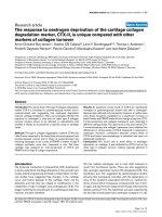
Apoptosis-related genesFigure 4
Apoptosis-related genes. (a) The set of apoptosis-related genes (as defined by Gene Ontology annotation) was used to extract gene expression data.
Genes with at least a 2.5-fold change from baseline in at least two arrays are displayed. (b) Transcript levels for tumor necrosis factor (ligand) superfamily,
member 10 (TNFSF10/TRAIL) at various times after infection. (c) Transcript levels of apoptosis-related genes, as determined by RNAase protection
assays at day 0 after infection (lanes A, C, G, I, K, M, O, and Q), day 1 after infection (lanes B and D), day 2 after infection (lane E), day 3 after infection
(lane F), day 4 after infection (lanes H and J), day 5 after infection (lanes L, N, and P), day 6 after infection (lane R). Colored bars represent multiple clones
on the array for a given gene.
Day 0
Day 1
Day 2
Day 3
Day 4
Day 5
Day 6
BAD
BCL-X
BCL2A1
TRAIL
BID
CFLAR
BIRC3
IL-1α
IL-1β
CASP1
BAK
CASP5
(a)
A B C D E F G H I J K L M N O P Q R
L32
BAK
BCL-X
BCL2A1
(b)
(c)
D0 D1 D2 D3 D4 D5 D6
0 5 10 15 20 25 30 35
TRAIL
Day post infection
Fold change above baseline
Genome Biology 2007, Volume 8, Issue 8, Article R174 Rubins et al. R174.7
comment reviews reports refereed researchdeposited research interactions information
Genome Biology 2007, 8:R174
MIP-1β [33], and MCP-1 [30]. In monkeys infected with
ZEBOV or Reston Ebola virus, increases in IL-1β [14,34], IL-
6 [14,33,34], MIP1-α [14,33], MIP-1β [14,33,34], and MCP-1
[14,34] have been reported. Monocytes and macrophages
represent a major cellular target for infection and dissemina-
tion of EBOV in monkeys [14,35-37]. Infection of monocytes
and macrophages leads to increased production and release
of proinflammatory cytokines, leading in turn to recruitment
of macrophages to areas of inflammation, which may contrib-
ute to viral proliferation and eventually an overwhelming sep-
sis-like syndrome [14,38,39].
Serum levels of TNF-α, in particular, are demonstrably
increased in human [12,29], primate [14,33], and in vitro [30-
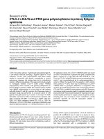
Interferon-responsive genesFigure 5
Interferon-responsive genes. (a) A list of known interferon (IFN) genes was compiled from the literature. The gene expression data for these genes was
arranged by a self-organzing map, using ten nodes. (b) Myxovirus resistance protein (MX) expression in circulating cells. MX protein (red) was detected in
circulating cells; cell nuclei are stained with DAPI (blue).
Day 2
MX2
IP-10
IFIT1
IP-10
GBP1
OAS3
GBP1
GBP2
IRF2
STAT1
OAS2
ISG15
OAS1
ISG15
IP-10
IFI16
MX1
OAS1
IFITM3
IRF7
IFITM1
IRF2
IFI16
PRK
IFNGR1
IFNAR1
IFNG
Day 0
Day 1
Day 3
Day 4
Day 5
Day 6
(a)
(b)
R174.8 Genome Biology 2007, Volume 8, Issue 8, Article R174 Rubins et al. />Genome Biology 2007, 8:R174
33] EBOV infection. Wahl-Jensen and coworkers [40]
recently showed that the virus-like particle induced decrease
in endothelial barrier function was further enhanced by TNF-
α, which is known to induce a long-lasting decrease in
endothelial cell barrier function and is hypothesized to play a
key role in EBOV pathogenesis [40]. We detected an increase
not only in TNF-α but also in the downstream transcriptional
response that is regulated by TNF-α and NF-κB (Figure 3),
providing evidence that the circulating cells are responding to
the large amounts of TNF-α that are induced during infection.
Induction of the NF-κB pathway by TNF-α usually induces an
anti-apoptotic response and cell survival [41], possibly
reflecting a mechanism by which EBOV counteracts host
apoptotic defenses in the infected cell, thereby contributing to
viral spread.
Despite the known role of the NF-κB pathway in an anti-
apoptotic response, we found that transcripts for many pro-
apoptotic genes were induced (Figure 4). Genes for the Bcl
antagonists BCL2-antagonist of cell death, BH3 interacting
domain death agonist, BCL-X, and BAK appeared to be
induced in the later stages of infection; all of these factors
promote apoptosis by inhibiting Bcl-2. Expression levels of
IL-1α and IL-1β were also increased; these cytokines are pro-
teolytically processed and released in response to cell injury
and induce apoptosis. Both forms of IL-1 are proteolytically
processed to their active form by caspase 1, which was also
expressed. In addition, transcript levels of TRAIL were mark-
edly increased (Figure 4b). TRAIL expression early during
infection and induction by IFN-α may contribute to lym-
phocyte apoptosis [33]. In view of the increased transcript
levels for a group of pro-apoptotic genes, the decrease in
lymphocyte related transcripts, including CD3, CD8, CD19,
CD64, major histocompatibility complex class II, T cell recep-
tor β, integrins, and granzymes (Figure 1) in ZEBOV infection
may result from 'bystander' lymphocyte apoptosis and subse-
quent depletion of lymphocytes in circulating peripheral
blood [14,23-25]. Thus, it appears that although the infected
monocyte/macrophage lineages can survive and carry virus
to secondary infection sites in the tissues, cells important for
the adaptive immune response are decimated through
bystander lymphocyte apoptosis, preventing an effective
adaptive immune response, and enabling further virus prop-
agation and spread.
Fibrin deposition and dissolutionFigure 6
Fibrin deposition and dissolution. (a) Transcripts of genes known to be involved in the coagulation cascade (intrinsic and extrinsic pathways) were selected
from the filtered dataset. Data were selected that showed a 2.5-fold change or greater in at least three arrays. (b) Protein levels of urokinase plasminogen
activator (uPA) in blood plasma, as determined by ELISA. (c and d) Transcript levels of uPA (c) and uPA receptor (uPAR) (d).
uPA
THBD
PAI
Factor VIII
uPA receptor
Fibrinogen
Day 0
Day 1
Day 2
Day 3
Day 4
Day 5
Day 6
(b)(a)
ELISA - uPA urokinase plasminogen activator
0
2
4
6
8
10
12
14
16
18
20
0123456
Day post-infection
ng/mL
(d)(c)
uPA receptor
urokinase plasminogen activator receptor
123456
Day post-infection
Fold change in gene expression
15
13
11
9
7
5
3
1
0
10
9
8
7
6
5
4
3
2
1
uPA
urokinase plasminogen activator
0123456
Day post-infection
Fold change in gene expression
Genome Biology 2007, Volume 8, Issue 8, Article R174 Rubins et al. R174.9
comment reviews reports refereed researchdeposited research interactions information
Genome Biology 2007, 8:R174
Although the major transcriptional changes appeared on days
4 to 6, corresponding to the initial appearance of clinical
signs, a strong IFN response was evident at day 3 after infec-
tion (Figure 5a), and transcripts levels for a subset of IFN
genes increased as early as 24 hours after infection (Figure 8).
In addition, expression of the classical IFN induced protein
MxA was detected in circulating cells (Figure 5b). Several
studies have reported the detection of IFN-α in serum from
EBOV-infected humans [12] and monkeys [14,33], and our
results provide evidence that cells in circulating peripheral
blood can mount a robust transcriptional response to the IFN
stimulus, despite the presence of EBOV proteins (VP24 and
VP35), which are thought to function as type I IFN antago-
nists [42,43]. This might imply that the major role of the
ZEBOV type I IFN antagonists is to act locally to influence the
microenvironment of the infected cell, rather than to shut
down a systemic IFN response. The majority of cells in the
peripheral blood sample (PBMCs) are uninfected, because no
evidence of EBOV infection of lymphocytes has been
observed [14,23] and the circulating population of infected
monocytes/macrophages constitutes only 1% to 13% of
PBMCs in these primates. Both VP35 and VP24 act in a cell
autonomous manner; VP35 blocks activation of the IFN reg-
ulatory factor 3 and the transcriptional responses of the IFN
regulatory factor 3 responsive promoters [44], and VP24
blocks nuclear accumulation of tyrosine phosphorylated
STAT through interaction with karyopherin α
1
[43]. Because
of the cell autonomous nature of the EBOV IFN antagonists,
uninfected cells should still be capable of producing a tran-
scriptional response to the large amounts of circulating IFN,
as shown in Figure 5a.
Disseminated intravascular coagulation, caused by over-acti-
vation of the coagulation system and resulting in microvascu-
lar thrombosis [45], may contribute to the lethal multi-
system organ failure in EBOV infection. Over-expression of
tissue factor in EBOV infected monocytes/macrophages has
been shown to produce fibrin deposition in the spleen, liver,
and blood vessels of infected macaques [46], and inhibition of
the tissue factor/factor VIIa pathway resulted in a decrease of
D-dimers (fibrin degradation products) and an increased sur-
vival rate in rhesus macaques [47]. In this study we found
evidence of cellular responses that would be expected to lead
to increased fibrin degradation. There was an increase in both
uPA and uPA receptor transcripts in PBMCs (Figure 6a,c,d),
accompanied by an increase in serum concentrations of uPA
protein (Figure 6b). uPA acts to convert plasminogen to plas-
min; the uPA receptor mediates the proteolysis independent
signal transduction activation effects of uPA, also promoting
plasmin formation. However, we also observed an increase in
transcripts encoding plasminogen activator inhibitor, per-
haps caused by negative feedback regulation. Thus, the over-
all impact of the observed transcriptional response on the
coagulation cascade is not self-evident. Nevertheless,
Expression levels of the metalloprotease responsible for cleavage of Ebola glycoproteinFigure 7
Expression levels of the metalloprotease responsible for cleavage of Ebola glycoprotein. Shown are (a) expression levels of tumor necrosis factor (TNF)-
α converting enzyme/α-disintegrin and metalloproteinase (ADAM)-17 from the overview cluster and (b) in graph form. (c) Glycoprotein (GP) in the
serum from infected rhesus macaques over the course of infection. Serum was diluted 1:3 in NP40 lysis buffer. Samples were run on a 10% Bis-Tris gel
under reducing conditions, as shown. Mock cell lysate from 293T cells transfected with vector only (pDisplay) is shown as a negative control (lane 1); Zaire
Ebola virus (ZEBOV; lane 2) is supernatant from in vitro Ebola infected Vero E6 cells at day 8 after infection. Lanes 3 and 4 are transfection controls
expressing glycoprotein (GP)
1,2
(cell lysate) and GP
1,2Δ
supernatant) [31]. Serum from infected rhesus macaques, before infection (lane 5), and on day 4 and
6 after infection (lanes 6 to 9) were diluted 1:3 in NP40 lysis buffer and 22.5 μl was loaded per lane. Samples included two animals per day (after infection)
analyzed. Note the lack of GP in the prebleed control sample (lane 5). GP
2Δ
is seen in the transfection control (lane 4) and NHP sera samples from days 4
(lane 6, albeit weakly) and 6 days after infection (lanes 8 and 9).
Day 2
ADAM17
Day 0
Day 1
Day 3
Day 4
Day 5
Day 6
ADAM 17
0
0.5
1
1.5
2
2.5
0246
Day post-infection
Fold change in gene expression
3.5
3
2.5
2
1.5
1
(a)
(b)
(c)
R174.10 Genome Biology 2007, Volume 8, Issue 8, Article R174 Rubins et al. />Genome Biology 2007, 8:R174
although the majority of the coagulation and fibrinolytic cas-
cade is regulated at the protein level through processing,
transcriptional induction of genes that are involved in fibrin
degradation may be a factor in the coagulopathy during
EBOV infection.
EBOV GP is regulated by complex transcriptional editing and
post-translational cleavage processes. The authentic tran-
script of the GP gene is expressed as a polypeptide, which is
cleaved into soluble glycoprotein (sGP) and the secreted delta
peptide [48,49]. Through RNA editing, the transmembrane
form of GP is expressed (GP
1,2
) and then cleaved into GP
1
and
GP
2
disulfide linked fragments, which are present on the sur-
face of virus particles [50-52]. The role of EBOV GP and its
contribution to pathogenesis has been the subject of much
investigation. GP can decrease the expression of cell adhesion
molecules, interfering with cell attachment and inducing
cytotoxicity [53-56], but mutant viruses that fail to produce
sGP are more cytotoxic, suggesting a negative regulation by
sGP of the GP induced cytotoxicity [57]. In vitro studies sug-
gest that GP
1,2
on the surface of virus-like particles, but not
sGP, activates target cells [31] and decreases endothelial bar-
rier function [40]. However, EBOV replication does not
induce direct cytolysis of endothelial cells either in vitro or in
animal models of EBOV infection [13], although cytolytic
infection of human umbilical cord vein endothelial cells has
been demonstrated with Marburg virus [58].
TNF-α converting enzyme/ADAM-17 was recently found to
mediate proteolytic processing and shedding of the ectodo-
main of Ebola GP (GP
1,2Δ
) [28]. We found that transcript lev-
els for ADAM-17 increased on days 4 to 6 after infection,
peaking at day 5 after infection (Figure 7a,b), which is consist-
ent with a role for ADAM-17 in shedding of GP
1,2Δ
during in
vivo primate infection. In addition, we also detected elevated
concentrations of cleaved GP
2Δ
in sera from late-stage ZEBOV
infected animals compared with uninfected controls (Figure
7c), demonstrating that cleavage of GP also takes place during
in vivo infection in a nonhuman primate model of EBOV
hemorrhagic fever. The relationship of shed GP
1,2Δ
to patho-
genesis/disease severity is unclear, and its role during in vivo
infection remains to be investigated. It is possible that GP
1,2Δ
can act as a decoy and soak up anti-EBOV antibodies, effec-
tively shielding the virus from the immune system [28].
The composite gene expression pattern assayed in a mixed
cell population, such as PBMCs, gives a rich and multidimen-
sional picture of the systemic host responses to infection,
reflecting many interconnected responses of a complex sys-
tem. However, because the observed gene expression pattern
represents a composite of diverse influences, data from mixed
cell populations can be more difficult to interpret. In samples
of whole blood or PBMCs, large variations in the cellular com-
position are often the largest source of overall variation in the
observed gene expression patterns [59]. We correlated the
gene expression vector for each individual gene in the dataset
with complete blood count and differential data on relative
levels of individual cell populations (Additional data file 3).
Correlation scores were highest between cell populations that
increased during the course of infection and the cluster of
genes whose transcripts levels were increasing, and also cell
populations that decreased and the cluster of genes whose
transcripts levels were decreasing. As an example, the deple-
tion of circulating lymphocytes during EBOV infection in vivo
[24] correlates with the decrease in lymphocyte-related tran-
scripts in our microarray dataset (Figure 1). However, given
the mathematical simplicity of the gene expression and
Preclinical gene expressionFigure 8
Preclinical gene expression. (a) Genes with transcripts whose abundance
shows at least a twofold increase or decrease from baseline (day 0) in at
least three of the samples for day 1 or day 2 are shown. The expression
patterns of 317 elements (202 unique named genes) were hierarchically
clustered; rows represent individual genes and columns represent samples.
These patterns reflect changes in gene expression before symptoms
appear. (b) Virus isolation from plasma. Infectious virus in EDTA plasma
was assayed by counting plaqueson Vero cells maintained as monolayers in
six-well plates under agarose, as previously described [70].
Day 2
Day 1
Day 0
EGR1
FOS
JUN
IL1β
MX1/2
IP-10
GBP1/2
STAT1
Plasma viremia
0
1
2
3
4
5
6
7
8
d 0 d 1 d 2 d 3 d 4 d 5 d 6
Days post challenge
Virus titer log
10
PFU
(a)
(b)
Genome Biology 2007, Volume 8, Issue 8, Article R174 Rubins et al. R174.11
comment reviews reports refereed researchdeposited research interactions information
Genome Biology 2007, 8:R174
differential parameters (elements either increase or decrease
uniformly and uniderectionally with respect to time), the
correlation scores could be due to temporal coincidence of
processes with similar directionality.
These correlations indicate that there is a certain proportion
of the variation in gene expression that might be explained by
changes in cell population. However, it is difficult to deter-
mine whether the variation in gene expression is directly
caused by changes in the composition of the mixed cell popu-
lation, activation of cells, or coincidence of temporal proc-
esses. Although correlative analysis based on numbers of
PBMC types may provide the basis for attributing variation in
expression to specific cell subsets, future experiments
examining filovirus infection in more homogenous cell sub-
populations will be essential for reliable identification of cell
type specific responses. Likewise, examination of different
tissues during the course of infection could provide a more
comprehensive picture of the molecular anatomy of host
responses to EBOV on an organism-wide basis.
Early detection and diagnosis of EBOV infection would be
invaluable, as many of the symptoms and signs are nonspe-
cific at presentation [2], and some recently described inter-
ventions have benefit when given early after filovirus
infection [47,60]. To determine whether we could detect any
changes in gene expression before appearance of symptoms,
we examined the gene expression profile of peripheral blood
in the early preclinical stages of infection (days 1 and 2). We
observed changes in expression of over 200 genes before any
clinical signs were evident and before plasma viremia was
detectable in the ZEBOV infected nonhuman primate model
(Figure 8a,b). This gene set is a possible starting point for the
identification of early diagnostic markers. Any of these genes
alone may provide little specificity, but combinations of these
and/or others may allow differentiation among different etio-
logic agents, all of which may otherwise demonstrate similar
clinical pictures. Although these early response genes are all
responsive to diverse infections and inflammatory conditions
and thus are of limited specificity for detection and diagnosis,
the pattern of response to EBOV infection, in a setting with a
high index of suspicion, may provide useful early warning for
triage, aggressive treatment, and/or quarantine.
Analysis of global gene expression as disease pathogenesis
unfolds provides a multifaceted picture of the complex inter-
play between host and pathogen. Examination of early events
during infection may help to identify and provide insight into
the specific molecular processes that initiate the cascade of
host damage during EBOV infection. The ability to detect
gene expression patterns before clinical symptoms may pro-
vide an opportunity for early diagnosis.
Materials and methods
Nonhuman primate model of Ebola infection
Fifteen healthy, adult male cynomolgus macaques were inoc-
ulated intramuscularly in the left or right caudal thigh with
1,000 plaque forming units of ZEBOV [61]. Animals were
killed on days 1, 2, 3, 4, 5, and 6 after infection [13,14,46].
Infection studies were performed under biosafety level 4 con-
tainment at the US Army Medical Research Institute of Infec-
tious Diseases. Research was conducted in compliance with
the Animal Welfare Act and other federal statues and regula-
tions relating to animals and experiments involving animals,
and adheres to the principles stated in the Guide for the Care
and Use of Laboratory Animals (National Research Council,
1996). The biosafety level 4 facility used is fully accredited by
the Association for Assessment and Accreditation of Labora-
tory Animal Care International.
Sample acquisition and RNA preparation
Peripheral blood samples (2.5 ml) were collected on days 1, 4,
and 6 before infection, in order to define a robust baseline,
and then on successive days after infection until death
(immediately before their death) or recovery. All samples
were collected at the same time of day (± 2 hours) to minimize
differences in expression caused by diurnal variation. PBMCs
were isolated from 1.5 ml of peripheral blood using by centrif-
ugation on Histopaque (Sigma, St. Louis, MO, USA) at 250 g
for 30 min. Cells at the interface were harvested, washed
twice in phosphate-buffered saline, and placed in TRIzol
(Invitrogen Corporation, Carlsbad, CA, USA). Total RNA was
extracted using TRIzol. RNA was linearly amplified using the
Ambion MessageAmp kit (Ambion Inc, Austin, TX, USA).
cDNA microarrays and hybridization
We used human cDNA microarrays containing 37,632 ele-
ments that represent approximately 18,000 unique genes,
which efficiently capture monkey transcripts [20]. Arrays
were produced as described previously [62-64]. Fluorescently
labeled cDNA prepared from amplified RNA was hybridized
to the array in a two-color comparative format [62,65], with
the experimental samples labeled with one fluorophore (Cy5)
and a reference pool of mRNA labeled with a second fluoro-
phore (Cy3). The reference pool (Universal Human Refer-
ence; Stratagene Inc., La Jolla, CA, USA) provided an internal
standard to enable reliable comparison of relative transcript
levels in multiple samples [62,63,66]. The microarrays were
submitted to the Gene Expression Omnibus database under
series record GSE8317.
Data filtering and analysis
Array images were scanned using an Axon Scanner 4000A
(Axon Instruments, Union City, CA, USA), and image analysis
was performed using GenePix Pro version 3.0.6.89 (Axon
Instruments). Data were expressed as the log
2
ratio of fluores-
cence intensities of the sample and the reference, for each ele-
ment on the array [62,65]. Data were filtered to exclude
elements that did not have a regression correlation of Cy5 to
R174.12 Genome Biology 2007, Volume 8, Issue 8, Article R174 Rubins et al. />Genome Biology 2007, 8:R174
Cy3 signal over the pixels spanning the array element of ≥0.6
and intensity/background ratio of ≥2.5 in at least 80% of the
arrays. For each gene, the expression levels over the time
course for each monkey were 'time zero-transformed' by
subtracting the average of the pre-infection baseline expres-
sion level from that animal, so that the values of each time
point represent changes relative to the uninfected samples.
The genes whose expression varied from the uninfected base-
line by at least threefold in at least three samples were
selected for further analysis. There was relatively little varia-
tion in the pre-infection baseline samples; in the 39 pre-expo-
sure samples only 769 elements (439 unique named genes)
varied at least threefold in at least three samples, as compared
with 3,670 elements (1,832 named genes) that varied at least
threefold in three post-exposure samples. The data were hier-
archically clustered using the Cluster program [67] and dis-
played using TreeView [68].
Hematology
Total white blood cell counts, lymphocyte counts, red blood
cell counts, platelet counts, hematocrit values, total hemo-
globin, mean cell volume, mean corpuscular volume, and
mean corpuscular hemoglobin concentration were deter-
mined from blood samples collected in tubes containing
EDTA, using a laser-based hematological Analyzer (Coulter
Electronics, Hialeah, FL, USA). White blood cell differentials
were measured manually on Wright-stained blood smears.
Cytokine and chemokine ELISAs
Cytokine/chemokine levels in monkey sera/plasma were
assayed using commercially available ELISA kits according to
manufacturer's directions. Cytokines/chemokines assayed
included monkey TNF-α (BioSource International, Inc.,
Camarillo, CA, USA). ELISAs for human proteins known to be
compatible with cynomolgus macaques included IL-6, MIP-
1α, and MIP-1β (BioSource International, Inc.), and human
IL-18 and MCP-1 (R&D Systems, Minneapolis, MN, USA).
RNase protection assays
PBMCs were prepared through a Histopaque gradient as
described above, washed in RPMI 1640, and placed in TRIzol.
The Multiprobe RNase Protection Assay was performed in
accordance with the manufacturer's directions (Pharmingen,
San Diego, CA, USA) with minor modifications as described
previously [33].
Immunofluorescence
De-paraffinized tissue sections were pretreated with protein-
ase K (20 μg/ml; DAKO, Carpinteria, CA, USA) for 30 min at
room temperature and incubated in normal goat serum for 20
minutes (DAKO). Sections were then incubated with an anti-
MxA antibody (mouse monoclonal antibody M143 directed
against a conserved epitope in the amino-terminal half of the
MxA molecule [69], courtesy of Otto Haller) for 30 min at
room temperature. After incubation, sections were placed in
Alexa Fluor
®
594 goat anti-mouse IgG
1
(Molecular Probes,
Carlsbad, CA, USA) for 30 minutes at room temperature and
rinsed. After rinsing in phosphate-buffered saline, sections
were mounted in an aqueous mounting medium containing
4',6'-diamidino-2-phenylindole (Vector Laboratories, Burlin-
game, CA, USA) and examined with a Nikon E600 fluores-
cence microscope (Nikon Instech Co., Ltd., Kanagawa,
Japan).
Western blotting for truncated glycoprotein 2 (GP
2Δ
)
in ZEBOV infected animals
Monkey sera were diluted 1:3 in NP-40 lysis buffer (10 mmol/
l Tris [pH 7.5], 3% 5 mol/l NaCl, 1% NP40 and complete pro-
tease inhibitor tablet [Roche Applied Science, Indianapolis,
IN, USA]). Controls included ZEBOV seed stock diluted 1:3
with NP40 lysis buffer, and glycoprotein controls were gener-
ated by transfecting 293T cells with GP
1,2
or GP
1,2Δ
plasmids,
as described previously [31]. Cells and supernatants were har-
vested at 48 hours after transfection. Samples were diluted in
NuPage LDS sample buffer and NuPage sample reducing
agent (Invitrogen Corporation), boiled and then loaded on a
10% Bis-Tris acrylamide gel, and run using NuPage MES
buffer (Invitrogen Corporation). Samples were run with a
SeeBlue Plus 2 standard (Invitrogen Corporation). The gel
was transferred to a nitrocellulose membrane and blocked
overnight at 4°C with Tris-buffered saline with 0.05% Tween-
20 (TBS-T) containing 10% milk. The membrane was washed
three times with TBS-T for 5 min per wash. Following these
washes, membranes were incubated with the primary anti-
EBOV glycoprotein (GP
2Δ
) antibody (1:500) for 2 hours at
room temperature (rabbit anti-GP
2
IgG [28]; antibody kindly
provided by V Volchkov, Lyon, France). Membranes were
washed three times for 5 min with TBS-T then incubated with
the secondary anti-rabbit IgG horseradish peoxidase anti-
body (1:30,000) at room temperature. Following secondary
antibody incubation, the membranes were washed twice for 5
min with TBS-T and three times for 5 min with Tris-buffered
saline only, and analyzed using the SuperSignal West Femto
maximum sensitivity chemiluminescent substrate following
the manufacturer's instructions (Pierce, Rockford, IL, USA).
Abbreviations
ADAM, α-disintegrin and metalloproteinase; BCL-X, BCL2-
like 1; EBOV, Ebola virus; ELISA, enzyme-linked immuno-
sorbent assay; GP, glycoprotein; IFN, interferon; IL, inter-
leukin; MCP, monocyte chemoattractant protein; MIP,
macrophage inflammatory protein; MX, myxovirus resist-
ance protein; NF-κB, nuclear factor-κB; PBMC, peripheral
blood mononuclear cell; sGP, soluble glycoprotein; STAT,
signal transducer and activators of transcription; TBS-T,
Tris-buffered saline with 0.05% Tween-20; TNF, tumor
necrosis factor; TRAIL, TNF related apoptosis inducing lig-
and; uPA, urokinase plasminogen activator; ZEBOV, Zaire
Ebola virus.
Genome Biology 2007, Volume 8, Issue 8, Article R174 Rubins et al. R174.13
comment reviews reports refereed researchdeposited research interactions information
Genome Biology 2007, 8:R174
Authors' contributions
KHR conceived, designed, and executed the experiments
described in this report and wrote the manuscript. LEH and
TWG designed and performed the animal studies. VJ and
KMD performed the Western blot experiment. HAY per-
formed the RNase protection assays. POB, DAR, LEH and
TWG oversaw completion of the studies as well as the final
manuscript. All authors read and approved the final version
of the manuscript.
Additional data files
The following additional data are available with the online
version of this paper. Additional data file 1 provides animal
numbers for blood samples. Additional data file 2 shows the
bleed schedule. Additional data file 3 shows the gene expres-
sion correlation with immune cell types.
Additional data file 1Animal numbers for blood samplesAnimal tattoo number for each blood sample listed by Day. Gene expression profiles in all figures are arranged from left to right for each day post-infection, as listed in the table.Click here for fileAdditional data file 2Bleed scheduleEach bleed day for each animal is indicated with an X. Serial sam-ples for all animals on all days were not taken, due to Laboratory Animal care and Use Committee restrictions on maximum blood volume amounts.Click here for fileAdditional data file 3Gene expression correlation with immune cell typesCorrelation coefficients were calculated between the expression pattern of each gene and each clinical parameter. The correlation coefficients are plotted as moving averages of 41 genes.Click here for file
Acknowledgements
The authors wish to thank Joseph Marquis, Kristi Coolley, Denise Braun,
and Joan Geisbert for expert technical assistance.
Opinions, interpretations, conclusions and recommendations are those of
the authors and are not necessarily endorsed by the US Army.
This investigation was supported by NIH Grant AI54922, DARPA Grant
N65236-99-1-5428, and a gift from the Horn Foundation (DAR and POB),
as well as the Howard Hughes Medical Institute (POB). POB is an Investi-
gator of the Howard Hughes Medical Institute. Work at the US Army Med-
ical Research Institute of Infectious Diseases was supported by the Defense
Threat Reduction Agency and the Medical Chemical/Biological Defense
Research Program, US Army Medical Research and Material Command
(project number 02-4-4J-081).
References
1. Feldmann H, Geisbert TW, Jahrling PB, Klenk HD, Netesov SV, Peters
CJ, Sanchez A, Swanepoel R, Volchkov VE: Filoviridae. In Virus Tax-
onomy: VIIIth Report of the International Committee on Taxonomy of
Viruses Edited by: Fauquet CM, Mayo MA, Maniloff J, Desselberger U,
Ball LA. London, UK: Elsevier Academic Press; 2005:645-653.
2. Sanchez A, Kahn AS, ZS R, Nabel GL, Ksiazek TG, Peters CJ: Filoviri-
dae. In Fields Virology Edited by: Knipe DM, Howley PM. Philadelphia:
Lippincott, Williams & Wilkins; 2001:1279-1304.
3. Jahrling PB, Geisbert TW, Dalgard DW, Johnson ED, Ksiazek TG, Hall
WC, Peters CJ: Preliminary report: isolation of Ebola virus
from monkeys imported to USA. Lancet 1990, 335:502-505.
4. Jahrling PB, Geisbert TW, Jaax NK, Hanes MA, Ksiazek TG, Peters CJ:
Experimental infection of cynomolgus macaques with Ebola-
Reston filoviruses from the 1989-1990 U.S. epizootic. Arch
Virol Suppl 1996, 11:115-134.
5. Dalgard DW, Hardy RJ, Pearson SL, Pucak GJ, Quander RV, Zack PM,
Peters CJ, Jahrling PB: Combined simian hemorrhagic fever and
Ebola virus infection in cynomolgus monkeys. Lab Anim Sci
1992, 42:152-157.
6. Le Guenno B, Formentry P, Wyers M, Gounon P, Walker F, Boesch
C: Isolation and partial characterisation of a new strain of
Ebola virus. Lancet 1995, 345:1271-1274.
7. Ebola infection in Côte d'Ivoire/Liberia [ />csr/don/1996_01_22c/en/index.html]
8. Borio L, Inglesby T, Peters CJ, Schmaljohn AL, Hughes JM, Jahrling PB,
Ksiazek T, Johnson KM, Meyerhoff A, O'Toole T, et al.: Hemor-
rhagic fever viruses as biological weapons: medical and pub-
lic health management. JAMA 2002, 287:2391-2405.
9. Stroher U, Feldmann H: Progress towards the treatment of
Ebola haemorrhagic fever. Expert Opin Investig Drugs 2006,
15:1523-1535.
10. Hensley LE, Jones SM, Feldmann H, Jahrling PB, Geisbert TW: Ebola
and Marburg viruses: pathogenesis and development of
countermeasures. Curr Mol Med 2005, 5:761-772.
11. Feldmann H, Jones SM, Schnittler HJ, Geisbert T: Therapy and
prophylaxis of Ebola virus infections.
Curr Opin Investig Drugs
2005, 6:823-830.
12. Villinger F, Rollin PE, Brar SS, Chikkala NF, Winter J, Sundstrom JB,
Zaki SR, Swanepoel R, Ansari AA, Peters CJ: Markedly elevated
levels of interferon (IFN)-gamma, IFN-alpha, interleukin
(IL)-2, IL-10, and tumor necrosis factor-alpha associated
with fatal Ebola virus infection. J Infect Dis 1999, 179:S188-S191.
13. Geisbert TW, Young HA, Jahrling PB, Davis KJ, Larsen T, Kagan E,
Hensley LE: Pathogenesis of Ebola hemorrhagic fever in pri-
mate models: evidence that hemorrhage is not a direct
effect of virus-induced cytolysis of endothelial cells. Am J
Pathol 2003, 163:2371-2382.
14. Geisbert TW, Hensley LE, Larsen T, Young HA, Reed DS, Geisbert
JB, Scott DP, Kagan E, Jahrling PB, Davis KJ: Pathogenesis of Ebola
hemorrhagic fever in cynomolgus macaques: evidence that
dendritic cells are early and sustained targets of infection.
Am J Pathol 2003, 163:2347-2370.
15. Zaki SR, Goldsmith CS: Pathologic features of filovirus infec-
tions in humans. Curr Top Microbiol Immunol 1999, 235:97-116.
16. Jahrling PB, Geisbert J, Swearengen JR, Jaax GP, Lewis T, Huggins JW,
Schmidt JJ, LeDuc JW, Peters CJ: Passive immunization of Ebola
virus-infected cynomolgus monkeys with immunoglobulin
from hyperimmune horses. Arch Virol Suppl 1996, 11:135-140.
17. Fisher-Hoch SP, Brammer TL, Trappier SG, Hutwagner LC, Farrar
BB, Ruo SL, Brown BG, Hermann LM, Perez-Oronoz GI, Goldsmith
CS, et al.: Pathogenic potential of filoviruses: role of geo-
graphic origin of primate host and virus strain. J Infect Dis
1992, 166:753-763.
18. Geisbert TW, Pushko P, Anderson K, Smith J, Davis KJ, Jahrling PB:
Evaluation in nonhuman primates of vaccines against Ebola
virus. Emerg Infect Dis 2002, 8:503-507.
19. Sullivan NJ, Sanchez A, Rollin PE, Yang ZY, Nabel GJ: Development
of a preventive vaccine for Ebola virus infection in primates.
Nature 2000, 408:605-609.
20. Rubins KH, Hensley LE, Jahrling PB, Whitney AR, Geisbert TW, Hug-
gins JW, Owen A, Leduc JW, Brown PO, Relman DA: The host
response to smallpox: analysis of the gene expression pro-
gram in peripheral blood cells in a nonhuman primate
model. Proc Natl Acad Sci USA 2004, 101:15190-15195.
21. Jahrling PB, Hensley LE, Martinez MJ, Leduc JW, Rubins KH, Relman
DA, Huggins JW: Exploring the potential of variola virus infec-
tion of cynomolgus macaques as a model for human
smallpox. Proc Natl Acad Sci USA 2004, 101:15196-15200.
22. Boldrick JC, Alizadeh AA, Diehn M, Dudoit S, Liu CL, Belcher CE,
Botstein D, Staudt LM, Brown PO, Relman DA: Stereotyped and
specific gene expression programs in human innate immune
responses to bacteria. Proc Natl Acad Sci USA 2002, 99:972-977.
23. Geisbert TW, Hensley LE, Gibb TR, Steele KE, Jaax NK, Jahrling PB:
Apoptosis induced in vitro and in vivo during infection by
Ebola and Marburg viruses. Lab Invest 2000, 80:171-186.
24. Reed DS, Hensley LE, Geisbert JB, Jahrling PB, Geisbert TW: Deple-
tion of peripheral blood T lymphocytes and NK cells during
the course of ebola hemorrhagic Fever in cynomolgus
macaques. Viral Immunol 2004, 17:390-400.
25. Baize S, Leroy EM, Georges-Courbot MC, Capron M, Lansoud-Sou-
kate J, Debre P, Fisher-Hoch SP, McCormick JB, Georges AJ: Defec-
tive humoral responses and extensive intravascular
apoptosis are associated with fatal outcome in Ebola virus-
infected patients. Nat Med 1999, 5:423-426.
26. Der SD, Zhou A, Williams BR, Silverman RH: Identification of
genes differentially regulated by interferon alpha, beta, or
gamma using oligonucleotide arrays. Proc Natl Acad Sci USA
1998, 95:15623-15628.
27. Boehm U, Klamp T, Groot M, Howard JC: Cellular responses to
interferon-gamma. Annu Rev Immunol 1997, 15:749-795.
28. Dolnik O, Volchkova V, Garten W, Carbonnelle C, Becker S, Kahnt
J, Stroher U, Klenk HD, Volchkov V: Ectodomain shedding of the
glycoprotein GP of Ebola virus. EMBO J 2004, 23:2175-2184.
29. Baize S, Leroy EM, Georges AJ, Georges-Courbot MC, Capron M,
Bedjabaga I, Lansoud-Soukate J, Mavoungou E: Inflammatory
responses in Ebola virus-infected patients. Clin Exp Immunol
2002, 128:163-168.
30. Gupta M, Mahanty S, Ahmed R, Rollin PE: Monocyte-derived
human macrophages and peripheral blood mononuclear
R174.14 Genome Biology 2007, Volume 8, Issue 8, Article R174 Rubins et al. />Genome Biology 2007, 8:R174
cells infected with ebola virus secrete MIP-1alpha and TNF-
alpha and inhibit poly-IC-induced IFN-alpha in vitro. Virology
2001, 284:20-25.
31. Wahl-Jensen V, Kurz SK, Hazelton PR, Schnittler HJ, Stroher U, Bur-
ton DR, Feldmann H: Role of Ebola virus secreted glycoproteins
and virus-like particles in activation of human macrophages.
J Virol 2005, 79:2413-2419.
32. Stroher U, West E, Bugany H, Klenk HD, Schnittler HJ, Feldmann H:
Infection and activation of monocytes by Marburg and Ebola
viruses. J Virol 2001, 75:11025-11033.
33. Hensley LE, Young HA, Jahrling PB, Geisbert TW: Proinflamma-
tory response during Ebola virus infection of primate mod-
els: possible involvement of the tumor necrosis factor
receptor superfamily. Immunol Lett 2002, 80:169-179.
34. Hutchinson KL, Villinger F, Miranda ME, Ksiazek TG, Peters CJ, Rollin
PE: Multiplex analysis of cytokines in the blood of cynomolgus
macaques naturally infected with Ebola virus (Reston
serotype). J Med Virol 2001, 65:561-566.
35. Davis KJ, Anderson AO, Geisbert TW, Steele KE, Geisbert JB, Vogel
P, Connolly BM, Huggins JW, Jahrling PB, Jaax NK: Pathology of
experimental Ebola virus infection in African green mon-
keys. Involvement of fibroblastic reticular cells. Arch Pathol Lab
Med 1997, 121:805-819.
36. Jaax NK, Davis KJ, Geisbert TJ, Vogel P, Jaax GP, Topper M, Jahrling
PB: Lethal experimental infection of rhesus monkeys with
Ebola-Zaire (Mayinga) virus by the oral and conjunctival
route of exposure. Arch Pathol Lab Med 1996, 120:140-155.
37. Geisbert TW, Jahrling PB, Hanes MA, Zack PM: Association of
Ebola-related Reston virus particles and antigen with tissue
lesions of monkeys imported to the United States. J Comp
Pathol 1992, 106:137-152.
38. Schnittler HJ, Feldmann H: Marburg and Ebola hemorrhagic
fevers: does the primary course of infection depend on the
accessibility of organ-specific macrophages? Clin Infect Dis
1998, 27:404-406.
39. Bray M, Mahanty S: Ebola hemorrhagic fever and septic shock.
J Infect Dis 2003, 188:1613-1617.
40. Wahl-Jensen VM, Afanasieva TA, Seebach J, Stroher U, Feldmann H,
Schnittler HJ: Effects of Ebola virus glycoproteins on endothe-
lial cell activation and barrier function. J Virol 2005,
79:10442-10450.
41. Wallach D, Varfolomeev EE, Malinin NL, Goltsev YV, Kovalenko AV,
Boldin MP: Tumor necrosis factor receptor and Fas signaling
mechanisms. Annu Rev Immunol 1999, 17:331-367.
42. Basler CF, Wang X, Muhlberger E, Volchkov V, Paragas J, Klenk HD,
Garcia-Sastre A, Palese P: The Ebola virus VP35 protein func-
tions as a type I IFN antagonist. Proc Natl Acad Sci USA 2000,
97:12289-12294.
43. Reid SP, Leung LW, Hartman AL, Martinez O, Shaw ML, Carbonnelle
C, Volchkov VE, Nichol ST, Basler CF: Ebola virus VP24 binds
karyopherin a1 and blocks STAT1 nuclear accumulation. J
Virol 2006, 80:5156-5167.
44. Basler CF, Mikulasova A, Martinez-Sobrido L, Paragas J, Muhlberger E,
Bray M, Klenk HD, Palese P, Garcia-Sastre A: The Ebola virus
VP35 protein inhibits activation of interferon regulatory fac-
tor 3. J Virol 2003, 77:7945-7956.
45. Mammen EF: Disseminated intravascular coagulation (DIC).
Clin Lab Sci 2000, 13:239-245.
46. Geisbert TW, Young HA, Jahrling PB, Davis KJ, Kagan E, Hensley LE:
Mechanisms underlying coagulation abnormalities in ebola
hemorrhagic fever: overexpression of tissue factor in pri-
mate monocytes/macrophages is a key event. J Infect Dis 2003,
188:1618-1629.
47. Geisbert TW, Hensley LE, Jahrling PB, Larsen T, Geisbert JB, Paragas
J, Young HA, Fredeking TM, Rote WE, Vlasuk GP: Treatment of
Ebola virus infection with a recombinant inhibitor of factor
VIIa/tissue factor: a study in rhesus monkeys. Lancet 2003,
362:1953-1958.
48. Volchkova VA, Klenk HD, Volchkov VE: Delta-peptide is the car-
boxy-terminal cleavage fragment of the nonstructural small
glycoprotein sGP of Ebola virus. Virology 1999, 265:164-171.
49. Volchkova VA, Feldmann H, Klenk HD, Volchkov VE: The nonstruc-
tural small glycoprotein sGP of Ebola virus is secreted as an
antiparallel-orientated homodimer. Virology 1998,
250:408-414.
50. Sanchez A, Trappier SG, Mahy BW, Peters CJ, Nichol ST: The virion
glycoproteins of Ebola viruses are encoded in two reading
frames and are expressed through transcriptional editing.
Proc Natl Acad Sci USA 1996, 93:3602-3607.
51. Volchkov VE, Becker S, Volchkova VA, Ternovoj VA, Kotov AN,
Netesov SV, Klenk HD: GP mRNA of Ebola virus is edited by
the Ebola virus polymerase and by T7 and vaccinia virus
polymerases. Virology 1995, 214:421-430.
52. Volchkov VE, Feldmann H, Volchkova VA, Klenk HD: Processing of
the Ebola virus glycoprotein by the proprotein convertase
furin. Proc Natl Acad Sci USA 1998, 95:5762-5767.
53. Sullivan NJ, Peterson M, Yang ZY, Kong WP, Duckers H, Nabel E,
Nabel GJ: Ebola virus glycoprotein toxicity is mediated by a
dynamin-dependent protein-trafficking pathway. J Virol 2005,
79:547-553.
54. Simmons G, Wool-Lewis RJ, Baribaud F, Netter RC, Bates P: Ebola
virus glycoproteins induce global surface protein down-mod-
ulation and loss of cell adherence. J Virol 2002, 76:2518-2528.
55. Chan SY, Ma MC, Goldsmith MA: Differential induction of cellu-
lar detachment by envelope glycoproteins of Marburg and
Ebola (Zaire) viruses. J Gen Virol 2000, 81:2155-2159.
56. Yang ZY, Duckers HJ, Sullivan NJ, Sanchez A, Nabel EG, Nabel GJ:
Identification of the Ebola virus glycoprotein as the main
viral determinant of vascular cell cytotoxicity and injury. Nat
Med 2000, 6:886-889.
57. Volchkov VE, Volchkova VA, Muhlberger E, Kolesnikova LV, Weik M,
Dolnik O, Klenk HD: Recovery of infectious Ebola virus from
complementary DNA: RNA editing of the GP gene and viral
cytotoxicity. Science 2001, 291:1965-1969.
58. Schnittler HJ, Mahner F, Drenckhahn D, Klenk HD, Feldmann H: Rep-
lication of Marburg virus in human endothelial cells. A possi-
ble mechanism for the development of viral hemorrhagic
disease. J Clin Invest 1993, 91:1301-1309.
59. Liu M, Popper SJ, Rubins KH, Relman DA: Early days: genomics
and human responses to infection. Curr Opin Microbiol 2006,
9:312-319.
60. Daddario-DiCaprio KM, Geisbert TW, Stroher U, Geisbert JB, Grolla
A, Fritz EA, Fernando L, Kagan E, Jahrling PB, Hensley LE, et al.: Pos-
texposure protection against Marburg haemorrhagic fever
with recombinant vesicular stomatitis virus vectors in non-
human primates: an efficacy assessment. Lancet 2006,
367:1399-1404.
61. Jahrling PB, Geisbert J, Swearengen JR, Jaax GP, Lewis T, Huggins JW,
Schmidt JJ, LeDuc JW, Peters CJ: Passive immunization of Ebola
virus-infected cynomolgus monkeys with immunoglobulin
from hyperimmune horses. Arch Virol 1996:135-140.
62. Alizadeh AA, Eisen MB, Davis RE, Ma C, Lossos IS, Rosenwald A,
Boldrick JC, Sabet H, Tran T, Yu X, et al.: Distinct types of diffuse
large B-cell lymphoma identified by gene expression
profiling. Nature 2000, 403:503-511.
63. Alizadeh A, Eisen M, Davis RE, Ma C, Sabet H, Tran T, Powell JI, Yang
L, Marti GE, Moore DT, et al.: The lymphochip: a specialized
cDNA microarray for the genomic-scale analysis of gene
expression in normal and malignant lymphocytes. Cold Spring
Harb Symp Quant Biol 1999, 64:71-78.
64. Brown Lab Protocols [ />cols/index.html]
65. Eisen MB, Brown PO: DNA arrays for analysis of gene
expression. Methods Enzymol 1999, 303:179-205.
66. Perou CM, Sorlie T, Eisen MB, van de Rijn M, Jeffrey SS, Rees CA, Pol-
lack JR, Ross DT, Johnsen H, Akslen LA, et al.: Molecular portraits
of human breast tumours. Nature 2000, 406:747-752.
67. Eisen MB, Spellman PT, Brown PO, Botstein D: Cluster analysis
and display of genome-wide expression patterns. Proc Natl
Acad Sci USA 1998, 95:14863-14868.
68. Saldanha AJ: Java Treeview: extensible visualization of micro-
array data.
Bioinformatics 2004, 20:3246-3248.
69. Flohr F, Schneider-Schaulies S, Haller O, Kochs G: The central
interactive region of human MxA GTPase is involved in
GTPase activation and interaction with viral target
structures. FEBS Lett 1999, 463:24-28.
70. Jahrling PB: Filoviruses and arenaviruses. In Manual of Clinical
Microbiology Edited by: Murray PR, Baron EJ, Pfaller M, Tenover FC,
Yolken RH. Washington, DC: ASM Press; 1999:1125-1136.