Báo cáo y học: "Distribution patterns of small-molecule ligands in the protein universe and implications for origin of life and drug discovery" doc
Bạn đang xem bản rút gọn của tài liệu. Xem và tải ngay bản đầy đủ của tài liệu tại đây (781.06 KB, 13 trang )
Genome Biology 2007, 8:R176
comment reviews reports deposited research refereed research interactions information
Open Access
2007Jiet al.Volume 8, Issue 8, Article R176
Research
Distribution patterns of small-molecule ligands in the protein
universe and implications for origin of life and drug discovery
Hong-Fang Ji, De-Xin Kong, Liang Shen, Ling-Ling Chen, Bin-Guang Ma
and Hong-Yu Zhang
Address: Shandong Provincial Research Center for Bioinformatic Engineering and Technique, Center for Advanced Study, Shandong University
of Technology, Zibo 255049, PR China.
Correspondence: Hong-Yu Zhang. Email:
© 2007 Ji et al.; licensee BioMed Central Ltd.
This is an open access article distributed under the terms of the Creative Commons Attribution License ( which
permits unrestricted use, distribution, and reproduction in any medium, provided the original work is properly cited.
Protein-ligand interactions<p>Ligand-protein mapping was found to follow a power law and the preferential attachment principle, leading to the identification of the molecules, mostly nucleotide-containing compounds, that are likely to have evolved earliest.</p>
Abstract
Background: Extant life depends greatly on the binding of small molecules (such as ligands) with
macromolecules (such as proteins), and one ligand can bind multiple proteins. However, little is
known about the global patterns of ligand-protein mapping.
Results: By examining 2,186 well-defined small-molecule ligands and thousands of protein domains
derived from a database of druggable binding sites, we show that a few ligands bind tens of protein
domains or folds, whereas most ligands bind only one, which indicates that ligand-protein mapping
follows a power law. Through assigning the protein-binding orders (early or late) for bio-ligands,
we demonstrate that the preferential attachment principle still holds for the power-law relation
between ligands and proteins. We also found that polar molecular surface area, H-bond acceptor
counts, H-bond donor counts and partition coefficient are potential factors to discriminate ligands
from ordinary molecules and to differentiate super ligands (shared by three or more folds) from
others.
Conclusion: These findings have significant implications for evolution and drug discovery. First,
the chronology of ligand-protein binding can be inferred by the power-law feature of ligand-protein
mapping. Some nucleotide-containing ligands, such as ATP, ADP, GDP, NAD, FAD, dihydro-
nicotinamide-adenine-dinucleotide phosphate (NDP), nicotinamide-adenine-dinucleotide
phosphate (NAP), flavin mononucleotide (FMN) and AMP, are found to be the earliest cofactors
bound to proteins, agreeing with the current understanding of evolutionary history. Second, the
finding that about 30% of ligands are shared by two or more domains will help with drug discovery,
such as in finding new functions from old drugs, developing promiscuous drugs and depending more
on natural products.
Background
Life is essentially a molecular network, not only in the indi-
vidual sense but also at the ecosystem level [1,2]. The network
depends greatly on the binding of small molecules (for exam-
ple, ligands and cofactors) with macromolecules (for exam-
ple, proteins). Small-molecule ligands not only participate in
Published: 29 August 2007
Genome Biology 2007, 8:R176 (doi:10.1186/gb-2007-8-8-r176)
Received: 4 February 2007
Revised: 22 August 2007
Accepted: 29 August 2007
The electronic version of this article is the complete one and can be
found online at />R176.2 Genome Biology 2007, Volume 8, Issue 8, Article R176 Ji et al. />Genome Biology 2007, 8:R176
many basic enzymatic reactions (as coenzymes or substrates)
to build metabolic networks, but also act as extra- and intra-
cellular signals to help construct regulation networks [3-9].
The great potential of small-molecule ligands to make links
between different proteins means that one ligand can bind to
diverse targets [10-13]. In fact, some ligands are extremely
powerful in contacting proteins, which are termed hubs of
biochemical networks [14-17]. However, little is known about
the global patterns of ligand-protein mapping, which stimu-
lated our interest to do a comprehensive analysis and explore
the biological and chemical bases underlying the mapping
patterns. Since ligand-protein binding is one of the most basic
biochemical processes, the present study has significant
implications for tracing the important events in the origin of
life and as well as for understanding the new paradigms in
drug discovery.
Results
Distribution patterns of ligands in the protein universe
Although considerable efforts have been devoted to con-
structing ligand databases [18-26], it is still a great challenge
to select clearly defined ligands from them. Thanks to the
endeavor of Rognan and co-workers, a well-defined ligand
database, the Annotated Database of Druggable Binding Sites
from the PDB (sc-PDB), was released recently [27]. For this
database, the ligands were collected according to the follow-
ing criteria: only host proteins with high-resolution (<2.5 Å)
crystal structures were considered; water molecule, metal
ions and other 'unwanted molecules' (for example, solvents,
detergents and covalently bound ligands) were removed; only
small-molecular-weight ligands (ranging from 70 to 800 Da
for heavy atoms) were selected; and only ligands with a lim-
ited solvent-exposed surface (that is, less than 50% of their
surface exposed to the solvent) were picked. In addition, the
corresponding binding sites were also extracted and were
defined by all of the protein residues with at least one atom
within 6.5 Å of any ligand atom. Taken together, the clear def-
inition for the ligands in sc-PDB guarantees the repeatability
of the present analysis, which gives sc-PDB an advantage over
other ligand databases.
Through searching sc-PDB, 2,186 small-molecule ligands
were selected, which are bound by 5,740 domains (the
domains were counted at a non-redundant level and consti-
tuted domain space; Additional data file 1). According to
SCOP 1.69 [28,29], these domains were classified into 591
folds. As one fold may cover multiple domains and bind more
than one ligand, the fold occurrences amounted to 3,224,
which constituted the fold universe.
As shown in Additional data file 1, ligands do not distribute
evenly in the domain space. A few ligands cover 100+
domains, 681 ligands (31.2%) are shared by 2 or more
domains and 1,505 (68.8%) bind only one. Moreover, ligands
also populate unevenly in the protein architecture universe.
For instance, 1,833 ligands (83.9%) are bound by only one
fold, 185 (8.5%) by two, while 24 ligands (1.1%) are bound by
10+ folds (Additional data file 1). The most common ligand,
ATP (adenosine-5'-triphosphate), is shared by 35 folds. As
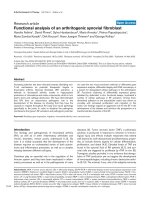
illustrated in Figure 1, the number of ligands (N) decays with
increasing number (L) of domains and folds that bind the lig-
and and follows the power law N = aL
-b
(P < 0.0001). It is
interesting to note that most of the widely shared ligands
(such as those shared by 15+ folds; Additional data file 1) are
hubs of metabolic networks [14-16] and are vital to metabo-
lism (especially energy metabolism).
Power-law behaviors of ligand-protein bindingFigure 1
Power-law behaviors of ligand-protein binding. The number of ligands (N)
decays with an increase in the number (L) of (a) domains and (b) folds
that bind the ligand and follows the equation N = aL
-b
. The figure illustrates
that a few ligands cover tens of protein domains or folds, while most
ligands bind only one domain or fold.
1 10 100
1
10
100
1000
a = 220.58
b = 0.05
R
2
= 0.81
P < 0.0001
Number of ligands (N)
Number of domains binding ligand (L)
011
1
10
100
1000
a = 630.60
b = 0.008
R
2
= 0.95
P < 0.0001
(a)
(b)
Number of ligands (N)
Number of domains binding ligand (L)
Genome Biology 2007, Volume 8, Issue 8, Article R176 Ji et al. R176.3
comment reviews reports refereed researchdeposited research interactions information
Genome Biology 2007, 8:R176
Biological basis underlying the power-law behaviors of
ligand-protein binding
Although power law is a central concept in network sciences
and has been implicated in most biological networks [14-16],
it is a challenge to elucidate the mechanisms underlying the
rule. The most popular theoretical models resort to preferen-
tial attachment principle, which attributes the different con-
nections of nodes to their different emerging orders, that is to
say, the more connected nodes originated earlier than the less
connected nodes [30]. Although the preferential attachment
principle has been justified for protein networks [31-33], it
remains unclear whether it can be applied to protein-ligand
binding.
As a large part of the sc-PDB-derived ligands are synthetic, to
explore the applicability of the preferential attachment prin-
ciple to protein-ligand binding, we extracted bio-ligands from
the ligand dataset. To do this, the MetaCyc database (9.5; a
metabolic-pathway database that contains 5,253 metabolites)
[34] was employed to filter the non-metabolic ligands. As a
result, 128 bio-ligands were obtained, which bind to 1,662
domains (counted at a non-redundant level). According to
SCOP 1.69 [28,29], these domains were classified into 207
folds. As one fold may cover multiple domains and bind more
than one ligand, the fold occurrences amounted to 574.
Although these ligands are only metabolism-relevant, they
also follow power-law distribution in the protein universe
(Additional data file 2).
As the quantity of bio-ligands is limited, to guarantee statisti-
cal significance, the 128 bio-ligands were classified into only
two categories: first, 70 early ligands, which are owned by
both prokaryotic (Escherichia coli) and eukaryotic (yeast or
higher) species; and second, 54 late ligands, which are owned
only by eukaryotic (yeast or higher) species (4 ligands failed
in age assignment) (Additional data file 3). It is interesting to
note that early ligands cover 7.1 folds on average, in contrast
to late ligands, which cover only 1.2 folds on average, and that
all (100%) super ligands (shared by 3+ folds) originated early,
while most (64.8%) ordinary ligands (bind to 3 or less folds)
appeared late. All of these findings strongly suggest that the
preferential attachment principle still holds for ligand-pro-
tein binding to a large extent.
Chemical basis underlying the power-law behaviors of
ligand-protein binding
It has been widely accepted that protein folds are among the
most conserved elements of life [35-37]. However, the
present analysis indicates that 353 ligands (16.1%) are shared
by 2 or more folds and 104 ligands (4.8%) can cover 3+ folds,
which suggests that ligand binding is not constrained by the
global architecture of proteins. This finding is consistent with
a recent concept that the local structures around an active site
are more basic than folds to describe a protein's biological
space (binding site for potential ligands) [38]. This phenom-
enon can be elucidated, at least in part, in terms of the struc-
ture-function relationships of proteins. First, binding sites
and ligands are quite flexible and plastic [39-41], and there-
fore, binding-site selection is, to certain extent, ligand
dependent [42-44]. Second, ligand binding is governed by a
few conserved residues and, thus, is a local rather than a glo-
bal property of proteins [10,11]. However, the structural fac-
tors underlying the strong protein-binding ability of the super
ligands still remain unknown. In addition, it is also of interest
to explore the structural features discriminating ligands from
ordinary molecules. Therefore, the chemical space consisting
of ligands and ordinary molecules was charted to reveal the
relationship between the ligand distribution patterns in the
protein universe and in the chemical space.
The chemical space is composed of 2,176 ligands derived from
sc-PDB (due to the lack of atomic parameters, 10 of the 2,186
ligands failed to go through the descriptor calculations) and
2,184 small molecules randomly selected from ACD-SC
(Available Chemicals Directory-Screening Compounds, Ver-
sion 2005.1, Molecular Design Ltd. Information Systems Inc.,
San Leardo, CA, USA; which collects chemicals that are com-
mercially available and is broadly regarded as a source of
ordinary molecules [45]). Seventy descriptors characterizing
the structural features of these molecules were calculated, of
which 13 were calculated by Sybyl (Tripos Inc., St Louis, Mis-
souri, USA [46]), 49 by Cerius2 (Version 4.10L, Accelrys Inc.,
San Diego, CA, USA [47]) and 8 by an in-house program writ-
ten in Perl (Table 1).
We used factor analysis to visualize the diversity of the mole-
cules. Factor analysis is widely used to study the patterns of
relationship among many dependent variables, with the goal
of discovering something about the nature of the independent
variables (called factors) that affect them [48,49]. In the
present analysis, two factors, which can explain 65.5% of the
variance, were extracted by principal component analysis and
rotated by the Varimax method [50] to chart the two-dimen-
sional chemical space of small molecules. The factor loadings
(Varimax normalized) are listed in Table 1.
From the factor loadings, we see that the first factor, explain-
ing 52.8% of the variance, contains high loadings (>0.9;
shown in bold in Table 1) from constitutional properties (such
as total molecular surface area, total molecular volume,
molecular weight, total bond counts, number of non-hydro-
gen atoms and number of carbons atoms) and topological
properties (such as Kappa topological indices, subgraph top-
ological counts, Kier and Hall Chi connectivity indices and
Zagreb topological Index). In comparison, the second factor,
explaining 12.7% of the variance, contains important contri-
butions (with loadings of higher than 0.8; shown in bold in
Table 1) from electronic properties, such as polar molecular
surface area, H-bond acceptor counts (whose loading is
0.799), H-bond donor counts and partition coefficient (meas-
ured by AlogP98 and LogP).
R176.4 Genome Biology 2007, Volume 8, Issue 8, Article R176 Ji et al. />Genome Biology 2007, 8:R176
Table 1
Descriptors of chemical space consisting of sc-PDB-derived ligands and ACD-SC-derived ordinary molecules and corresponding load-
ings (Varimax normalized) for the first two factors*
Descriptors Characterization Factor loadings Software
12
AREA Total molecular surface area 0.974 0.103 Sybyl
PSA Polar molecular surface area 0.255 0.892
PV Polar molecular volume 0.501 0.741
VOL Total molecular volume 0.991 0.062
MOLWEIGHT Molecular weight 0.958 0.206
Acceptor H-bond acceptor counts 0.464 0.799
Donor H-bond donor counts 0.376 0.817
BondCount Total bond counts 0.972 0.060
Chiral Counts of chiral center 0.367 0.617
Hydrophobe Hydrophobic fragment counts 0.767 -0.417
RingCount Ring counts 0.686 -0.069
RotBonds Number of rotatable bonds 0.630 0.428
HeavyAtoms Number of non-H atoms 0.978 0.149
Carbons Number of carbons atoms 0.943 -0.228 Perl
Oxygens Number of oxygen atoms 0.425 0.793
Nitrogens Number of nitrogen atoms 0.475 0.324
Sulfurs Number of sulfur atoms 0.141 -0.009
Phosphorus Number of phosphorus atoms 0.162 0.617
Halides Number of halide atoms 0.076 -0.170
DoubleBonds Number of double bonds 0.527 0.378
TripleBonds Number of triple bonds -0.009 -0.109
RadOfGyration Radius of gyration 0.888 0.004 Cerius 2
ShadowXY Surface area projections 0.967 0.076
ShadowXZ 0.951 0.053
ShadowYZ 0.877 0.093
ShadowXYfrac -0.610 -0.027
ShadowXZfrac -0.421 -0.002
ShadowYZfrac -0.289 0.039
Shadownu 0.268 -0.117
ShadowXlength 0.849 -0.008
ShadowYlength 0.798 0.075
ShadowZlength 0.756 0.059
Density Density -0.089 0.354
PMImag Principal moment of inertia 0.819 0.134
Genome Biology 2007, Volume 8, Issue 8, Article R176 Ji et al. R176.5
comment reviews reports refereed researchdeposited research interactions information
Genome Biology 2007, 8:R176
AlogP Log of the partition coefficient using Ghose and Crippen's method. 0.425 -0.727
AlogP98 Log of the partition coefficient, atom-type value, using latest parameters. 0.365 -0.852
Fh2o Desolvation free energy for water -0.479 -0.762
Foct Desolvation free energy for octanol -0.578 -0.617
LogP Log of the partition coefficient. -0.022 -0.892
MR Molar refractivity using Hopfinger's method. 0.835 -0.110
MolRef Molar refractivity using linear additive method based on AlogP atom types 0.986 -0.033
JX Balaban indices -0.567 0.027
Kappa1 Kappa topological indices 0.969 0.189
Kappa2 0.926 0.026
Kappa3 0.691 0.033
Kappa1AM 0.958 0.220
Kappa2AM 0.901 0.050
Kappa3AM 0.630 0.046
PHI Molecular flexibility index 0.800 0.078
SC0 Subgraph topological counts 0.980 0.147
SC1 0.973 0.125
SC2 0.943 0.186
SC3P 0.904 0.141
SC3C 0.749 0.389
SC3CH 0.016 -0.086
CHI0 Kier and Hall Chi connectivity indices 0.974 0.190
CHI1 0.983 0.115
CHI2 0.958 0.210
CHI3P 0.939 0.136
CHI3C 0.655 0.484
CHI3CH 0.015 -0.087
CHIV0 0.990 0.076
CHIV1 0.971 0.120
CHIV2 0.913 0.137
CHIV3P 0.838 0.096
CHIV3C 0.476 0.148
CHIV3CH 0.016 -0.088
Wiener Wiener topological index 0.854 0.186
logZ Logarithm of Hosoya topological index -0.220 -0.131
Zagreb Zagreb topological index 0.958 0.162
*The first factor explains 52.8% of the variance and the second explains 12.7%. Factors with high loadings (>0.9 for first factors and >0.8 for second
factors) are shown in bold.
Table 1 (Continued)
Descriptors of chemical space consisting of sc-PDB-derived ligands and ACD-SC-derived ordinary molecules and corresponding load-
ings (Varimax normalized) for the first two factors*
R176.6 Genome Biology 2007, Volume 8, Issue 8, Article R176 Ji et al. />Genome Biology 2007, 8:R176
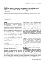
In the chemical space formed by the two factors (Figure 2),
one can find some differences between the distribution pat-
terns of ligands and ordinary molecules. That is, ligands (in
red) occupy the relatively upper part of the space, while ordi-
nary molecules (in blue) hold the relatively lower part, which
implies that it is the second factor that discriminates ligands
from ordinary molecules. As a consequence, it can be deduced
that polar molecular surface area, H-bond donor counts, H-
bond acceptor counts and partition coefficient are likely
responsible for the differences between ligands and ordinary
molecules, which agrees well with the current understanding
of the chemical basis of ligand-protein binding that electro-
static interactions (including H-bond) and hydrophobic
interactions make major contributions to the binding. More
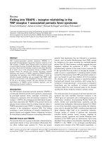
interestingly, as shown in Figure 3, super ligands (in blue and
red) do not distribute randomly in the chemical space, but
concentrate in the relatively upper part of the space, which
suggests that polar molecular surface area, H-bond donor
counts, H-bond acceptor counts and partition coefficient are
also key factors discriminating super ligands from others.
To shed more light on the above findings, the average values
of descriptors characterizing polar molecular surface area, H-
bond donors, H-bond acceptors and partition coefficient were
calculated for ordinary molecules, ligands and super ligands.
From Table 2, it can be seen that there indeed exist correla-
Chemical space consisting of ligands (derived from sc-PDB) and ordinary molecules (randomly selected from ACD-SC), defined by the first two factors derived from 70 descriptorsFigure 2
Chemical space consisting of ligands (derived from sc-PDB) and ordinary molecules (randomly selected from ACD-SC), defined by the first two factors
derived from 70 descriptors. The figure illustrates that ligands (in red) occupy the relatively upper part of the space, while ordinary molecules (in blue)
occupy the relatively lower part, which means that it is the second factor that discriminates ligands from ordinary molecules. From the loadings of the
second factor, it can be deduced that polar molecular surface area, H-bond donor counts, H-bond acceptor counts and partition coefficient are likely
responsible for the differences between ligands and ordinary molecules, which is supported by the different average values of the four kinds of parameters
for ligands and ordinary molecules (Table 2).
-2 -1 0 1 2 3 4
-3
-2
-1
0
1
2
3
4
5
Factor 2
Factor 1
Genome Biology 2007, Volume 8, Issue 8, Article R176 Ji et al. R176.7
comment reviews reports refereed researchdeposited research interactions information
Genome Biology 2007, 8:R176
tions between protein-binding ability and the four kinds of
parameters. The protein-binding potential of ligands is posi-
tively correlated with polar molecular surface area, H-bond
donor and acceptor counts, and negatively correlated with
partition coefficient (measured by AlogP98 and LogP).
Recently, through examining the conformational diversity of
some very common ligands (that is, ATP, NAD and FAD)
bound to proteins, Stockwell and Thornton [41] suggested
that molecular flexibility is important for ligands to bind
diverse proteins. This opinion is partially supported by the
present analysis. Although the contribution from the number
of rotatable bonds (RotBonds) to the second factor is not very
strong (the loading is 0.428; Table 1), there is a correlation
between the protein-binding ability of ligands and index Rot-
Bonds. As listed in Table 2, the average RotBonds for ligands
is significantly higher than that for ordinary molecules
(independent samples t-test shows that P < 0.0001), and it is
clear that the more folds the ligands cover, the higher the
average RotBonds are for the ligands.
Discussion
Since ligand-protein binding is one of the most basic bio-
chemical processes, the present findings have broad biologi-
cal and medical implications.
Chemical space consisting of sc-PDB-derived ligands, defined by the first two factors derived from 70 descriptorsFigure 3
Chemical space consisting of sc-PDB-derived ligands, defined by the first two factors derived from 70 descriptors. The figure illustrates that super ligands
(shared by 3+ folds; in blue), especially those that are shared by 10+ folds (in red), concentrate in the relatively upper part of the space (the area of the
circle is directly proportional to the number of folds that bind the ligand), which suggests that polar molecular surface area, H-bond donor counts, H-bond
acceptor counts and partition coefficient are responsible for the strong protein-binding potential of the super ligands, which is supported by the different
average values of the four kinds of parameters for ligands with different protein-binding potentials (Table 2).
-2 -1 0 1 2 3 4
-3
-2
-1
0
1
2
3
4
5
Factor 2
Factor 1
R176.8 Genome Biology 2007, Volume 8, Issue 8, Article R176 Ji et al. />Genome Biology 2007, 8:R176
Implications for tracing the chronology of ligand
binding to proteins
The most challenging issue in life sciences may be elucidating
how organisms originated from inorganic scratches (gases,
water and clays), during which one of the most important
missions is to establish the chronology of the important bio-
logical events. Thanks to the continuing efforts of chemists
and biologists, the chronologies of the evolution of amino
acids and proteins have been established in principle [37,51-
55]. However, as many proteins bind ligands that are essen-
tial for their functions and the ligands are likely to have orig-
inated independently of proteins [56-59], the binding of
ligands with primordial proteins would also be a critical step
in the origin of life. Thus, it is intriguing to explore the chro-
nology of ligand-protein binding and answer the following
questions: which ligand was first recognized by a protein and
what kind of architecture did the host protein have. Neverthe-
less, since there is no fossil of the last universal common
Table 2
Average values of descriptors characterizing polar molecular surface area, H-bond donors, H-bond acceptors, partition coefficient and
rotatable bonds for ordinary molecules, ligands and ligands with different protein-binding potentials
Descriptor* Small molecules
†
Average values Standard error Number of molecules
PSA Molecules 111.81 1.79 2,184
Ligands 230.59 2.79 2,176
Ligands (≤ 3) 225.71 2.80 2,072
Ligands (4-9) 304.28 15.67 80
Ligands (≥ 10) 406.83 33.10 24
Donor Molecules 1.51 0.04 2,184
Ligands 3.97 0.07 2,176
Ligands (≤ 3) 3.87 0.07 2,072
Ligands (4-9) 5.24 0.43 80
Ligands (≥ 10) 8.21 0.90 24
Acceptor Molecules 3.35 0.05 2,184
Ligands 5.87 0.09 2,176
Ligands (≤ 3) 5.74 0.09 2,072
Ligands (4-9) 7.69 0.53 80
Ligands (≥ 10) 11.00 1.18 24
AlogP98 Molecules 2.87 0.05 2,184
Ligands 0.81 0.06 2,176
Ligands (≤ 3) 0.92 0.06 2,072
Ligands (4-9) -1.33 0.25 80
Ligands (≥ 10) -1.80 0.38 24
LogP Molecules 0.77 0.08 2,184
Ligands -2.27 0.10 2,176
Ligands (≤ 3) -2.10 0.10 2,072
Ligands (4-9) -5.06 0.50 80
Ligands (≥ 10) -8.11 0.96 24
RotBond Molecules 4.88 0.09 2,184
Ligands 7.49 0.11 2,176
Ligands (≤ 3) 7.43 0.11 2072
Ligands (4-9) 8.00 0.50 80
Ligands (≥ 10) 11.33 1.19 24
* PSA, polar molecular surface area; Donor, H-bond donor counts; Acceptor, H-bond acceptor counts; AlogP98, log of the partition coefficient,
atom-type value, using latest parameters; LogP, log of the partition coefficient; RotBond, number of rotatable bonds.
†
Molecules, ACD-SC-derived
ordinary molecules; Ligands, sc-PDB-derived ligands; Ligands (≤ 3), ligands covering ≤ 3 folds; Ligands (4-9), ligands covering 4-9 folds; Ligands (≥ 10),
ligands covering ≥ 10 folds.
Genome Biology 2007, Volume 8, Issue 8, Article R176 Ji et al. R176.9
comment reviews reports refereed researchdeposited research interactions information
Genome Biology 2007, 8:R176
ancestor, let alone the more ancestral organisms, it is a great
challenge to trace the protein-binding history of early ligands.
As stated above, through determining the protein-binding
ages of ligands, a rough temporal order (early or late) for
ligand-protein binding can be inferred (as shown in Addi-
tional data file 3). However, considering the fact that fold dis-
tribution pattern in the sequence universe helps greatly to
reveal the chronology of the evolution of protein architecture
[37,53,54], we speculate that the power-law distribution of
ligands in the protein universe may implicate a more explicit
temporal order for ligand-protein binding. In fact, the prefer-
ential attachment principle underlying the power-law behav-
ior of ligand-protein mapping suggests that the more widely a
ligand is shared, the earlier it bound to proteins. As protein
architecture is more conserved than sequence [35-37], the
fold-based inference is believed to be more robust than the
domain-based one. Therefore, the nine bio-ligands that are
most popular in the fold universe (covering 15+ folds; Table
3) are considered to have bound their host proteins relatively
earlier than others and to follow the order (from early to late):
ATP, ADP (adenosine-5'-diphosphate), GDP (guanosine-5'-
diphosphate), NAD (nicotinamide-adenine-dinucleotide),
FAD (flavin-adenine dinucleotide), NDP (dihydro-nicotina-
mide-adenine-dinucleotide phosphate), NAP (nicotinamide-
adenine-dinucleotide phosphate), FMN (flavin mononucle-
otide) and AMP (adenosine monophosphate).
A close inspection of ATP's host proteins reveals that
although ATP covers 35 folds and 97 domains, most domains
belong to a small group of folds, indicating that power law is
still effective (Additional data file 4). According to the
preferential attachment principle of fold usage [37], it is rea-
sonable to infer that the most prevalent fold, P-loop hydrolase
(c.37), was employed by ATP's first host (Table 3). Interest-
ingly, c.37 is the most ancient fold predicted by a
phylogenomic analysis of protein architectures [37,53,54].
Similar analyses allowed us to deduce the most ancestral host
proteins of the other eight early ligands (Additional data file
4, Table 3). It is interesting to note that the predicted earliest
hosts for the nine bio-ligands appeared in roughly the same
order as the protein structures deduced by a phylogenomic
analysis (that is, c.37 is the earliest, followed by c.2, c.23, c.3
and c.26, all of which belong to the α/β class) [37,53,54].
Although no consensus has been reached on the exact tempo-
ral order of protein architectures, α/β is generally considered
to be the most ancient protein class [37,53,54,60-62]. In addi-
tion, based on an extensive analysis of sequences and struc-
tures of numerous proteins, Trifonov and co-workers [63-65]
also inferred that some P-loop ATP-binding domains repre-
sent the most ancient proteins. Recently, through a phyloge-
nomic analysis on protein architectures of modern metabolic
networks, Caetano-Anollés and co-workers [66] indicated
that enzymes with the P-loop hydrolase fold engaged in
nucleotide (especially purine) metabolism may be the most
primitive members of metabolic systems. Through examining
the structures and functions of these members, we found that
most (approximately 80%) of them need ATP to work nor-
mally. Therefore, the present speculations on the chronology
of ligand-protein binding are self-consistent and are in line
with the up-to-date knowledge on protein evolutionary
history.
To get a deeper insight into the evolutionary features of lig-
ands, the building block usage of 128 bio-ligands was ana-
lyzed. As shown in Additional data file 5, nucleic acid bases
are the most frequently used building blocks, followed by
carbohydrates and amino acids, which is in accordance with
Nobeli et al.'s [67] finding that nucleic acid bases are the most
common fragments of metabolites. More interestingly, many
early bio-ligands (45.0%) contain nucleic acid bases; in par-
ticular, the nine earliest bio-ligands all contain one or more
bases. In contrast, carbohydrates or amino acids are con-
tained by only a small proportion of early bio-ligands (25.0%
and 7.5%, respectively). This provides further evidence to
support the notion that early ligands are vestiges of the RNA
world [56].
Table 3
The most prevalent bio-ligands in the fold universe (shared by 15+ folds) and the most common folds used by host proteins of each ligand
Ligands Number of folds Most common folds
Adenosine-5'-triphosphate (ATP) 35 P-loop containing nucleoside triphosphate hydrolases (c.37)
Adenosine-5'-diphosphate (ADP) 31 P-loop containing nucleoside triphosphate hydrolases (c.37)
Guanosine-5'-diphosphate (GDP) 29 P-loop containing nucleoside triphosphate hydrolases (c.37)
Nicotinamide-adenine-dinucleotide (NAD) 27 NAD(P)-binding Rossmann-fold domains (c.2)
Flavin-adenine dinucleotide (FAD) 21 FAD/NAD(P)-binding domain (c.3)
Dihydro-nicotinamide-adenine-dinucleotide phosphate (NDP) 18 NAD(P)-binding Rossmann-fold domains (c.2)
Nicotinamide-adenine-dinucleotide phosphate (NAP) 16 NAD(P)-binding Rossmann-fold domains (c.2)
Flavin mononucleotide (FMN) 16 Flavodoxin-like (c.23)
Adenosine monophosphate (AMP) 15 Adenine nucleotide alpha hydrolase-like (c.26)
R176.10 Genome Biology 2007, Volume 8, Issue 8, Article R176 Ji et al. />Genome Biology 2007, 8:R176
As mentioned above, the presently revealed chronology of
early ligands' host proteins is roughly in line with the previ-
ously deduced evolutionary history of protein architectures
[37,53,54]. Thus, it is interesting to ask: is the accordance
between both events fortuitous? Our answer is maybe not.
Considering the prevalent ligand-induced protein folding
[68-72], we conjecture that early ligands might have facili-
tated protein formation as catalysts (to assemble amino acids
or peptide segments), as molecular chaperons (to help
protein folding) and/or as selectors (because of the important
functions of the early ligands), which naturally resulted in the
accordance between both events. This conjecture implicates
that the origin of primitive proteins benefited from ligand
binding, which is reasonable in terms of the thermodynamics
of ligand binding and protein folding.
It has been found that some early ligands, such as ADP and
GDP, can bind proteins related to the very old P-loop hydro-
lase fold (for example, preprotein translocase SecA (1M74),
ADP-ribosylation factor-like protein 3 (1FZQ) and GTP-bind-
ing protein (1A4R)) with an affinity (free energy) of 10-15
kcal/mol [73], which is just in the range of the free energy loss
(10-20 kcal/mol) during protein folding [74,75]. Thus, the
free energy release during ligand binding may meet the free
energy demand during protein folding. It is tempting to
examine the conjecture of ligand-induced formation and/or
folding of primordial proteins through experimentation. To
do that, in vitro selection may be an appropriate methodology
[76]. It is interesting to note that in vitro selection of proteins
(consisting of 80 residues) targeted to bind ATP has been per-
formed [77]. The randomly generated proteins indeed belong
to the α/β class, but are not related to P-loop hydrolases fold
[78]. However, considering the fact that the shortest protein
sequence for the P-loop hydrolase fold contains 94 residues
(according to the Protein Databank), we suggest that to
explore whether the formation of the most ancient proteins
was induced by ATP, one should adopt longer protein
sequences in the in vitro selection experiments and use small
amino acids as building blocks, because in the primordial
world only these amino acids were available [51,55].
Implications for understanding the new paradigms in
drug discovery
Nowadays, the pharmaceutical industry is facing an unprece-
dented challenge. Global research funding has doubled since
1991, whereas the number of approved new drugs has fallen
by 50% [79,80]. To meet the more-investment-less-outcome
challenge, some novel drug discovery strategies have
appeared in recent years, which include finding new func-
tions from old drugs, developing promiscuous drugs rather
than selective agents and depending more on natural prod-
ucts than on combinatorial libraries of synthetic compounds
to derive drug leads. Since the essence of drug action is the
binding between drugs and target biomolecules (most of
which are proteins), the ligand-protein binding features
revealed in the present study have important implications for
understanding these new drug discovery strategies.
As indicated above, approximately 30% of ligands are bound
by two or more domains (this number gets ~15%, if counted
on fold level), which suggests that if a ligand can bind to a pro-
tein, it has great potential to bind to others. Considering the
fact that the US Food and Drug Administration (FDA) has
approved approximately 2,000 drugs (chemical entities) and
there exist only 2,000-3,000 druggable genes and 600-1,500
drug targets [81,82], it is truly possible to find new functions
from these old 'safe' drugs, which supports an increasingly
shared notion in drug development that the most fruitful
basis for the discovery of a new drug is to start with an old
drug [83-85].
Since most human diseases, such as cancer, diabetes, heart
disease, arthritis and neurodegenerative diseases, involve
multiple pathogenetic factors, the more-investment-less-out-
come predicament is attributed in part to the limitations of
the current one-drug-one-target paradigm in drug discovery
[79,86]. Therefore, more and more efforts are devoted to
finding new therapeutics aimed at multiple targets [86],
which is becoming a new paradigm in drug discovery. To hit
the multiple targets implicated in complex diseases, two
strategies are conceivable. One is called the multicomponent
therapeutic strategy, which incorporates two or more active
ingredients in one drug [86-89], as was applied in some tra-
ditional medicines (in China and many other countries) and
in recently developed drug cocktails. The other is to hit the
multiple targets with a single component, which is termed the
one-ligand-multiple-targets strategy or promiscuous drug
strategy [89-99]. Compared with the former strategy, the lat-
ter might take advantage of lower risks of drug-drug interac-
tions and more predictable pharmacokinetic behaviors
[91,92] and thus has been paid more and more attention. The
feasibility of the one-ligand-multiple-targets strategy is sup-
ported by the present findings, because a certain proportion
of ligands do indeed bind to two or more domains (even
folds). In addition, the presently revealed structural features
of super ligands are of significance for selecting and/or
designing multipotent agents. Of course, the new strategy
should be treated with wariness, because of the potential side
effects of the promiscuous ligands.
Another feature of the recent drug discovery paradigm shift is
that more attention has been given to natural-product repos-
itories rather than combinatorial libraries of synthetic com-
pounds for finding novel drug leads [100,101]. Due to their
biosynthetic origin, natural products are natively bound to
proteins (synthases). In light of the present findings, one can
conclude that natural products have more potential than syn-
thetic compounds to bind proteins, including those of human,
which helps to understand the natural product-based drug
discovery strategy. In addition, it can be inferred that it is
rather easy to build a protein-ligand network on the basis of
Genome Biology 2007, Volume 8, Issue 8, Article R176 Ji et al. R176.11
comment reviews reports refereed researchdeposited research interactions information
Genome Biology 2007, 8:R176
naturally occurring small-molecule ligands, which definitely
benefits the birth of networked life and facilitates the forma-
tion of links within different species.
Materials and methods
Data selection/collection
Until June 2006, 2,721 ligands had been recorded in sc-PDB.
As our interest was focused on non-peptide ligands, 433 pep-
tides were eliminated. After removing 102 repeated ligands
(which have the same structures to others but were given dif-
ferent release names), 2,186 small-molecule ligands
remained (Additional data file 1), which bind to 5,740 non-
redundant domains (to remove the redundancy of domains,
only one domain was chosen from each species). Domain is
defined as an independently folded unit within a protein,
often joined by a flexible segment of the polypeptide chain
[102]. For a small proportion of ligands that are shared by two
domains, both domains were counted. According to SCOP
1.69 [28,29], these domains were classified into 591 folds. As
one fold may cover multiple domains and hold more than one
ligand, the fold occurrences amounted to 3,224.
Since sc-PDB is a subset of the PDB, one may be concerned
about the robustness of the conclusions derived when using
it. However, considering the facts that the present inferences
were made mainly on the level of protein fold and that folds
are much more conserved than domains, and thus fold
increase is much slower than that of domains in the PDB
[103], it is believed that the present conclusions are solid. In
fact, even if the latest data of the sc-PDB (containing 396 new
ligands and 827 new domains, which were kindly provided by
Dr Rognan and have not been uploaded on the website) are
considered, all of the present conclusions still hold.
Descriptor calculation
Seventy descriptors characterizing the structural features of
2,186 ligands selected from the sc-PDB and 2,184 small mol-
ecules randomly selected from ACD-SC were calculated by
Sybyl (13 descriptors) [46], Cerius 2 (49 descriptors) [47] and
an in-house program written in Perl (8 descriptors). Then, the
calculated data were linked together with Perl for further
analysis. Because of the lack of atomic parameters for ten lig-
ands (that is, 2,3,4,5,6-pentafluorobenzyl alcohol, 2-amino-
4-oxo-4,7-dihydro-3h-pyrrolo [2,3-d] pyrimidine-5-carboni-
trile, 3,5,3',5'-tetraiodo-l-thyronine, 6,7-dinitroquinoxaline-
2,3-dione, 9-hydroxy aristolochic acid, 3,5,7-trihydroxy-2-(4-
hydroxyphenyl)-4h-chromen-4-one, 5-hydroxy-2-(4-hydrox-
yphenyl)-1-benzofuran-7-carbonitrile, 3,3',5,5'-tetraiodothy-
roacetic acid, 3,5,7,3',4'-pentahydroxyflavone and radicicol),
some descriptors could not be calculated for these molecules.
Hence, only 2,176 ligands went through the calculation. How-
ever, as each of the ten ligands covers only one fold, their
absence has no impact on the conclusion of the present study.
Factor analysis
SPSS 13.0 (SPSS Inc., Chicago, IL, USA) was employed to do
the factor analysis. The factors were extracted by means of
principal component analysis [48,49] and the parameter set-
tings were as follows: a correlation matrix was used; and two
factors were extracted to visualize the two-dimensional
chemical space of ligands and ordinary molecules. In order to
simplify the interpretation of the extracted factors, factor
rotation was performed, during which the most popular
orthogonal rotation method, Varimax, developed by Kaiser
[50], was employed. For other variables, default parameters
were adopted.
Age assignment for bio-ligands
An early bio-ligand is defined as that owned by both prokary-
otic (E. coli) and eukaryotic (yeast or higher) species, while a
late bio-ligand is defined as that owned only by eukaryotic
(yeast or higher) species. As there is no direct information on
ligand ownership, we used the information of their host pro-
teins to deduce their ages. That is, a ligand is early, provided
that at least one of its host proteins is owned by both E. coli
and yeast (or higher species); and a ligand is late if none of its
host proteins is owned by E. coli but at least one is owned by
yeast or higher species. During the age-assigning process, not
only the host proteins recorded in sc-PDB were checked, but
also the corresponding homologous proteins retrieved from
Swiss-Prot [104] were considered.
Abbreviations
ACD-SC, Available Chemicals Directory-Screening Com-
pounds; RotBond, rotatable bond; sc-PDB, Annotated Data-
base of Druggable Binding Sites from the PDB.
Authors' contributions
H Y.Z. designed the study. H F.J., D X.K. and L.S. collected
the data and performed the calculation. All authors analyzed
the data. H Y.Z., H F.J. and L L.C. wrote the paper.
Additional data files
The following additional data are available with the online
version of this paper. Additional data file 1 lists ligands and
the numbers of domains and folds that bind them. Additional
data file 2 illustrates the power-law behaviors of metabolism-
relevant ligands. Additional data file 3 provides building
blocks and ownerships of metabolism-relevant ligands. Addi-
tional data file 4 illustrates the power-law behaviors of folds
for proteins binding ATP, ADP and NAD. Additional data file
5 illustrates the building block usage of bio-ligands.
Additional data file 1Ligands and the numbers of domains and folds that bind themLigands and the numbers of domains and folds that bind them.Click here for fileAdditional data file 2Power-law behaviors of metabolism-relevant ligandsPower-law behaviors of metabolism-relevant ligands.Click here for fileAdditional data file 3Building blocks and ownerships of metabolism-relevant ligandsBuilding blocks and ownerships of metabolism-relevant ligands.Click here for fileAdditional data file 4Power-law behaviors of folds for proteins binding ATP, ADP and NADPower-law behaviors of folds for proteins binding ATP, ADP and NAD.Click here for fileAdditional data file 5Building block usage of bio-ligandsBuilding block usage of bio-ligands.Click here for file
R176.12 Genome Biology 2007, Volume 8, Issue 8, Article R176 Ji et al. />Genome Biology 2007, 8:R176
Acknowledgements
We thank Prof. Antonio Lazcano and Dr Xin-Min Li for fruitful discussions
and Dr Didier Rognan for generously providing the sdf file of sc-PDB and
the latest data. This work was partially supported by National Basic
Research Program of China (2003CB114400) and National Natural Science
Foundation of China (30570383 and 30600119).
References
1. Barabási AL, Oltvai ZN: Network biology: understanding the
cell's functional organization. Nat Rev Genet 2004, 5:101-113.
2. Fewell JH: Social insect networks. Science 2003, 301:1867-1870.
3. Whittaker RH, Feeny PP: Allelochemics: chemical interactions
between species. Science 1971, 171:757-770.
4. Dixon RA: Natural products and plant disease resistance.
Nature 2001, 411:843-847.
5. Camilli A, Bassler BL: Bacterial small-molecule signaling
pathways. Science 2006, 311:1113-1116.
6. Baldwin IT, Halitschke R, Paschold A, von Dahl CC, Preston CA: Vol-
atile signaling in plant-plant interactions: 'talking trees' in
the genomics era. Science 2006, 311:812-815.
7. Keller L, Surette MG: Communication in bacteria: an ecological
and evolutionary perspective. Nat Rev Microbiol 2006, 4:249-258.
8. Bassler BL, Losick R: Bacterially speaking. Cell 2006, 125:237-246.
9. Ladurner AG: Rheostat control of gene expression by
metabolites. Mol Cell 2006, 24:1-11.
10. Cappello V, Tramontano A, Koch U: Classification of proteins
based on the properties of the ligand-binding site: The case
of adenine-binding proteins. Proteins 2002, 47:106-115.
11. Denessiouk KA, Johnson MS: When fold is not important: A
common structural framework for adenine and AMP binding
in 12 unrelated protein families. Proteins 2000, 38:310-326.
12. Anantharaman V, Aravind L, Koonin EV: Emergence of diverse
biochemical activities in evolutionarily conserved structural
scaffolds of proteins. Curr Opin Chem Biol 2003, 7:12-20.
13. Russell RB, Sasieni PD, Sternberg MJE: Supersites within super-
folds. Binding site similarity in the absence of homology. J
Mol Biol 1998, 282:903-918.
14. Jeong H, Tombor B, Albert R, Oltvai ZN, Barabási AL: The large-
scale organization of metabolic networks. Nature 2000,
407:651-654.
15. Wagner A, Fell DA: The small world inside large metabolic
networks. Proc R Soc Lond Ser B 2001, 268:1803-1810.
16. Ma HW, Zeng AP: Reconstruction of metabolic networks from
genome data and analysis of their global structure for vari-
ous organisms. Bioinformatics 2003, 19:270-277.
17. Arita M: The metabolic world of Escherichia coli is not small.
Proc Natl Acad Sci USA 2004, 101:1543-1547.
18. Michalsky E, Dunkel M, Goede A, Preissner R: SuperLigands-a
database of ligand structures derived from the Protein Data
Bank. BMC Bioinformatics 2005, 6:122.
19. Hendlich M: Databases for protein-ligand complexes. Acta
Crystallogr 1998, D54:1178-1182.
20. Goto S, Nishioka T, Kanehisa M: LIGAND: chemical database for
enzyme reactions. Bioinformatics 1998, 14:591-599.
21. Shin JM, Cho DH: PDB-Ligand: a ligand database based on PDB
for the automated and customized classification of ligand-
binding structures. Nucleic Acids Res 2005, 33:D238-D241.
22. Gold ND, Jackson RM: A searchable database for comparing
protein-ligand binding sites for the analysis of structure-func-
tion relationships. J Chem Inf Model 2006, 46:736-742.
23. Feldmana HJ, Snydera KA, Ticolla A, Pintiliea G, Hogue CWV: A
complete small molecule dataset from the protein data
bank. FEBS Lett 2006, 580:1649-1653.
24. Gold ND, Jackson RM: SitesBase: a database for structure-
based protein-ligand binding site comparisons. Nucleic Acids
Res 2006, 34:D231-D234.
25. Block P, Sotriffer CA, Dramburg I, Klebe G: AffinDB: a freely
accessible database of affinities for protein-ligand complexes
from the PDB. Nucleic Acids Res 2006, 34:D522-D526.
26. Golovin A, Dimitropoulos D, Oldfeld T, Rachedi A, Henrick K: MSD-
site: A database search and retrieval system for the analysis
and viewing of bound ligands and active sites. Proteins 2005,
58:190-199.
27. Kellenberger E, Muller P, Schalon C, Bret G, Foata N, Rognan D: sc-
PDB: an annotated database of druggable binding sites from
the Protein Data Bank. J Chem Inf Model 2006, 46:717-727.
28. Murzin AG, Brenner SE, Hubbard T, Chothia C: SCOP: a structural
classification of proteins database for the investigation of
sequences and structures. J Mol Biol 1995, 247:536-540.
29. Andreeva A, Howorth D, Brenner SE, Hubbard TJP, Chothia C, Mur-
zin AG: SCOP database in 2004: refinements integrate struc-
ture and sequence family data. Nucleic Acid Res 2004,
32:D226-D229.
30. Barabási AL, Albert R: Emergence of scaling in random
networks. Science 1999, 286:509-512.
31. Eisenberg E, Levanon EY: Preferential attachment in the protein
network evolution. Phys Rev Lett 2003, 91:138701-138704.
32. Ekman D, Light S, Björklund ÅK, Elofsson A: What properties
characterize the hub proteins of the protein-protein interac-
tion network of Saccharomyces cerevisiae? Genome Biol 2006,
7:R45.
33. Prachumwat A, Li WH: Protein function, connectivity, and
duplicability in yeast. Mol Biol Evol 2006, 23:30-39.
34. Caspi R, Foerster H, Fulcher CA, Hopkinson R, Ingraham J, Kaipa P,
Krummenacker M, Paley S, Pick J, Rhee SY, et al.: MetaCyc: a mul-
tiorganism database of metabolic pathways and enzymes.
Nucleic Acids Res 2006, 34:D511-D514.
35. Qian J, Luscombe NM, Gerstein M: Protein family and fold
occurrence in genomes: power-law behaviour and evolution-
ary model. J Mol Biol 2001, 313:673-681.
36. Koonin EV, Wolf YI, Karev GP: The structure of the protein uni-
verse and genome evolution. Nature 2002, 420:218-223.
37. Caetano-Anollés G, Caetano-Anollés D: An evolutionarily struc-
tured universe of protein architecture. Genome Res 2003,
13:1563-1571.
38. McArdle BM, Quinn RJ: Identification of protein fold topology
shared between different folds inhibited by natural products.
ChemBioChem 2007, 8:788-798.
39. Todd AE, Orengo CA, Thornton JM: Plasticity of enzyme active
sites. Trends Biochem Sci 2002, 27:419-426.
40. Macchiarulo A, Nobeli I, Thornton JM: Ligand selectivity and
competition between enzymes in silico. Nat Biotechnol 2004,
22:1039-1045.
41. Stockwell GR, Thornton JM: Conformational diversity of ligands
bound to proteins. J Mol Biol 2006, 356:928-944.
42. Van Regenmortel MHV: Molecular recognition in the post-
reductionist era. J Mol Recognit 1999, 12:1-2.
43. Ma B, Kumar S, Tsai CJ, Nussinov R: Folding funnels and binding
mechanisms. Protein Eng 1999, 12:713-720.
44. Ma B, Shatsky M, Wolfson HJ, Nussinov R: Multiple diverse ligands
binding at a single protein site: a matter of pre-existing
populations. Protein Sci 2002, 11:184-197.
45. Available Chemicals Directory-Screening Compounds
[ />46.
SYBYL 7.0 [ />ules,SimplePage,,,&page=comp_informatics]
47. Cerius2 [ />48. Kim JO, Mueller CW: Factor Analysis: Statistical Methods and Practical
Issues Thousand Oaks, CA: Sage Publications; 1978.
49. Reyment RA, Joreskog KG: Applied Factor Analysis in the Natural
Sciences Cambridge: Cambridge University Press; 1993.
50. Kaiser HF: The varimax criterion for analytic rotation in fac-
tor analysis. Psychometrika 1958, 23:187-200.
51. Trifonov EN, Gabdank I, Barash D, Sobolevsky Y: Primordia vita
deconvolution from modern sequences. Orig Life Evol Biosph
2006, 36:559-565.
52. Wong JT-F: Coevolutionary theory of the genetic code at age
thirty. BioEssays 2005, 27:416-425.
53. Caetano-Anollés G, Caetano-Anollés D: Universal sharing pat-
terns in proteomes and evolution of protein fold architec-
ture and life. J Mol Evol 2005, 60:484-498.
54. Wang M, Boca SM, Kalelkar R, Mittenthal JE, Caetano-Anollés G:
Phylogenomic reconstruction of the protein world based on
a genomic census of protein fold architecture. Complexity
2006, 12:27-40.
55. Zhang H-Y: Exploring the evolution of standard amino-acid
alphabet: when genomics meets thermodynamics. Biochem
Biophys Res Commun 2007, 359:403-405.
56. White HB: Coenzymes as fossils of an earlier metabolic state.
J Mol Evol 1976, 7:101-104.
57. Miller SL, Schlesinger G: Prebiotic syntheses of vitamin coen-
zymes: I. Cysteamine and 2-mercaptoethanesulfonic acid
(coenzyme M). J Mol Evol 1993, 36:302-307.
Genome Biology 2007, Volume 8, Issue 8, Article R176 Ji et al. R176.13
comment reviews reports refereed researchdeposited research interactions information
Genome Biology 2007, 8:R176
58. Miller SL, Schlesinger G: Prebiotic syntheses of vitamin coen-
zymes: II. Pantoic acid, pantothenic acid, and the composi-
tion of coenzyme A. J Mol Evol 1993, 36:308-314.
59. Huang F, Bugg CW, Yarus M: RNA-catalyzed CoA, NAD, and
FAD synthesis from phosphopantetheine, NMN, and FMN.
Biochemistry 2000, 39:15548-15555.
60. Winstanley HF, Abeln S, Deane CM: How old is your fold? Bioinfor-
matics 2005, 21(Suppl 1):i449-i458.
61. Abeln S, Deane CM: Fold usage on genomes and protein fold
evolution. Proteins 2005, 60:690-700.
62. Ji H-F, Zhang H-Y: Protein architecture chronology deduced
from structures of amino acid synthases. J Biomol Struct Dyn
2007, 24:321-323.
63. Berezovsky IN, Kirzhner VM, Kirzhner A, Rosenfeld VR, Trifonov EN:
Protein sequences yield a proteomic code. J Biomol Struct Dyn
2003, 21:317-325.
64. Berezovsky IN, Kirzhner A, Kirzhner VM, Trifonov EN: Spelling
protein structure. J Biomol Struct Dyn 2003, 21:327-339.
65. Trifonov EN: Early molecular evolution. Israel J Ecol Evol
2006:375-387.
66. Caetano-Anollés G, Kim HS, Mittenthal JE: The origin of modern
metabolic networks inferred from phylogenomic analysis of
protein architecture. Proc Natl Acad Sci USA 2007, 104:9358-9363.
67. Nobeli I, Ponstingl H, Krissinel EB, Thornton JM: A structure-based
anatomy of the E. coli metabolome. J Mol Biol 2003,
334:697-719.
68. Wright PE, Dyson HJ: Intrinsically unstructured proteins: re-
assessing the protein structure-function paradigm. J Mol Biol
1999, 293:321-331.
69. Dyson HJ, Wright PE: Coupling of folding and binding for
unstructured proteins. Curr Opin Struct Biol 2002, 12:54-60.
70. Fink AL: Natively unfolded proteins. Curr Opin Struct Biol 2005,
15:35-41.
71. Dyson HJ, Wright PE: Intrinsically unstructured proteins and
their functions. Nat Rev Mol Cell Biol 2005, 6:197-208.
72. Grandori R, Schwarzinger S, Müller N: Cloning, overexpression
and characterization of micro-myoglobin, a minimal heme-
binding fragment. Eur J Biochem 2000, 267:1168-1172.
73. Wang RX, Fang XL, Lu YP, Yang CY, Wang SM: The PDBbind data-
base: methodologies and updates. J Med Chem 2005,
48:4111-4119.
74. Dobson CM, Šali A, Karplus M: Protein folding: a perspective
from theory and experiment. Angew Chem Int Ed 1998,
37:868-893.
75. Fersht A: Structure and Mechanism in Protein Science: A Guide to Enzyme
Catalysis and Protein Folding New York: Freeman; 1999.
76. Wilson DS, Szostak JW: In vitro selection of functional nucleic
acids. Ann Rev Biochem 1999, 68:611-648.
77. Keefe AD, Szostak JW: Functional proteins from a random-
sequence library. Nature 2001, 410:715-718.
78. Lo Surdo P, Walsh MA, Sollazzo M: A novel ADP- and zinc-bind-
ing fold from function-directed in vitro evolution. Nat Struct
Mol Biol 2004, 11:382-383.
79. Buehler LK: Advancing drug discovery-beyond design. Phar-
maGenomics 2004, 4:24-26.
80. Ruffolo RR: Why has R&D productivity declined in the phar-
maceutical industry? Expert Opin Drug Discov 2006, 1:99-102.
81. Russ AP, Lampel S: The druggable genome: an update.
Drug Dis-
cov Today 2005, 10:1607-1610.
82. Hopkins AL, Groom CR: The druggable genome. Nat Rev Drug
Discov 2002, 1:727-730.
83. Wermuth CG: Selective optimization of side activities:
another way for drug discovery. J Med Chem 2004,
47:1303-1314.
84. Lipinski C, Hopkins A: Navigating chemical space for biology
and medicine. Nature 2004, 432:855-861.
85. O'Connor KA, Roth BL: Finding new tricks for old drugs: an effi-
cient route for public-sector drug discovery. Nat Rev Drug
Discov 2005, 4:1005-1014.
86. Fitzgerald JB, Schoeberl B, Nielsen UB, Sorger PK: Systems biology
and combination therapy in the quest for clinical efficacy.
Nat Chem Biol 2006, 2:458-466.
87. Zimmermann GR, Lehár J, Keith CT: Multi-target therapeutics:
when the whole is greater than the sum of the parts. Drug Dis-
cov Today 2007, 12:34-42.
88. Kitano H: A robustness-based approach to systems-oriented
drug design. Nat Rev Drug Discov 2007, 6:202-210.
89. Keith CT, Borisy AA, Stockwell BR: Multicomponent therapeu-
tics for networked systems. Nat Rev Drug Discov 2005, 4:71-78.
90. Mencher SK, Wang LG: Promiscuous drugs compared to selec-
tive drugs (promiscuity can be a virtue). BMC Clin Pharmacol
2005, 5:3.
91. Morphy R, Kay C, Rankovic Z: From magic bullets to designed
multiple ligands. Drug Discov Today 2004, 9:641-651.
92. Morphy R, Rankovic Z: Designed multiple ligands. An emerging
drug discovery paradigm. J Med Chem 2005, 48:6523-6543.
93. Zhang H-Y:
One-compound-multiple-targets strategy to com-
bat Alzheimer's disease. FEBS Lett 2005, 579:5260-5264.
94. Frantz S: Drug discovery:Playing dirty. Nature 2005,
437:942-943.
95. Hopkins AL, Mason JS, Overington JP: Can we rationally design
promiscuous drugs? Curr Opin Struct Biol 2006, 16:127-136.
96. Roth BL, Sheffler DJ, Kroeze WK: Magic shotguns versus magic
bullets: selectively non-selective drugs for mood disorders
and schizophrenia. Nat Rev Drug Discov 2004, 3:353-359.
97. Hampton T: 'Promiscuous' anticancer drugs that hit multiple
targets may thwart resistance. J Am Med Assoc 2004,
292:419-422.
98. Zhang H-Y, Yang D-P, Tang G-Y: Multifunctional antioxidants:
from screening to design. Drug Discov Today 2006, 11:749-754.
99. Morphy R, Rankovic Z: Fragments, network biology and
designing multiple ligands. Drug Discov Today 2007, 12:156-160.
100. Breinbauer R, Vetter IR, Waldmann H: From protein domains to
drug candidates-natural products as guiding principles in the
design and synthesis of compound libraries. Angew Chem Int Ed
2002, 41:2878-2890.
101. Paterson I, Anderson EA: The renaissance of natural products
as drug candidates. Science 2005, 310:451-453.
102. Rose GD: Hierarchic organization of domains in globular
proteins. J Mol Biol 1979, 134:447-470.
103. Levitt M: Growth of novel protein structural data. Proc Natl
Acad Sci USA 2007, 104:3183-3188.
104. Bairoch A, Apweiler R: The SWISS-PROT protein sequence
data bank and its supplement TrEMBL in 1999. Nucleic Acids
Res 1999, 27:49-54.