Báo cáo y học: "Functional analysis of an arthritogenic synovial fibroblast" docx
Bạn đang xem bản rút gọn của tài liệu. Xem và tải ngay bản đầy đủ của tài liệu tại đây (1.06 MB, 18 trang )
R140
Introduction
The etiology and pathogenesis of rheumatoid arthritis
(RA), as well as of other inflammatory arthritides and
chronic disorders, remain poorly understood [1,2]. By
now, it is widely accepted that the development of the
disease requires an orchestrated series of both autoim-
mune and inflammatory processes, as well as a complex
interplay between different cell types.
Cytokines play an essential role in the regulation of the
immune system and they have been implicated in inflam-
matory processes as well as in the pathogenesis of many
diseases [3]. Tumor necrosis factor (TNF), a pleiotropic
cytokine, is produced in response to infection or immuno-
logical injury and effects multiple responses that extend
well beyond its well-characterized proinflammatory proper-
ties, to include diverse signals for cellular differentiation,
proliferation, and death [4,5]. Elevated levels of TNF are
found in the synovial fluid of RA patients [6,7], and syn-
ovial cells are triggered to proliferate by rTNF in vitro [8].
Transgenic studies provided in vivo evidence that deregu-
lation of TNF production per se triggers the development
of immunopathologies, including chronic destructive arthri-
tis [9,10]. The minimal, if any, role of the adaptive immunity
Abbreviations: BSA = bovine serum albumin; DD = differential display; DD-RT-PCR = differential display reverse transcriptase polymerase chain
reaction; DMEM = Dulbecco’s modified Eagle’s medium; ECM = extracellullar matrix; ELISA = enzyme-linked immunosorbent assay; FACS = fluo-
rescence-activated cell sorter; FBS = fetal bovine serum; FCS = fetal calf serum; H & E = hematoxylin and eosin; hTNF = human tumor necrosis
factor; LF = lung fibroblast; MHC = major histocompatibility complex; MMP = matrix metalloproteinase; PBS = phosphate-buffered saline; PCR =
polymerase chain reaction; RA = rheumatoid arthritis; RT = reverse transcriptase; SCID = severe combined immunodeficiency; SDS = sodium
dodecyl sulfate; SF = synovial fibroblast; SPARC = secreted protein acidic and rich in cysteine; SSC = standard saline citrate; SSPE = standard
sodium phosphate EDTA; SV40 = simian virus 40; TAg = large tumor antigen; TIMP = tissue inhibitor of metalloproteinases; TNF = tumor necrosis
factor; tsTAg = temperature-sensitive large tumor antigen; VCAM = vascular cell adhesion molecule; wt = wild-type.
Arthritis Research & Therapy Vol 5 No 3 Aidinis et al.
Research article
Functional analysis of an arthritogenic synovial fibroblast
Vassilis Aidinis
1
, David Plows
2
, Sylva Haralambous
2
, Maria Armaka
1
, Petros Papadopoulos
1
,
Maria Zambia Kanaki
1
, Dirk Koczan
3
, Hans Juergen Thiesen
3
and George Kollias
1
1
Institute of Immunology, Biomedical Sciences Research Center ‘Alexander Fleming’, Athens, Greece
2
Laboratory of Molecular Genetics, Hellenic Pasteur Institute, Athens, Greece
3
Institute of Immunology, University of Rostock, Rostock, Germany
Corresponding author: Vassilis Aidinis and George Kollias (e-mail: and )
Received: 1 Oct 2002 Revisions requested: 18 Oct 2002 Revisions received: 13 Feb 2003 Accepted: 20 Feb 2003 Published: 14 Mar 2003
Arthritis Res Ther 2003, 5:R140-R157 (DOI 10.1186/ar749)
© 2003 Aidinis et al., licensee BioMed Central Ltd (Print ISSN 1478-6354; Online ISSN 1478-6362). This is an Open Access article: verbatim
copying and redistribution of this article are permitted in all media for any purpose, provided this notice is preserved along with the article's original
URL.
Abstract
Increasing attention has been directed towards identifying non-
T-cell mechanisms as potential therapeutic targets in
rheumatoid arthritis. Synovial fibroblast (SF) activation, a
hallmark of rheumatoid arthritis, results in inappropriate
production of chemokines and matrix components, which in turn
lead to bone and cartilage destruction. We have demonstrated
that SFs have an autonomous pathogenic role in the
development of the disease, by showing that they have the
capacity to migrate throughout the body and cause pathology
specifically to the joints. In order to decipher the pathogenic
mechanisms that govern SF activation and pathogenic potential,
we used the two most prominent methods of differential gene
expression analysis, differential display and DNA microarrays, in
a search for deregulated cellular pathways in the arthritogenic
SF. Functional clustering of differentially expressed genes,
validated by dedicated in vitro functional assays, implicated a
number of cellular pathways in SF activation. Among them,
diminished adhesion to the extracellullar matrix was shown to
correlate with increased proliferation and migration to this
matrix. Our findings support an aggressive role for the SF in the
development of the disease and reinforce the perspective of a
transformed-like character of the SF.
Keywords: fibroblast, gene expression, migration, rheumatoid arthritis, tumor necrosis factor
Open Access
Available online />R141
in the development of arthritis in these models has been
confirmed in studies showing that the course of the
disease in these transgenic mice is not affected by the
absence of mature T and B cells [5,10]. The demonstra-
tion of the importance of TNF in synovial inflammation and
disease progression has led to the successful therapeutic
use of anti-TNF agents in RA [11], yet the precise molecu-
lar and cellular mechanisms of TNF function in disease
have remained vague.
Increasing attention has been directed towards identifying
non-T-cell mechanisms as potential therapeutic targets in
RA. There is little disagreement that macrophages and
fibroblasts, the majority of cells in both the normal and the
hyperplastic synovium, which line diarthoidal joints, should
play an essential part by providing the cytokine networks
and destructive processes for the initiation and mainte-
nance of disease [12–14]. Synovial fibroblasts (SFs), or
fibroblast-like type B synoviocytes (FLS), are mesenchymal,
nonvascular, nonepithelial, CD45-negative cells that
display heterogeneous tissue localization (intimal and
subintimal) [15]. Their physiological function is to provide
nutrients for the cartilage and proteoglycans that lubricate
the articular surfaces. They also express a variety of surface
adhesion receptors that, presumably, help anchor them to
the extracellular matrix (ECM) and regulate the flux of cells
that pass into the synovial fluid space. In RA and under the
influence of inflammatory cytokines, small-molecular-weight
mediators, as well as from the interaction with other cell
types and the extracellullar matrix, intimal SFs become acti-
vated and hyperplastic [16], while releasing a number of
effector signals. These include proinflammatory and anti-
inflammatory factors, chemoattractants, and factors that
promote angiogenesis, matrix degradation and tissue
remodeling, bone formation, and osteoclastogenesis [17].
Isolated human RA SFs were able to induce arthritis upon
transfer to the knee of healthy SCID mice (mice with
severe combined immunodeficiency) even in the absence
of a functioning immune system. Similarly, in the present
study, immortalized SFs, from an immune-independent
animal model of RA [9,5], were shown to be able to
induce an SF-specific, T/B-cell independent, TNF-depen-
dent, arthritis-like disease in healthy mice upon transfer to
the knee joint. Moreover, we employed two of the most
prominent methods of differential gene expression analy-
sis, differential display reverse transcriptase polymerase
chain reaction (DD-RT-PCR) and DNA microarrays, in a
search of pathways involved in SF activation and disease
pathogenesis. Predicted deregulated functions were then
validated in vitro.
Materials and methods
Animals
All mice were bred and maintained on a mixed
CBA × C57BL/6 genetic background and kept at the
animal facilities of the Biomedical Sciences Research
Center ‘Alexander Fleming’ or the Hellenic Pasteur Insti-
tute under specific pathogen-free conditions, in compli-
ance with the Declaration of Helsinki principles.
Cell isolation and culture
SFs were isolated from 6- to 8-week-old mice essentially
as described previously [18]. Fibroblasts were selected by
continuous culturing for at least 21 days and a minimum of
4 passages. Cells were grown at 37°C, 5% CO
2
in com-
plete Dulbecco’s modified Eagle’s medium (DMEM)
(Gibco/Invitrogen, Paisley, UK) supplemented with 10%
fetal calf serum (FCS) and 100 Units/ml of penicillin/strep-
tomycin. Conditionally immortalized cells were grown simi-
larly at the permissive conditions (33°C, 10 Units/ml of
murine recombinant interferon gamma). For the generation
of clones, SF populations were counted and diluted to
0.5 cells per well in a 96-well plate. To ensure clonicity,
growth (which was observed in 30% of the plated wells, a
statistical prerequisite for clonicity under these conditions)
was monitored microscopically every day.
hTNF ELISA and measurement of TNF bioactivity
The enzyme-linked immunosorbent assay (ELISA) for
hTNF (human tumor necrosis factor) was kindly provided
by Dr Wim Buurman (University of Limburg, the Nether-
lands) and performed as described earlier [19]. TNF
bioactivity was measured in tissue-culture supernatants by
standard L929 cytotoxicity assay [20]. One unit of TNF
bioactivity was taken as the amount of activity for LD
50
(median lethal dose). Values are reported as units of TNF
bioactivity/10
6
cells.
Transfer and blockade of disease
Single-cell suspensions of SF clones (2.5 × 10
6
cells per
20 µl) in phosphate-buffered saline (PBS) were injected
into the right knee joint of adult RAG-1-deficient mice
(mice deficient in recombination activating gene). Injection
was from an anterolateral position using a Hamilton
syringe with a 30G × ½ gauge needle (Becton Dickinson,
Madrid, Spain). After the mice had been humanely killed,
joints were fixed, embedded in paraffin wax, and assessed
for histopathology, as previously described [9,10]. Sec-
tions were examined for histological signs of arthritis and
classified accordingly, as previously described [9,10].
Disease induction occurred from 2 to 8 weeks after injec-
tion, with maximal incidence at around 4 weeks after injec-
tion. In order to block the transferred disease, mice were
treated (2 weeks after transfer) with weekly intraperitoneal
injections of anti-hTNF antibody (CB0006 5 µg/g) kindly
provided by Celltech Ltd (Slough, UK).
Detection of tsTAg transgene by PCR
Tissue was removed by dissection, digested overnight
with 20 µg/ml proteinase K (Sigma, L’Isle d’Abeau,
France) in 50 mM Tris, 100 mM NaCl, 100mM EDTA, 1%
Arthritis Research & Therapy Vol 5 No 3 Aidinis et al.
R142
sodium dodecyl sulfate (SDS) pH 8.0, at 55°C. Precipi-
tated DNA was screened by PCR for the presence of the
SV40 tsTAg (simian virus 40 temperature-sensitive large
tumor antigen) transgene using the following primers:
5′-CAC TGC CAT CCA AAT AAT CCC-3′ and 5′-CAG
CCC AGC CAC TAT AAG TAC C-3′. Amplification was
performed for 30 cycles of 93°C for 1 min, 55°C for 1 min,
and 72°C for 1 min.
Analysis by fluorescence-activated cell sorter (FACS)
Cells (10
5
–10
6
) were washed extensively in PBS and
incubated in the presence of 0.2% bovine serum albumin
(BSA) with the 429 (MVCAM.A) monoclonal antibody
(PharMingen) for 20 min at 4°C. After being washed in
PBS (3 times), cells were incubated with a fluorescein-
isothiocyanate-conjugated antirat secondary antibody
(Southern Biotechnology Associates, Birmingham, AL,
USA) for 20 min at 4°C in the dark, washed, and resus-
pended in 1 ml of PBS and analyzed with a FACSCal-
ibur
TM
cytometer.
RNA extraction and differential display RT-PCR
Total RNA was extracted from subconfluent (70–80%)
cultured SFs with the RNAwiz reagent (Ambion Inc,
Austin, TX, USA), in accordance with the manufacturer’s
instructions. For Affymetrix gene chip hybridizations, RNA
was extracted using the guanidinium isothiocynate/acid
phenol protocol [21], followed by single passage through
an RNeasy column from QIAGEN GmbH (Hilden,
Germany), in accordance with the manufacturer’s instruc-
tions. RNA integrity was assessed by electrophoresis on
denaturing 1.2% agarose/formaldehyde gels. DNase treat-
ment, first-strand cDNA synthesis, and differential-display
PCR were executed with the Delta Differential Display kit
PT1173-1 from Clontech/BD Biosciences (Palo Alto, CA,
USA), in accordance with the manufacturer’s instructions
[22]. The (α-
32
P)dATP-labeled (Amersham Pharmacia
Biotech GmbH, Freiburg, Germany) PCR products were
analyzed on 5% polyacrylamide (19:1)/8 M urea denatur-
ing gels run at a constant power of 60 W. Gels were dried
and exposed to film (X-omat AR, Kodak, Hannover,
Germany). Differentially expressed bands were located,
excised from the gel, amplified by PCR, and cloned in the
pT/Adv vector using the AdvanTage PCR cloning kit
(Clontech), in accordance with the manufacturer’s instruc-
tions. Positive plasmid clones were selected on
LB/X-gal/IPTG plates containing 100 µg/ml ampicillin.
Reverse Northern slot blot and Northern blot analysis
0.5–1 µg of 4–6 positive plasmid clones for each differen-
tially expressed band were denatured in 0.4 N NaOH for
15 min and slot blotted to nitrocellulose filter in duplicates
(Protran, Schleicher & Schuell Biosciences GmbH,
Dassel/Relliehausen, Germany) after the addition of
1 volume of cold 2 M ammonium acetate. After washing
with 1 M ammonium acetate, the nitrocellulose filter was
air-dried and baked for 2 hours at 80°C. The two sets of
filters were then hybridized separately with the two differ-
ent DD-RT-PCR reactions from where the differentially
expressed band was detected. Hybridization was per-
formed at 65°C for 12–17 hours, in 3 ×standard saline
citrate (SSC), 0.1% SDS, 10 × Denhardt’s solution, 10%
(w/v) dextran sulfate, 100 µg/ml single-stranded salmon-
sperm DNA. Filters were sequentially washed with
3 × SSC/0.1% SDS, 1×SSC/0.1% SDS, and
0.3 × SSC/0.1% SDS for 15 min at 65°C and exposed to
film (Kodak X-omat AR). For Northern blot analysis, 15 µg
of total RNA was electrophoresed on denaturing 1.2%
agarose/formaldehyde gels alongside a ribosomal RNA
marker and visualized by ethidium bromide staining
(0.5 µg/ml). The gel was then soaked sequentially in: H
2
O
for 20 min (twice), 50 mM NaOH/150 mM NaCl for
20 min, 100 mM Tris-HCl pH 7.6/150 mM NaCl for
20 min, and 6 × SSC for 20 min and was transferred to
nylon membranes (Hybond, Amersham Pharmacia Biotech
GmbH) with 20 × SSC for 12–17 hours. Membranes were
prehybridized at 65°C for 60 min in 5 × standard sodium
phosphate EDTA (SSPE)/5 × Denhardt’s solution/0.5%
SDS in the presence of 20 µg/ml single-stranded salmon-
sperm DNA. The denatured radiolabelled probe (α-
32
P
dATP, Amersham Pharmacia Biotech GmbH; random
primers/Klenow fragment of DNA polymerase, Fermentas
UAB, Vilnius, Lithuania) was then added and hybridization
was carried on at 65°C for 17–20 hours. Membranes were
washed sequentially in 1 ×SSPE/0.1% SDS at 65°C for
10 min, 0.3 ×SSPE/0.1% SDS at 65°C for 10 min, and
0.1 ×SSPE/0.1% SDS at 65°C for 10 min, depending on
the probe, and exposed to film (Kodak X-omat AR).
RT-PCR
First-strand cDNA synthesis was performed with an oligo
(dT)
15
primer and the M-MLV reverse transcriptase from
PROMEGA Biosciences Inc (Mannheim, Germany), in
accordance with the manufacturer’s instructions. PCR
was performed on a thermal cycler (PTC-200, MJ
Research, Waltham, MA, USA) using 25–30 cycles
(depending on the primers) of 93°C for 1 min, 55°C for
1 min, and 72°C for 1 min with a custom-made Taq poly-
merase.
High-density oligonucleotide array hybridization
cRNA probes were generated and hybridized to the
Mu11K (A,B) chip set in accordance with the manufactur-
er’s instructions (Affymetrix, Santa Clara, CA, USA) and as
previously described [23]. Data were normalized on the
basis of total intensity with the Affymetrix GeneChip soft-
ware, and data analysis was performed with the Affymetrix
GeneChip and the Microsoft Excel software.
Proliferation assay
2×10
3
SFs, grown in monolayers and harvested by
trypsinization, were placed in 24-well tissue-culture plates
Available online />R143
in DMEM medium (Gibco/Invitrogen) supplemented with
10% FCS and 100 Units/ml of penicillin/streptomycin.
After 3 hours at 37%, 5% CO
2
, for cell attachment,
0.5 µCi of [
3
H]thymidine was added and incubation was
continued for 24 and/or 48 hours. Cells were then
washed, harvested by trypsinization, transferred to glass-
fiber filters, and counted in a liquid scintillation counter.
Adhesion, migration, and wound-healing assays
Adhesion assays were performed on Cytometrix adhesion
strips (Chemicon International, Temecula, CA, USA)
coated with human fibronectin, vitronectin, laminin, and
collagen I, in accordance with the manufacturer’s instruc-
tions. Assays of cell migration were performed by using
modified Boyden chambers with 8-µm pores (Transwell
polycarbonate, Corning/Costar, Corning, NY, USA). The
lower surface of the membrane was coated with 10 µg/ml
human fibronectin (Becton and Dickinson) for 2 hours at
37°C. The lower chamber was filled with 0.6 ml of DMEM
with 10% fetal bovine serum (FBS) or 0.5% BSA. Cells
were harvested with trypsin/EDTA, washed with PBS, and
resuspended to 1 × 10
6
cells per ml. The suspension
(100 µl) was added to the upper chamber, and the cells
were allowed to migrate at 37°C, 5% CO
2
, for 2–4 hours.
The upper surface of the membrane was wiped with a
cotton bud to mechanically remove nonmigratory cells.
The migrant cells attached to the lower surface were
extensively washed with PBS and stained with 0.2%
crystal violet in 10% ethanol for 10 min. After extensive
washing in H
2
O, the cells were lysed in 1% SDS for
5 min. The absorbance at 550 nm was determined on a
microplate reader (SPECTRAmax PLUS
384
, Molecular
Devices, Sunnyvale, CA, USA). Assays of wound healing
were performed by scraping a confluent culture of cells (in
DMEM supplemented with 10% FCS and 100 Units/ml of
penicillin/streptomycin at 37%, 5% CO
2
), with the edge of
a pipette tip, forming a straight line. Cells were then
allowed to continue to grow and a picture was taken at
each of 0, 12, 24, and 48 hours after the scraping.
Results
Generation of conditionally immortalized synovial
fibroblasts
In order to create an in vitro cell system for analysis of the
functional properties of the activated SF, we first gener-
ated conditionally immortalized SFs. The hTNF-expressing
transgenic mice (Tg197) and their normal littermates were
mated with the H-2K
b
-tsA58 SV40-TAg (simian virus
40 large tumor antigen) transgenic mice [24]. This system
has become a standard tool for isolation of specific condi-
tionally immortalized cell lines and has proved useful for
isolating diverse cell lines such as lung epithelial [25],
osteoblast [26], osteoclast [27], and neuronal [27] cell
lines. Adult mice carrying both transgenes or just the
SV40 tsTAg transgene were identified by PCR as
described previously for hTNF [9]. SFs were isolated from
ankle joints and cultured under permissive conditions, as
described in Materials and methods. All the isolated SFs
were able to grow indefinitely without a change in the mor-
phology and exhibited no signs of terminal differentiation,
senescence, or death (after more than 40 passages). All
the isolated SFs corresponded, most likely, to the intimal
subpopulation of SFs [15], since they all expressed
VCAM-1 (vascular cell adhesion molecule 1), as shown by
FACS analysis (Fig. 1). Immortalized SFs were expanded
by limiting dilution (under conditions that guarantee clonic-
ity, as described in Materials and methods), and a number
of hTNF/TAg SF clones, along with wild-type (wt)/TAg SF
clones, were selected for the study. All selected clones
were stained homogeneously with various surface markers
(MHC class I, VCAM, data not shown), thus confirming
that they were indeed monoclonal. Production of bioactive
human TNF from hTNF/TAg SF clones was confirmed by
hTNF-specific ELISA (Fig. 2) and L929 cytotoxicity assay
(data not shown). Because of the lack of a definitive cellu-
lar marker for murine SFs, all clones were confirmed as
SFs based on culture conditions (adherence for a
minimum of 21 days/4 passages), morphology (spindle
shape), and absence of specific cellular markers (F4/80,
CD11b/Mac-1, MOMA-2, CD45), as determined by
immunocytochemical and FACScan analysis (data not
shown).
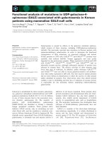
Figure 1
Expression of VCAM-1 by all the isolated synovial fibroblasts, as
detected by FACS analysis. Similar results were obtained whether the
cells were grown in permissive or nonpermissive conditions. FACS =
fluorescence-activated cell sorter; VCAM = vascular cell adhesion
molecule.
Transfer of hTNF/TAg SFs into normal murine joints
induces a T/B-cell-independent, SF-specific, TNF-driven
form of arthritis
Isolated human RA SFs were shown to be able to induce
arthritis upon transfer to the knee of healthy SCID mice –
that is, even in the absence of a functioning immune
system [28]. In order to examine if the established SF
clones have similar functional properties, age-matched
female nontransgenic F1 (C57BL/6 × CBA) mice were
injected intra-articularly in the right knee with cloned SFs.
Animals were humanely killed 4 to 8 weeks after the injec-
tion. Clinical manifestations were usually not detectable.
However, histological analysis of injected joints revealed a
high incidence of disease transfer (Table 1), characterized
by variable degrees of synovitis, soft-tissue inflammation,
synovial hyperplasia, cartilage disruption characterized by
pyknotic chondrocytes, and bone erosion. None of the
control TAg-injected mice showed disease by the end of
the study period. In addition, histological examination of
other tissues such as liver, lung, spleen, and kidney failed
to show evidence of tissue injury.
Despite the similar genetic backgrounds of the donor and
recipient mice (C57BL/6 × CBA), the presence of the
human transgene might be expected to elicit an immune
response, which might account for disease development.
To assess this, we repeated our transfer procedure into
immunodeficient RAG
–/–
mice [29]. We observed disease
induction in the host mice, with incidence (see Table 1)
and pathology similar to those in the previous experiments
in immunocompetent animals.
Remarkably, the levels of transgenic TNF production by
the transferred SFs did not alter the efficiency of disease
transfer in these experiments. The three hTNF-expressing
clones, although expressing different levels of hTNF (see
Fig. 2), all gave similar incidences of disease (see Table 1).
To investigate whether the transferred disease was driven
by transgene expression, an additional group of mice was
injected with the arthritogenic hTNF/TAg SF clone B2 and
then treated with a neutralizing, nondepleting anti-hTNF
antibody 2 weeks after transfer (see Table 1). Antibody
treatment was continued weekly for a further 6 weeks
before the mice were humanely killed for histopathological
analysis. The absence of histological evidence of disease
in any of these mice at the end of the study period shows
that hTNF blockade was able to block disease progres-
sion. The ability of anti-hTNF therapy to block disease sug-
gested that disease pathology is TNF-driven. To
investigate whether TNF-mediated disease could be
Arthritis Research & Therapy Vol 5 No 3 Aidinis et al.
R144
Figure 2
Expression of human TNF by SF clones. Anti-hTNF enzyme-linked
immunosorbent assay from cell-culture supernatants was carried out
as described in Materials and methods. Values are normalized for
hTNF production per 1 × 10
6
cells/ml over a 24-hour period. Mean
averages of triplicates with t-test P values less than 0.01. ‘Recovered’
refers to SFs derived from the diseased ankle of hTNF/TAg SF B2
injected mice (hTNF) or the nondiseased ankle of wt/TAg SF F6
injected mice (wt). hTNF production was assayed after
20 days/4 passages in culture. hTNF = human tumor necrosis factor;
SF = synovial fibroblast; TNF = tumor necrosis factor; tsTAg =
temperature-sensitive large tumor antigen.
Table 1
Summary of arthritis induction by transfer of TNF-expressing
SFs
Host Incidence
Derived Transgene Clone genotype of arthritis
Synovium hTNF/TAg B2 wt 31/65 (47.6%)
Synovium hTNF/TAg B1 wt 5/8 (62.5%)
Synovium hTNF/TAg A4 wt 4/8 (50.0%)
Synovium TAg F6 wt 0/54
Synovium TAg B2 wt 0/8
Synovium TAg A2 wt 0/8
Synovium hTNF/TAg + Ab
a
B2 wt 0/16
Synovium hTNF/TAg B2 RAG
–
/
–
6/10 (60.0%)
Synovium TAg F6 RAG
–
/
–
0/9
Lung
b
hTNF/TAg LFs wt 0/6
Lung
b
hTNF/TAg LFs RAG
–
/
–
0/5
Mice were classified as arthritic upon positive confirmation by
histological analysis.
a
+Ab denotes group injected with arthritogenic
clone B2 and then treated with anti-hTNF antibody 2 weeks after
injection.
b
‘Lung’ refers to a population of hTNF-secreting lung
fibroblasts derived from hTNF/TAg double transgenic mice.
hTNF, human tumor necrosis factor; RAG, recombinant activating
gene; TAg, large tumor antigen; wt, wild-type.
induced by a mere transfer of locally produced TNF or,
rather, involves an imprinted property of SFs, we isolated
hTNF-expressing (see Fig. 2) lung fibroblasts (LFs) from
double transgenic hTNF/TAg mice and injected them
intra-articularly into both immunocompetent and immunod-
eficient hosts of similar genetic backgrounds
(C57BL/6 × CBA). We did not observe any pathology in
recipient mice at any time point examined (see Table 1).
Synovial fibroblasts migrate to cause disease in distal
joints
Remarkably, noninjected hind ankles from mice injected
with hTNF/TAg SFs, both draining and opposing, as well
as other distal joints such as the wrist joints, showed man-
ifestations characteristic of arthritis in most cases.
Histopathological examination of the affected joints
showed variably synovitis, soft-tissue inflammation (mostly
polymorphonuclear leukocytes), synovial hyperplasia, and
cartilage disruption characterized by pyknotic chondro-
cytes (Fig. 3a).
In order to confirm that transfer of disease to distal joints
involves the physical presence of the arthritic input cells,
mice injected intra-articularly with either the arthritogenic
hTNF/TAg SF clone B2 or the control SF clone wild-type
(wt)/TAg F6, as well as with hTNF/TAg LFs, were
humanely killed 4 weeks after transfer and total genomic
DNA was isolated from all joints and various tissues.
Samples were then screened by PCR for the presence of
the TAg transgene, as described in Materials and
methods. In mice injected with SFs (both hTNF/TAg B2
and wt/TAg F6) the presence of the transgene was
detected in almost all tissues examined, including injected
and noninjected joints (Fig. 3b), suggesting that input SFs
survive for at least 4 weeks after transfer and that they
migrate throughout the body. In contrast, in mice injected
with TNF-expressing lung fibroblasts (hTNF/TAg LFs) the
presence of the transgene could be detected in only the
injected knee. Careful analysis of the fibroblast-containing
organs did not show any evidence of tissue pathology; this
finding suggests that the ability of the input (hTNF/TAg)
fibroblasts to cause disease is specific to joints.
In order to confirm that the induced disease observed in
the hind paws was initiated by the transferred hTNF-
expressing SFs, ankle joints showing clinical signs of
disease 4 weeks after injection with hTNF/TAg SF clone
B2 were used to generate primary cellular cultures in vitro
and supernatants were tested for the presence of the
transgene product by anti-hTNF ELISA. Only those cells
derived from the diseased hTNF/Tag-injected mice were
able to secrete detectable hTNF in culture (see Fig. 2), an
observation providing strong evidence that the transferred
SFs had migrated to the ankle joint.
Identification of differentially expressed genes and
pathways
In order to understand on a molecular level the differences
between the arthritic and normal SF clones and identify
cellular pathways that govern SF activation, total RNA
extracted from the arthritic (hTNF/TAg) SF clone B2 and
the corresponding wt (wt/TAg) SF clone F6 was used for
analysis of differential gene expression by differential
display, as described in Materials and methods. The selec-
tion of the clone was arbitrary, since the levels of TNF pro-
duction did not alter the efficiency of disease transfer (see
Table 1). The disease induction potential of the SF clone
B2 and the up-regulation of matrix metalloproteinase 1
(MMP1) and MMP9 (a hallmark of SF activation in RA)
(Fig. 4) indicate that our in vitro (ex vivo) system has func-
tional in vivo characteristics, thus validating the system for
the discovery of new genes and/or pathways.
Available online />R145
Figure 3
Transfer of arthritis into distal joints with hTNF-expressing SFs.
(a) Histopathological analysis (H & E) of an ankle + 4 weeks after
injection with hTNF/TAg SF clone B2 or wt/TAg SF clone F6.
Representative diseased ankle joint shows arthritic features of synovitis
and signs of chondrocyte loss. Original magnification × 95. (b) PCR
amplification of TAg transgene from various tissue samples taken from
mice injected in the right knee with the hTNF/TAg SF clone B2, the
wt/TAg SF clone F6, and hTNF/TAg lung fibroblasts. +/– = positive
and negative controls, respectively; GAPDH = glyceraldehyde-3-
phosphate dehydrogenase; H = heart; hTNF = human tumor necrosis
factor; LA = left ankle; Li = liver; LK = left knee; Lu = lung; RA = right
ankle; RK = right knee; SF = synovial fibroblast; Sp = spleen; TAg =
large tumor antigen; Th = thymus; tsTAg = temperature-sensitive large
tumor antigen; wt = wild-type. Bars: 100µm.
Two different RNA preparations, which were isolated from
cells that were cultured for different times (10 and 20 pas-
sages, resepctively), were used as duplicates. We per-
formed a total of 80 reactions, using 35 different
combinations of primers [22]. A representative reaction,
with one set of primers, is shown in Fig. 4a. DD-RT-PCR
products (50–100/reaction) ranged from 100 to 2000
nucleotides. On average, 1 to 3 differentially displayed
bands were selected per reaction, based on the following
criteria:
1) differential expression between B2 (arthritic) versus F6
(normal);
2) expression in both serial dilutions a and b of the
sample (Fig. 5a); and
3) expression in both duplicate samples (Fig. 5a, I,II) iso-
lated from different cell-culture passages/RNA prepa-
rations.
Before sequencing, cloning of the differentially displayed
bands (and not of some underlying ones in the gel) was
verified by reverse Northern slot blot, as described in Mate-
rials and methods (Fig. 5b). The differential expression of
most of the selected clones (Table 2) was verified by
Northern blot and/or in some cases RT-PCR (Fig. 5c and d,
respectively) as described in Materials and methods. Of
the 73 selected differentially expressed genes, 13 were
found to be false positives (17%) and 11 clones were
found redundant (after sequencing). Overall, 49 genes
were identified, 39 up-regulated in arthritis (SF clone B2)
and 10 down-regulated (see Table 2).
Total RNA extracted from the same clones (hTNF/TAg SF
B2, wt/TAg SF F6) used for the differential display, grown
under identical conditions, was used to hybridize the
Mu11K (A,B) high-density oligonucleotide chip set from
Affymetrix. The hybridizations were repeated twice from
different cell-culture passages/RNA preparations. 91% of
the genes gave similar intensities between the two
samples and all genes represented more than once on the
chip always gave similar values (data not shown). The
gene expression levels of the duplicate samples were
plotted against each other in order to find a reliable range
of hybridization signal intensity and fold induction levels.
Such a range lay above signal intensities of 3500 (arbi-
trary hybridization signal units) and above fourfold induc-
tion levels (data not shown). On the basis of the above
criteria and of various significance criteria from Afffymetrix
(absolute call, difference call, baseline call), 85 up-regu-
lated and 287 down-regulated genes were selected. The
known genes (26 up-regulated and 118 down-regulated)
are shown in Table 3. All genes that were tested by RT-
PCR for confirmation of deregulation (11 expressed
sequence tags) were found to have been correctly pre-
dicted by the DNA chip hybridization (data not shown).
Only 11 of the genes selected by differential display were
included in the DNA chips (five with the same accession
number). Of these 11, six fell within the noninformative
range of deregulation (< 2), three were in the doubtful
range of two- to fivefold deregulation (and gave the same
prediction of deregulation), and two were on the listed of
those selected by the DNA chip method (> fivefold dereg-
ulation). Of these last two, MEKK4 was predicted to be
up-regulated with both platforms, while SPARC (secreted
protein acidic and rich in cysteine) was predicted to be
up-regulated by differential display (and Northern blot) and
down-regulated by DNA chip hybridization.
Functional clustering of deregulated genes
Known genes whose expression was found to be deregu-
lated in either differential display or DNA chip hybridiza-
tions were clustered collectively, where possible,
according to their function (Table 4) to reveal deregulated
functions or cellular pathways of the arthritogenic SFs.
Classifications were redundant, since some genes were
included in more than one class of functions. The most
prominent deregulated cellular functions of the arthritic
SF, equally predicted by both methods, include stress
response, energy production, transcription, RNA process-
ing, protein synthesis, protein degradation, growth control,
adhesion, cytoskeletal organization, Ca
2+
binding, and
antigen presentation.
Decreased ECM adhesion of the arthritic SF clone
correlates with increased proliferation and migration
in vitro
The most prominent functional class of genes found to be
deregulated with both differential display and DNA chip
hybridization is a class comprising genes encoding for
proteins involved in either the ECM, cell–substratum and
Arthritis Research & Therapy Vol 5 No 3 Aidinis et al.
R146
Figure 4
MMP1 and MMP9 are up-regulated in arthritic SF clone B2. RT-PCR
of hTNF/TAg SF clone B2 and wt/TAg SF clone F6, as described in
Materials and methods. F6/mTNF: wt/TAg SF clone F6 stably
transfected with mouse TNF, acting as positive control. hTNF = human
tumor necrosis factor; MMP = matrix metalloproteinase; RT-PCR =
reverse transcriptase polymerase chain reaction; SF = synovial
fibroblast; TAg = large tumor antigen; TNF = tumor necrosis factor;
wt = wild-type.
cell–cell adhesion, or the cytoskeleton (see Table 4, ECM/
Adhesion, Cytoskeleton organization). Several genes
involved in cell–cell and cell–ECM adhesion were found to
be deregulated, suggesting deregulated adhesion of the
arthritogenic SF clone. In order to test the hypothesis
functionally, the adherence of both the RA SF clone (B2)
and the normal SF clone (F6) to various ECM proteins
(fibronectin, vitronectin, laminin, and collagen I) was tested
in vitro. The arthritic SF clone adhered less well to all ECM
proteins tested than did the normal SF clone (Fig. 6).
The ability of cells to adhere to the ECM is a critical deter-
minant of cytoskeletal organization and cellular morphol-
ogy [30], as well as of the ability of a cell to proliferate and
migrate [31]. Several genes that control the proliferation
rate of the cell were found to be deregulated upon differ-
ential gene expression analysis (see Table 4, Growth
control), suggesting an altered proliferation capacity. In
order to test the hypothesis functionally, the proliferation
rate of the two SF clones (arthritic versus normal) was
examined in vitro by the [
3
H]thymidine incorporation/DNA
synthesis assay. The arthritogenic SF clone was indeed
found to proliferate faster, confirming the expression-
based hypothesis (Fig. 7a).
Because it has been suggested that an intermediate state
of adhesion (as opposed to strong adhesion or none at all)
favors cell motility [32], we investigated the motility of the
arthritogenic SF clone by studying its ability to migrate to
fibronectin. The arthritic SF clone migrated to fibronectin
(through Boyden chambers) more efficiently than its
normal counterpart (Fig. 7b). Moreover, the ability of the
two clones to ‘heal a wound’ was also assayed; this is a
combined measure of both migration and proliferation. The
arthritic SF clone was able to heal the wound much more
efficiently, further confirming its increased rate of prolifera-
tion and migration (Fig. 7c).
Discussion
Fibroblasts are ubiquitous connective tissue cells of mes-
enchymal origin, whose primary function is to provide
mechanical strength to tissues by secreting a supporting
framework of ECM. Chemokines secreted by fibroblasts
are an important link between the innate and acquired
immune responses and play a crucial role in determining
the nature and magnitude of the inflammatory infiltrate. As
a result of their activation and inappropriate production of
chemokines and matrix components during inflammation
and disease, fibroblasts actively define tissue microenvi-
Available online />R147
Figure 5
(a) Representative differential display RT-PCR of the arthritic SF (hTNF/TAg) clone B2 versus the normal (wt/TAg) SF clone F6. I and II are
duplicate experiments; b is a duplicate reaction of a, starting with a 1:5 dilution of RNA/cDNA sample. Representative (b) reverse Northern blot,
(c) Northern blot, and (d) RT-PCR respectively, as described in Materials and methods. hTNF = human tumor necrosis factor; RT-PCR = reverse
transcriptase polymerase chain reaction; SF = synovial fibroblast; TAg = large tumor antigen; wt = wild-type.
ronments and are thought to be responsible for the transi-
tion from acute to chronic inflammation and/or acquired
immunity [33].
In RA, several potential mechanisms independent of T and
B cells have been suggested as the mechanism for
disease induction, including those involving macrophage
Arthritis Research & Therapy Vol 5 No 3 Aidinis et al.
R148
Table 2
Deregulated genes in the arthritic SF as revealed by differential display RT-PCR
Clone no. RN N Deregulation
a
RT-PCR Gene ID Accession no.
6 + + > 1 NADH dehydrogenase S2 M22756
7 + + 1.3 (+) FIN 13 U42383
8 + + > 1 Aldose Reductase U93231.1
9,17,19 + + 2.1/4.8/8.6 Ribosomal protein L3 U89417
10 + + 2.7 NADH dehydrogenase S4 AF100726
13 + 4 SPARC/osteonectin X04017
14,39,71 + + –5 MHC-1b H2-T23 (Qa1-like) U12822
15,35 + + 2.2 EST ~ GMP reductase AA240130
16 + + – 70 mt. cytochrome b AF159396.1
18 + + > 1 ATP-specific succinyl-CoA synthetase β AF058955
20,25 + + 1.6/5 Hsp70 M34561
21,58 + + 1.8/6.3 HnRNP D-like / JKTBP AB017020
22,24 + + 2.26 + ZO-2 U75916
23 + + 10 + HSPC194 AF151028
27 + + 4 Unknown
28 + + –14.7 Smoothelin large isoform L2 AF064236.1
29 + ? < –1 + EST ~ RNA binding protein L17076/S72641
29b + ? < –1 EST AI013881
31 (+) + > 1 + Unknown
32,33 + + 3 + Unknown
34 + (+) > 1 Unknown
36 + ? > 1 Unknown
37 + ? > 1 Hypothalamus protein HT001 AF113539
38 + ? < –1 Ran-GTP binding protein Y08890
Karyopherin b3 NM002271.1
41 + (+) > 1 Huntingtin int. prot. 1 family AF049613
HSPC136 AF161485
42 + + > 1 Unknown
43 (+) + < –1 Pyruvate kinase (PK3)-M2 subunit NM011099
44 + > 1 HSPC030 AF170920
45,46,47 + + 2.1 Ribosomal protein L7a X15013.1
48 + + 36 (+) Homologue to eIF6/integrin b4 int. prot. AF081140
49 + + 8.8 + HSPC249 (from CD34+ stem cells) AF151083
50 + > 1 Unknown
51 + ? > 1 Unknown
52 + + 72? (+) EAP330 of ELL NM007241
53 + + > 1 Ferritin heavy chain NM010239
54 (+) + – 7.9 Ly-6E.1 alloantigen X04653
TAP (Tcells activating pr) M59713.1
55 (+) ? < –1 E124 (etoposide induced/+p53) U41751
56 + ? > 1 EST AA960119
57 (+) ? > 1 mt DNA polymerase γ U53584
60 + ? > 1 EST AA963457
61 (+) + 2.3 Karyopherin a4 (importin a3)? NM002268
62 + + 3.5 + LIM-protein? AF037208
65 + + 22 MEKK4? NM011948
66 + ? > 1 Human mRNA expr. in thyroid gland D83198
66b + ? > 1 Human cDNA FLJ20657 fis AK000664
67 + ? > 1 Unknown
68 + ? > 1 Unknown
69 + (+) > 1 Unknown
70 + ? < –1 MHC class II AF110520
a
Fold of up-/down-regulation, as calculated from Northern blots after normalization against glyceraldehyde-3-phosphate dehydrogenase.
N, Northern; RN, reverse Northern; RT-PCR, reverse transcriptase polymerase chain reaction.
Available online />R149
Table 3
Deregulated genes in the arthritic synovial fibroblast as revealed by DNA microarrays
Fold change
AB P
a
Gene ID Accession no.
25 20 0.099 peroxisome proliferator activated protein-gamma-2 U09138
14 20 0.119 c-erbA alpha2 for thyroid hormone receptor X07751
17 16 0.000 clusterin L08235
12 12 0.085 matricin L20509
8 13 0.031 laminin B1 M15525
8 10 0.064 RNA-binding protein AUF1 U11274
11 7 0.304 ribosomal protein L41 U93862
9 8 0.065 type II DNA topoisomerase beta isoform D38046
8 8 0.122 ZO-1 D14340
7 6 0.012 Ca
2+
-dependent activator protein for secretion D86214
8 5 0.317 ryanodine receptor type 3 X83934
4 8 0.051 serine/threonine-protein kinase PRP4m (PRP4m) U48737
5 6 0.038 multifunctional aminoacyl-tRNA synthetase AA048927
6 5 0.017 p53-associated cellular protein PACT U28789
5 6 0.021 alpha-adaptin (C) X14972
5 5 0.007 splicing factor; arginine/serine-rich 7 (SFRS7) AA408185
5 5 0.053 Y box transcription factor (MSY-1) M62867
5 5 0.014 ASF X66091
4 6 0.053 stromelysin PDGF responsive element binding protein transcription factor U20282
6 4 0.127 translation initiation factor (Eif4g2) U63323
5 4 0.153 ubiquitin-conjugating enzyme UbcM2 AF003346
5 4 0.065 DNA topoisomerase I D10061
6 3 0.082 calcyclin M37761
5 4 0.055 activin receptor (ActR) M65287
5 3 0.112 putative RNA helicase and RNA dependent ATPase (mDEAH9) AF017153
3 5 0.051 small nuclear RNA (Rnu1a-1) L15447
–5 –3 0.014 gC1qBP gene AJ001101
–5 –3 0.013 primase small subunit D13544
–5 –3 0.071 T-cell specific protein S L38444
–5 –3 0.064 complement receptor (Crry) gene M34173
–2 –6 0.168 primary response gene B94 L24118
–5 –3 0.033 ferritin L-subunit L39879
–2 –6 0.414 C/EBP delta X61800
–5 –4 0.092 tropomyosin isoform 2 M22479
–6 –3 0.136 serine proteinase inhibitor (SPI3) U25844
–7 –2 0.145 interferon beta (type 1) V00755
–3 –6 0.019 TSC-22 mRNA X62940
–3 –7 0.013 novel GTP-binding protein D10715
–4 –6 0.039 core-binding factor L03279
–8 –2 0.125 G-protein-like LRG-47 U19119
–5 –5 0.016 SIG41 X80232
–5 –5 0.017 BAP31 X81816
–6 –5 0.000 Rat translational initiation factor (eIF-2) alpha subunit AA408104
–3 –8 0.048 latent TGF-beta binding protein-2 AF004874
–7 –4 0.011 endothelial monocyte-activating polypeptide I U41341
–7 –4 0.007 beta proteasome subunit (Lmp3) U65636
–7 –4 0.075 histone H2A.Z (H2A.Z) U70494
–9 –3 0.029 NAD-dependent methylenetetrahydrofolate dehydrogenase- J04627
methenyltetrahydrofolate cyclohydrolase
–7 –5 0.059 fibrillin (Fbn-1) L29454
–7 –5 0.031 calumenin U81829
–8 –4 0.023 TIMP-3 gene for metalloproteinase-3 tissue inhibitor Z30970
–7 –5 0.006 Cctb mRNA for CCT (chaperonin containing TCP-1) beta subunit Z31553
–7 –6 0.036 Nedd5 mRNA for DIFF6- or CDC3,10,11,12-like D49382
–8 –5 0.060 cadherin-associated protein (CAP102/alpha catenin) D90362
–9 –4 0.338 OTS-8 M73748
–7 –6 0.183 20S proteasome subunit Lmp7 (Lmp7d allele) U22031
–5 –8 0.202 ornithine aminotransferase X64837
–4 –10 0.050 Chromosome segregation protein CUT3 AA241064
–7 –7 0.183 small heat-shock protein (HSP25) L07577
Continued overleaf
Arthritis Research & Therapy Vol 5 No 3 Aidinis et al.
R150
Table 3
Continued
Fold change
AB P
a
Gene ID Accession no.
–5 –9 0.009 triosephosphate isomerase X53333
–10 –4 0.024 Sec61 protein complex gamma subunit U11027
–9 –5 0.010 SPARC X04017
–11 –4 0.025 Mer D73368
–8 –7 0.002 IFN-gamma induced (Mg11) U15635
–15 –2 0.129 lysyl oxidase L04262
–9 –8 0.100 proteasome (Lmp2) L11613
–13 –4 0.059 DNA topoisomerase II D12513
–9 –9 0.006 cytochrome c gene (MC1) X01756
–12 –6 0.004 destrin W08453
–12 –7 0.027 deleted in split hand/split foot 1 homologue (Dss1) U41626
–13 –7 0.013 adenine nucleotide translocase-1 (Ant1) U27315
–18 –2 0.215 major excreted protein (MEP) X06086
–15 –5 0.137 osteopontin X51834
–11 –11 0.075 integral membrane phosphoprotein band U17297
–18 –4 0.027 phosphatase 2A B′alpha3 regulatory subunit U59418
–11 –11 0.038 p85SPR U96634
–12 –11 0.036 ubiquitinating enzyme E2-20K U19854
–11 –12 0.006 brain factor-1 (Hfhbf1) U36760
–13 –11 0.072 synaptonemal complex protein Sc65 AA028785
–12 –12 0.009 mevalonate pyrophosphate decarboxylase AA059528
–14 –11 0.061 MO15-associated kinase (MO15) U11822
–12 –13 0.031 overexpressed and amplified in teratocarcinoma cell line ECA39 X17502
–13 –12 0.109 DNA-polymerase delta catalytic subunit. Z21848
–12 –14 0.114 alanyl-tRNA synthetase AA254996
–15 –11 0.035 pituitary tumor-specific transforming factor AA711028
–19 –7 0.041 neural precursor mRNA D85414
–15 –11 0.107 protective protein (Mo54) J05261
–15 –11 0.044 C57BL/6 Ly-49D-GE antigen U10090
–12 –15 0.169 histone H2A.Z AA285607
–11 –16 0.031 bcl-2 binding protein BAG-1 U17162
–17 –11 0.005 antigen (homologue of human CD9 antigen) C80730
–18 –10 0.002 adenine nucleotide translocase-1 (Ant1) U27315
–18 –11 0.080 laminin J02870
–14 –15 0.036 protective protein (Mo54) J05261
–11 –18 0.210 keratinocyte lipid-binding protein X70100
–14 –16 0.253 manganese superoxide dismutase (MnSOD) X04972
–12 –18 0.155 MyD118, a myeloid differentiation primary response gene X54149
–15 –16 0.007 glutamate dehydrogenase X57024
–14 –18 0.505 cytosolic aspartate aminotransferase isoenzyme1 J02623
–16 –16 0.010 voltage-dependent anion channel 1 mRNA U30840
–17 –15 0.059 fractalkine U92565
–17 –15 0.044 alpha glucosidase II beta subunit U92794
–15 –17 0.038 glutamate dehydrogenase X57024
–21 –12 0.069 p53 cellular tumor antigen K01700
–19 –14 0.020 bcl-2 binding protein BAG-1 U17162
–22 –12 0.051 FKBP65 binding protein mRNA, complete cds L07063
–18 –16 0.022 hepatoma transmembrane kinase ligand L38847
–19 –16 0.022 hypothetical E. coli protein AA271603
–23 –12 0.091 talin X56123
–24 –12 0.086 alpha-1 type IV collagen (Col4a-1) J04694
–19 –17 0.007 S-adenosyl homocysteine hydrolase (ahcy) L32836
–16 –20 0.054 UBcM4 protein X97042
–21 –16 0.098 cadherin-11 AA184551
–17 –20 0.012 YL-1 protein D43643
–23 –14 0.199 alpha-B crystallin M63170
–23 –14 0.155 TDAG51 U44088
–18 –19 0.088 chop-10 X67083
–21 –17 0.033 3-oxoacyl-CoA thiolase C79215
–23 –15 0.171 alpha-B2-crystallin M73741
Continued overleaf
or SF-driven disease [12,34]. SFs derived from RA
patients display unique properties and secrete a distinct
pattern of cytokines, chemokines, matrix proteases, and
many other effector molecules. Isolated human RA SFs
were able to induce arthritis upon transfer to the knee of
healthy SCID mice even in the absence of a functioning
immune system [28]. Similarly, in the present study, trans-
fer to the knee joint of immortalized SFs from an immune-
independent animal model of RA [9,5] induced an
SF-specific, T/B-cell independent, TNF-dependent, arthri-
tis-like disease in healthy mice. An intriguing finding in this
report is the ability of SFs to migrate to most tissues
throughout the body, including peripheral joints, where
only the hTNF-activated/expressing SFs induced signs of
the disease. The observed migratory potential could
provide an alternative explanation of the origins of the
polyarticular nature of RA.
SF activation in arthritis leads to alterations in a number of
signalling pathways, which stem from changes in gene
expression. The study of differences in gene expression
patterns is one of the most promising approaches for
understanding mechanisms of differentiation, develop-
ment, and disease pathogenesis. In the present study, we
have used two of the most prominent methods, DD-RT-
PCR and DNA microarrays, to compare the gene expres-
sion of an arthritogenic SF clone with that of the
corresponding wt clone, in a search for novel genes
and/or pathways involved in the pathogenesis of RA. We
chose to analyze a single clone rather than a population of
cells because we needed to establish a monoclonal
in vitro cell system for functional validation of gene expres-
sion studies, because the SF clone we used was able to
transfer the disease to healthy animals and, most impor-
tantly, because of the suggested heterogeneity of SFs
[13,35]. In accordance with the suggested SF subpopula-
tions in the synovium [13,35], we noticed functional differ-
ences (in adhesion to ECM, proliferation rates,
cytoskeletal organization) between SF and LF populations
and SF clones, as well as between SF clones themselves
(unpublished results). TNF-expressing populations of SFs
(as well as LFs) adhered more strongly to ECM than their
wt counterparts (and than the arthritogenic clone analyzed
in this study), suggesting that only a subpopulation of SFs
have a pathogenic potential characterized by diminished
adhesion. In accordance, the observed down-regulation of
MHC class II in the arthritogenic clone (see Table 4,
Antigen presentation), which was confirmed by FACS
Available online />R151
Table 3
Continued
Fold change
AB P
a
Gene ID Accession no.
–23 –15 0.134 reduced folate carrier (RFC1) U32469
–28 –11 0.079 Ubiquinol-cytochrome c reductase complex 6.4 kDa protein AA198790
–27 –13 0.255 epimorphin D10475
–22 –18 0.004 alpha-2 type IV collagen J04695
–21 –19 0.027 acrogranin M86736
–29 –11 0.013 endogenous murine leukemia virus modified polytropic provirus DNA M17327
–20 –20 0.023 C3H cytochrome P450 (Cyp1b1) U03283
–21 –21 0.005 delta-aminolevulinate dehydratase X13752
–22 –21 0.058 U1 snRNP-specific protein C U70315
–27 –16 0.058 HN1 U90123
–28 –17 0.145 mMIS5 D86726
–37 –11 0.104 proteasome subunit MECL1 D85561
–22 –26 0.063 growth factor-induced delayed early response protein L02914
–21 –27 0.121 TAP2-d U60087
–27 –23 0.007 RNA polymerase I 40kDa subunit D31966
–16 –34 0.212 Ma X62742
–38 –13 0.282 argininosuccinate synthetase (Ass) M31690
–31 –22 0.087 Gas 5 growth arrest specific protein X59728
–27 –26 0.006 mama X67809
–26 –27 0.031 Cctz mRNA for CCT (chaperonin containing TCP-1) zeta Z31557
–34 –20 0.185 hypothetical 28.4 kDa protein AA617493
–22 –37 0.148 H2-M alpha, H2-M beta 2, H2-M beta 1, Lmp2 U35323
–39 –35 0.056 metaxin L36962
–41 –39 0.060 Ubiquinol-cytochrome c reductase complex 7.2 kDa protein AA237529
–49 –43 0.022 hypothetical 26.5 kDa protein AA122622
Deregulation is expressed as fold change of gene expression after global normalization, which is an estimate of the transcript abundance between
the control and experimental samples, as determined by the Affymetrix software (www.affymetrix.com). Negative values indicate downregulation A
and B refer to duplicate samples that differ by 10 passages.
a
Calculated with paired t-test for the average difference changes between the samples
(www.affymetrix.com).
Arthritis Research & Therapy Vol 5 No 3 Aidinis et al.
R152
Table 4
Functional clustering of deregulated genes in the arthritic synovial fibroblasts
Stress response Protein synthesis
DD
(M34561) Hsp70
DD
U89417) Ribosomal protein L3 (x3)
DD
(AF170920) HSPC030
DD
(X15013.1) Ribosomal protein L7a (x3)
DD
(AA240130) EST~GMP reductase Reductase
MA
(U93862) Ribosomal protein L41
DD
(U93231.1) Aldose
MA
(AA048927) Aminoacyl-tRNA synthetase
DD
(NM010239) Ferritin heavy chain
DD
(AF081140) Homologue to eIF6/integrin b4 int. prot.
Energy production
MA
(U63323) Translation initiation factor (Eif4g2)
DD,.MA
(U53584) Mt DNA polymerase γ
MA
(AA408104) Translational initiation factor (eIF-2)
αα
DD
(M22756) NADH dehydrogenase S2
MA
(AA254996) Alanyl-tRNA synthetase
DD
(AF100726) NADH dehydrogenase S4 Protein degardation
DD
(AF058955) Succinyl-CoA synthetase β
MA
(U19854) ubiquitinating enzyme E2-20K
DD
(AF159396.1) Mt. Cytochrome b
MA
(X97042) UBcM4
MA
(X01756) Cytochrome c (MC1)
MA
(AA198790)ubiquinol-cytochrome c reductase (6.4)
MA
(U03283) C3H cytochrome P450 (Cyp1b1)
MA
(AA237529)ubiquinol-cytochrome c reductase (7.2)
Transcription
Growth control
DD
(NM007241) EAP330 of ELL
DD, MA
(U42383) FIN 13
MA
(U09138) PPAR γ2
MA
(M65287) ActR
MA
(M62867) MSY-1
MA
(K01700) p53
MA
(U20282) SPBP
MA
(U28789) PACT
MA
(D31966) RNA polymerase I 40kDa subunit
MA
(X59728) gas5
MA
(X67083) Chop-10
MA
(X54149) MyD118
MA
(X61800) C/EBP
δδ
MA
(L02914) Aquaporin
MA
(X62940) TSC-22
MA
(M86736) Acrogranin
MA
(L03279) CBF ECM /Adhesion
MA
(D43643) YL-1
MA
(M15525) Laminin B1
RNA processing
MA
(M65287) ActR
DD
(AB017020) HnRNP D-like/JKTBP (x2)
MA
(L08235) Clusterin
MA
(U11274) AUF1
DD
(X040170) SPARC
MA
(AA408185) SFRS7
MA
(D14340) ZO-1
MA
(X66091) ASF
DD
(U75916) ZO-2
MA
(L15447) Rnu1a-1
MA
(X56123) Talin
MA
(U48737) PRP4m
MA
(L02918) procollagen type V alpha 2
MA
(AF017153) mDEAH9
MA
(J04694)
αα
-1 type IV collagen
MA
(X80232) SIG41
MA
(J04695)
αα
-2 type IV collagen
MA
(U70315) U1 snRNP- C
MA
(L29454) Fibrillin
Calcium binding
MA
(Z30970) TIMP-3
MA
(M37761) Calcyclin
MA
(AA184551) Cadherin-11
MA
(D86214) Ca
2+
dep. activated. Prot. for secretion
MA
(D90362) CAP102/alpha catenin
MA
(X83934) Ryanodine receptor type 3 Antigen presentation
DD
(X04017) SPARC
DD
(M35244) MHC-1b H2-TL-T10-129
MA
(U81829) Calumenin
MA
(U10090) C57BL/6 Ly-49D-GE antigen
MA
(L29454) Fibrillin
DD,MA
(AF110520) MHC class II
Cytoskeleton organization
DD
(X04653) T-cells activating protein (TAP)
MA
(L20509) Matricin
MA
(U60087) TAP2-d
N
β-actin
MA
(X62742) Ma
DD
(AF064236.1) Smoothelin large isoform L2
MA
(U35323) H2-M
αα
, H2-M
ββ
2, H2-M
ββ
1, Lmp2
MA
(X56123) Talin
MA
(U65636) beta proteasome subunit (Lmp3)
MA
(Z31553) Cctz (Chaperonin containing TCP-1
ββ
z)
MA
U22031) 20S proteasome subunit (Lmp7)
MA
(D49382) Nedd5 (Septin)
MA
(L11613) proteasome (Lmp2)
MA
(W08453) Destrin
MA
(D85561) proteasome subunit MECL1
Numbers in parentheses refer to accession numbers; superscript prefixes indicate the method of gene selection, as follows: DD, differential
display; MA, microarrays; N, Northern blot. Bold text denotes down-regulated genes.
analysis (utilizing the M5/114 antibody; data not shown),
was also discovered in a fraction (44%) of the SF popula-
tion. A large number of SF clones from two different
animal models of RA have been prepared and are cur-
rently being analyzed in order to correlate gene expression
with functional characteristics and the ability to transduce
the disease, as means to functionally define subpopula-
tions in the (arthritic or not) synovium.
A number of genes already known to be involved in arthri-
tis or to be regulated by TNF were selected in the differen-
tial screen, thus validating our system and approach,
together with the discovery of large number of novel
genes. Genes, which were found to be deregulated with a
high degree of confidence in both DD-RT-PCR and
microarray hybridizations, were clustered together accord-
ing to function (see Table 4), to reveal deregulation of par-
ticular cellular functions. Activated SFs have been shown
to constitutively up-regulate the expression of transcription
factors such as AP-1 (activating protein-1) and nuclear
factor kappa B (NF-κB) [36], which are known to control
the expression of genes involved in RA such as the matrix
metalloproteinases [37,38]. It is expected that up- or
down-regulation of numerous other transcription factors
during SF activation will be discovered, and that such dis-
covery will lead indirectly to important genes that get acti-
vated or deactivated during pathogenesis. Various
transcription and splicing factors were found to be either
up-regulated or down-regulated in the arthritogenic SF
clone, in accord with the observed massive reprogram-
ming of gene expression. Among these factors,
Available online />R153
Figure 6
The arthritogenic SF clone B2 exhibits diminished adhesion to proteins
of the extracellular matrix. Adhesion assays as described in Materials
and methods. Mean averages of triplicates with mean background
(adhesion to BSA) values subtracted. Representative experiment out of
three. t-test P values were always less than 0.05. BSA = bovine serum
albumin; hTNF = human tumor necrosis factor; SF = synovial
fibroblast; TAg = large tumor antigen; wt = wild-type.
Figure 7
The arthritogenic SF clone B2 has a higher proliferation rate and
exhibits increased migration to fibronectin. (a) DNA synthesis/
proliferation assay as described in Materials and methods in the
presence or absence of 10% FBS. Values represent [
3
H]thymidine
incorporation (× 10
3
). Mean averages of triplicates. Representative
experiment out of three. t-test P values were always less than 0.01.
(b) Migration assays as described in Materials and methods. Migration
shown was for 2 hours; similar results were found for 4 hours. Mean
averages of triplicates with mean background (adhesion to BSA)
values subtracted. Representative experiment out of three. t-test
P values were always less than 0.05. (c) Assay of wound healing, as
described in Materials and methods. Pictures were taken at 0 and
48 hours after the wound. BSA = bovine serum albumin; FBS = fetal
bovine serum; hTNF = human tumor necrosis factor; SF = synovial
fibroblast; TAg = large tumor antigen; wt = wild-type.
stromelysin-1 platelet-derived growth factor-responsive
element binding protein (SPBP) is a putative transcription
factor that binds to the promoter region of stromelysin
[39], a metalloproteinase known to be up-regulated in the
arthritic synovium [40]. Surprisingly, peroxisome prolifera-
tor-activated receptor-γ (PPAR-γ), a transcription factor
involved in the differentiation of adipocytes [41] (which
originate from the same progenitor cells as SFs), was
found to be up-regulated in the arthritogenic SF.
A number of stress-response genes were found to be up-
regulated in the arthritogenic SF clone, where most of
them can be linked directly or indirectly to TNF-mediated
cytotoxicity and have been reported in the literature. Ele-
vated levels of ferritin heavy chain have been found to be
elevated in the synovial fluid, but not in the serum, of RA
patients [42,43]. Ferritin heavy chain, an iron homeostasis
protein, was shown to be specifically induced by TNF in
fibroblasts, most likely through NF-κB activation, in
response to oxidative stress [44,45]. Aldose reductase is
a NADPH-dependent aldo-keto reductase, implicated in
cellular osmoregulation and detoxification. TNF has been
shown to induce aldose reductase through NF-κB binding
to the osmotic response elements [46]. Heat-shock
protein 70 is also a stress-response protein, which was
reported to protect cells against TNF-mediated cytotoxic-
ity [47]. Similarly, the deregulation of several mitochondrial
genes or enzymes can be imputed to TNF cytotoxicity,
which is known to be mediated by early damage of mito-
chondrial functions through generation of free radicals
[48].
The up-regulation of ribosomal protein mRNA, which is
seen in the arthritogenic SF clone, has been reported in
various pathological states, with unknown etiology. Inter-
estingly, a number of genes involved in ubiquitination,
which targets proteins for degradation, were found to be
down-regulated. Therefore, it seems that there is
increased protein synthesis and stability in the arthrito-
genic SF clone. A gross look at the proteome of the two
cell types did indeed reveal massive differences (data not
shown).
The deregulation of several regulators of calcium levels
(ryanodine receptor) or calcium-binding proteins (calu-
menin) might indicate changes in the intracellular levels of
calcium. An increase in the intracellular calcium ion con-
centration controls a diverge range of cellular functions,
including adhesion, motility, gene expression, and prolifer-
ation [49]. Calcium crystals have been implicated in the
pathogenesis of disease through various pathways [50]. In
lymphocytes, calcium plays an essential role in signal
transduction upon receptor cross-linking, through activa-
tion of many transcription factors, including nuclear factor
of activated T cells (NFAT), NF-κB, c-Jun N-terminal
kinase 1 (JNK1), myocyte enhancer factor-2 (MEF-2) and
cAMP-response-element-binding protein (CREB) [51]. A
similar situation could also be envisaged for fibroblasts.
Constant tissue remodeling is a characteristic feature of
the synovium. The phenomenon is based on, among other
factors (i.e. osteoblast/osteoclast ratio), a balance
between production of matrix-degrading enzymes (metal-
loproteinases, cathepsins) on the one hand and, on the
other hand, their inhibitors (TIMPs [tissue inhibitors of met-
alloproteinases]) and matrix components (collagens,
fibronectin). In RA, the balance is tilted towards the former
side, resulting in tissue destruction. The down-regulation
of the expression of a number of collagen genes, fibrillin,
and TIMP-3 (see Table 4) and the up-regulation of the
expression of MMP1 and MMP9 (see Fig. 4) show that the
arthritic SF clone has functional characteristics typical of
an RA SF. Moreover, laminin B1, a basement-membrane-
specific glycoprotein found to be up-regulated in the
arthritic synovium [52], was also found to be up-regulated
in the arthritogenic SF clone. In addition, several genes
involved in cell–ECM adhesion were found to be deregu-
lated, suggesting decreased adhesion of the arthritic SF
clone, a hypothesis functionally confirmed in vitro (see
Fig. 6). SPARC (or BM-40 or osteonectin), a matricellular
glycoprotein that modulates the interaction of cells with
the ECM [53] and whose levels were found to be elevated
in the synovial fluids of RA patients [54], was also found to
be up-regulated in the arthritogenic SF clone. Transient
transfection of the arthritogenic clone with a plasmid over-
expressing the antisense mRNA of SPARC did not have
any appreciable effects on cell adhesion to ECM proteins
(data not shown). Similarly, overexpression of SPARC in
the normal SF clone had no effects either. Moreover, in
order to decipher the role of SPARC in the transgenic
animal model of RA, the hTNF-overexpressing arthritic
mouse was crossed with a SPARC-deficient mouse [55].
Knocking out SPARC expression did affect the severity or
onset of disease (data not shown).
The ability of cells to adhere to the ECM is a critical deter-
minant of cytoskeletal organization and cellular morphol-
ogy [30], and of the ability of a cell to proliferate and
migrate [31]. Several cytoskeletal genes were found to be
deregulated in the arthritic SF clone (see Table 4). Given
the differences in the cell shape between the arthritic and
normal clones (with the arthritic clone being more
rounded, less spread; Fig. 7c and data not shown), the
data suggest that the demonstrated altered adhesion to
ECM most likely results in reorganization of the cytoskele-
ton, which in turn is reflected in the altered cellular mor-
phology of the arthritic SF clone. On the other hand,
several genes that control the proliferation status of the
cell were found to be deregulated in the arthritogenic SF
clone. FIN13, a phosphatase inducible by fibroblast
growth factor, is found predominately in tissues undergo-
ing proliferation [56]. Levels of activin, a cytokine with
Arthritis Research & Therapy Vol 5 No 3 Aidinis et al.
R154
potential effects on fibroblast proliferation and structural
remodeling [57,58], were found to be elevated in the syn-
ovial fluid of RA patients [59]. Both FIN13 and the recep-
tor for activin were found to be up-regulated in the
arthritogenic RA SF clone, indicating an enhanced prolifer-
ative status. It is consistent with this hypothesis that a
number of genes that negatively regulate cell growth (p53,
MyD118, gas5, aquaporin, acrogranin) were found to be
down-regulated. Most importantly, the hypothesis was
tested functionally in vitro, where the arthritogenic SF
clone was found to proliferate faster than its normal coun-
terpart (see Fig. 7), thus correlating decreased adhesion
with increased proliferation. Moreover, the arthritic SF
clone was found to migrate to ECM in vitro much faster
than its normal counterpart (see Fig. 7). Since both SFs
were able to migrate throughout the body when injected at
the knee (see Fig. 3), differential migration to ECM could
explain their observed differential pathogenic potential.
The discovery in activated RA SFs of somatic mutations in
key regulatory genes, such as H-ras and that for p53, has
led to alternative perspectives on synovial biology [12].
Accumulating evidence suggests that SFs exhibit charac-
teristics of transformed cells that contribute to the patho-
genesis of RA, namely, expression of several oncogenes
(i.e. c-myc) [60,61], anchorage-independent growth and
loss of contact inhibition [62], and increased proliferation
and reduced apoptosis [63–65]. In agreement, the
observed correlation of decreased adhesion with
increased proliferation and migration in the arthritic SF has
been recently observed in highly metastatic melanoma
cells, where a very similar set of genes was found to be
deregulated [66]. This analogy suggests that activated
fibroblasts utilize similar mechanisms to those of metasta-
tic cancer cells, strengthening the view that the SF has a
transformed-like character.
Conclusion
Our results demonstrate an autonomous pathogenic role
for TNF-expressing synovial fibroblasts (SFs) in the devel-
opment of polyarthritis, by showing that these cells have
the capacity to migrate throughout the body and cause
pathology specifically in joints. These findings provide a
possible explanation for the polyarticular nature of rheuma-
toid arthritis and introduce a novel, simplified model
system, which may facilitate the functional dissection of
the SF’s contribution to RA. Moreover, expression analysis
of the arthritogenic SF clone and functional clustering of
the deregulated genes revealed a number of cellular path-
ways that get deregulated in RA. Of these, decreased
adhesion to ECM was shown to correlate with increased
proliferation and migration, extending the analogies of acti-
vated SFs to cancer cells. Expression analysis combined
with functional clustering and validation proved to be an
indispensable tool in identifying pathogenic mechanisms.
Clearly, extending the analysis to include various points in
the development of the disease and cross-reference to
samples from human patients will undoubtedly lead to
deciphering of pathogenic mechanisms and implicate
other specific cellular pathways and genes.
Competing interests
None declared.
Acknowledgements
The authors would like to thank Celltech Ltd for providing the CB0006
anti-hTNF antibody and Dr Wim Buurman (University of Limburg, The
Netherlands) for providing the hTNF ELISA. VA would like to thank Dr
Dimitris Kontoyiannis for critical reading of the manuscript and support.
A special thank-you to Ms S Papandreou and Mr S Lalos for technical
assistance. This work was supported by European Commission grants
QLG1-CT-1999-00202 and QLG1-CT-2001-01407. DP was a holder
of a Marie Curie Research Fellowship.
References
1. Choy EH, Panayi GS: Cytokine pathways and joint inflamma-
tion in rheumatoid arthritis. N Engl J Med 2001, 344:907-916.
2. Feldmann M: Pathogenesis of arthritis: recent research
progress. Nat Immunol 2001, 2:771-773.
3. Arai KI, Lee F, Miyajima A, Miyatake S, Arai N, Yokota T:
Cytokines: coordinators of immune and inflammatory
responses. Annu Rev Biochem 1990, 59:783-836.
4. Locksley RM, Killeen N, Lenardo MJ: The TNF and TNF receptor
superfamilies: integrating mammalian biology. Cell 2001, 104:
487-501.
5. Kollias G, Douni E, Kassiotis G, Kontoyiannis D: On the role of
tumor necrosis factor and receptors in models of multiorgan
failure, rheumatoid arthritis, multiple sclerosis and inflamma-
tory bowel disease. Immunol Rev 1999, 169:175-194.
6. Saxne T, Palladino MA, Jr., Heinegard D, Talal N, Wollheim FA:
Detection of tumor necrosis factor alpha but not tumor necro-
sis factor beta in rheumatoid arthritis synovial fluid and serum.
Arthritis Rheum 1988, 31:1041-1045.
7. Di Giovine FS, Nuki G, Duff GW: Tumour necrosis factor in syn-
ovial exudates. Ann Rheum Dis 1988, 47:768-772.
8. Butler DM, Piccoli DS, Hart PH, Hamilton JA: Stimulation of
human synovial fibroblast DNA synthesis by recombinant
human cytokines. J Rheumatol 1988, 15:1463-1470.
9. Keffer J, Probert L, Cazlaris H, Georgopoulos S, Kaslaris E, Kious-
sis D, Kollias G: Transgenic mice expressing human tumour
necrosis factor: a predictive genetic model of arthritis. EMBO J
1991, 10:4025-4031.
10. Kontoyiannis D, Pasparakis M, Pizarro TT, Cominelli F, Kollias G:
Impaired on/off regulation of TNF biosynthesis in mice
lacking TNF AU-rich elements: implications for joint and gut-
associated immunopathologies. Immunity 1999, 10:387-398.
11. Moreland LW, Baumgartner SW, Schiff MH, Tindall EA, Fleis-
chmann RM, Weaver AL, Ettlinger RE, Cohen S, Koopman WJ,
Mohler K, Widmer MB, Blosch CM: Treatment of rheumatoid
arthritis with a recombinant human tumor necrosis factor
receptor (p75)-Fc fusion protein. N Engl J Med 1997, 337:141-
147.
12. Firestein GS: Invasive fibroblast-like synoviocytes in rheuma-
toid arthritis. Passive responders or transformed aggressors?
Arthritis Rheum 1996, 39:1781-1790.
13. Kontoyiannis D, Kollias G: Fibroblast biology: Synovial fibrob-
lasts in rheumatoid arthritis: leading role or chorus line?
Arthritis Res 2000, 2:342-343.
14. Pap T, Muller-Ladner U, Gay RE, Gay S: Fibroblast biology: Role
of synovial fibroblasts in the pathogenesis of rheumatoid
arthritis. Arthritis Res 2000, 2:361-367.
15. Edwards JC: Fibroblast biology: Development and differentia-
tion of synovial fibroblasts in arthritis. Arthritis Res 2000, 2:
344-347.
16. Konttinen YT, Li TF, Hukkanen M, Ma J, Xu JW, Virtanen I: Fibrob-
last biology: Signals targeting the synovial fibroblast in arthri-
tis. Arthritis Res 2000, 2:348-355.
17. Ritchlin C: Fibroblast biology: Effector signals released by the
synovial fibroblast in arthritis. Arthritis Res 2000, 2:356-360.
Available online />R155
18. Brennan FM, Chantry D, Jackson A, Maini R, Feldmann M: Inhibitory
effect of TNF alpha antibodies on synovial cell interleukin-1 pro-
duction in rheumatoid arthritis. Lancet 1989, 2:244-247.
19. Bemelmans MH, Gouma DJ, Buurman WA: LPS-induced sTNF-
receptor release in vivo in a murine model. Investigation of
the role of tumor necrosis factor, IL-1, leukemia inhibiting
factor, and IFN-gamma. J Immunol 1993, 151:5554-5562.
20. Alexopoulou L, Pasparakis M, Kollias G: A murine transmem-
brane tumor necrosis factor (TNF) transgene induces arthritis
by cooperative p55/p75 TNF receptor signaling. Eur J Immunol
1997, 27:2588-2592.
21. Chomczynski P, Sacchi N: Single-step method of RNA isolation
by acid guanidinium thiocyanate-phenol-chloroform extrac-
tion. Anal Biochem 1987, 162:156-159.
22. Clonetech: Delta Differential Display kit PT1173-1. Palo Alto, CA,
USA: Clonetech Laboratories Inc; 2000.
23. Ibrahim SM, Mix E, Bottcher T, Koczan D, Gold R, Rolfs A,
Thiesen HJ: Gene expression profiling of the nervous system
in murine experimental autoimmune encephalomyelitis. Brain
2001, 124:1927-1938.
24. Jat PS, Noble MD, Ataliotis P, Tanaka Y, Yannoutsos N, Larsen L,
Kioussis D: Direct derivation of conditionally immortal cell
lines from an H-2Kb-tsA58 transgenic mouse. Proc Natl Acad
Sci USA 1991, 88:5096-5100.
25. deMello DE, Mahmoud S, Padfield PJ, Hoffmann JW: Generation
of an immortal differentiated lung type-II epithelial cell line
from the adult H-2K(b)tsA58 transgenic mouse. In Vitro Cell
Dev Biol Anim 2000, 36:374-382.
26. Divieti P, Lanske B, Kronenberg HM, Bringhurst FR: Conditionally
immortalized murine osteoblasts lacking the type 1
PTH/PTHrP receptor. J Bone Miner Res 1998, 13:1835-1845.
27. Takeshita S, Kaji K, Kudo A: Identification and characterization
of the new osteoclast progenitor with macrophage pheno-
types being able to differentiate into mature osteoclasts. J
Bone Miner Res 2000, 15:1477-1488.
28. Lehmann J, Jungel A, Lehmann I, Busse F, Biskop M, Saalbach A,
Emmrich F, Sack U: Grafting of fibroblasts isolated from the
synovial membrane of rheumatoid arthritis (RA) patients
induces chronic arthritis in SCID mice-A novel model for
studying the arthritogenic role of RA fibroblasts in vivo. J
Autoimmun 2000, 15:301-313.
29. Spanopoulou E, Roman CA, Corcoran LM, Schlissel MS, Silver
DP, Nemazee D, Nussenzweig MC, Shinton SA, Hardy RR, Balti-
more D: Functional immunoglobulin transgenes guide ordered
B-cell differentiation in Rag-1-deficient mice. Genes Dev
1994, 8:1030-1042.
30. Gumbiner BM: Cell adhesion: the molecular basis of tissue
architecture and morphogenesis. Cell 1996, 84:345-357.
31. Lelievre SA, Bissell MJ, Pujuguet P: Cell nucleus in context. Crit
Rev Eukaryot Gene Expr 2000, 10:13-20.
32. Palecek SP, Loftus JC, Ginsberg MH, Lauffenburger DA, Horwitz
AF: Integrin-ligand binding properties govern cell migration
speed through cell-substratum adhesiveness. Nature 1997,
385:537-540.
33. Buckley CD, Pilling D, Lord JM, Akbar AN, Scheel-Toellner D,
Salmon M: Fibroblasts regulate the switch from acute resolv-
ing to chronic persistent inflammation. Trends Immunol 2001,
22:199-204.
34. Burmester GR, Stuhlmuller B, Keyszer G, Kinne RW: Mononu-
clear phagocytes and rheumatoid synovitis. Mastermind or
workhorse in arthritis? Arthritis Rheum 1997, 40:5-18.
35. Jelaska A, Strehlow D, Korn JH: Fibroblast heterogeneity in
physiological conditions and fibrotic disease. Springer Semin
Immunopathol 1999, 21:385-395.
36. Han Z, Boyle DL, Manning AM, Firestein GS: AP-1 and NF-
kappaB regulation in rheumatoid arthritis and murine colla-
gen-induced arthritis. Autoimmunity 1998, 28:197-208.
37. Vincenti MP, Coon CI, Brinckerhoff CE: Nuclear factor
kappaB/p50 activates an element in the distal matrix metallo-
proteinase 1 promoter in interleukin-1beta-stimulated syn-
ovial fibroblasts. Arthritis Rheum 1998, 41:1987-1994.
38. Benbow U, Brinckerhoff CE: The AP-1 site and MMP gene reg-
ulation: what is all the fuss about? Matrix Biol 1997, 15:519-
526.
39. Sanz L, Moscat J, Diaz-Meco MT: Molecular characterization of
a novel transcription factor that controls stromelysin expres-
sion. Mol Cell Biol 1995, 15:3164-3170.
40. Johnson LL, Dyer R, Hupe DJ: Matrix metalloproteinases. Curr
Opin Chem Biol 1998, 2:466-471.
41. Tontonoz P, Hu E, Graves RA, Budavari AI, Spiegelman BM:
mPPAR gamma 2: tissue-specific regulator of an adipocyte
enhancer. Genes Dev 1994, 8:1224-1234.
42. Ota T, Katsuki I: Ferritin subunits in sera and synovial fluids
from patients with rheumatoid arthritis. J Rheumatol 1998, 25:
2315-2318.
43. Kumon Y, Suehiro T, Nishiya K, Hashimoto K, Nakatani K, Sipe JD:
Ferritin correlates with C-reactive protein and acute phase
serum amyloid A in synovial fluid, but not in serum. Amyloid
1999, 6:130-135.
44. Miller LL, Miller SC, Torti SV, Tsuji Y, Torti FM: Iron-independent
induction of ferritin H chain by tumor necrosis factor. Proc Natl
Acad Sci USA 1991, 88:4946-4950.
45. Kwak EL, Larochelle DA, Beaumont C, Torti SV, Torti FM: Role
for NF-kappa B in the regulation of ferritin H by tumor necro-
sis factor-alpha. J Biol Chem 1995, 270:15285-15293.
46. Iwata T, Sato S, Jimenez J, McGowan M, Moroni M, Dey A, Ibaraki
N, Reddy VN, Carper D: Osmotic response element is required
for the induction of aldose reductase by tumor necrosis
factor-alpha. J Biol Chem 1999, 274:7993-8001.
47. Jaattela M, Wissing D, Bauer PA, Li GC: Major heat shock
protein hsp70 protects tumor cells from tumor necrosis factor
cytotoxicity. EMBO J 1992, 11:3507-3512.
48. Schulze-Osthoff K, Bakker AC, Vanhaesebroeck B, Beyaert R,
Jacob WA, Fiers W: Cytotoxic activity of tumor necrosis factor
is mediated by early damage of mitochondrial functions. Evi-
dence for the involvement of mitochondrial radical generation.
J Biol Chem 1992, 267:5317-5323.
49. Berridge MJ, Lipp P, Bootman MD: The versatility and universal-
ity of calcium signalling. Nat Rev Mol Cell Biol 2000, 1:11-21.
50. Cheung HS: Calcium crystal effects on the cells of the joint:
implications for pathogenesis of disease. Curr Opin Rheuma-
tol 2000, 12:223-227.
51. Dolmetsch RE, Lewis RS, Goodnow CC, Healy JI: Differential
activation of transcription factors induced by Ca2+ response
amplitude and duration. Nature 1997, 386:855-858.
52. Konttinen YT, Li TF, Xu JW, Tagaki M, Pirila L, Silvennoinen T,
Santavirta S, Virtanen I: Expression of laminins and their inte-
grin receptors in different conditions of synovial membrane
and synovial membrane-like interface tissue. Ann Rheum Dis
1999, 58:683-690.
53. Lane TF, Sage EH: The biology of SPARC, a protein that modu-
lates cell-matrix interactions. FASEB J 1994, 8:163-173.
54. Nakamura S, Kamihagi K, Satakeda H, Katayama M, Pan H,
Okamoto H, Noshiro M, Takahashi K, Yoshihara Y, Shimmei M,
Okada Y, Katu Y: Enhancement of SPARC (osteonectin) syn-
thesis in arthritic cartilage. Increased levels in synovial fluids
from patients with rheumatoid arthritis and regulation by
growth factors and cytokines in chondrocyte cultures. Arthritis
Rheum 1996, 39:539-551.
55. Gilmour DT, Lyon GJ, Carlton MB, Sanes JR, Cunningham JM,
Anderson JR, Hogan BL, Evans MJ, Colledge WH: Mice deficient
for the secreted glycoprotein SPARC/osteonectin/BM40
develop normally but show severe age-onset cataract forma-
tion and disruption of the lens. EMBO J 1998, 17:1860-1870.
56. Guthridge MA, Bellosta P, Tavoloni N, Basilico C: FIN13, a novel
growth factor-inducible serine-threonine phosphatase which
can inhibit cell cycle progression. Mol Cell Biol 1997, 17:5485-
5498.
57. Ohga E, Matsuse T, Teramoto S, Ouchi Y: Activin receptors are
expressed on human lung fibroblast and activin A facilitates
fibroblast-mediated collagen gel contraction. Life Sci 2000,
66:1603-1613.
58. Ohga E, Matsuse T, Teramoto S, Katayama H, Nagase T, Fukuchi
Y, Ouchi Y: Effects of activin A on proliferation and differentia-
tion of human lung fibroblasts. Biochem Biophys Res Commun
1996, 228:391-396.
59. Yu EW, Dolter KE, Shao LE, Yu J: Suppression of IL-6 biologi-
cal activities by activin A and implications for inflammatory
arthropathies. Clin Exp Immunol 1998, 112:126-132.
60. Muller-Ladner U, Kriegsmann J, Gay RE, Gay S: Oncogenes in
rheumatoid arthritis. Rheum Dis Clin North Am 1995, 21:675-
690.
61. Cutolo M, Sulli A, Barone A, Seriolo B, Accardo S: Sex hor-
mones, proto-oncogene expression and apoptosis: their
Arthritis Research & Therapy Vol 5 No 3 Aidinis et al.
R156
Available online />R157
effects on rheumatoid synovial tissue. Clin Exp Rheumatol
1996, 14:87-94.
62. Lafyatis R, Remmers EF, Roberts AB, Yocum DE, Sporn MB,
Wilder RL: Anchorage-independent growth of synoviocytes
from arthritic and normal joints. Stimulation by exogenous
platelet-derived growth factor and inhibition by transforming
growth factor-beta and retinoids. J Clin Invest 1989, 83:1267-
1276.
63. Nakajima T, Aono H, Hasunuma T, Yamamoto K, Shirai T, Hirohata
K, Nishioka K: Apoptosis and functional Fas antigen in rheuma-
toid arthritis synoviocytes. Arthritis Rheum 1995, 38:485-491.
64. Hashiramoto A, Sano H, Maekawa T, Kawahito Y, Kimura S,
Kusaka Y, Wilder RL, Kato H, Kondo M, Nakajima H: C-myc anti-
sense oligodeoxynucleotides can induce apoptosis and
down-regulate Fas expression in rheumatoid synoviocytes.
Arthritis Rheum 1999, 42:954-962.
65. Qu Z, Garcia CH, O’Rourke LM, Planck SR, Kohli M, Rosenbaum
JT: Local proliferation of fibroblast-like synoviocytes con-
tributes to synovial hyperplasia. Results of proliferating cell
nuclear antigen/ cyclin, c-myc, and nucleolar organizer region
staining. Arthritis Rheum 1994, 37:212-220.
66. Clark EA, Golub TR, Lander ES, Hynes RO: Genomic analysis of
metastasis reveals an essential role for RhoC. Nature 2000,
406:532-535.
Correspondence
V Aidinis or G Kollias, Institute of Immunology, Biomedical Sciences
Research Center ‘Alexander Fleming’, 14-16 Al. Fleming Street, 166
72 Athens, Greece. Tel: +30 210 9654335; fax: +30 210 9656563;
e-mail: or