Báo cáo y học: "Drawing the tree of eukaryotic life based on the analysis of 2,269 manually annotated myosins from 328 species" ppsx
Bạn đang xem bản rút gọn của tài liệu. Xem và tải ngay bản đầy đủ của tài liệu tại đây (1.02 MB, 23 trang )
Genome Biology 2007, 8:R196
comment reviews reports deposited research refereed research interactions information
Open Access
2007Odronitz and KollmarVolume 8, Issue 9, Article R196
Research
Drawing the tree of eukaryotic life based on the analysis of 2,269
manually annotated myosins from 328 species
Florian Odronitz and Martin Kollmar
Address: Department of NMR-based Structural Biology, Max-Planck-Institute for Biophysical Chemistry, Am Fassberg, 37077 Goettingen,
Germany.
Correspondence: Martin Kollmar. Email:
© 2007 Odronitz and Kollmar; licensee BioMed Central Ltd.
This is an open access article distributed under the terms of the Creative Commons Attribution License ( which
permits unrestricted use, distribution, and reproduction in any medium, provided the original work is properly cited.
The eukaryotic tree of life<p>The tree of eukaryotic life was reconstructed based on the analysis of 2,269 myosin motor domains from 328 organisms, confirming some accepted relationships of major taxa and resolving disputed and preliminary classifications.</p>
Abstract
Background: The evolutionary history of organisms is expressed in phylogenetic trees. The most
widely used phylogenetic trees describing the evolution of all organisms have been constructed
based on single-gene phylogenies that, however, often produce conflicting results. Incongruence
between phylogenetic trees can result from the violation of the orthology assumption and
stochastic and systematic errors.
Results: Here, we have reconstructed the tree of eukaryotic life based on the analysis of 2,269
myosin motor domains from 328 organisms. All sequences were manually annotated and verified,
and were grouped into 35 myosin classes, of which 16 have not been proposed previously. The
resultant phylogenetic tree confirms some accepted relationships of major taxa and resolves
disputed and preliminary classifications. We place the Viridiplantae after the separation of
Euglenozoa, Alveolata, and Stramenopiles, we suggest a monophyletic origin of Entamoebidae,
Acanthamoebidae, and Dictyosteliida, and provide evidence for the asynchronous evolution of the
Mammalia and Fungi.
Conclusion: Our analysis of the myosins allowed combining phylogenetic information derived
from class-specific trees with the information of myosin class evolution and distribution. This
approach is expected to result in superior accuracy compared to single-gene or phylogenomic
analyses because the orthology problem is resolved and a strong determinant not depending on
any technical uncertainties is incorporated, the class distribution. Combining our analysis of the
myosins with high quality analyses of other protein families, for example, that of the kinesins, could
help in resolving still questionable dependencies at the origin of eukaryotic life.
Background
Reconstructing the tree of life is one of the major challenges
in biology [1]. Although several attempts to derive the phylo-
genetic relationships among eukaryotes have been published
[2,3], the validity of many taxonomic groupings is still heavily
debated [1]. The major reason for this is the fact that molecu-
lar phylogenies based on single genes often lead to apparently
conflicting results (for a review, see [4]). Only recently has the
application of genome-scale approaches to phylogenetic
inference (phylogenomics) been introduced to overcome this
Published: 18 September 2007
Genome Biology 2007, 8:R196 (doi:10.1186/gb-2007-8-9-r196)
Received: 6 March 2007
Revised: 17 September 2007
Accepted: 18 September 2007
The electronic version of this article is the complete one and can be
found online at />R196.2 Genome Biology 2007, Volume 8, Issue 9, Article R196 Odronitz and Kollmar />Genome Biology 2007, 8:R196
limitation [5,6]. In this context, large and diverse gene fami-
lies are often considered unhelpful for reconstructing ancient
evolutionary relationships because of the accompanying diffi-
culties in distinguishing homologs from paralogs and
orthologs [7]. However, if the different homologs can be
resolved, the analysis of a large gene family provides several
advantages compared to a single gene analysis, because it
provides additional information on the evolution of gene
diversity for reconstructing organismal evolution. In addi-
tion, direct information on duplication events involving part
of a genome or whole genomes can be obtained. Such an anal-
ysis requires a large and divergent gene family and sufficient
taxon sampling. It is advantageous if the taxa are closely
related, to provide the necessary statistical basis for sub-
families, as well as spread over many branches of eukaryotic
life, to cover the highest diversity possible. Today, sequencing
of more than 300 genomes from all branches of eukaryotic
life has been completed [8]. In addition, many of these
sequences are derived from comparative genomic sequencing
efforts (for example, the sequencing of 12 Drosophila spe-
cies), providing the statistical basis for excluding artificial
relationships.
The myosins constitute one of the largest and most divergent
protein families in eukaryotes [9]. They are characterized by
a motor domain that binds to actin in an ATP-dependent
manner, a neck domain consisting of varying numbers of IQ
motifs, and amino-terminal and carboxy-terminal domains of
various lengths and functions [10]. Myosins are involved in
many cellular tasks, such as organelle trafficking [11], cytoki-
nesis [12], maintenance of cell shape [13], muscle contraction
[14], and others. Myosins are typically classified based on
phylogenetic analyses of the motor domain [15].
Recently, two analyses of myosin proteins describing conflict-
ing findings have been published [16,17]. Both disagree with
previously established models of myosin evolution (reviewed
in [18]). These analyses are based on 150 myosins from 20
species grouped into 37 myosin classes [17] and 267 myosins
from 67 species in 24 classes [16], respectively. However, the
number of taxa and sequences included was not sufficient to
provide the necessary statistical basis for myosin classifica-
tion and for reconstructing the tree of eukaryotic life.
Here, we present the comparative genomic analysis of 2,269
myosins found in 328 organisms. Based on the myosin class
content of each organism and the positions of each organ-
ism's single myosins in the phylogenetic tree of the myosin
motor domains, we reconstructed the tree of eukaryotic life.
Results
Identification of myosin genes
Wrongly predicted genes are the main reason for wrong
results in domain predictions, multiple sequence alignments
and phylogenetic analyses. Therefore, we have taken special
care in the identification and annotation of the myosin
sequences. We have collected all myosin genes that have
either been derived from the isolation of single genes and sub-
mitted to the nr database at NCBI, or that we obtained by
manually analysing the data of whole genome sequencing and
expressed sequence tag (EST)-sequencing projects. Gene
annotation by manually inspecting the genomic DNA
sequences was the only way to get the best dataset possible
because the sequences derived by automatic annotation proc-
esses contained mispredicted exons in almost all genes (for an
in-depth discussion of the problems and pitfalls of automatic
gene annotation, gene collection, domain prediction and
sequence alignment, see Additional data file 1). These pre-
dicted genes contain errors derived from including intronic
sequence and/or leaving out exons, as well as wrong predic-
tions of start and termination sites. Automatic gene predic-
tion programs are also not able to recognize that parts of a
gene belong together if these are spread over two or several
different contigs. Often they also fail to identify all homologs
in a certain organism. The only way to circumvent these prob-
lems is to perform a manual comparative genomic analysis. In
addition, datasets with automatically predicted model tran-
scripts are available for only a small part of all sequenced
genomes.
The basis of our analysis was a very accurate multiple
sequence alignment. In cases of less conserved amino acid
stretches, the corresponding DNA regions of several organ-
isms have been analyzed in parallel, aiming to identify coding
regions and shared intron splice sites. Thus, our dataset was
generated by an iterative gene identification (using
TBLASTN) and gene annotation process, meaning that most
of the myosin sequences have been reanalyzed as soon as data
from closely related organisms or further species specific data
(new cDNA/EST data or a new assembly version) became
available. In addition to manually annotating the myosins
from genomic data, it was also absolutely necessary to reana-
lyze previously published data, as these also contain many
sequencing errors (especially sequences produced in the last
century) and wrongly predicted translations.
The myosin dataset contains 2,269 sequences from 328
organisms (Table 1), of which 1,941 have been derived from
181 whole genome sequencing (WGS) projects. Of all myosin
sequences, 1,634 are complete (from the amino terminus to
the carboxyl terminus) while parts of the sequence are miss-
ing for 635. Sequences for which a small part is missing (up to
5%) were termed 'Partials' while sequences for which a con-
siderable part is missing were termed 'Fragments'. This dif-
ference has been introduced because Partials are not expected
to considerably influence the phylogenetic analysis. Indeed,
even long loops like the approximately 300 amino acid loop-
1 of the Arthropoda variant C class-I myosins can either be
included or excluded from the analysis without changing the
resulting trees (data not shown). Eight of the myosins were
termed pseudogenes because they contain proven single
Genome Biology 2007, Volume 8, Issue 9, Article R196 Odronitz and Kollmar R196.3
comment reviews reports refereed researchdeposited research interactions information
Genome Biology 2007, 8:R196
frame shifts in exons (for example, in the HsMhc20 gene) or
many frame shifts and missing sequences that cannot be
attributed to sequencing or assembly errors.
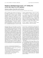
Class-I and class-II by far comprise the most myosins (Figure
1a). Class-I myosins were found in almost all organisms, and
class-II myosins have undergone several gene duplications
(either resulting from whole genome or single gene duplica-
tions), leading to up to 22 class-II myosins per vertebrate
organism. Although the total numbers of myosins per class
are biased by the sequenced species, we expect class-I and
class-II to remain the largest classes even if many other spe-
cies not containing any of these classes (for example, the
plants and Alveolata) are sequenced in the future (Figure 1b).
For example, the numbers of species of the Chordata and the
Viridiplantae lineage for which myosin data are available are
similar. However, the number of myosins for each of these
species is very different, with the Chordata species encoding
up to three times more myosins. In contrast, the number of
sequenced Fungi species (over 90 organisms) is almost twice
as high as the number of Chordata species, but the number of
Fungi myosins is only a quarter of that of the Chordata
myosins.
Table 1
Data statistics
Sequences 2,269 Total
1,941 From WGS
1,634 Complete sequences
38 Domains
3,441,237 Amino acids
8 Total pseudogenes
2 Pseudogenes without sequence
Classes 35 Classes
149 Unclassified myosins
Motor domain position 1,806 Amino-terminal
1 Carboxy-terminal
305 Middle
157 Unknown
Completeness 1,834 Heads complete
150 Head partials
277 Head fragments
149 Only head sequence
6 Only tail sequence
1,725 Tails complete
183 Tail partials
210 Tail fragments
Extremes 4,407 Amino acids in BrMyo15B*
495 kDa is the weight of BrMyo15B*
61 Myosin homologs in Br*
23 Homologs for OlMhc*
13 Classes in Br, Dap, Gg, Xt*
Species 328 Total
181 WGS-projects
127 EST-projects
80 WGS- and EST-projects
3 Species without myosin heavy chain
*Br, Brachydanio rerio; Ol, Oryzias latipes; Dap, Daphnia pulex; Gg, Gallus gallus; Xt, Xenopus tropicalis.
R196.4 Genome Biology 2007, Volume 8, Issue 9, Article R196 Odronitz and Kollmar />Genome Biology 2007, 8:R196
Nomenclature
The amount of produced data spread over all eukaryotic king-
doms now allows and demands a consistent, systematic, and
extendable nomenclature. Here, we introduce the following
nomenclature, which builds on the already established sys-
tem [15,18-20] and tries to keep as many of the existing
names as possible. Nevertheless, it changes some of the
already used names, thus getting rid of sequence-specific and
species-specific exceptions. We are aware of the confusion
that this might introduce about the names of some sequences,
but given the fact that the amount of annotated data known
before finishing this analysis (about 250-300 sequences) was
very small compared to the data presented here, it was neces-
sary for us to introduce an appropriate nomenclature. Other-
wise the number of exceptions would soon exceed the number
of consistently named sequences. We are also aware that dif-
ferent names and classifications have recently been intro-
duced in the literature [16,17]. However, these results were
Taxon and class related statistics of the myosin datasetFigure 1
Taxon and class related statistics of the myosin dataset. (a) The pie-chart shows the number of myosins for each class. (b) The charts show the number
of species and the number of myosins for a set of selected taxa. Exact numbers are given in brackets.
Chordata (56)
Arthropoda (35)
Nematoda (17)
Mollusca (11)
Viridiplantae (39)
Apicomplexa (21)
Basidiomycota (16)
Ascomycota (71)
Microsporidia (2)
Rest (60)
Chordata (910)
Arthropoda (293)
Nematoda (93)
Mollusca (20)
Viridiplantae (180)
Apicomplexa (114)
Basidiomycota (51)
Ascomycota (246)
Microsporidia (4)
Rest (358)
Numer of Species per taxon
Numer of Myosins per taxon
Numer of Myosins per class
Myo1 (381)
Mhc (617)
Myo3 (41)
Myo4 (1)
Myo5 (197)
Myo6 (59)
Myo7 (91)
Myo8 (53)
Myo9 (60)
Myo10 (37)
Myo11 (127)
Myo12 (6)
Myo13 (8)
Myo14 (28)
Myo15 (45)
Myo16 (16)
Myo17 (70)
Myo18 (61)
Myo19 (27)
Myo20 (20)
Myo21 (20)
Myo22 (14)
Myo23 (15)
Myo24 (23)
Myo25 (8)
Myo26 (14)
Myo27 (22)
Myo28 (9)
Myo29 (6)
Myo30 (12)
Myo31 (7)
Myo32 (4)
Myo33 (3)
Myo34 (4)
Myo35 (14)
Orph (149)
(a)
(b)
Genome Biology 2007, Volume 8, Issue 9, Article R196 Odronitz and Kollmar R196.5
comment reviews reports refereed researchdeposited research interactions information
Genome Biology 2007, 8:R196
derived from analyses of small datasets based on many incor-
rectly assembled sequences and, thus, wrongly annotated
myosins, and we have not found a way to incorporate the
small part of matching data into our system. We also think
that even if we introduce some confusion to certain research-
ers in the field, there is a strong necessity to have an appropri-
ate nomenclature to manage existing and upcoming data.
CyMoBase, which we have developed to provide access to all
myosin sequence data [21], uses the new nomenclature, pro-
vides links to previously used names, and can be used as
reference.
The nomenclature is simply as follows and in agreement with
what most people in the field already use. The names of the
sequences consist of four parts: the abbreviation of the spe-
cies' systematic name; the abbreviation of the protein; the
class designation; and the variant designation.
Abbreviation of the species' systematic name
In general, species are abbreviated by using the first letters of
their systematic names (for example, Dm for Drosophila mel-
anogaster). However, there are many species, that would
have the same abbreviation, and in these cases we added the
second letter of the first part of the name (for example, Drm
for Drosophila mimetica). Different strains of the same spe-
cies are differentiated by adding lowercase letters separated
by an underscore (for example, Pf_a for Plasmodium falci-
parum 3D7, Pf_b for Plasmodium falciparum Ghanaian Iso-
late, Pf_c for Plasmodium falciparum HB3, Pf_d for
Plasmodium falciparum Dd2).
Abbreviation of the protein
The abbreviation of the protein is Myo. In the case of the
class-II myosins, the abbreviations Mhc and Mys are used in
the literature. As class-II comprises by far the most sequences
and as numbers have very often been introduced as variant
designations (for example, human Mys1, Mys2, and so on),
we decided to keep the class-II abbreviation as an exception
of the proteins general abbreviation. We decided to use Mhc
as protein abbreviation for class-II myosins as the abbrevia-
tion Mys has been used only for mammalian members while
all other class-II myosins have been named Mhc. If the class-
II myosins were named Myo2 (in accordance with the other
myosin classes) we would have to also rename their variant
designations to avoid confusion with other classes (for exam-
ple, Myo21 could be a class-II myosin variant 1 or a class-XXI
myosin).
Class designation
Classes are numbered according to their discovery. Thus, we
keep all previously accepted class designations [18]. Recent
further class designations [16,22] are based on data analyses
of very small datasets of wrongly annotated myosins and will
not be considered. Richards and Cavalier-Smith [17] have
also used wrongly annotated myosins in their analysis and
have developed a completely new classification not consistent
with any previous classification. As has been agreed upon in
the past, new classes should be designated only if members of
different organisms contribute. We have been very conserva-
tive in our analysis in designating new classes, assigning new
classes only if several species contribute (for example, class-
XXI, all Arthropoda), or very divergent species contribute (for
example, class-XXIX, Thallassiosira pseudonana, Phytoph-
thora sp. and others), or, if the species are closely related, sev-
eral homologs of each species contribute (for example, class-
XXX, Phytophthora sp. and Hyaloperonospora parasitica).
It is obvious, that class separation improves as more and
more divergent sequences are added. In particular, the
myosins of very divergent species (for example, Phytoph-
thora sp., Thallassiosira pseudonana, Tetrahymena ther-
mophila, Paramecium tetrarelia) tend to group mainly with
the homologs of the same organism. Our experience showed
that if more sequences of closely related species are added
(for example, sequences of Phytophthora ramorum, Phy-
tophthora infestans, and Phytophthora sojae), the class sep-
aration improves, and improves further if sequences of more
divergent species are added (Hyaloperonospora parasitica).
But in most of these cases the separation is still not good
enough to distinguish between a class separation and just a
variant separation. Thus, we designated only classes that are
well-supported and separated. There are 24 classes supported
by bootstrap values higher than 985 (out of 1,000; Additional
data file 2) and 5 are supported by bootstrap values higher
than 874. Class-I has the widest taxonomic distribution and is
supported by a bootstrap value of 788. Class-XXVIII (boot-
strap value of 750), class-V (593) class-XXIII (463) and class-
XV (305) show the lowest bootstrap values, but are well sep-
arated from any neighboring class. We left groups of
sequences (for example, the Tetrahymena thermophila and
Paramecium tetrarelia myosins) unclassified, although their
first node in the tree might be supported by a relatively high
bootstrap value. A similar situation would exist if only five
sequences of class-VII, class-X, and class-XV myosins were
known; in this case, these sequences would certainly group
together, supported by a high bootstrap value of the first
node, as they are far more similar to each other than to the
other myosins. Adding more homologs showed these myosins
to be separated into three classes, and we expect a similar
class separation for the myosins of, for example, Tetrahy-
mena thermophila and Paramecium tetrarelia if more
sequences of closely related species are added.
Variant designation
If several myosin homologs exist for the same class, they are
distinguished by a variant designation, a letter starting with
A. Variants with numbers may be used only for the class-II
myosins (see above).
Additional qualification
If both alleles of an organism have been assembled independ-
ently, providing two versions for each myosin gene, the differ-
ent versions are distinguished by adding alpha and beta to the
R196.6 Genome Biology 2007, Volume 8, Issue 9, Article R196 Odronitz and Kollmar />Genome Biology 2007, 8:R196
sequence name. Alternative splice forms of the same gene get
the same protein name. All myosins that cannot be classified
at the moment will be considered as 'orphan' myosins. If sev-
eral orphans exist in a species, they get a variant designation.
Orphan names are considered to be preliminary names. Thus,
orphan myosins will be renamed as soon as more sequences
are available that allow a well-supported classification.
Classification
The basis for the classification of the myosins is the phyloge-
netic relation of their myosin motor domains [15,18]. The
data for the myosins is now strong enough that all designated
classes are well supported. Including or excluding sets of
myosins (for example, the orphans) does not change the phy-
logeny of the other classes as has been observed for the small
dataset used in previous analyses [16]. Also, including or
excluding large insertions like the loop-1 insertion of the
class-I variant C myosins of Arthropoda does not change the
tree.
In contrast to other suggestions, we do not agree with the idea
that the tail domain architectures should also be considered
in the classification process [16,17]. Our analysis shows that
the motor domains and the tails coevolved in most of the
assigned classes, but there are many exceptions now where
the separation of organismal lineages occurred before the
adaptation of further tail domains. It does not make sense to
artificially 'force' sequences together only because there is not
enough sequence data for a better classification. If, for exam-
ple, the class-XII myosins should be related to the class-XV
myosins only because they also contain MyTH4 and Ferm
domains [16], then they could also be grouped with the class-
VII, class-X, or class-XXII myosins. Many other myosins
from Stramenopiles or Amoeba would also have to be
grouped with these classes as they also contain MyTH4 and
Ferm domains. This seems very arbitrary. Also, several
domains, such as the PH domain, Ankyrin repeats or the Pki-
nase domain, are found on either the amino terminus or the
carboxyl terminus of the myosins. Many of the tail regions
have also not been analyzed specifically (domains have not
been defined yet). Thus, as soon as further domains are
defined other myosin classes might unexpectedly share tail
regions. It is also not reasonable to consider the organismal
distribution of myosins as a classification helper as has been
proposed [16]. The species sequenced cover only an extremely
small part of all organisms, and their selection has also been
biased in favor of financial, medical and other interests. It is
not reasonable, therefore, to assume that the organisms that
we have data for are the best representatives with regard to
the myosin diversity of their taxa. For example, even the well-
studied Drosophila melanogaster has lost the class-XXII
myosin that the closely related species Drosophila willistoni
and other Drosophila species still have. Other Arthropoda
(Daphnia, Apis, Anopheles) have additional myosins belong-
ing to well established classes (for example, a class-III myosin
and a class-IX myosin) that all Drosophila species (that have
been sequenced so far) have lost. The same is true for nema-
todes, where a class-XVIII myosin is found in Brugia malayi
and not in Caenorhabditis species. It is very unlikely, there-
fore, that myosins that do not group to any of the other
assigned metazoan myosins (for example, the class-XII
myosins) are closely related to one of the metazoan classes,
although they might share some domains in the tail regions.
It is far more likely that a class-XII myosin will be found in
another metazoa species (as, for example, a class-XX myosin
has been found in Echinodermata in addition to Arthropoda),
or that a class-XV myosin, to which the class-XII myosins
have artificially been grouped [16], will be found in another
nematode (as, for example, a class-XVIII myosin has been
found in Brugia malayi). Both possibilities will support the
current class designation. Nevertheless, at the moment it
seems that all sequenced lineages have developed their own
specific myosin, for example, the class-XVI myosins in verte-
brates, the class-XXI myosins in Arthropoda, and the class-
XII myosins in Nematoda.
Fragments have been classified and named based on their
obvious homology at the amino acid level. Those Fragments
that did not obviously group to one of the assigned classes
have sequentially been added to the dataset used to construct
the major tree. Some of these Fragments could subsequently
be classified; others have to be considered as orphans. Note
that even very short fragments of only 100 amino acids are
sufficient for proper classification. Thus, it is very unlikely
that the orphan Fragments will group to one of the estab-
lished 35 classes if their full-length sequences become
available.
Renamed myosins
Change of previous classification
Class-IV contains only one myosin. According to the nomen-
clature guidelines outlined above, this myosin would not be
designated as a class but would be considered as an orphan.
So as not to cause confusion, we did not change its classifica-
tion from class-IV myosin, expecting that more members will
be added as soon as further genomes are sequenced. How-
ever, our phylogenetic tree shows that the former class-XIII
myosins (of the algae Acetabularia cliftonii) belong to the
class-XI myosins, supported by a bootstrap value of 999.
Therefore, we reclassified the former Acetabularia class-XIII
myosins as class-XI myosins, and assigned the class-XIII to a
Kinetoplastida specific myosin class. The Drosophila mela-
nogaster NinaC protein has previously been classified as a
class-III myosin. However, other Arthropoda contain real
class-III myosins (or more precisely, homologs to the mam-
malian class-III myosins) and NinaC as well as the NinaC
homologs of the other Arthropoda form a distinct class. We
decided not to rename all the mammalian class-III myosins
but to rename NinaC and introduce the new class-XXI.
Genome Biology 2007, Volume 8, Issue 9, Article R196 Odronitz and Kollmar R196.7
comment reviews reports refereed researchdeposited research interactions information
Genome Biology 2007, 8:R196
Change of previous names
The apicomplexan myosins have traditionally been named
alphabetically [16,23]. However, even different splice forms
of the same gene received different protein names. In
addition, gene and genome duplication events have led to,
and will continue to lead to, confusing naming. Thus, it is not
possible to name these myosins consistently in an alphabeti-
cal manner and to provide consistency for the future. We
renamed the apicomplexan myosins according to our nomen-
clature, introducing some apicomplexan-specific myosin
classes. Nevertheless, we tried to keep the former letters as
variants where possible.
The Saccharomyces cerevisiae myosins have previously been
named numerically [24], thus leading to confusion with class
numbers. In addition, several yeast species have now been
sequenced that separated before some of the gene and whole
genome duplication events happened during yeast evolution.
Most of the sequenced yeast species contain only one version
of the class-I and class-V myosins, and Naumovia castellii
contains one class-I but two class-V myosins. It is not possible
to name the newly identified yeast myosins according to the
Saccharomyces cerevisiae myosins. Therefore, we renamed
the Saccharomyces cerevisiae myosins according to our
nomenclature.
Some of the plant and algae myosins were given arbitrary
names in the past, especially those from Helianthus annuus
and Arabidopsis thaliana. This happened before genome
data became available but has not been changed since [25].
We have renamed these few myosins. Some of the vertebrate
class-II myosins have also been renamed based on their hom-
ology to myosins from closely related organisms. In particu-
lar, descriptive names (for example, 'nonmuscle myosin II' or
'fast skeletal muscle myosin') have been disbanded in favor of
numerical variant designations as suggested [18].
Thirty-five myosin classes
The analysis of the phylogenetic tree of the 2,269 myosin
motor domain sequences resulted in the definition of 35
myosin classes (Figures 2 and 3; Additional data file 2), of
which 19 classes have been assigned and described previously
[18]. Our analysis supports and retains the existing classifica-
tion except for the former class-XIII, which consisted of two
myosins from the chlorophyte Acetabularia peniculus
(Acetabularia cliftonii). The former class-XIII was substi-
tuted by a Kinetoplastide-specific class consisting of myosins
with an amino-terminal SH3-like domain, a coiled-coil
region, and two tandem UBA domains. Five new classes,
class-XX, class-XXI, class-XXII, class-XXVIII, and class-
XXXV, are specific to Metazoan species. So far, class-XX has
been found only in arthropods and the sea urchin Strongylo-
centrotus purpuratus and consists of myosins with a long,
coiled-coil region containing an amino-terminal domain and
a short neck composed of one IQ motif. The myosins of class-
XXI are very similar to the class-III myosins in their domain
organization but contain distinct motor domains. The class-
XXII myosins are defined by two tandem MyTH4 and FERM
domains. Most Metazoan species have lost their class-XXVIII
myosin. So far, class-XXVIII myosins have been identified
only in the sea anemone Nematostella vectensis, the frog
Xenopus tropicalis, Gallus gallus, and some fishes. From the
data available it seems that the species of the Acanthopterygii
branch of the fishes (including Takifugu rubripes and Gas-
terosteus aculeatus) have lost the class-XXVIII myosins. The
tail regions of class-XXVIII myosins consist of an IQ motif, a
short coiled-coil region and an SH2 domain.
Five of the new myosin classes (class-XXIII to class-XXVII)
are composed solely of Apicomplexan myosins. The domain
organizations of these myosins have been described else-
where [16] but classes have not been assigned yet. Another six
new myosin classes were attributed to Stramenopiles myosins
(class-XXIX to class-XXXIV). Class-XXIX shows the highest
taxonomic sampling, consisting of members from all Stra-
menopiles species. Class-XXIX myosins have very long tail
domains consisting of three IQ motifs, short coiled-coil
regions, up to 18 CBS domains, a PB1 domain, and a carboxy-
terminal transmembrane domain. The myosin classes XXX to
XXXIV contain only members from Phytophthora species
and the closely related Hyaloperonospora parasitica.
Although the taxonomic sampling is quite low, these classes
have distinct motor domains and unique tail domain organi-
zations. Myosins of class-XXX are composed of an amino-ter-
minal SH3-like domain, two IQ motifs, a coiled-coil region
and a PX domain. Class-XXXI myosins have a very long neck
region consisting of 17 IQ motifs and two tandem Ankyrin
repeats separated by a PH domain. Class-XXXII myosins do
not contain any IQ motifs but a tandem MyTH4 and FERM
domain. The myosins of class-XXXIII have long amino-ter-
minal regions with an amino-terminal PH domain. Class-
XXXIV myosins are composed of one IQ motif, a short coiled-
coil region, five tandem Ankyrin repeats, and a carboxy-ter-
minal FYVE domain.
Orphan myosins
Fungi/Metazoa lineage
The domain organizations of the orphan myosins of the
Fungi/Metazoa lineage are shown in Figure 4. The Micro-
sporida have two myosins, one class-II myosin and an orphan
myosin containing a DIL domain that is also shared by class-
V and class-XI myosins. In contrast to these classes, the
Microsporida orphan myosins do not have any IQ motifs,
thus lacking the ability to bind calmodulin-like light chains.
The wasp Nasonia vitripennis has an orphan myosin that has
a similar domain organization to the class-V and class-XI
myosins, although it has less IQ motifs and its coiled-coil
region is considerably shorter. This myosin is unique to all
Arthropoda species sequenced so far. A myosin very similar in
domain organization to the fungal class-XVII myosins has
been found in the mollusc Atrina rigida. It has 12 transmem-
brane domains separated by a chitin synthetase domain. The
R196.8 Genome Biology 2007, Volume 8, Issue 9, Article R196 Odronitz and Kollmar />Genome Biology 2007, 8:R196
Figure 2 (see legend on next page)
Nematoda
Vertebrata
1000
962
705
820
921
704
Urochordata
Echinodermata
Anthozoa
Protostomia
Choanoflagellida
0.35
Mhc
0.30 0.25 0.20 0.15 0.10 0.05 0
Myo5
Myo27
Myo34
Myo6
Myo30
Myo26
Myo23
Myo14
Myo24
Myo25
Myo20
Myo17
Myo18
Myo32
Myo12
Myo16
Myo21
Myo33
Myo35
Myo1
Orphan Sequences
Myo19
Myo28
Myo3
Myo9
Myo7
Myo15
Myo10
Myo22
Myo13
Myo8
Myo11
Myo31
Myo4
Myo29
Genome Biology 2007, Volume 8, Issue 9, Article R196 Odronitz and Kollmar R196.9
comment reviews reports refereed researchdeposited research interactions information
Genome Biology 2007, 8:R196
choanoflagellate Monosiga brevicollis has 16 orphan myosins
of different domain organizations. Due to missing genome
sequence data of closely related species, all these gene predic-
tions are preliminary (especially the tail regions) and might
change in the future. Some of the predicted orphan myosins
contain domains unique to all myosins analyzed so far, like
the SAM and the Vicilin-N domains. Seven sequences contain
SH2 domains as have been found in the class-XXVIII
myosins.
Alveolata lineage
Several of the Alveolata myosins could not be classified (Fig-
ure 5). All Tetrahymena thermophila and Paramecium
tetraurelia myosins remain ungrouped. The tails of the Par-
amecium tetraurelia myosins contain only IQ motifs, coiled-
coil regions, and RCC1 domains, while some of the Tetrahy-
mena thermophila myosins also contain FERM or MyTH4
domains. However, the FERM and MyTH4 domains never
appear in tandem like in class-VII, class-X, or class-XXII
myosins.
Orphan myosins from Stramenopiles
Although they share only the class-I myosins, the Strameno-
piles species show a similar myosin diversity as the metazoan
species (Figure 6). So far, three Phytophthora species and the
closely related Hyaloperonospora parasitica have been
sequenced; all share the same set of myosins. The orphan
myosins of this group have not been classified because it is
not clear from the phylogenetic tree where to draw class
boundaries. However, it is obvious that the Myo-A to Myo-H
and the Myo-Q to Myo-U orphans form distinct groups. The
domain organizations of the myosins within these groups are
also very different. To resolve their classification, further data
from more distantly related species are needed. The genome
sequences of two diatoms, Phaeodactylum tricornutum and
Thalassiosira pseudonana, have also been finished. Both
species share several sequences, but Thalassiosira pseudo-
nana has a higher myosin diversity, having myosins with
HEAT or Mis14 domains that do not exist in any other
myosin.
Orphan myosins from other taxa
Orphan myosins from other taxa are shown in Figure 7. The
Dictyostelium discoideum orphan myosins have been dis-
cussed elsewhere [26]. The amoeba-flagellate Naegleria gru-
beri has three orphan myosins having only coiled-coil regions
in the tail. The unicellular red alga Galdieria sulphuraria
contains one myosin with a unique domain organization con-
sisting of at least nine IQ motifs followed by an AAA domain
and a DnaJ domain. Both alleles of Trypanosoma cruzi have
been assembled independently, providing two slightly differ-
ent versions for each myosin gene. The seven orphan myosins
of Trypanosoma cruzi contain amino-terminal SH3-like
domains, IQ motifs, or coiled-coil regions.
Species that do not contain myosins
There are three species whose genome sequences are availa-
ble and that do not contain any myosin: the unicellular red
alga Cyanidioschyzon merolae, the flagellated protozoan
parasite Giardia lamblia, and the protozoan parasite Tri-
chomonas vaginalis.
Discussion
All myosin protein sequences have been derived by manually
inspecting the corresponding DNA, either the published
cDNA or genomic DNA, or the genomic DNA provided by
sequencing centers. Published sequences contained errors in
many cases, either from sequencing or from manual annota-
tion, while automatic annotations provided by the sequencing
centers resulted in mispredicted exons in almost all tran-
scripts. For many sequences, the prediction of the correct
exons was only possible with the help of the analysis of the
homologs of related species. Thus, not only has the quantity
of myosin data increased as more and more genomes have
been analyzed but also the quality as all ambiguous regions
could be resolved for those sequences for which data from a
closely related organism are available. Therefore, mispre-
dicted exons may be limited to a few orphan myosins.
For the phylogenetic analysis of the myosin motor domains
we created a structure-guided manual sequence alignment
whose quality is far beyond any computer-generated align-
ment. It is obvious that all secondary structure elements of
the class-II myosin motor domain structure remain con-
served in all myosins, even in the most divergent homologs.
Sequence motifs that would not have been aligned at first
glance were placed based on the analysis of their supposed
three-dimensional counterparts, which always maintained
the structural integrity of the respective region. Thus, strong
sequence variation and sequence insertions were limited to
loop regions. Based on the phylogenetic tree constructed from
1,984 myosin motor domains, 35 classes have been assigned
(Figures 2 and 3; Additional data files 2 and 3). There are 149
myosins that still remain unclassified due to our conservative
view on designating classes but it is anticipated that sequenc-
ing of further genomes will result in their classification and
will substantially increase the existing number of classes. For
Phylogenetic tree of the myosin motor domainsFigure 2 (see previous page)
Phylogenetic tree of the myosin motor domains. The phylogenetic tree was built from the multiple sequence alignment of 1,984 myosin motor domains.
The complete tree with bootstrap values and sequence descriptors is available as Additional data file 2. The expanded view shows the myosin sequences
of class-VI and their distribution in taxa. Every other myosin class has been analyzed in a similar way. Labels at branches are bootstrap values (1,000 total
boostraps). The scale bar corresponds to estimated amino acid substitutions per site. The tree was drawn using FigTree v1.0 [40].
R196.10 Genome Biology 2007, Volume 8, Issue 9, Article R196 Odronitz and Kollmar />Genome Biology 2007, 8:R196
Figure 3 (see legend on next page)
0 500 1000 1500 2000
aa
2500 3000
HsMyo7A
TicMyo22
Pf_aMyo23
Pf_aMyo27
Pf_aMyo26
TepMyo25
Pf_aMyo24B
AcMyo4
HsMyo10
HsMyo5A
HsMyo3A
DmMyo15
EnMyo17
DmMyo21
HsMyo16
AtMyo8A
HsMyo1A
HsMhc1
LemMyo13
HsMyo6
HsMyo19
IpMyo28
DmMyo20
HsMyo9A
AtMyo11A
TgMyo14
CeMyo12
HsMyo18A
PhrMyo29
IQ motif
SH3
SH2
C1
Coiled-coil
MyTH4
MyTH1
FERM
chitin synthase
DIL
PH
Cyt-b5
Pkinase
RhoGAP
N-terminal SH3-like
RA
PX
Ankyrin repeat
WD40 repeat
CBS
RCC1
FYVE
PB1
Transmembrane domain
PDZ
UBA
PhrMyo30A
PhrMyo31A
PhrMyo32
PhrMyo33
PhrMyo34
HsMyo35
Genome Biology 2007, Volume 8, Issue 9, Article R196 Odronitz and Kollmar R196.11
comment reviews reports refereed researchdeposited research interactions information
Genome Biology 2007, 8:R196
generating the tree it does not matter whether long loop
regions (for example, the 300 amino acid loop-1 of the
Arthropoda Myo1C proteins) are included in the alignment or
not (data not shown). So far, almost all orphan myosins
belong to taxa that have not undergone large-scale compara-
tive sequencing efforts. Only short sequence fragments have
been found for 277 myosins. These sequences were excluded
from the phylogenetic analysis but have been classified based
on their similarity in the multiple sequence alignment. Never-
theless, these data are important for defining myosin diver-
sity in as many organisms as possible.
The highest number of myosins in a single organism has been
found in Brachydanio rerio (61 myosins grouped into 13
classes) while the broadest class distribution is expected for
the Phytophthora species (25 myosins grouped into at least 15
classes). The high numbers of vertebrate myosin genes in
general are due to several whole genome duplications that
happened after the separation from the Craniata and Uro-
chordata [27].
Our survey of the myosin gene family now allows the recon-
struction of the tree of 328 eukaryotes (Figure 8). The organ-
isms of the major clades Fungi/Metazoa, Euglenozoa,
Stramenopiles and Alveolata have distinct sets of myosin
classes (except class-I), showing that horizontal gene transfer
of myosins has not happened in later stages of eukaryotic evo-
lution. However, we cannot exclude yet that horizontal gene
transfer of myosins has not happened at the origin of eukary-
otic evolution. Hence, only paralogs and orthologs have to be
resolved. Figure 8 represents a schematic reconstruction of
both the phylogenetic relationships of major taxa recon-
structed from class-specific trees as well as the information
on myosin class evolution and distribution. For example, Tet-
rahymena thermophila, Perkinsus marinus, Toxoplasma
gondii, Plasmodium falciparum, and Babesia bovis have all
been classified as Alveolata. However, the relation between
Ciliophora (Tetrahymena thermophila), Perkinsea (Perkin-
sus marinus), and Apicomplexa (Toxoplasma gondii, Plas-
modium falciparum, and Babesia bovis) has not been
resolved yet. Tetrahymena thermophila does not share any
myosin with the other Alveolata and should, therefore, have
diverged before the other species. Perkinsus marinus shares
two myosin classes with the Apicomplexa. Thus, they must
have had a common ancestor. The Apicomplexa developed
three further common classes, of which single classes have
been lost by different species. The myosin class-specific trees
show that the Coccidia, the Haemosporida, and the
Piroplasmida form distinct lineages. However, their relation
cannot be resolved further. This principle for reconstructing
the tree has been applied to all species.
The class-I myosins show the widest taxonomic distribution
and are devoid of the amino-terminal SH3-like domain and
are thus suggested to be the first myosins to have evolved (see
below). Only two major lineages, the Viridiplantae and the
Alveolata, do not contain class-I myosins (Figure 8). The
Alveolata have either lost the class-I myosin, or their class-I
myosin diverged so far that a common ancestor could not be
reconstructed. The Apicomplexa developed several specific
classes, while the Ciliophora myosins cannot be classified yet.
The evolutionary history of the Euglenozoa and
Stramenopiles cannot be further resolved because both do
not share any further myosin classes with other species, and
their taxonomic sampling is not high enough for a more pre-
cise grouping.
The second myosin class to develop during the evolution of
the Fungi and Metazoa kingdoms was class-V. The plants
have developed two kingdom-specific classes. However, the
domain organization of the plant-specific class-XI is similar
to that of class-V, suggesting that both had a common ances-
tor. In contrast to the class-I myosins, the class-V and class-
XI myosins have diverged so far that a common ancestry is
not visible beyond their general domain organization. After
separation of the plant lineage, the class-II myosins arose.
The protists Entamoeba
sp., Acanthamoeba castellanii, Nae-
gleria gruberi, and Dictyostelium discoideum have closely
related myosins, suggesting that they share a common ances-
tor that diverged shortly before the Fungi and Metazoa split.
While the Entamoebidae have lost their class-V myosin,
retaining only a class-I and a class-II myosin, the Acan-
thamoebidae, Dictyosteliida, and Heterolobosea have devel-
oped several additional specific myosins with unique domain
organizations, in addition to the increase in the number of
myosin genes through single gene or whole genome
duplications. The Acanthamoebidae and Dictyosteliida
already contain the combination of the myosin motor domain
and the MyTH4 domain that is also widely found in the
metazoan lineage. However, a lack of genomic data prevents
the designation of a common myosin motor domain-MyTH4
containing ancestor. The fungi developed the class-XVII
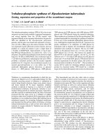
Schematic diagram of the domain structures of representative members of the 35 myosin classesFigure 3 (see previous page)
Schematic diagram of the domain structures of representative members of the 35 myosin classes. The sequence name of the representative member is
given in the motor domain of the respective myosin. A color key to the domain names and symbols is given on the right except for the myosin domain,
which is colored in blue. The abbreviations for the domains are: C1, protein kinase C conserved region 1; CBS, cystathionine-beta-synthase; Cyt-b5,
cytochrome b5-like Heme/Steroid binding domain; DIL, dilute; FERM, band 4.1, ezrin, radixin, and moesin; FYVE, zinc finger in Fab1, YOTB/ZK632.12,
Vac1, and EEA1; IQ motif, isoleucine-glutamine motif; MyTH1, myosin tail homology 1; MyTH4, myosin tail homology 4; PB1, Phox and Bem1p domain;
PDZ, PDZ domain; PH, pleckstrin homology; Pkinase, protein kinase domain; PX, phox domain; RA, Ras association (RalGDS/AF-6) domain; RCC1,
regulator of chromosome condensation; RhoGAP, Rho GTPase-activating protein; SH2, src homology 2; SH3, src homology 3; UBA, ubiquitin associated
domain; WD40, WD (tryptophan-aspartate) or beta-transducin repeats.
R196.12 Genome Biology 2007, Volume 8, Issue 9, Article R196 Odronitz and Kollmar />Genome Biology 2007, 8:R196
myosin that consists of a functionally restricted myosin motor
domain fused with a highly conserved chitin synthetase [28].
While the Ascomycetes, Basidiomycetes, and Chytridiomy-
cota have retained one member of each of the four myosin
classes, the Zygomycotes Rhizopus arrhizus and Phycomyces
blakesleeanus have undergone several single gene or whole
genome duplications. The Saccharomycetes, Schizosaccharo-
mycetes, and Microsporidia have lost their class-XVII
myosin.
Two different models can be proposed for the further evolu-
tion of the Metazoa (Figures 8 and 9). In both models a con-
siderable boost of myosin diversity happened at the early
evolution of Metazoa. The most reasonable model based on
the myosin class distribution suggests an increase of the
myosin diversity in three steps. After separation of the Fungi,
the Metazoa developed four new classes, class-VI, class-VII,
class-IX, and class-XVIII. These classes are shared by species
of all Metazoa taxa sequenced so far, except the choanoflagel-
late Monosiga brevicollis, which does not contain class-IX
and class-XVIII myosins. However, single species of the other
taxa have also lost their members of these four classes; for
example, the nematode Trichinella spiralis contains only a
class-VII myosin, the Caenorhabditis species have lost their
Schematic diagram of the domain structures of the orphan myosins of the Fungi/Metazoa lineageFigure 4
Schematic diagram of the domain structures of the orphan myosins of the Fungi/Metazoa lineage. The sequence names of the ophan myosins are given in
the motor domain of the respective myosins. Color keys to the domain names and symbols are given on the right except for the myosin domain, which is
colored in blue. Myosin names next to domain representations list orthologs from closely related species or orthologs from the same species. These
sequences have a similar domain organization. Sequences that are not orthologs and have not resulted from recent gene duplications are shown separately,
although their domain organizations might be very similar. The myosin domains without names on the bottom symbolize that only head fragments are
available for the sequences listed on the right. The exclamation mark on the left side of some sequences signifies that the corresponding sequences
(especially the tail regions) have not completely been validated because of missing comparative genome sequences. Those sequences and corresponding
tail domain predictions might change with upcoming genome sequences of related species. Abbreviations for the domains are: SAM, sterile alpha motif;
Vicilin-N, Vicilin amino-terminal region; WW, tryptophan-tryptophan motif domain; Y phosphatase, protein tyrosine phosphatase, catalytic domain.
0 500 1000 1500 2000
aa
2500 3000
WW
AnlMyo-A
MbMyo-O
EcMyo-A
MbMyo-B
NavMyo-A
AtrMyo-A
MbMyo-A
SAM
MbMyo-C
Y phosphatase
MbMyo-K
MbMyo-L
MbMyo-D
MbMyo-F
StpMyo-A, StpMyo-B, MyMyo-A, MbMyo-P
MbMyo-E
MbMyo-G
MbMyo-H
MbMyo-I
MbMyo-J
Vicilin-N
MbMyo-M
MbMyo-N
IQ motif
Coiled-coil
N-terminal SH3-like
DIL
chitin synthase
Transmembrane domain
SH3
SH2
MyTH1
Pkinase
Ankyrin repeat
Genome Biology 2007, Volume 8, Issue 9, Article R196 Odronitz and Kollmar R196.13
comment reviews reports refereed researchdeposited research interactions information
Genome Biology 2007, 8:R196
Schematic diagram of the domain structures of the orphan myosins from the Alveolata lineageFigure 5
Schematic diagram of the domain structures of the orphan myosins from the Alveolata lineage. The sequence names of the ophan myosins are given in the
motor domain of the respective myosins. Color keys to the domain names and symbols are given on the right except for the myosin domain, which is
colored in blue. Myosin names next to domain representations list orthologs from closely related species or orthologs from the same species. These
sequences have a similar domain organization. Sequences that are not orthologs and have not resulted from recent gene duplications are shown separately,
although their domain organizations might be very similar. The myosin domains without names on the bottom symbolize that only head fragments are
available for the sequences listed on the right. The exclamation mark on the left side of some sequences signifies that the corresponding sequences
(especially the tail regions) have not completely been validated because of missing comparative genome sequences. Those sequences and corresponding
tail domain predictions might change with upcoming genome sequences of related species. Abbreviations for the domains are: HDAC interact, histone
deacetylase (HDAC) interacting.
0 500 1000 1500 2000
IQ motif
SH3
Coiled-coil
MyTH4
FERM
aa
2500 3000
EtMyo-A
TgMyo-B
N-terminal SH3-like
CpMyo-A
ChMyo-A
TetMyo-D
CpMyo-B
ChMyo-B
PtMyo-B
PtMyo-E
PtMyo-H
TetMyo-G
TetMyo-J
TetMyo-A
TgMyo-A
PtMyo-C
TetMyo-K
TetMyo-N
TetMyo-C
TetMyo-L
TetMyo-I
TetMyo-M
TetMyo-E
TetMyo-H
TetMyo-F
PtMyo-A
PtMyo-F
PtMyo-D
PtMyo-G
PtMyo-J, PtMyo-K, TetMyo-B, PrmMyo-A, EtMyo-B
RCC1
HDAC interact
R196.14 Genome Biology 2007, Volume 8, Issue 9, Article R196 Odronitz and Kollmar />Genome Biology 2007, 8:R196
Schematic diagram of the domain structures of the orphan myosins from StramenopilesFigure 6
Schematic diagram of the domain structures of the orphan myosins from Stramenopiles. The sequence names of the ophan myosins are given in the motor
domain of the respective myosins. Color keys to the domain names and symbols are given on the right except for the myosin domain, which is colored in
blue. Myosin names next to domain representations list orthologs from closely related species or orthologs from the same species. These sequences have
a similar domain organization. Sequences that are not orthologs and have not resulted from recent gene duplications are shown separately, although their
domain organizations might be very similar. Abbreviations for the domains are: CH, Calponin homology domain; GAF, domain present in phytochromes
and cGMP-specific phosphodiesterases; HEAT repeat, named after the proteins huntingtin, elongation factor 3 (EF3), the 65 kDa alpha regulatory subunit
of protein phosphatase 2A (PP2A) and the yeast PI3-kinase TOR1; Mis14, kinetochore protein Mis14 like.
0 500 1000 1500 2000
aa
2500 3000
PhrMyo-A
PhrMyo-B
WW
CH
GAF
PhrMyo-C
PhrMyo-D
PhrMyo-E
PhrMyo-F
PhrMyo-G
PhrMyo-H
PhrMyo-Q
PhrMyo-R
PhrMyo-S
PhrMyo-T
PhrMyo-U
ThpMyo-A
PhtMyo-D
PhtMyo-I
PhtMyo-B, ThpMyo-C, ThpMyo-D
PhtMyo-C, PhtMyo-E, PhtMyo-G
PhtMyo-F
ThpMyo-H
ThpMyo-B
ThpMyo-I
ThpMyo-J
ThpMyo-E
ThpMyo-G
PhtMyo-H
PhtMyo-A
ThpMyo-F
Mis14
HEAT
HDAC interact
Phytophthora species,
Hyaloperonospora parasitica
IQ motif
Coiled-coil
N-terminal SH3-like
PX
Ankyrin repeat
FYVE
PDZ
Pkinase
Genome Biology 2007, Volume 8, Issue 9, Article R196 Odronitz and Kollmar R196.15
comment reviews reports refereed researchdeposited research interactions information
Genome Biology 2007, 8:R196
class-XVIII myosins, and the Drosophila species have lost
their class-IX myosin. Our model places the choanoflagellates
to the Coelomata that invented the related class-X, class-XV,
and class-XXII myosins. After separation of the choanoflagel-
lates, the Bilateria gained another three classes, class-III,
class-XIX, and class-XX. The Deuterostomia, to which we
placed the Cnidaria, invented the class-XXVIII myosins and
lost class-XXII myosins. Later in evolution, the Chordata lost
class-XX myosins. This model proposes the continuous
invention of new myosin classes over a relatively long time
and the subsequent loss of single myosin classes by certain
species and lineages. The placement of the Cnidaria to the
Deuterostomia is surprising as the Cnidaria are commonly
considered to be a sister group of the Bilateria. However, the
analysis of the Nematostella vectensis genome showed that,
from a genomic perspective, Nematostella more closely
resembles modern vertebrates than the fruit fly or nematodes
[29], which is consistent with our analysis. But as long as
genome sequences of further Cnidaria species are not availa-
ble, this placement could also be the result of long branch
attraction effects in the phylogenetic tree. Sequencing of fur-
ther species of the lineages Choanoflagellida, Cnidaria, and
Echinodermata, which are as yet represented only by single
species, will provide a better picture of these taxa, as has been
obtained for the nematodes, Arthropoda, and vertebrates,
which show a wide distribution of the myosin content
between their member species. For example, during the evo-
lution of the Arthropoda, the Insecta lost the class-XIX
myosin. Later in evolution the ancestor of all Drosophila spe-
cies lost the class-III and class-IX myosins, and finally most
Drosophila species lost the class-XXII myosin. Most of the
lineages like the Nematoda, Arthropoda and Vertebrata have
developed further branch-specific myosins. We propose that
sequencing of related organisms to Strongylocentrotus pur-
puratus and Monosiga brevicollis will result in the classifica-
tion of their orphan myosins and, thus, also of branch-specific
myosins for these lineages.
Schematic diagram of the domain structures of the orphan myosins of species not belonging to one of the other taxaFigure 7
Schematic diagram of the domain structures of the orphan myosins of species not belonging to one of the other taxa. Both alleles of Trypanosoma cruzi
have been assembled independently, providing two slightly different copies of each myosin gene. None of the Myo-F versions is complete and the
presented domain organization of Myo-F is the result of a merged version of both myosins. The sequence names of the ophan myosins are given in the
motor domain of the respective myosins. Color keys to the domain names and symbols are given on the right except for the myosin domain, which is
colored in blue. Myosin names next to domain representations list orthologs from closely related species or orthologs from the same species. These
sequences have a similar domain organization. Sequences that are not orthologs and have not resulted from recent gene duplications are shown separately,
although their domain organizations might be very similar. Abbreviations for the domains are: AAA, ATPase family associated with various cellular
activities; DnaJ domain, named after the prokaryotic heat shock protein DnaJ; RhoGEF, Rho GDP/GTP exchange factor.
0 500 1000 1500 2000
IQ motif
SH3
DnaJ
Coiled-coil
MyTH4
FERM
RhoGEF
PH
DdMyo-M
aa
2500 3000
DdMyo-I
DdMyo-G
N-terminal SH3-like
NgMyo-A
NgMyo-C
NgMyo-B
GsMyo-A
AAA
TrcMyo-Aalpha
TrcMyo-Balpha
TrcMyo-Calpha
TrcMyo-Dalpha
TrcMyo-Galpha
TrcMyo-Ealpha
TrcMyo-F
R196.16 Genome Biology 2007, Volume 8, Issue 9, Article R196 Odronitz and Kollmar />Genome Biology 2007, 8:R196
Figure 8 (see legend on next page)
a
26 23
Amoebozoa
Metazoa
Chordata
Bilateria
Coelomata
Pseudocoelomata
13
1
Euglenozoa
O
Trypanosoma cruzi
Leishmania major
29 301
Stramenopiles
O
Phytophthora ramorum
31 32 33 34
Thalassiosira pseudonana
Phaeodactylum tricornutum
5
8 11
Streptophyta
1
Oryza sativa
Populus trichocarpa
5
8 11
Chlorophyta
Viridiplantae
Ostreococcus tauri
Chlamydomonas reinhardtii
Arabidopsis thaliana
1 2 5
4
Acanthamoebidae
1 2 5
Entamoebidae
1 2 5
O
Dictyosteliida
Dictyostelium discoideum
Acanthamoeba castellanii
Entamoeba histolytica
5
1 2
O
Heterolobosea
Naegleria gruberi
17
1 2 5
Basidiomycota
Ustilago maydis
Phanerochaete chrysosporium
17
1 2 5
Ascomycota
Magnaporthe grisea
17
1 2 5
Chytridiomycota
Batrachochytrium dendrobatidis
17
1 2 5
Schizo-
saccharomycetes
Schizosaccharomyces pombe
17
1 2 5
Zygomycota
Rhizopus arrhizus
Phycomyces blakesleeanus
O
1 2 5
17
Microsporidia
Encephalitozoon cuniculi
1 2 5
Saccharomycetes
17
Saccharomyces cerevisiae
Pichia stipitis
Naumovia castellii
Choanoflagellida
2215
10
6 7 9 18
1 2 5
12
18
6 7 9
1 2 5
Nematoda
Trichinella spiralis
Caenorhabditis elegans
Brugia malayi
O
Monosiga brevicollis
212215 28
10 3 19 20
6 7 9 18
1 2 5
Arthropoda
10 28 16 35
15 3 19
6 7 9 18
1 2 5
Vertebrata
Brachydanio rerio
Takifugu rubripes
Xenopus tropicalis
Mus musculus
Monodelphis domestica
Homo sapiens
Gallus gallus
10
315 19
6 7 9 18
1 2 5
Urochordata
Ciona intestinalis
15
10 3 19 28
6 7 9 18
1 2 5
Echinodermata
20
28
O
Strongylocentrotus
purpuratus
15 28
10 3 19
6 7 9 18
1 2 5
Anthozoa
Cnidaria
Protoostomia
Deuterostomia
Nematostella vectensis
20
5
17
6 7 9 18
15
10
22
19
2028
3
22
20
Fungi / Metazoa
Fungi
Drosophila melanogaster
Daphnia pulex
Anopheles gambiae
2
Eukaryota
1
Alveolata
14 23 2526 27
Coccidia
Apicomplexa
O
2325
14
Ciliophora
O
14 23 242526 27
Haemosporida
1
Cryptosporidium parvum
Eimeria tenella
14 2527
Piroplasmida
Babesia bovis
Theileria annulata
Toxoplasma gondii
Plasmodium gallinaceum
Plasmodium falciparum
Tetrahymena thermophila
Paramecium tetraurelia
26 27
Perkinsea
O
Perkinsus marinus
X
First Occurence
Loss
Genome survey incomplete
1 Gene
5 Genes
Accepted Taxon
Proposed Taxon
Myosin Class X
2627
Amoebozoa
Cnidaria
Pseudocoelomata
Protostomia
Genome Biology 2007, Volume 8, Issue 9, Article R196 Odronitz and Kollmar R196.17
comment reviews reports refereed researchdeposited research interactions information
Genome Biology 2007, 8:R196
In contrast, the metazoan tree based on classical taxa and
nodes shows the invention of ten myosin classes in a very
short time scale (Figure 9). The evolution of the Metazoa
would thus mainly be characterized by gene losses. While the
Anthozoa Nematostella vectensis shares all its 12 myosin
classes with vertebrates, the nematodes must have lost 6 of
the 13 common Metazoa myosin classes. The nematode
Trichinella spiralis has lost another three of the remaining
classes, sharing only four classes with the other Metazoa. The
Arthropoda must also have lost at least two of the common
Metazoa myosin classes. This scenario, the invention of ten
myosin classes during the evolution of only two taxa nodes
and the subsequent major losses of myosin classes until the
final speciation, seems very unlikely compared to the other
model that proposes the invention of new myosin classes over
a long period with the subsequent loss of single classes.
In both models, the tree of myosin diversity gives clear sup-
port for the classical Coelomata hypothesis that groups
Arthropoda with Deuterostomia in a monophyletic class. The
Nematoda sequenced so far lack four classes that the Arthro-
poda share with the vertebrates. It is very unlikely that the
Nematoda have lost just these four classes and not one or
more of the others. The class specific phylogenetic trees show
that the Nematoda myosins always separate before the
Arthropoda-Deuterostomia split, except for the class-IX
myosins, where the Nematoda and Arthropoda homologs
group separately from the Deuterostomia homologs. These
findings illustrate the advantage of analyzing the diversity of
a large protein family in contrast to looking at single-gene
phylogenies that have supported the monophyletic grouping
of Nematoda and Arthropoda in some cases [30].
The comparative analysis of the phylogenetic relationship of
the species in single myosin classes showed several
incongruities. We hypothesized that the myosin genes of the
corresponding organisms might have evolved asynchro-
nously, as has been observed for a number of yeast genes [31].
From the phylogenetic tree we therefore determined the dis-
tances between pairs of sequences. To compensate for differ-
Schematic drawing of the evolution of myosin diversityFigure 8 (see previous page)
Schematic drawing of the evolution of myosin diversity. The tree has been constructed based on the combination of the phylogenetic information obtained
from the analysis of single myosin classes as well as the analysis of the class distribution of major taxa (see Materials and methods). Thus, branch lengths do
not correspond to any scale. Nodes that have already been suggested are symbolized by filled circles. Nodes that we propose base on the analysis of the
myosins are represented by open circles. The exact myosin contents of several representative organisms are given. The myosin inventory of all 328
organisms is available from Additional data file 3.
Schematic drawing of the evolution of myosin diversity in the Fungi/Metazoa lineage based on the 'accepted' taxonomyFigure 9
Schematic drawing of the evolution of myosin diversity in the Fungi/Metazoa lineage based on the 'accepted' taxonomy. The inventions and losses of the
myosin classes have been plotted onto the 'accepted' phylogeny of the Eukaryotes available at NCBI. Branch lengths do not correspond to any scale.
10
Metazoa
Chordata
Bilateria
Coelomata
Pseudocoelomata
Choanoflagellida
2215
10
6 7
1 2 5
12
18
6 7 9
1 2 5
Nematoda
Trichinella spiralis
Caenorhabditis elegans
Brugia malayi
O
Monosiga brevicollis
2193 20
22 18 19 28
6 7 10 15
1 2 5
Arthropoda
10
28 16 35
15 3 19
6 7 9 18
1 2 5
Vertebrata
Brachydanio rerio
Takifugu rubripes
Xenopus tropicalis
Mus musculus
Monodelphis domestica
Homo sapiens
Gallus gallus
10 15 3 19
6 7 9 18
1 2 5
Urochordata
Ciona intestinalis
Strongylocentrotus purpuratus
3
9 18 28
6 7 10 15
1 2 5
Echinodermata
20
19
O
9 28
3 18 19
6 7 10 15
1 2 5
Anthozoa
Cnidaria
Fungi
Protostomia
Deuterostomia
Nematostella vectensis
6 7
9 18
6 7
15
22
15
10
22
19
20
28
3
22
22
10 15
3
22
19 28
20
Fungi / Metazoa
Drosophila melanogaster
Daphnia pulex
Anopheles gambiae
28
X
First Occurence
Loss
Genome survey incomplete
1 Gene
5 Genes
Myosin Class X
R196.18 Genome Biology 2007, Volume 8, Issue 9, Article R196 Odronitz and Kollmar />Genome Biology 2007, 8:R196
Asynchronous evolution of mammalian myosin proteinsFigure 10
Asynchronous evolution of mammalian myosin proteins. The matrix illustrates the normalized distances between corresponding sequences. Asynchronous
evolution is observed if the pattern of the deviation from the mean is different. For example, the pattern from rat to the other mammalian species is very
similar, illustrating their synchronous evolution in general. However, there are differences in the patterns of some class-I myosins between rat and mouse
and opossum, indicating their asynchronous evolution. In contrast, the sequence comparison patterns of cow and the other mammals are very different,
indicating the asynchronous evolution of all cow myosin genes. The abbreviations for the organisms are: Rn, Rattus norvegicus; Mm, Mus musculus; Pat, Pan
troglodytes; Hs, Homo sapiens; Mam, Macaca mulatto; Caf, Canis familiaris; Bt, Bos Taurus; Md, Monodelphis domestica.
Normalized Distance
Md
Bt
Caf
Mam
Rn
Hs
Pat
Mm
0
3.6
(distant)
(close)
No Data
Myo1A
Myo1B
Myo1C
Myo1D
Myo1E
Myo1F
Myo1G
Myo1H
Myo3A
Myo3B
Myo5A
Myo5B
Myo5C
Myo6
Myo7A
Myo7B
Myo9A
Myo9B
Myo10
Myo15
Myo16
Myo18A
Myo18B
Myo19
Myo35
times mean distance
within class
Genome Biology 2007, Volume 8, Issue 9, Article R196 Odronitz and Kollmar R196.19
comment reviews reports refereed researchdeposited research interactions information
Genome Biology 2007, 8:R196
Asynchronous evolution of fungi myosin proteinsFigure 11
Asynchronous evolution of fungi myosin proteins. The matrix is shown in a similar way as in Figure 10. The consensus tree from the analysis of the single
myosin class trees is shown. The obtained polytomic tree is the result of the asynchronous evolution of the different species. The abbreviations for the
organisms are: Bad, Batrachochytrium dendrobatidis JEL423; Sc_c, Saccharomyces cerevisiae S288c; Sc_a, Saccharomyces cerevisiae YJM789; Sc_b, Saccharomyces
cerevisiae RM11-1a; Sap, Saccharomyces paradoxus NRRL Y-17217; Smi, Saccharomyces mikatae IFO 1815; Sak, Saccharomyces kudriavzevii IFO 1802; Sab_b,
Saccharomyces bayanus MCYC 623; Sab_a, Saccharomyces bayanus 623-6C; Sk_a, Saccharomyces kluyveri NRRL Y-12651; Kw, Kluyveromyces waltii NCYC 2644;
Kl, Kluyveromyces lactis NRRL Y-1140; Erg, Eremothecium gossypii ATCC 10895; Nac, Naumovia castellii NRRL Y-12630; Cgl, Candida glabrata CBS138; Loe,
Lodderomyces elongisporus NRLL YB-4239; Deh, Debaryomyces hansenii CBS767; Cad, Candida dubliniensis CD36; Ca_a, Candida albicans SC5314; Ca_b, Candida
albicans WO-1; Ct_a, Candida tropicalis MYA-3404; Cap, Candida parapsilosis; Cll, Clavispora lusitaniae ATCC 42720; Pig, Pichia guilliermondii ATCC 6260; Pcs,
Pichia stipitis CBS 6054; Rha, Rhizopus arrhizus RA 99-880; Phb, Phycomyces blakesleeanus; Fnd_c, Filobasidiella neoformans var. neoformans JEC21; Fnd_b,
Filobasidiella neoformans var. neoformans B-3501A; Fna_b, Filobasidiella neoformans var. neoformans H99; Fnb_b, Filobasidiella neoformans var. bacillispora R265;
Lab, Laccaria bicolor S238N; Cpc, Coprinopsis cinerea okayama7#130; Phc, Phanerochaete chrysosporium RP-78; Um_a, Ustilago maydis 521; Um_b, Ustilago
maydis FB1; Spr, Sporobolomyces roseus IAM 13481; Alb,
Alternaria brassicicola ATCC 96836; Pn, Phaeosphaeria nodorum SN15; Mg, Mycosphaerella graminicola;
Chg, Chaetomium globosum CBS 148.51; Poa, Podospora anserina; Nc, Neurospora crassa OR74A; Mag, Magnaporthe grisea 70-15; Gz, Gibberella zeae PH-1;
Gim, Gibberella moniliformis 7600; Nh, Nectria haematococca MPVI; Scs, Sclerotinia sclerotiorum 1980; Bof, Botryotinia fuckeliana B05.10; Hj, Hypocrea jecorina
QM9414; Cop, Coccidioides posadasii C735; Coi_a, Coccidioides immitis RS; Coi_c, Coccidioides immitis RMSCC 2394; Ur, Uncinocarpus reesii 1704; Ajc_b,
Ajellomyces capsulatus NAmII G217B; Ajc_a, Ajellomyces capsulatus NAmII G186AR; Ajc_c, Ajellomyces capsulatus NAmI WU24; Nef, Neosartorya fischeri NRRL
181; Asf, Aspergillus fumigatus Af293; Asc, Aspergillus clavatus NRRL 1; An, Aspergillus niger ATCC 1015; Ast, Aspergillus terreus NIH2624; Ao, Aspergillus oryzae
RIB40; Af, Aspergillus flavus NRRL3357; En, Emericella nidulans FGSC A4; Asa, Ascosphaera apis USDA-ARSEF 7405; Yl, Yarrowia lipolytica CLIB99; Sp,
Schizosaccharomyces pombe 972h-; Sj, Schizosaccharomyces japonicus yFS275.
Bad
Sc_c
Sc_a
Sc_b
Sap
Smi
Sak
Sab_b
Sab_a
Sk_a
Kw
Kl
Erg
Nac
Cgl
Loe
Deh
Cad
Ca_a
Ca_b
Ct_a
Cap
Cll
Pig
Pcs
Rha
Phb
Fnd_c
Fnd_b
Fna_b
Fnb_b
Lab
Cpc
Phc
Um_a
Um_b
Spr
Alb
Pn
Mg
Chg
Poa
Nc
Mag
Gz
Gim
Nh
Scs
Bof
Hj
Cop
Coi_a
Coi_c
Ur
Ajc_b
Ajc_a
Ajc_c
Nef
Asf
Asc
An
Ast
Ao
Af
En
Asa
Yl
Sp
Sj
Sj
Sp
Yl
Asa
En
Af
Ao
Ast
An
Asc
Asf
Nef
Ajc_c
Ajc_a
Ajc_b
Ur
Coi_c
Coi_a
Cop
Hj
Bof
Scs
Nh
Gim
Gz
Mag
Nc
Poa
Chg
Mg
Pn
Alb
Spr
Um_b
Um_a
Phc
Cpc
Lab
Fnb_b
Fna_b
Fnd_b
Fnd_c
Phb
Rha
Pcs
Pig
Cll
Cap
Ct_a
Ca_b
Ca_a
Cad
Deh
Loe
Cgl
Nac
Erg
Kl
Kw
Sk_a
Sab_a
Sab_b
Sak
Smi
Sap
Sc_b
Sc_a
Sc_c
Bad
Normalized Distance
Myo1
Mhc
Myo5
Myo17
0
1.8
times mean distance
within class
(distant)
(close)
No Data
Sj
Sp
Yl
Asa
En
Af
Ao
Ast
An
Asc
Asf
Nef
Ajc_c
Ajc_a
Ajc_b
Ur
Coi_c
Coi_a
Cop
Hj
Bof
Scs
Nh
Gim
Gz
Mag
Nc
Poa
Chg
Mg
Pn
Alb
Spr
Um_b
Um_a
Phc
Cpc
Lab
Fnb_b
Fna_b
Fnd_b
Fnd_c
Phb
Rha
Pcs
Pig
Cll
Cap
Ct_a
Ca_b
Ca_a
Cad
Deh
Loe
Cgl
Nac
Erg
Kl
Kw
Sk_a
Sab_a
Sab_b
Sak
Smi
Sap
Sc_b
Sc_a
Sc_c
Bad
Chytridiomycota
Saccha
rom
ycotin
a
Zygomycota
Basidio
mycota
Basidiomycota
Basidiomycota
Ascomycota
Schizosaccharomycetes
R196.20 Genome Biology 2007, Volume 8, Issue 9, Article R196 Odronitz and Kollmar />Genome Biology 2007, 8:R196
ences in general diversity within each class, all distances were
normalized. Asynchronous evolution is visualized by the
comparison of the deviation from the mean distances. As
examples we analyzed the myosins of completely sequenced
mammalian (Figure 10) and fungal (Figure 11) genomes. As
expected, all primates are very closely related, with the chim-
panzee generally closer to Homo sapiens than to macaca. The
myosin proteins from dog and cow are more closely related to
those of the primates than to those from rodents. The opos-
sum Monodelphis domestica is, in general, the most diver-
gent mammal with respect to myosins, although in the case of
Myo1E and Myo16, it is most closely related to the dog and the
primates than to the rodents. The myosin proteins from cow
show the most asynchronous phylogenetic relationship of the
analyzed mammalian genomes. They either diverge before
the split of the rodents and primates/dog, after this split, or
form a monophyletic class with the corresponding dog
orthologs. Hence, it is not possible either to resolve the phyl-
ogenetic grouping of the cow in general, or to do so by using
the myosin proteins, or sequences from additional mammals
have to be added to better resolve the tree.
The fungal myosins show several distinct groups that are
related to the established taxa. However, the analysis resolves
some so far unrecognized relationships. The Saccharomy-
cotina do not group to the Ascomycota in all myosin classes,
but have evolved asynchronously. Based on our analysis of
the myosins, the Saccharomycotina should be considered as
an independent clade that evolved from Fungi, in parallel
with the Ascomycota, the Basidiomycota, the Zygomyocota,
and the Schizosaccharomycetes. These clades developed very
asynchronously so that their phylogeny cannot be resolved. In
addition, the species in these clades have undergone consid-
erable asynchronous development. Yarrowia lipolytica,
which has been considered a yeast species, is more closely
related to the Ascomycota than to the Saccharomycotina,
based on both the phylogenetic relation of the respective
myosin homologs and its myosin content as it contains a
class-XVII myosin that all Saccharomycotina have lost.
What did the very first myosin look like? In the beginning of
eukaryotic evolution, the myosin motor domain had been
developed (Figure 12). During subsequent early evolution, an
extensive process of domain fusions started, during which the
carboxy-terminal IQ motif was added first. After duplication
of this gene, the amino-terminal SH3-like domain was fused
to the motor domain. These two domain organizations are
shared by myosins of all species. The class-I myosins show the
widest taxonomic distribution, are devoid of the amino-ter-
minal SH3-like domain and, thus, are suggested to be the first
distinct myosin class to have evolved. We propose that the
most ancient myosin motor domain had a sequence very close
to that of the class-I myosins.
Evolution of the first myosinsFigure 12
Evolution of the first myosins. The first myosin, called ur-myosin, is expected to consist only of the myosin motor domain. By domain fusion it generated
the IQ motif either directly carboxy-terminal to the motor domain (2), or after a gene duplication event (1). After a further gene duplication event, this
myosin developed to the class-I myosins as well as the ancestor of most of the other myosin classes after fusion with an SH3 domain (which developed
into the amino-terminal SH3-like domain).
?class-I
Ur-myosin
??
1
2
Genome Biology 2007, Volume 8, Issue 9, Article R196 Odronitz and Kollmar R196.21
comment reviews reports refereed researchdeposited research interactions information
Genome Biology 2007, 8:R196
Conclusion
Here, we present the phylogenetic analysis of 2,269 manually
annotated myosin proteins. The previously assigned 19
myosin classes were confirmed and 16 new classes with
unique domain organizations defined. A phylogenetic tree
has been constructed that includes information about the
class distribution and evolution in certain taxa as well as the
phylogenetic information contained in class-specific sub-
trees. The analysis shows the Choanoflagellida as part of the
Metazoa lineage and the cnidaria (Nematostella vectensis) as
having diverged after the separation of Deuterostomia and
Protostomia. The myosin data show that several taxa have
evolved asynchronously, for example the Mammalia and the
Fungi.
The presented tree will increase in resolution as more organ-
isms are sequenced. To increase the fine resolution, more
sequences of intermediate taxa, for example, in the metazoan
lineage, are needed. For some major taxa a significant
amount of species has to be sequenced to get the resolution
already obtained for the fungi and metazoans. For example,
only eight species of the Viridiplantae have completely been
sequenced so far. In particular, sequencing of further under-
represented taxa will increase myosin diversity. The myosin
data presented here will allow the correct annotation and
classification of all upcoming homologs. We hope that CyMo-
Base [21,32], which stores and presents all related informa-
tion, will be an invaluable tool in all areas of myosin and
motor protein research, and in classical taxonomy.
Materials and methods
Identification of myosin family proteins
Myosin genes were identified in iterated TBLASTN searches
of the completed genomes of 181 organisms starting with the
protein sequence of DdMhcA. All hits were manually
analyzed at the genomic DNA level. The correct coding
sequences were identified with the help of the multiple
sequence alignment of the myosins. As the amount of myosin
sequences increased (especially the number of sequences in
classes with few representatives), many of the initially pre-
dicted sequences were reanalyzed to correctly identify all
exon borders. Where possible, EST data have been analyzed
to help in the annotation process. Now, all designated myosin
classes contain enough members to correctly predict any
additional member sequence in the future. However, some of
the orphan myosins (for example, from Tetrahymena ther-
mophila and Paramecium tetraurelia) might still contain
wrongly predicted exons in the tail regions, because sufficient
comparative genomic data are not yet available. For some
organisms only EST data are available to date, and myosin
sequences identified in these databases have been included in
the analysis as long as the sequences contains at least 100 res-
idues. These short sequences cannot be, and have not been,
used in the phylogenetic analysis but are important to define
the myosin inventory with as many organisms as possible. In
addition to the analysis of these large-scale sequencing
projects, all myosin sequences in the nr database at NCBI
have been collected and reanalyzed. Many of these sequences
contain sequencing errors, mispredicted exons, and wrongly
predicted gene borders.
Some of the genes contain alternative splice forms for the
motor domain. The different splice forms were not
considered independently in the analysis but in all cases the
same splice forms were taken for homologous myosins. All
sequence-related data (names, corresponding species,
GenBank IDs, alternative names, corresponding publica-
tions, domain predictions, and sequences) and references to
genome sequencing centers are available through CyMoBase
[21,32]. The annotated sequences and the alignment of the
motor domain sequences are available as Additional data files
5 and 6, respectively.
Building trees
The phylogenetic tree was built based on a manually con-
structed and maintained structure-guided multiple sequence
alignment. The phylogenetic tree is unrooted and was gener-
ated using neighbor joining and the bootstrap (1,000 repli-
cates) method as implemented in ClustalW (standard
settings) [33]. The phylogenetic tree presented in Additional
data file 2 was visualized using TreeDyn [34]. The schematic
tree was constructed using the following criteria. The myosins
are separated into 35 classes. Thus, we assigned a class inven-
tory to every organism. The myosin classes are not only well-
separated based on their motor domains but also due to the
unique composition of their tail domains. Thus, we conclude
that species having myosins of the same class must have had
a common ancestor. It is extremely if not completely unlikely
that myosins with these distinct features were invented inde-
pendently. The other criterion is the analysis of the different
trees of the myosin classes. Looking at the tree of a single
class, for example, the class VI myosins, it is obvious that cer-
tain taxa always separated earlier than others, for example,
the Arthropoda myosins always separated before the mam-
malian myosins. In the next step we ordered all species
according to their class inventory, minimizing the number of
myosin class inventions and losses. If taxa have the same class
inventory we used the data from the single class trees to
resolve their phylogenetic relationship. If the phylogenetic
relationships of taxa in single classes contradicted each other,
we hypothesized asynchronous evolution and did not resolve
the relationship between these taxa.
Distance maps
The distances between two sequences of one class/variant
were obtained from the distance matrix produced by Clus-
talW using default substitution matrix BLOSUM62 [35]. We
collected the distances between all sequences of each class/
variant. The distances within each class/variant were normal-
ized by dividing by the mean distance of the class/variant.
This set the mean distance of each class/variant to 1. In the
R196.22 Genome Biology 2007, Volume 8, Issue 9, Article R196 Odronitz and Kollmar />Genome Biology 2007, 8:R196
distance maps the normalized distances were visualized in
blocks, each block representing the distances of all sequences
from two species. Asynchronous phylogenetic relationships
are visible as color fluctuations within a block.
Domain and motif predictions
Protein domains were predicted using the SMART [36,37]
and Pfam [38,39] web server. The prediction of coiled-coils is
based on the coils program. The IQ motifs and amino-termi-
nal domains were predicted manually based on the homology
to similar domains of the other myosins. The recognition
motifs included in the SMART and Pfam databases are too
restrictive, as the motifs have been created based on the small
datasets available some years ago. The domain profiles of the
other domains have not been revised yet.
Abbreviations
EST, expressed sequence tag; WGS, whole genome
sequencing.
Authors' contributions
FO performed data analysis. MK designed the study, assem-
bled and annotated all sequences, and performed data analy-
sis. Both authors wrote and approved the manuscript.
Additional data files
The following additional data are available with the online
version of this paper. Additional data file 1 outlines the prob-
lems and pitfalls of automatic gene annotation, gene collec-
tion, domain prediction, and sequence alignment. Additional
data file 2 shows the complete phylogenetic tree of 1,984
myosins. Additional data file 3 is a table of the complete
myosin inventory of all 328 species. Additional data file 4
provides references for sequence data, listing of all organisms
and related publications with references and links to genome
sequencing centers. Additional data file 5 lists the myosin
sequences. These sequences are the basis for the myosin
inventory and the domain prediction figures. Additional data
file 6 shows the alignment of the myosin head domains used
to compute the phylogenetic tree shown in Additional data
file 2.
Additional data file 1Problems and pitfalls of automatic gene annotation, gene collec-tion, domain prediction, and sequence alignmentProblems and pitfalls of automatic gene annotation, gene collec-tion, domain prediction, and sequence alignment.Click here for fileAdditional data file 2Complete phylogenetic tree of 1,984 myosinsComplete phylogenetic tree of 1,984 myosins.Click here for fileAdditional data file 3Complete myosin inventory of all 328 speciesComplete myosin inventory of all 328 species.Click here for fileAdditional data file 4All organisms and related publications with references and links to genome sequencing centersAll organisms and related publications with references and links to genome sequencing centers.Click here for fileAdditional data file 5Myosin sequencesAll annotated myosin sequences in fasta formatClick here for fileAdditional data file 6Motor domain alignmentAlignment of the myosin head domains used to compute the phyl-ogenetic tree shown in Additional data file 2Click here for file
Acknowledgements
We would like to thank HD Schmitt and M Schliwa for invaluable discus-
sions and C Griesinger for discussions and continuous support. MK was
supported by a Liebig Stipendium of the Fonds der Chemischen Industrie,
which is in part financed by the BMBF. This work has been funded by grant
I80798 of the VolkswagenStiftung and grant KO 2251/3-1 of the Deutsche
Forschungsgemeinschaft.
References
1. Embley TM, Martin W: Eukaryotic evolution, changes and
challenges. Nature 2006, 440:623-630.
2. Yang S, Doolittle RF, Bourne PE: Phylogeny determined by pro-
tein domain content. Proc Natl Acad Sci USA 2005, 102:373-378.
3. Doolittle RF: Evolutionary aspects of whole-genome biology.
Curr Opin Struct Biol 2005, 15:248-253.
4. Jeffroy O, Brinkmann H, Delsuc F, Philippe H: Phylogenomics: the
beginning of incongruence? Trends Genet 2006, 22:225-231.
5. Ciccarelli FD, Doerks T, von Mering C, Creevey CJ, Snel B, Bork P:
Toward automatic reconstruction of a highly resolved tree
of life. Science 2006, 311:1283-1287.
6. Delsuc F, Brinkmann H, Philippe H: Phylogenomics and the
reconstruction of the tree of life. Nat Rev Genet 2005, 6:361-375.
7. Koonin EV: Orthologs, paralogs, and evolutionary genomics.
Annu Rev Genet 2005, 39:309-338.
8. Odronitz F, Hellkamp M, Kollmar M: diArk - a resource for
eukaryotic genome research. BMC Genomics 2007, 8:103.
9. Krendel M, Mooseker MS: Myosins: tails (and heads) of
functional diversity. Physiology (Bethesda) 2005, 20:239-251.
10. Schliwa M, Woehlke G: Molecular motors. Nature 2003,
422:759-765.
11. Vale RD: The molecular motor toolbox for intracellular
transport. Cell 2003, 112:467-480.
12. Scholey JM, Brust-Mascher I, Mogilner A: Cell division. Nature 2003,
422:746-752.
13. Yumura S, Uyeda TQ: Myosins and cell dynamics in cellular
slime molds. Int Rev Cytol 2003, 224:
173-225.
14. Geeves MA, Holmes KC: The molecular mechanism of muscle
contraction. Adv Protein Chem 2005, 71:161-193.
15. Cheney RE, Riley MA, Mooseker MS: Phylogenetic analysis of the
myosin superfamily. Cell Motil Cytoskeleton 1993, 24:215-223.
16. Foth BJ, Goedecke MC, Soldati D: New insights into myosin evo-
lution and classification. Proc Natl Acad Sci USA 2006,
103:3681-3686.
17. Richards TA, Cavalier-Smith T: Myosin domain evolution and the
primary divergence of eukaryotes. Nature 2005,
436:1113-1118.
18. Berg JS, Powell BC, Cheney RE: A millennial myosin census. Mol
Biol Cell 2001, 12:780-794.
19. Gillespie PG, Albanesi JP, Bahler M, Bement WM, Berg JS, Burgess DR,
Burnside B, Cheney RE, Corey DP, Coudrier E, et al.: Myosin-I
nomenclature. J Cell Biol 2001, 155:703-704.
20. Hodge T, Cope MJ: A myosin family tree. J Cell Sci 2000,
113:3353-3354.
21. CyMoBase [ />22. Williams SA, Gavin RH: Myosin genes in Tetrahymena. Cell Motil
Cytoskeleton 2005, 61:237-243.
23. Heintzelman MB, Schwartzman JD: Myosin diversity in
Apicomplexa. J Parasitol 2001, 87:429-432.
24. Brown SS: Myosins in yeast. Curr Opin Cell Biol 1997, 9:44-48.
25. Reddy AS, Day IS: Analysis of the myosins encoded in the
recently completed Arabidopsis thaliana genome sequence.
Genome Biol 2001, 2:. RESEARCH0024
26. Kollmar M: Thirteen is enough: the myosins of Dictyostelium
discoideum
and their light chains. BMC Genomics 2006, 7:183.
27. Jaillon O, Aury JM, Brunet F, Petit JL, Stange-Thomann N, Mauceli E,
Bouneau L, Fischer C, Ozouf-Costaz C, Bernot A, et al.: Genome
duplication in the teleost fish Tetraodon nigroviridis reveals
the early vertebrate proto-karyotype. Nature 2004,
431:946-957.
28. Fujiwara M, Horiuchi H, Ohta A, Takagi M: A novel fungal gene
encoding chitin synthase with a myosin motor-like domain.
Biochem Biophys Res Commun 1997, 236:75-78.
29. Putnam NH, Srivastava M, Hellsten U, Dirks B, Chapman J, Salamov
A, Terry A, Shapiro H, Lindquist E, Kapitonov VV, et al.: Sea anem-
one genome reveals ancestral eumetazoan gene repertoire
and genomic organization. Science 2007, 317:86-94.
30. Adoutte A, Balavoine G, Lartillot N, Lespinet O, Prud'homme B, de
Rosa R: The new animal phylogeny: reliability and
implications. Proc Natl Acad Sci USA 2000, 97:4453-4456.
31. Langkjaer RB, Cliften PF, Johnston M, Piskur J: Yeast genome dupli-
cation was followed by asynchronous differentiation of dupli-
cated genes. Nature 2003, 421:848-852.
32. Odronitz F, Kollmar M: Pfarao: a web application for protein
family analysis customized for cytoskeletal and motor pro-
teins (CyMoBase). BMC Genomics 2006, 7:300.
33. Chenna R, Sugawara H, Koike T, Lopez R, Gibson TJ, Higgins DG,
Thompson JD: Multiple sequence alignment with the Clustal
series of programs. Nucleic Acids Res 2003, 31:3497-3500.
34. Chevenet F, Brun C, Banuls AL, Jacq B, Christen R: TreeDyn:
Genome Biology 2007, Volume 8, Issue 9, Article R196 Odronitz and Kollmar R196.23
comment reviews reports refereed researchdeposited research interactions information
Genome Biology 2007, 8:R196
towards dynamic graphics and annotations for analyses of
trees. BMC Bioinformatics 2006, 7:439.
35. Henikoff S, Henikoff JG: Amino acid substitution matrices from
protein blocks. Proc Natl Acad Sci USA 1992, 89:10915-10919.
36. Letunic I, Copley RR, Schmidt S, Ciccarelli FD, Doerks T, Schultz J,
Ponting CP, Bork P: SMART 4.0: towards genomic data
integration. Nucleic Acids Res 2004, 32:D142-144.
37. SMART [ />38. Bateman A, Coin L, Durbin R, Finn RD, Hollich V, Griffiths-Jones S,
Khanna A, Marshall M, Moxon S, Sonnhammer EL, et al.: The Pfam
protein families database. Nucleic Acids Res 2004, 32:D138-141.
39. Pfam [ />40. FigTree [ />