Báo cáo y học: "Making the most of high-throughput protein-interaction data" ppsx
Bạn đang xem bản rút gọn của tài liệu. Xem và tải ngay bản đầy đủ của tài liệu tại đây (223.41 KB, 10 trang )
Genome Biology 2007, 8:112
Opinion
Making the most of high-throughput protein-interaction data
Robert Gentleman* and Wolfgang Huber
†
Addresses: *Fred Hutchinson Cancer Research Center, Seattle, WA 98109, USA.
†
European Bioinformatics Institute, European Molecular
Biology Laboratory, Cambridge CB10 1SD, UK.
Published: 2 November 2007
Genome Biology 2007, 8:112 (doi:10.1186/gb-2007-8-10-112)
The electronic version of this article is the complete one and can be
found online at />© 2007 BioMed Central Ltd
Most protein functions involve their interaction with other
molecules, often with other proteins in the assembly of opera-
tional complexes. A better understanding of protein inter-
actions is fundamental to the study of biological systems. As
many drugs act on proteins, it is also a prerequisite for
understanding intended, and unintended, drug effects. Over
the past few years a number of large-scale experiments have
set out to map protein interactions systematically [1-15].
While there is interest in combining the resulting data, there
appear to be substantial discrepancies between experiments,
and evaluation studies have reported large error rates, lack of
overlap and apparent contradictions between the different
datasets [16-21].
The purpose of this article is to critically assess the metho-
dology used to analyze protein-interaction datasets. When
interpreting individual experiments or combining datasets
from different experiments, we need to consider three
questions: first, what do we want to know and which experi-
ments provide data that can be used to answer our questions;
second, which types of protein interactions were assayed and
under what conditions; and third, what types of measure-
ment errors may have occurred and what is their prevalence.
In this article we will discuss how the formulation of appro-
priate statistical models can allow investigators to clearly
identify and estimate quantities of interest.
We will consider two particular types of protein interactions:
physical interactions, and interactions between members of
a protein complex - which we shall call ‘complex membership
interactions’. A physical interaction is a direct and specific
contact between a pair of proteins [22]. We regard two
proteins in a complex as having a physical interaction if they
share an interaction surface. A complex membership
interaction exists between proteins that are part of the same
multiprotein complex and does not necessarily imply a
physical interaction.
Sampling and coverage
The two most widely used experimental techniques for
detecting protein-protein interactions are the yeast two-
hybrid (Y2H) system [23] and affinity purification followed
by mass spectrometry (AP-MS) [24]. The Y2H system assays
whether proteins can physically interact with each other.
Large-scale experiments are carried out in a colony-array
format, in which each yeast colony expresses a defined pair
of ‘bait’ and ‘prey’ proteins that can be scored for reporter
gene activity - indicating interaction - in an automated
manner [1,6,25]. The type of information obtained from a
Y2H experiment is shown in Figure 1. In an AP-MS experi-
ment, a tagged protein is expressed in yeast and then ‘pulled
down’ from a cell extract, along with any proteins associated
with it, by co-immunoprecipitation or by tandem affinity
purification. The set of pulled-down proteins is identified by
MS. In a laborious and expensive process, this procedure has
been systematically applied to large sets of yeast proteins
[7-11]. The tagged protein in AP-MS is also sometimes called
Abstract
We review the estimation of coverage and error rate in high-throughput protein-protein interaction
datasets and argue that reports of the low quality of such data are to a substantial extent based on
misinterpretations. Probabilistic statistical models and methods can be used to estimate properties
of interest and to make the best use of the available data.
the bait and the proteins it pulls down the prey. The
information on protein complexes given by Y2H and AP-MS
experiments is compared in Figure 2.
An appreciation of the concepts of sampling and coverage is
vital for interpreting the data from these types of experiments
[26,27]. The term ‘sampling’ is used for experimental designs
where only a subset of the population is interrogated.
Representative sampling techniques are used in many fields of
science, but they are not common in the generation of protein-
interaction datasets, where sampling has often been guided by
biological priorities. The ‘coverage’ summarizes which part of
the total set of possible interactions has actually been tested.
Even when genome-wide screening was intended [1,10,11],
coverage was in fact well below 100%, and the success for each
bait seems to depend on nonrandom biological, technological
and economic factors. For example, Gavin et al. [10] used all
6,466 open reading frames (ORFs) that were at that time
annotated in the Saccharomyces cerevisiae genome and
obtained tandem affinity purifications for 1,993 of those. The
remaining 4,473 (69%) failed at various stages, because, for
example, the tagged protein failed to express or protein bands
were not well separated by gel electrophoresis. Thus, neither
the set of tested baits nor the set of tested prey in current
experiments are random subsets of all proteins in the
organism and in general, it is not valid to make inferences
about the ‘population’, that is, the set of all physical
interactions that take place in a cell under the conditions being
studied, by assuming the available experimental data from a
Y2H or AP-MS experiment to be a representative sample. We
are not arguing that random sampling be used, as it would not
be appropriate in this setting, but rather that the data need
to be interpreted more judiciously.
Genome Biology 2007, Volume 8, Issue 10, Article 112 Gentleman and Huber 112.2
Genome Biology 2007, 8:112
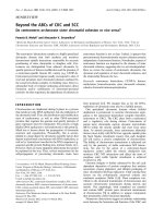
Figure 2
The manifestation of protein complexes in Y2H and AP-MS data. AP-MS
experiments measure complex co-membership, and the fact that a prey is
found by a certain bait means that there is either a direct physical
interaction or an indirect physical interaction mediated by a protein
complex. The set of proteins pulled down by a particular bait cannot
therefore be equated with a single complex: if the bait is part of several
different complexes, then the set of prey will be the union of all proteins in
all complexes. (a) Protein B is involved in three different multiprotein
complexes. In two of these it directly interacts with C, which itself can also
interact with proteins F, G or H, whereas in the third complex, B interacts
with D and E. (b) Assuming there are no other interactions under the
conditions of the experiment, the bipartite graph between proteins B, H
and complexes 1, 2, and 3 will look like this. (c,d) The result of a
hypothetical AP-MS experiment with no false positives and no false
negatives when (c) B is used as a bait and (e) F is used as a bait. (e,f) Result
from a hypothetical Y2H experiment with a genome-wide set of preys and
with no false positives and false negatives when (d) B is used as a bait and (f)
F is used as a bait. (g,h) The results of (g) an ideal AP-MS experiment and
(h) an ideal Y2H experiment if all proteins were used as baits. The Y2H
data in (e,f,h) identifies the direct interactions, but it does not contain
information on the number and architecture of the complexes. The
maximal cliques identified by the AP-MS experiment in (g) correspond to
the complexes in (a). However, the AP-MS data do not contain information
on the topology of the direct interactions within each complex.
(a)
1
2
3
B
C
D
E
F
G
H
(b)
C
D
E
F
G
H
(c)
(e)
B
(d)
C
E
B
D
BC
G
H
F
C
F
(f)
(g)
C
F
G
E
D
B
H
(h)
G
C
F
H
B
D
E
D
B
E
G
C
B
F
B
C
H
F
3
2
1
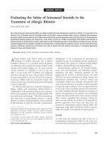
Figure 1
Interpreting results on direct physical interactions from Y2H
experiments. (a) The observation of interactions A-B and B-C in a Y2H
experiment does not indicate whether the two interactions can take place
simultaneously (center) or whether they are exclusive of each other
(right). (b) The ability of two proteins to interact may depend on post-
translational modifications whose presence or absence may be actively
regulated. Proteins D and E interact (center) in the absence of a certain
post-translational modification (red shape), whose presence inhibits the
interaction (right).
B
C
A
A
B
C
D
E
D
E
B
A
C
(a)
(b)
B
E
D
One problem in evaluating large-scale protein-interaction
experiments is that the published data are often not
sufficiently detailed to allow accurate description of the sets
of baits and prey that were actually tested. As a proxy, we
introduced the concept of ‘viable baits’ and ‘viable prey’ [28].
The first is the set of baits that were reported to have
interacted with at least one prey, and the latter are those
proteins reported to be found by at least one bait. Numbers
for these can be unambiguously obtained from the reported
data and provide surrogate measures for the tested baits and
tested prey. The set of all pairs between viable bait and
viable prey are the interactions that we are confident were
experimentally tested and could, in principle, have been
detected. The failure to detect an interaction between a
viable bait and a viable prey is informative, whereas the
absence of an observed interaction between an untested bait
and prey is not. We note that the set of viable prey is a subset
of the tested prey, and viable baits are a subset of the tested
baits. This approach might introduce bias, because negative
data from baits that were tested but found no prey, as well as
from prey that were present but did not interact with any
bait, are not recorded. On the other hand, presuming that
combinations were tested, when in fact they were not, can
also result in bias. Gilchrist et al. [29] used a randomization
approach to estimate the size of the prey populations for the
datasets in [7] and [8]. Their estimates are about double
those of the number of viable prey.
Representation as graphs
Graph theory offers a convenient and useful set of terms and
concepts to represent relationships between entities. Graphs
most commonly represent binary relationships and these
can be either directed or undirected. A further type of graph
is needed to represent the membership of proteins in
complexes: this relationship is not binary and requires a type
of graph called a bipartite graph. Box 1 gives precise
definitions of these concepts and an overview of how they
apply to protein-interaction data.
Undirected graphs are often used as a model for physical
interactions. True relationships are symmetric: if protein A
interacts with B, then B interacts with A. The observed
experimental data, however, often display asymmetry, which
is a consequence of the experimental asymmetry between
bait and prey. Protein A may identify protein B as an inter-
actor when A is used as a prey, but B as a prey may not find
A. To represent asymmetric data, we suggest using a
directed-graph model. This is a point on which we diverge
from much of the current practice. We argue that although
the quantity of interest is an unknown undirected graph, it
must be estimated from the observed data, which should be
represented as a directed graph.
“All models are wrong, but some are useful.” This maxim of
George Box [30] reminds us that we should not expect these
models to adequately represent all possible aspects of
protein interactions in a satisfactory way. For the current
types of data and questions, graph models are useful. As the
data and the questions that we ask become more sophisti-
cated, more complicated models are likely to be needed.
Some limitations of the graph models described here are
related to their lack of resolution in time and space, failure
to distinguish between different protein isoforms or post-
translational modifications, and to the fact that experiments
do not record interactions between individual protein
molecules but between populations. It is the lack of such
information that makes it difficult to use Y2H data to make
inference about the composition of protein complexes (see
Figure 1) or to use AP-MS data to identify the physical
interactions of the proteins within a complex and their
stoichiometry (see Figure 2).
Error statistics
Whether two proteins physically interact in vivo is not always
simple to determine: the range of binding affinities of
biologically relevant protein interactions spans many orders of
magnitude [31], and interactions can be dynamic, transient
and highly regulated. Nevertheless, the simple measurement
model used to interpret the results of protein-interaction
experiments presumes that for each pair of proteins, the
question of whether or not they interact can be answered as
either yes or no. The aim of making a measurement is to
record the true, typically unknown, value of a physical
quantity, but in practice there will be deviations -
measurement errors. In such circumstances, statistical
methods can be used to infer the true value of a quantity, given
the data and some assumptions about how the measurement
tool works. In this sense, the Y2H system or an AP-MS screen
are simply measurement tools that provide imperfect data
from which we make inferences about the true state of nature.
Standard definitions of various error statistics [32] are given
in Box 2. We give them to enable a coherent dialog and to
address some of the confusion in the literature. For example,
a widely cited evaluation study by Edwards et al. [17]
reported a “false positive rate” defined as FP/(TP + FP):
where FP is the number of false positives and TP the number
of true positives. However, the more common name for this
quantity is the ‘false-discovery rate’ (see Box 2). The differ-
ence between the false-positive rate, as usually defined by
FP/N, and the false-discovery rate can be substantial, as
their denominators are very different, N being the true tested
non-interactions, given by TN + FP (see Box 2). Incompatible
terminology leads to confusion and makes comparison of
error rates reported in different studies difficult.
Measurement errors can be decomposed into two compo-
nents: stochastic and systematic errors. Stochastic errors are
associated with random variability, whereas systematic
errors are recurrent. Stochastic errors are simpler to
Genome Biology 2007, Volume 8, Issue 10, Article 112 Gentleman and Huber 112.3
Genome Biology 2007, 8:112
address: they can be controlled by replication, can be
eventually eliminated if the experiment is repeated many
times, and they can often readily be described using
probability models. Systematic errors give rise to bias: the
quantity being measured is consistently different from the
truth. Their identification is difficult, but if it can be done,
they can be addressed either by improving the experimental
procedures or by developing appropriate methods for post-
experiment data processing.
Statistical models for the analysis of
protein-interaction data
Statistical models can integrate the information from
repeated or related measurements and quantify the (un)cer-
tainty that we have about the conclusions. Here we consider
how statistical techniques have been applied to two distinct
problems: estimating membership of a protein complex and
the integration of data from different experiments (cross-
experiment integration of data).
Genome Biology 2007, Volume 8, Issue 10, Article 112 Gentleman and Huber 112.4
Genome Biology 2007, 8:112
Box 1. The terminology of graphs
Undirected graphs
An undirected graph consists of a set of nodes V and a set of edges E and is denoted as G = (V,E). Each element of the
edge set E is an unordered pair (u,v) of nodes, and the two nodes in a pair are called ‘adjacent’. The neighborhood of a
node v is the set of nodes N(v) to which it is adjacent, and its ‘degree’
δ
(v) is the number of its neighbors,
δ
(v) = |N(v)|. A
subgraph S of a graph G contains a node set V
S
⊆ V and an edge set E
S
= {(u,v) ∈ E|u,v ∈ V
S
}. The unordered pairs
defining each edge e ∈ E represent symmetric binary relationships between the elements of the node set. Undirected
graphs can succinctly model physical protein interactions. The node set of a protein-protein interaction graph consists of
all the individual proteins in the biological system of interest, and the edge set indicates which pairs of proteins
physically interact.
Directed graphs
The definition of a directed graph builds upon that of undirected graphs, the only difference being that the edges are
ordered. By convention, the direction of an edge (u,v) originates from u towards v. The edges (u,v) and (v,u) are distinct,
and a graph may contain either one or both. The notion of degree in a directed graph is separated into two distinct
concepts: ‘indegree’ and ‘outdegree’. The outdegree,
δ
o
(v), of a node v is the number of directed edges that originate at v
(out-edges). Its indegree,
δ
i
(v), is the number of edges that flow towards v (in-edges). Directed graphs can be used to
represent Y2H data as well as AP-MS data. An edge A → B indicates that an interaction was tested with protein A as a
bait and protein B when used as a prey. The result of the measurement is either positive or negative and can be
represented as an edge attribute.
Bipartite graphs
Bipartite graphs or membership graphs are useful to represent the grouping of objects. They have two distinct types of
nodes, and edges only connect a node of one type to a node of the other. For example, the proteins of a biological system
could be the nodes of one type, its functional modules that of the other, and an edge in the bipartite graph represents
membership of a protein in a module. Proteins can be members of multiple modules, and some proteins might not be
assigned to any module.
One-mode graphs
Two graphs called one-mode graphs can be derived from a bipartite graph. If U and W are the node partitions of a
bipartite graph G, then the edges in the one-mode graph on U (in respect of W) are determined by whether or not the two
nodes both have edges in G to a common element of W (in respect of U). If A is the |U| × |W| adjacency matrix of the
bipartite graph, then the one-mode graph for the node set U can be obtained by A⊗A
t
and the one-mode graph for W by
A
t
⊗A. The symbol ⊗ represents matrix multiplication under Boolean algebra and the superscript t indicates matrix
transposition. The one-mode graph of the proteins is the complex membership graph: two nodes are connected if they
are members of the same complex. Similarly, the one-mode graph of the complexes is the complex overlap graph: two
complexes are connected to each other in this graph if there is at least one protein that is a member of both.
Maximal cliques
A clique is a fully connected subgraph. A maximal clique is a cligue that is not s proper subset of another clique.
Estimating membership of a protein complex
Russell and colleagues [10] have developed a heuristic that
they term the ‘socioaffinity index’, A
ij
. It quantifies the
confidence that proteins i and j share complex membership,
given a set of protein purifications each with its bait and a
number of prey. The score is the logarithm of the product of
three odds-ratios. The first odds-ratio compares the frequency
with which bait i pulled down prey j to the frequency that
would be expected if prey came down randomly; the second
is the corresponding value for bait j pulling down prey i; and
the third is the ratio of frequency of co-occurrence of i and j
in a pull-down to what would be expected under random
sampling. The authors then apply a customized clustering
algorithm to the matrix A
ij
to estimate sets of protein
complexes from AP-MS data.
Scholtens and colleagues took a different route [33,34]. They
explicitly modeled the underlying bipartite graph of member-
ship of proteins in protein complexes. They estimated the
bipartite graph from the observed data using a penalized
likelihood method. Their method explicitly differentiates
between tested and untested edges in the data, and it deals
with the possibility that some proteins can be members of
multiple complexes and others may not be assignable to any.
Cross-experiment integration of data
Turning to the issue of the cross-experiment integration of
data, Gilchrist and colleagues [29] described a statistical
model for identifying stochastic errors in protein-protein
interaction datasets that is based on the Binomial distribu-
tion. They assumed that there is a true underlying graph of
protein interactions in the biological system under study and
that multiple experimental runs are performed, each result-
ing in a set of observed edges. A true edge is observed with
probability 1 - p
FN
and missed with the false-negative
probability p
FN
. Similarly, a true non-edge is observed as an
edge with false-positive probability p
FP
and not observed
with probability 1 - p
FP
. They assumed that all these stochas-
tic events are independent of each other, and governed only by
the two Binomial rates p
FP
and p
FN
. The statistical distribution
of the number of observed edges S between two proteins, given
n
t
trials, and conditional on whether or not they truly interact,
is then simply given by Binomial distributions:
S | true edge ∼ Bin(n
t
, 1 - p
FN
) (1)
S | true non-edge ∼ Bin(n
t
, p
FP
) (2)
From this, the authors constructed a maximum likelihood
estimator of p
FP
and p
FN
, and a likelihood-ratio test to
decide, for any pair of proteins, whether the data suggest an
interaction between them.
Krogan and colleagues [11,35] took an approach that is
similar in spirit to that of Gilchrist et al. [29]. Their formula-
tion uses a Bayes factor that compares the probability of the
observed data under the two possible alternatives, and a
further component that represents the prior odds of an
interaction. The use of a Bayes factor in this context is
entirely appropriate, but given that the selection of baits is
typically not a simple random sample from the population of
potential baits, it is somewhat difficult to interpret the role of
Genome Biology 2007, Volume 8, Issue 10, Article 112 Gentleman and Huber 112.5
Genome Biology 2007, 8:112
Box 2. Standard definitions of error terms
True positives (TP): Number of cases in which a true
interaction is experimentally observed.
True negatives (TN): Number of cases in which two
proteins do not interact (truly absent interaction); their
interaction is tested but not observed.
False positives (FP): Number of cases in which two
proteins do not interact, but an interaction is
experimentally observed.
False negatives (FN): Number of cases in which a
true interaction is experimentally tested and not
observed.
True tested interactions (P): TP + FN
True tested non-interactions (N): TN + FP
False-positive rate (p
FP
): Probability that a truly
absent interaction is detected. It can be estimated by
FP/N.
False-negative rate (p
FN
): Probability that a true
interaction is not detected. It can be estimated by FN/P.
Sensitivity: Probability that a true interaction is
detected. It can be estimated by TP/P.
Specificity: Probability that a truly absent interaction
is not detected, estimated by TN/N.
False-discovery rate (FDR): Informally, the
expected value of FP/(TP + FP) [42].
Positive predictive value (PPV): Probability that
an observed interaction is indeed true. It can be
estimated by TP/(TP + FP).
Negative predictive value (NPV): Probability that
an observed non-interaction is truly absent. It can be
estimated by TN/(TN + FN).
See [32] for a more extensive discussion of these
concepts. The probabilities are conditional on whether
the interaction is tested.
the prior, and it seems some justification is needed. The two
approaches [29,35] differ somewhat in how specific
quantities, such as p
FP
and p
FN
, are estimated. An important
difference is that Krogan and colleagues [35] were specifically
interested in combining AP-MS datasets to solve the problem
of identifying protein complexes.
Internal error rate estimation using reciprocity
The direction of an observed bait-prey interaction is infor-
mative for the estimation of error rates and the
identification of systematic errors. If two proteins A and B
are each tested both as bait and prey, then ideally we
expect reciprocity in their interaction data: if they truly
interact, bait A should find prey B and bait B should find
prey A. If they truly do not interact, there should be no
observed interaction in either direction. In real data there
will be many pairs of proteins for which reciprocity does
not hold, and these cases imply that either a false positive
or a flase negative measurement was made. Comparing the
prevalence of reciprocally measured interactions amung
the reciprocally tested edges can tell us something about
error rates, both stochastic and systematic.
As the set of reciprocally tested edges is usually not explicitly
recorded, we have used the concept of viable baits and viable
prey to produce Table 1, which gives the numbers of viable
bait and prey proteins, and based on this, the numbers of
reciprocated and unreciprocated interaction measurements
for several large-scale Y2H and AP-MS experiments. We can
represent these data for each experiment as a directed
subgraph G
BP
, with nodes being the intersection of viable
baits and viable prey, and with directed edges each
representing an observed interaction of a bait with a prey.
There are several experiments in which G
BP
is sufficiently
large for statisical analysis, and the usefulness of the
reciprocity criterion can be used to measure the internal
consistency of a datset [28].
To identify proteins that are likely to be subject to
systematic experimental error, we can compare their in-
edges and out-edges (see Box 1) within the directed
subgraph G
BP
. Ideally, theses edges should all reciprocate
each other; if a certain protein has very many
unreciprocated edges, this indicates that it is likely to be
affected by a systematic error. To quantify this, the number
of unreciprocated edges, n
unr
, originating from or pointing
to a particular protein can be compared with the number of
reciprocated edges that it has and to the false-positive and
false-negative rates p
FP
and p
FN
. Precise estimation of
these rates is difficult, however, and a simple and effective
criterion can instead be derived from considering
symmetry.
Genome Biology 2007, Volume 8, Issue 10, Article 112 Gentleman and Huber 112.6
Genome Biology 2007, 8:112
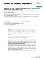
Table 1
Overview of seven Y2H and five AP-MS experiments
Reference VB CB TB VP VBP VBP/BP TI TI/VB REC UNR
Ito et al. [1] 1,522 6,604 2,493 773 0.51 4,524 3.0 75 803
Cagney et al. [2] 19 31 40 11 0.58 54 2.9 3 4
Tong et al. [3] 20 22 59 5 0.25 115 5.8 1 1
Hazbun et al. [4] 66 100 1,940 28 0.42 2,524 38 4 13
Zhao et al. [5] 1 1 90 0 0.00 90 90 0 0
Uetz et al. Experiment 1 [6] 508 6,604 630 142 0.28 952 1.9 10 47
Uetz et al. Experiment 2 [6] 139 192 400 36 0.26 524 3.8 18 7
Gavin et al. [7] 455 600 725 1,179 271 0.60 3,419 7.5 192 314
Ho et al. [8] 493 589 1,739 1,316 231 0.47 3,687 7.5 69 297
Krogan et al. [9] 153 165 165 483 151 0.99 1,132 7.4 89 157
Gavin et al. [10] 1,752 1,993 6,466 1,790 991 0.57 19,105 10.9 1,077 4,297
Krogan et al. [11] 2,264 2,357 4,562 5,323 2,226 0.98 63,360 28.0 1,969 34,363
VB, the number of viable baits; CB, the number of cloned (hybridized) baits, if available; TB, the total number of baits that the experimenters were
initially aiming at; VP, the number of viable prey; VBP, the number of proteins observed as both bait and prey; TI, the total number of interactions
observed; REC, the number of reciprocated interactions between proteins that were observed as both bait and prey; UNR, the number of
unreciprocated interactions between proteins that were observed as both bait and prey. Not all of the experiments were genome-wide - some were
focused on particular aspects of the cellular machinery [2-5,9]. Even in the so-called genome-wide studies [1,6-8,10,11], however, the viable baits cover
only around a third of the yeast genes. This means that the largest part of interaction space by far, containing interactions between proteins not used as
baits, was not sampled in any of these experiments. We can also see that TI/VB, the average number of interactions per viable bait, varies markedly
between experiments. In the more focused studies, this will certainly be a result of different criteria for the selection of baits. In the genome-wide
screens it may indicate the application of different, experiment-specific cutoffs.
For a given number of unreciprocated edges, n
unr
, if there
are no systematic errors then the unreciprocated edges
should be in-edges and out-edges in approximately equal
numbers. If we denote their numbers by n
in
and n
out
,
respectively, then n
in
+ n
out
= n
unr
, and we expect that
n
in
∼ Bin(n
unr
, 0.5) (3)
If n
in
and n
out
are significantly different from each other,
according to the Binomial distribution we would conclude
that the protein behaved differently in the experiment
when used as bait compared with prey, and would use this
as an indication of systematic error affecting at least part of
the data for that protein. An application of this criterion to
the subgraph G
BP
of the data of Krogan et al. [11] is shown
in Figure 3.
Estimation of the properties of the interaction graph
in this setting
There are two basic approaches to estimation: one is to
estimate the true underlying graph, given the data and some
modeling assumptions, then to calculate properties of inter-
est from the estimated graph. The other is to directly
estimate the quantities of interest without making an
attempt to estimate the true underlying graph. For protein-
interaction data we suggest that the latter is often preferable,
as it can deal better with the low coverage of the datasets. As
new methods and models for integrating datasets are
developed it will be important to reassess the situation.
We distinguish between two different types of quantities to
be estimated. The first type are single numeric values, such
as degree, clustering coefficient or diameter. The second are
more general structures, such as modules or subgraphs. The
tools for estimation are more developed for numeric
quantities than for modules, and there is agreement on the
definitions of the different quantities. For modules, or
cohesive subgroups, there is little agreement on what is
being sought or how to find it.
The integration of data from different
independent experiments
No single experiment has provided complete information on
all interactions in a system of interest and so data from
different experiments need to be integrated. Integration
promises to increase coverage and reduce the effects of
stochastic errors. Table 1 summarizes experiments done on
the yeast protein interactome that are candidates for inte-
gration. The overlap between experiments is examined in
Tables 2 and 3.
An essential step before integration of data is to assess their
quality in terms of specificity, sensitivity and coverage. Such
an assessment should provide reliable estimates of the false-
positive and false-negative error rates. There are three main
computational approaches: comparison to a benchmark or
‘gold standard’ data, within-experiment or internal valida-
tion, and between-experiment validation.
When direct physical interactions are being measured (for
example, by Y2H), crystal structures of the interacting
proteins can be used as the gold standard for the validity of
the interaction. This was one of the approaches used in [17].
Only a handful of crystal structures of interacting proteins
are known, however, and such data are still difficult and
expensive to obtain. Some physical interactions and protein
complexes have also been characterized through detailed
biochemical investigations, and are collected in databases
such as MIPS [36] and GO [37]. Circularity needs to be
avoided, however; for example, the data from [7] and [9] are
now reported as known complexes in some of the public
protein complex databases.
Genome Biology 2007, Volume 8, Issue 10, Article 112 Gentleman and Huber 112.7
Genome Biology 2007, 8:112
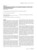
Figure 3
Scatterplot of n
in
and n
out
for the AP-MS data of Krogan et al. [11]. Each
point in the plot corresponds to one protein. n
in
is the number of times
that the protein was found as a prey; n
out
the number of prey it found
when used as a bait. The two lines mark contours of probability p =10
-4
according to the Binomial model in Equation (3). Outlying proteins (dark
blue) show a significantly large difference between n
in
and n
out
, suggesting
that at least one of them is wrong. For example, if n
out
>> n
in
, one
possible reason is that a protein is not expressed when used as prey or of
such low abundance that it is outcompeted, but when tagged and
expressed as a bait, it will identify and pull down its interaction partners
as prey. Further validation experiments are needed to determine in each
case whether the unreciprocated interactions correspond to false-
positive or false-negative observations.
●
●
●
●
●
●
●
●
●
●
●
●
●
●
●
●
●
●
●
●
●
●
●
●
●
●
●
●
●
●
●
●
●
●
●
●
●
●
●
●
●
●
●
●
●
●
●
●
●
●
●
●
●
●
●
●
●
●
●
●
●
●
●
●
●
●
●
●
●
●
●
●
●
●
●
●
●
●
●
●
●
●
●
●
●
●
●
●
●
●
●
●
●
●
●
●
●
●
●
●
●
●
●
●
●
●
●
●
●
●
●
●
●
●
●
●
●
●
●
●
●
●
●
●
●
●
●
●
●
●
●
●
●
●
●
●
●
●
●
●
●
●
●
●
●
●
●
●
●
●
●
●
●
●
●
●
●
●
●
●
●
●
●
●
●
●
●
●
●
●
●
●
●
●
●
●
●
●
●
●
●
●
●
●
●
●
●
●
●
●
●
●
●
●
●
●
●
●
●
●
●
●
●
●
●
●
●
●
●
●
●
●
●
●
●
●
●
●
●
●
●
●
●
●
●
●
●
●
●
●
●
●
●
●
●
●
●
●
●
●
●
●
●
●
●
●
●
●
●
●
●
●
●
●
●
●
●
●
●
●
●
●
●
●
●
●
●
●
●
●
●
●
●
●
●
●
●
●
●
●
●
●
●
●
●
●
●
●
●
●
●
●
●
●
●
●
●
●
●
●
●
●
●
●
●
●
●
●
●
●
●
●
●
●
●
●
●
●
●
●
●
●
●
●
●
●
●
●
●
●
●
●
●
●
●
●
●
●
●
●
●
●
●
●
●
●
●
●
●
●
●
●
●
●
●
●
●
●
●
●
●
●
●
●
●
●
●
●
●
●
●
●
●
●
●
●
●
●
●
●
●
●
●
●
●
●
●
●
●
●
●
●
●
●
●
●
●
●
●
●
●
●
●
●
●
●
●
●
●
●
●
●
●
●
●
●
●
●
●
●
●
●
●
●
●
●
●
●
●
●
●
●
●
●
●
●
●
●
●
●
●
●
●
●
●
●
●
●
●
●
●
●
●
●
●
●
●
●
●
●
●
●
●
●
●
●
●
●
●
●
●
●
●
●
●
●
●
●
●
●
●
●
●
●
●
●
●
●
●
●
●
●
●
●
●
●
●
●
●
●
●
●
●
●
●
●
●
●
●
●
●
●
●
●
●
●
●
●
●
●
●
●
●
●
●
●
●
●
●
●
●
●
●
●
●
●
●
●
●
●
●
●
●
●
●
●
●
●
●
●
●
●
●
●
●
●
●
●
●
●
●
●
●
●
●
●
●
●
●
●
●
●
●
●
●
●
●
●
●
●
●
●
●
●
●
●
●
●
●
●
●
●
●
●
●
●
●
●
●
●
●
●
●
●
●
●
●
●
●
●
●
●
●
●
●
●
●
●
●
●
●
●
●
●
●
●
●
●
●
●
●
●
●
●
●
●
●
●
●
●
●
●
●
●
●
●
●
●
●
●
●
●
●
●
●
●
●
●
●
●
●
●
●
●
●
●
●
●
●
●
●
●
●
●
●
●
●
●
●
●
●
●
●
●
●
●
●
●
●
●
●
●
●
●
●
●
●
●
●
●
●
●
●
●
●
●
●
●
●
●
●
●
●
●
●
●
●
●
●
●
●
●
●
●
●
●
●
●
●
●
●
●
●
●
●
●
●
●
●
●
●
●
●
●
●
●
●
●
●
●
●
●
●
●
●
●
●
●
●
●
●
●
●
●
●
●
●
●
●
●
●
●
●
●
●
●
●
●
●
●
●
●
●
●
●
●
●
●
●
●
●
●
●
●
●
●
●
●
●
●
●
●
●
●
●
●
●
●
●
●
●
●
●
●
●
●
●
●
●
●
●
●
●
●
●
●
●
●
●
●
●
●
●
●
●
●
●
●
●
●
●
●
●
●
●
●
●
●
●
●
●
●
●
●
●
●
●
●
●
●
●
●
●
●
●
●
●
●
●
●
●
●
●
●
●
●
●
●
●
●
●
●
●
●
●
●
●
●
●
●
●
●
●
●
●
●
●
●
●
●
●
●
●
●
●
●
●
●
●
●
●
●
●
●
●
●
●
●
●
●
●
●
●
●
●
●
●
●
●
●
●
●
●
●
●
●
●
●
●
●
●
●
●
●
●
●
●
●
●
●
●
●
●
●
●
●
●
●
●
●
●
●
●
●
●
●
●
●
●
●
●
●
●
●
●
●
●
●
●
●
●
●
●
●
●
●
●
●
●
●
●
●
●
●
●
●
●
●
●
●
●
●
●
●
●
●
●
●
●
●
●
●
●
●
●
●
●
●
●
●
●
●
●
●
●
●
●
●
●
●
●
●
●
●
●
●
●
●
●
●
●
●
●
●
●
●
●
●
●
●
●
●
●
●
●
●
●
●
●
●
●
●
●
●
●
●
●
●
●
●
●
●
●
●
●
●
●
●
●
●
●
●
●
●
●
●
●
●
●
●
●
●
●
●
●
●
●
●
●
●
●
●
●
●
●
●
●
●
●
●
●
●
●
●
●
●
●
●
●
●
●
●
●
●
●
●
●
●
●
●
●
●
●
●
●
●
●
●
●
●
●
●
●
●
●
●
●
●
●
●
●
●
●
●
●
●
●
●
●
●
●
●
●
●
●
●
●
●
●
●
●
●
●
●
●
●
●
●
●
●
●
●
●
●
●
●
●
●
●
●
●
●
●
●
●
●
●
●
●
●
●
●
●
●
●
●
●
●
●
●
●
●
●
●
●
●
●
●
●
●
●
●
●
●
●
●
●
●
●
●
●
●
●
●
●
●
●
●
●
●
●
●
●
●
●
●
●
●
●
●
●
●
●
●
●
●
●
●
●
●
●
●
●
●
●
●
●
●
●
●
●
●
●
●
●
●
●
●
●
●
●
●
●
●
●
●
●
●
●
●
●
●
●
●
●
●
●
●
●
●
●
●
●
●
●
●
●
●
●
●
●
●
●
●
●
●
●
●
●
●
●
●
●
●
●
●
●
●
●
●
●
●
●
●
●
●
●
●
●
●
●
●
●
●
●
●
●
●
●
●
●
●
●
●
●
●
●
●
●
●
●
●
●
●
●
●
●
●
●
●
●
●
●
●
●
●
●
●
●
●
●
●
●
●
●
●
●
●
●
●
●
●
●
●
●
●
●
●
●
●
●
●
●
●
●
●
●
●
●
●
●
●
●
●
●
●
●
●
●
●
●
●
●
●
●
●
●
●
●
●
●
●
●
●
●
●
●
●
●
●
●
●
●
●
●
●
●
●
●
●
●
●
●
●
●
●
●
●
●
●
●
●
●
●
●
●
●
●
●
●
●
●
●
●
●
●
●
●
●
●
●
●
●
●
●
●
●
●
●
●
●
●
●
●
●
●
●
●
●
●
●
●
●
●
●
●
●
●
●
●
●
●
●
●
●
●
●
●
●
●
●
●
●
●
●
●
●
●
●
●
●
●
●
●
●
●
●
●
●
●
●
●
●
●
●
●
●
●
●
●
●
●
●
●
●
●
●
●
●
●
●
●
●
●
●
●
●
●
●
●
●
●
●
●
●
●
●
●
●
●
●
●
●
●
●
●
●
●
●
●
●
●
●
●
●
●
●
●
●
●
●
●
●
●
●
●
●
●
●
●
●
●
●
●
●
●
●
●
●
●
●
●
●
●
●
●
●
●
●
●
●
●
●
●
●
●
●
●
●
●
●
●
●
●
●
●
●
●
●
●
●
●
●
●
●
●
●
●
●
●
●
●
●
●
●
●
●
●
●
●
●
●
●
●
●
●
●
●
●
●
●
●
●
●
●
●
●
●
●
●
●
●
●
●
●
●
●
●
●
●
●
●
●
●
●
●
●
●
●
●
●
●
●
●
●
●
●
●
●
●
●
●
●
●
●
●
●
●
●
●
●
●
●
●
●
●
●
●
●
●
●
●
●
●
●
●
●
●
●
●
●
●
●
●
●
●
●
●●
●
●
●
●
●
●
●
●
●
●
●
●
●
●
●
●
●
●
●
●
●
●
●
●
●
●
●
●
●
●
●
●
●
●
●
●
●
●
●
●
●
●
●
●
●
●
●
●
●
●
●
●
●
●
●
●
●
●
●
●
●
●
●
●
●
●
●
●
●
●
●
●
●
●
●
●
●
●
●
●
●
●
●
●
●
●
●
●
●
●
●
●
●
●
●
●
●
●
●
●
●
●
●
●
●
●
●
●
●
●
●
●
●
●
●
●
●
●
●
●
●
●
●
●
●
●
●
●
●
●
●
●
●
●
●
●
●
●
●
●
●
●
●
●
●
●
●
●
●
●
●
●
●
●
●
●
●
●
●
●
●
●
●
●
●
●
●
●
●
●
●
●
●
●
●
●
●
●
●
●
●
●
●
●
●
●
●
●
●
●
●
●
●
●
●
●
●
●
●
●
●
●
●
●
●
●
●
●
●
●
●
●
●
●
●
●
●
●
●
●
●
●
●
●
●
●
●
●
●
●
●
●
●
●
●
●
●
●
●
●
●
●
●
●
●
●
●
●
●
●
●
●
●
●
●
●
●
●
●
●
●
●
●
●
●
●
●
●
●
●
●
●
●
●
●
●
●
●
●
●
●
●
●
●
●
●
●
●
●
●
●
●
●
●
●
●
●
●
●
●
●
●
●
●
●
●
●
●
●
●
●
●
●
●
●
●
●
●
●
●
●
●
●
●
●
●
●
●
●
●
●
●
●
●
●
●
●
●
●
●
●
●
●
●
●
●
●
●
●
●
●
●
●
●
●
●
●
●
●
●
●
●
●
●
●
●
●
●
●
●
●
●
●
●
●
●
●
●
●
●
●
●
●
●
●
●
●
●
●
●
●
●
●
●
●
●
●
●
●
●
●
0102030
0
10
20
30
n
out
n
in
Within-experiment validation relies on internal properties of
the data, such as redundancies or symmetries that are not
used in the experiment, and that can therefore be used to
validate the experimental results. One such property is
reciprocity, as discussed above. Deviations from expectation
can be used to estimate stochastic error rates, and they can
also be used to identify individual proteins whose data
appear to be subject to systematic artifacts (see Figure 3).
Reported replicate measurements can also be used to help
validate experimental data and to estimate error rates. The
basic idea is that if edges are tested multiple times under the
same conditions, those that are found frequently can be
termed true positives and can be used to estimate the false-
negative rate from those cases when they were missed.
Similarly, those that are seldom found can be deemed true
negatives, and from the positive data points the false-
positive rate can be estimated. This approach is complicated
by possible dependencies between the replicate measure-
ments and by systematic errors that, if present, will affect all
replicates. These complications may render the statistical
model intractable. Further caution is warranted. Was the
choice of replicates measures made a priori or because of
anomalous results obtained during the experiment? Do they
provide equal coverage of all important conditions and of all
types of proteins that were studied?
Between-experiment comparisons rely on the experimental
conditions being sufficiently similar to ensure that the
measurements are made on the same underlying set of true
interactions. However, as we see in Tables 2 and 3, in many
cases there is relatively little overlap in bait selection and in
observed prey. For two recent experiments with at least
some overlap, a comparison was presented by [20]. These
authors found a moderate overlap between the primary
data, for example the proteins identified by each successful
Genome Biology 2007, Volume 8, Issue 10, Article 112 Gentleman and Huber 112.8
Genome Biology 2007, 8:112
Table 2
Pairwise comparison of Y2H datasets
Uetz et al. [6] Uetz et al. [6]
References Ito et al. [1] Cagney et al. [2] Tong et al. [3] Hazbun et al. [4] Zhao et al. [5] Experiment 1 Experiment 2
[1] - 9 7 24 1 224 47
[2] 28-00073
[3] 340- 0047
[4] 856 14 25 - 0 15 12
[5] 43 1 2 38 - 0 0
[6] Experiment 1 388 14 22 272 15 - 36
[6] Experiment 2 200 9 26 204 13 108 -
The values above the diagonal give the number of viable baits in common between each pair of experiments, and the values below the diagonal give the
number of viable prey in common. We see that the overlap between experiments in the sampled fractions of protein-interaction space is in all cases very
small, given that thousands of interactions were assayed.
Table 3
Pairwise comparison of AP-MS datasets
References Gavin et al. [7] Ho et al. [8] Krogan et al. [9] Gavin et al. [10] Krogan et al. [11]
[7] - 82 51 442 334
[8] 516 - 25 222 286
[9] 299 246 - 121 151
[10] 1,143 717 371 - 1,128
[11] 1,149 1,277 478 1,732 -
As in Table 2 the values above the diagonal give the number of viable baits in common between each pair of experiments, and the values below the
diagonal give the number of viable prey in common. Again, the overlap is very small. Consider the two largest experiments carried out so far: with a set
of 2,264 viable baits and 5,323 viable prey, Krogan et al. [11] tested for the presence of at least 12 million complex membership interactions. Gavin et al.
[10], with 1,752 viable baits and 1,790 viable prey, tested for at least 3.1 million interactions. However, even for these two datasets, the largest so far, the
known overlap is only 1,128 × 1,732 ≈ 2.0 million. One of the possible explanations for these low estimates of coverage and overlap is that our
definitions of viable baits and viable prey are restrictive and that indeed a much larger space of interactions might have been tested. For example,
Gilchrist et al. [29] estimated a value about twice ours for the number of tested prey in [7]. This situation will hopefully be alleviated as researchers
report more complete data on which interactions were actually tested.
bait, but a low everlap of the computed protein complexes
by each group.
When integrating data from different experiments our
recommendation is that validation to a gold standard and
within-experiment validation should first be done on each
experiment separately. Once the data are sufficiently well
understood and as many of the systematic errors as possible
have been resolved, integration becomes worthwhile. If
there is little agreement on the existence of interactions for
edges tested in different experiments, then one must
question the prudence of their integration: it may be that the
biological conditions were too different to allow their
integration into a single meaningful dataset.
There is room for much more research here. Evidence in
favor of, or against, experimentally detected interactions can
often be obtained from other sources, such as data from
other organisms, dependencies of different types of inter-
actions on each other (for example, coexpression, co-
localization and physical interaction), evolutionary conser-
vation [38], protein structure [39] and amino-acid binding
motifs [40]. The challenge is to ensure that the evidence is
applicable and that it does bear relationship to the assay and
system under study.
Our purpose in writing this article was to address the
observation that the many different protein-interaction
datasets available appear to have very little in common, and
also to address reports that the data were inherently noisy
and of low quality (for example [17,41]). Our investigations
suggest that the data themselves, while problematic in some
cases, are not the real issue, but rather there is often mis-
interpretation of the data, methods to address noisiness are
often inadequate, and the lack of substantive comparisons
between methods applied to the data has led to a situation
where the data, rather than the methods, are treated with
suspicion. As seen from Tables 2 and 3, low coverage, and
not the false-positive rate, is responsible for the small
amount of overlap between datasets.
The separation of errors into stochastic and systematic
components is potentially of great benefit. Comparison of
experimental data should be based on stochastic error rates.
The identification of systematic errors can help to identify
problems with the experimental techniques and hopefully
suggest solutions to those problems. We believe that when
more standard, and sound, statistical practices are adopted
for preprocessing the data, it will be possible to estimate
quantities of interest and to make substantial comparisons.
An essential prerequisite is the adoption of standard
methods for estimation of stochastic error rates and where
possible the identification of systematic errors. Standardized
preprocessing is also required in order to be able to
synthesize different experimental datasets. Combining data
requires attention to the differing error rates, and the
discounting of information from more variable experiments.
Given the numbers in Tables 2 and 3, there is much to be
gained by combining the different experimental datasets. We
believe that the data, while noisy, are in fact very useful, and
with appropriate preprocessing and statistical modeling they
can provide deep insight into the functioning of cellular
machineries.
Acknowledgements
We thank Richard Bourgon, Michael Boutros, Tony Chiang, Denise
Scholtens and Lars Steinmetz for helpful comments on the manuscript.
This work was supported by HFSP research grant RGP0022/2005 to W.H.
and R.G.
References
1. Ito T, Chiba T, Ozawa R, Yoshida M, Hattori M, Sakaki Y: A com-
prehensive two-hybrid analysis to explore the yeast protein
interactome. Proc Natl Acad Sci USA 2001, 98:4569-4574.
2. Cagney G, Uetz P, Fields S: Two-hybrid analysis of the Saccha-
romyces cerevisiae 26S proteasome. Physiol Genomics 2001, 7:
27–34.
3. Tong AH, Drees B, Nardelli G, Bader GD, Brannetti B, Castagnoli L,
Evangelista M, Ferracuti S, Nelson B, Paoluzi S, et al.: A combined
experimental and computational strategy to define protein
interaction networks for peptide recognition modules.
Science 2002, 295:321-324.
4. Hazbun TR, Malmström L, Anderson S, Graczyk BJ, Fox B, Riffle M,
Sundin BA, Aranda JD, McDonald WH, Chiu CH, et al.: Assigning
function to yeast proteins by integration of technologies. Mol
Cell 2003, 12:1353-1365.
5. Zhao R, Davey M, Hsu YC, Kaplanek P, Tong A, Parsons AB, Krogan
N, Cagney G, Mai D, Greenblatt J, et al.: Navigating the chaper-
one network: an integrative map of physical and genetic
interactions mediated by the hsp90 chaperone. Cell 2005,
120:715-727.
6. Uetz P, Giot L, Cagney G, Mansfield TA, Judson RS, Knight JR, Lock-
shon D, Narayan V, Srinivasan M, Pochart P, et al.: A comprehen-
sive analysis of protein-protein interactions in
Saccharomyces cerevisiae. Nature 2000, 403:623-627.
7. Gavin AC, Bösche M, Krause R, Grandi P, Marzioch M, Bauer A,
Schultz J, Rick JM, Michon AM, Cruciat CM, et al.: Functional orga-
nization of the yeast proteome by systematic analysis of
protein complexes. Nature 2002, 415:141-147.
8. Ho Y, Gruhler A, Heilbut A, Bader GD, Moore L, Adams SL, Millar
A, Taylor P, Bennett K, Boutilier K, et al.: Systematic identifica-
tion of protein complexes in Saccharomyces cerevisiae by
mass spectrometry. Nature 2002, 415:180-183.
9. Krogan NJ, Peng WT, Cagney G, Robinson MD, Haw R, Zhong G,
Guo X, Zhang X, Canadien V, Richards DP, et al.: High-definition
macromolecular composition of yeast RNA-processing
complexes. Mol Cell 2004, 13:225-239.
10. Gavin AC, Aloy P, Grandi P, Krause R, Boesche M, Marzioch M, Rau
C, Jensen LJ, Bastuck S, Dümpelfeld B, et al.: Proteome survey
reveals modularity of the yeast cell machinery. Nature 2006,
440:631-636.
11. Krogan NJ, Cagney G, Yu H, Zhong G, Guo X, Ignatchenko A, Li J,
Pu S, Datta N, Tikuisis AP, et al.: Global landscape of protein
complexes in the yeast Saccharomyces cerevisiae. Nature 2006,
440:637-643.
12. Giot L, Bader JS, Brouwer C, Chaudhuri A, Kuang B, Li Y, Hao YL,
Ooi CE, Godwin B, Vitols E, et al.: A protein interaction map of
Drosophila melanogaster. Science 2003, 302:1727-1736.
13. Li S, Armstrong CM, Bertin N, Ge H, Milstein S, Boxem M, Vidalain
PO, Han JD, Chesneau A, Hao T, et al.: A map of the interac-
tome network of the metazoan C. elegans. Science 2004, 303:
540-543.
14. Rual JF, Venkatesan K, Hao T, Hirozane-Kishikawa T, Dricot A, Li N,
Berriz GF, Gibbons FD, Dreze M, Ayivi-Guedehoussou N, et al.:
Towards a proteome-scale map of the human protein-
protein interaction network. Nature 2005, 437:1173-1178.
Genome Biology 2007, Volume 8, Issue 10, Article 112 Gentleman and Huber 112.9
Genome Biology 2007, 8:112
15. Stelzl U, Worm U, Lalowski M, Haenig C, Brembeck FH, Goehler H,
Stroedicke M, Zenkner M, Schoenherr A, Koeppen S, et al.: A
human protein-protein interaction network: a resource for
annotating the proteome. Cell 2005, 122:957-968.
16. Mrowka R, Patzak A, Herzel H: Is there a bias in proteome
research? Genome Res 2001, 11:1971-1973.
17. Edwards AM, Kus B, Jansen R, Greenbaum D, Greenblatt J, Gerstein
M: Bridging structural biology and genomics: assessing
protein interaction data with known complexes. Trends Genet
2002, 18:529-536.
18. von Mering C, Krause R, Snel B, Cornell M, Oliver SG, Fields S, Bork
P: Comparative assessment of large-scale data sets of
protein-protein interactions. Nature 2002, 417:399-403.
19. Goll J, Uetz P: The elusive yeast interactome. Genome Biol 2006,
7:223.
20. Gagneur J, David L, Steinmetz LM: Capturing cellular machines
by systematic screens of protein complexes. Trends Microbiol
2006, 14:336–339.
21. Hart GT, Ramani AK, Marcotte EM: How complete are current
yeast and human protein-interaction networks? Genome Biol
2006, 7:120.
22. Jones S, Thornton JM: Principles of protein-protein interac-
tions. Proc Natl Acad Sci USA 1996, 93:13-20.
23. Fields S, Song O: A novel genetic system to detect protein-
protein interactions. Nature 1989, 340:245-246.
24. Kumar A, Snyder M: Protein complexes take the bait. Nature
2002, 415:123-124.
25. Uetz P: Two-hybrid arrays. Curr Opin Chem Biol 2002, 6:57-62.
26. Han JD, Dupuy D, Bertin N, Cusick ME, Vidal M: Effect of sam-
pling on topology predictions of protein-protein interaction
networks. Nat Biotechnol 2005, 23:839-844.
27. Stumpf MPH, Wiuf C: Sampling properties of random graphs:
the degree distribution. Phys Rev E Stat Nonlin Soft Matter Phys
2005, 72:036118.
28. Chiang T, Scholtens D, Sarkar D, Gentleman R, Huber W. Cover-
age and error models or protein-protein interaction data by
directed graph analysis. Genome Biol 2007, 8:R186.
29. Gilchrist MA, Salter LA, Wagner A:
A statistical framework for
combining and interpreting proteomic datasets. Bioinformatics
2004, 20:689–700.
30. Box GEP, Draper NR: Empirical Model-Building and Response Surfaces.
New York: Wiley; 1987.
31. Aloy P, Russell RB: Structural systems biology: modelling
protein interactions. Nat Rev Mol Cell Biol 2006, 7:188-197.
32. Kelsey JL, Whittemore AS, Evans AS, Thompson WD: Methods in
observational epidemiology. In Monographs in Epidemiology and
Biostatistics, New York: Oxford University Press; 1996.
33. Scholtens D, Gentleman R: Making sense of high-throughput
protein-protein interaction data. Stat Appl Genet Mol Biol 2004,
3:39.
34. Scholtens D, Vidal M, Gentleman R: Local modeling of global
interactome networks. Bioinformatics 2005, 21:3548-3557.
35. Collins SR, Kemmeren P, Zhao XC, Greenblatt JF, Spencer F, Hol-
stege FC, Weissman JS, Krogan NJ: Toward a comprehensive
atlas of the physical interactome of Saccharomyces
cerevisiae. Mol Cell Proteomics 2007, 6:439-450.
36. Mewes HW, Frishman D, Mayer KF, Münsterkötter M, Noubibou O,
Pagel P, Rattei T, Oesterheld M, Ruepp A, Stümpflen V: MIPS:
analysis and annotation of proteins from whole genomes in
2005. Nucleic Acids Res 2006, 34(Database issue):D169-D172.
37. Harris MA, Clark J, Ireland A, Lomax J, Ashburner M, Foulger R,
Eilbeck K, Lewis S, Marshall B, Mungall C, et al.: The Gene Ontol-
ogy (GO) database and informatics resource. Nucleic Acids Res
2004, 32(Database issue):D258-D261.
38. Poyatos JF, Hurst LD: How biologically relevant are interac-
tion-based modules in protein networks? Genome Biol 2004, 5:
R93.
39. Aloy P, Böttcher B, Ceulemans H, Leutwein C, Mellwig C, Fischer S,
Gavin AC, Bork P, Superti-Furga G, Serrano L, Russell RB: Struc-
ture-based assembly of protein complexes in yeast. Science
2004, 303:2026-2029.
40. Neduva V, Russell RB: Peptides mediating interaction net-
works: new leads at last. Curr Opin Biotechnol 2006, 17:465-471.
41. Chen J, Hsu W, Lee ML, Ng SK: Increasing confidence of
protein interactomes using network topological metrics.
Bioinformatics 2006, 22:1998-2004.
42. Storey J: A direct approach to false discovery rates. J R Stat Soc
Ser B 2002, 64:479-498.
Genome Biology 2007, Volume 8, Issue 10, Article 112 Gentleman and Huber 112.10
Genome Biology 2007, 8:112