Báo cáo y học: "Genomic chart guiding embryonic stem cell cardiopoiesis" docx
Bạn đang xem bản rút gọn của tài liệu. Xem và tải ngay bản đầy đủ của tài liệu tại đây (2.18 MB, 16 trang )
Open Access
Volume
et al.
Faustino
2008 9, Issue 1, Article R6
Research
Genomic chart guiding embryonic stem cell cardiopoiesis
Randolph S Faustino*, Atta Behfar*, Carmen Perez-Terzic*† and
Andre Terzic*
Addresses: *Marriott Heart Disease Research Program, Division of Cardiovascular Diseases, Departments of Medicine, Molecular
Pharmacology and Experimental Therapeutics, and Medical Genetics, Mayo Clinic, First Street SW, Rochester, Minnesota 55905, USA.
†Department of Physical Medicine and Rehabilitation, Mayo Clinic, First Street SW, Rochester, Minnesota 55905, USA.
Correspondence: Andre Terzic. Email:
Published: 9 January 2008
Received: 27 September 2007
Revised: 20 November 2007
Accepted: 9 January 2008
Genome Biology 2008, 9:R6 (doi:10.1186/gb-2008-9-1-r6)
The electronic version of this article is the complete one and can be
found online at />
© 2008 Faustino et al.; licensee BioMed Central Ltd.
This is an open access article distributed under the terms of the Creative Commons Attribution License ( which
permits unrestricted use, distribution, and reproduction in any medium, provided the original work is properly cited.
committing to cardiac cell fate.
Gene expression
ES cell cardiopoiesis analysis of embryonic stem cells undergoing guided cardiogenic differentiation reveals the molecular fingerprint for
Abstract
Background: Embryonic stem cells possess a pluripotent transcriptional background with the
developmental capacity for distinct cell fates. Simultaneous expression of genetic elements for
multiple outcomes obscures cascades relevant to specific cell phenotypes. To map molecular
patterns critical to cardiogenesis, we interrogated gene expression in stem cells undergoing guided
differentiation, and defined a genomic paradigm responsible for confinement of pluripotency.
Results: Functional annotation analysis of the transcriptome of differentiating embryonic stem cells
exposed downregulated components of DNA replication, recombination and repair machinery, cell
cycling, cancer mechanisms, and RNA post-translational modifications. Concomitantly,
cardiovascular development, cell-to-cell signaling, cell development and cell movement were
upregulated. These simultaneous gene ontology rearrangements engaged a repertoire switch that
specified lineage development. Bioinformatic integration of genomic and gene ontology data further
unmasked canonical signaling cascades prioritized within discrete phases of cardiopoiesis.
Examination of gene relationships revealed a non-stochastic network anchored by integrin, WNT/
β-catenin, transforming growth factor β and vascular endothelial growth factor pathways, validated
by manipulation of selected cascades that promoted or restrained cardiogenic yield. Moreover,
candidate genes within anchor pathways acted as nodes that organized correlated expression
profiles into functional clusters, which collectively orchestrated and secured an overall cardiogenic
theme.
Conclusion: The present systems biology approach reveals a dynamically integrated and tractable
gene network fundamental to embryonic stem cell specification, and represents an initial step
towards resolution of a genomic cardiopoietic atlas.
Genome Biology 2008, 9:R6
/>
Genome Biology 2008,
Background
stem cells, the recognized cardioinductive potential of the
cytokine tumor necrosis factor (TNF)α-induced, endodermally derived paracrine factors was reduced to a collective
cocktail, that is, bone morphogenetic protein (BMP), transforming growth factor (TGF)β, interleukin (IL)-13 (IL13),
IL3, insulin-like growth factor (IGF1), vascular endothelial
growth factor (VEGF), epidermal growth factor (EGF),
fibroblast growth factor (FGF) and IL6 [18]. Cardiogenic
cocktail-primed embryonic stem cells responded by structural metamorphosis and progressive up-regulation in
canonical cardiac markers, with distinct phenotypes resolved
by sequential field emission scanning electron microscopy
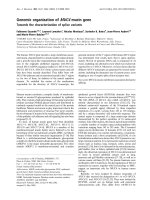
(Figure 1a, left) and immunofluorescence (Figure 1a, right).
Embryonic stem cells, initially maintained in the undifferentiated proliferative state in the presence of the mitogenic
leukemia inhibitory factor [23], assumed a spheroid shape
with high nuclear-to-cytoplasmic volumes, and lacked the
cardiac sarcomeric protein α-actinin with marginally detectable cytosolic levels of the cardiac transcription factor myocyte enhancer factor 2C (MEF2C; Figure 1a). From this
original state, mitogen removal initiated differentiation,
characterized by a progressive decrease in the nuclear-tocytoplasmic volume ratio and an increased expression of
MEF2C accompanied by cytosolic-to-nuclear translocation
(Figure 1a). Developmentally regulated nuclear import of cardiac transcription factors is indicative of definitive commitment to cardiac differentiation [19]. Accordingly, these
intermediate cell types have been termed cardiopoietic stem
cells [18]. Sustained nuclear import of MEF2C and formation
of sarcomeres expressing cardiac α-actinin after 12 days identified mature, functional cardiomyocyte morphology. The
degree of purity for derived progenitors and cardiomyocytes
reached 85 ± 5% and 90 ± 5%, respectively (see Materials and
methods). Interrogation of the developing transcriptome
revealed 8,656 quality-filtered genes underlying guided cardiopoietic lineage specification, resolved into distinct groups
of increasing, decreasing or unchanging profiles (Figure 1b).
Concomitant with dynamic trends of lineage specification,
each stage of cardiac differentiation demonstrated discrete
molecular fingerprints revealed by unsupervised agglomerative clustering (Figure 1c). Gene sets were highly similar
within, but significantly distinct between, stages of cardiac
differentiation. Hierarchical categorization using Euclidean
distance was used to measure differences between expression
profiles to determine dissimilarity among replicates (Figure
1c). Unbiased confidence levels for these reproducible transcriptional profiles were assessed by bootstrapping, used to
determine the accuracy of statistical estimates [24]. All distance measurements possessed a 100% confidence level and
demonstrated increasing similarity towards the smaller, terminal branches of the condition tree. Small distances (≤0.33)
reflected close association among replicate gene profiles,
which were virtually inseparable at each stage of differentiation (Figure 1c). Larger Euclidean distances of 0.491 and
0.610 indicated greater dissimilarity between embryonic
stem cells in the presence and absence of mitogen, as well as
Expression patterns characterize the production and proliferation of stem cells [1,2]. In particular, unique genetic profiles
are concealed in the rich pluripotent transcriptional background of embryonic stem cells and support their inherent
potential for multiple and diverse cell fates [3-6]. Genomewide profiling and system analyses, used to distinguish markers identifying stemness [7,8], and high-throughput
approaches applied to categorize large scale transcriptional
dynamics during stem cell development and specification
provide an initial insight into the global genomics evolving in
response to inductive stimuli [2,9,10]. Beyond identification
of stemness markers, however, integration of genes promoting tissue-restricted differentiation becomes a priority
[11,12]. Mapping genetic relationships underlying metamorphosis of a pluripotent into a monopotent stem cell would
allow for directional control over developmental fate, enhancing targeted derivation of phenotype-specified cell types.
Indeed, the broad potential for regenerative therapy based on
embryonic stem cell technology is hampered by the threat of
neoplastic transformation associated with unsupervised
pluripotency, mandating unipotential commitment prior to
application [13,14]. A case in point is the need to secure controlled cardiogenesis of embryonic stem cells for safe heart
repair [15-17]. Guided pro-cardiac programming has been
established as a strategy to suppress the risk for uncontrolled
tumorigenic growth outside the natural milieu of a developing embryo [18]. Cardiopoietic induction allowed activation
of the cardiac program on a monolayer of stem cells, eliminating the confounding contribution of trigerminal differentiation [18,19]. Privileged access to the cardiac transcriptional
program, otherwise camouflaged within the stem cell
genomic background [20,21], provides an opportunity to
selectively examine gene interrelationships vital for pluripotent streamlining into cardiopoiesis.
Here, a transcriptome profiling and tandem network analysis
of embryonic stem cells during guided cardiogenic differentiation identified a molecular fingerprint, synthesized from an
ontological functional switch, that commits the cells to a cardiac fate. Pathway prioritization of signaling axes during cardiopoiesis resolved a non-stochastic organization of genes
underlying cardiac specification. Manipulation of high-priority nodes within this deconvoluted pro-cardiac gene network
commanded cardiomyocyte derivation from primordial stem
cells, demonstrating a responsive program amenable to
molecular calibration during directed cardiogenesis.
Results
Distinct transcriptomes define transitions in stem cell
cardiogenic restriction
Pluripotency is a labile characteristic of embryonic stem cells
amenable to specification by distinct inductive stimuli [9,22].
Here, to initiate cardiac commitment in undifferentiated
Genome Biology 2008, 9:R6
Volume 9, Issue 1, Article R6
Faustino et al. R6.2
/>
(a) ES-LIF(+)
Genome Biology 2008,
ES-LIF(+)
Normalized intensity (log scale)
MEF2C
DAPI
ES-LIF(-)
0
1
0.1
(c)
CP
>5
10
ES-LIF(+)
CP
Faustino et al. R6.3
(b) 100
<-5
ES-LIF(-)
Volume 9, Issue 1, Article R6
ES-LIF(+)
<-5
ES-LIF(-)
0
CP
CM
>5
0.292
0.330
0.491
ES-LIF(-)
0.295
0.261
CM
CM
CP
CM
0.885
0.242
0.265
0.285
0.303
0.610
actinin
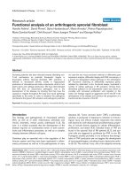
Figure 1
Phenotypic changes and transcriptome dynamism during cardiac stem cell differentiation
Phenotypic changes and transcriptome dynamism during cardiac stem cell differentiation. (a) Electron microscopy visualized morphological changes
occurring during guided stem cell cardiogenesis (left column) with associated expression and distribution of the selected cardiac transcription factor
MEF2C and the cardiac contractile protein α-actinin (right column). Cell stage is given in the top left corner of each panel with associated scale bars at the
bottom right. First column: ES-LIF(+), 2.5 μm; ES-LIF(-), 5 μm; cardiopoietic cell (CP), 25 μm; cardiomyocyte (CM), 5 μm. All scale bars in the second
column indicate 10 μm. Nuclei were counterstained with DAPI. (b) Transcriptional profiling of samples from each stage of stem cell-derived
cardiomyocyte formation. Changes in gene expression were plotted on a semi-log scale graph using normalized intensity values as a function of the stage
of differentiation. The color scale indicates increased expression (red), no change (yellow) and decreased expression (blue). Associated numbers indicate
fold change, where red and blue indicate a respective minimum five-fold up- or downregulation in expression value. (c) Hierarchical clustering of changing
genes during differentiation. The condition tree on right illustrates similarity of replicates within each stage. Numbers above branches are the calculated
Euclidean distances between the two samples at the left termini. Smaller numbers indicate less dissimilarity between samples while higher numbers indicate
an increase in dissimilarity. The shaded box identifies emergence of cardiac specficity (orange, CP) with transition to stem cell derived cardiomyocyte
(cyan, CM). The color scale indicates relative changes in gene expression as described previously.
between cardiopoietic precursors and derived cardiomyocytes, allowing for separation of respective genomic fingerprints (Figure 1c). The largest measurement (0.885) reflected
macroscopic differences between undifferentiated stem cells
and lineage-specified populations (Figure 1c). Thus, discrete
clustering of transcriptome dynamics during guided cardiogenesis genetically delimits precursor phenotype underlying
cardiac confinement of stem cells.
Tailored gene ontology directing cardiopoiesis
Restrictive quality filtering of the transcriptome to genes with
dynamics exceeding a >1.5-fold change in cardiac precursors
relative to undifferentiated embryonic stem cells yielded
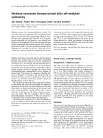
1,069 (12%) and 4,632 (54%) genes up- and downregulated,
respectively, with 2,955 (34%) transcripts changing by <1.5fold (Figure 2a). Analyses of subthreshold genes below the
1.5-fold limit revealed no predominant functional overrepre-
Genome Biology 2008, 9:R6
/>
(a)
Genome Biology 2008,
(d)
Downregulated (54%)
Volume 9, Issue 1, Article R6
Faustino et al. R6.4
RNA-PTM Cancer Cell Cycle DNA-RRR
-0.5
-1.5
Log P-value
-2.5
Upregulated (12%)
Changing less than 1.5 fold (34%)
(b)
Nucleotide
binding
(17%)
Enzyme
regulator
activity
(8%)
-6.5
Translation
regulator
activity
(2%)
mRNA
binding
(2%)
LIF(-)
CP
-9.5
(e)
LIF(-)
6
Helicase
activity
(3%)
7
-Log P-value
Other
(16%)
Enzyme regulator
activity
(8%)
Structural
constituent of
muscle
(1%)
-5.5
-8.5
ATP binding
(15%)
RNA
binding
(8%)
(c)
-4.5
-7.5
Nucleic acid binding (31%)
Structural component
of ribosome
(2%)
Ligase
activity
(4%)
-3.5
CP
5
4
3
2
1
Other (30%)
0
Protein binding
(25%)
Cardiac Cell-Cell Embryo Cellular
Dev
Signaling Dev Movement
(f)
Metal ion
binding Actin binding
(4%)
(13%)
Cytoskeletal
protein
binding
Calcium ion
(6%)
binding
(9%)
Gene
Pou5f1
Mybl2
Mycn
Myocd
Lbh
ES-LIF(+) ES-LIF(-)
1.04
0.65
1.02
0.73
1.00
0.84
1.01
0.55
1.02
0.80
CP
0.18*
0.24*
0.42*
35.0*
46.8*
CM
0.03*
0.09*
0.15*
261*
362*
Figure 2
Enrichment analysis of functional groups within the stem cell-derived cardiopoietic transcriptome
Enrichment analysis of functional groups within the stem cell-derived cardiopoietic transcriptome. (a) Approximately half of all expression profiles in
cardiopoietic cells are downregulated while a third do not change more than 1.5-fold compared to unstimulated embryonic stem cells. Upregulated genes
account for >10% of all genes. (b, c) Ontological analysis of downregulated and upregulated biological processes in cardiopoietic cells. (d, e) Identification
of overrepresented canonical functions in cardiopoietic cells (CP) using Ingenuity Pathways Analysis (IPA) in downregulated and upregulated gene lists.
Significance as determined by IPA was plotted as log P value for downregulated genes and -log P value for those upregulated to emphasize direction of
change. The dashed line indicates the threshold where the P value = 0.05. Embryonic stem cells in the presence of mitogenic LIF were taken as baseline and
significant functional enrichment in cardiopoietic cells are shown in comparison with stem cells cultured without LIF. (f) Gene validation using quantitative
PCR. Candidate genes representing pluripotent (Pou5f1), oncogenic (Mybl2, Mycn) and cardiac (Myocd, Lbh) phenotypes were assayed by Taqman.
Transcriptional profile changes were expressed as fold change relative to ES-LIF(+). CM, cardiomyocyte.
sentation within ontologically annotated families (data not
shown). In contrast, genes identified as up- or downregulated
beyond 1.5-fold unmasked overrepresented molecular functions in each gene set (Figure 2b,c). Genetic metabolism,
identified by nucleotide binding, helicase and ligase activity,
ribosomal structure, and translation regulator activity, was
downregulated in cardiac precursors (Figure 2b). Alternative
corroboration reported functional reductions in RNA post-
translational modifications, oncogenic processes (for example, Aurkb and Hmgb1), cell cycling, and DNA replication,
recombination and repair (Figure 2d). Decreased nucleotide
metabolic machinery was paralleled by emergence of myogenic structural constituents, actin and calcium binding activities, and protein modification mechanisms regulating
enzyme function (Figure 2c). Independent validation demonstrated that upregulated transcripts functionally overrepre-
Genome Biology 2008, 9:R6
/>
Genome Biology 2008,
sented cardiovascular development, cell-to-cell signaling,
embryonic development and cellular movement (Figure 2e).
Collectively, this ontological switch indicates congruent
genetic losses and gains that define a departure from oncogenicity associated with pluripotency towards acquisition of
tissue-specificity and cardiopoietic elaboration. Gene chip
and functional categorization analyses were verified by quantitative genetic amplification of markers for pluripotency
(Pou5f1/Oct4), oncogenesis (Mybl2, Mycn) and cardiogenesis (Myocd, Lbh). Pou5f1 transcription, prototypical of
pluripotent stem cells [25], was decreased as embryonic stem
cells underwent differentiation (Figure 2f). Transcription of
Mybl2 and Mycn, markers for neoplastic growth and tumor
susceptibility [26,27], paralleled Pou5f1 expression and
decreased as the cardiac program progressed (Figure 2f). In
contrast, developmental expression of myocardial Myocd
[28] and Lbh [29] genes increased during cardiac specification (Figure 2f). Thus, concomitant genetic streamlining with
targeted induction of a focused transcriptome defines essential requirements for cardiopoietic lineage establishment.
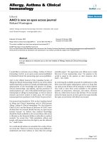
(Figure 3c). Thus, discrete cascades anchor the molecular cardiopoietic network.
Cardiopoiesis-associated signaling cascades
Analysis of genes associated with the ontological 'Cardiac
development' class in the specialized precursor transcriptome
was composed of 65 upregulated genes (Table 1). Of these, 49
integrated into a cardiopoietic network (Figure 3a), while 16
did not possess curated interactions (Table 1). Inspection of
network topology through degree and clustering coefficient
distribution analysis suggested non-arbitrary architecture
with hierarchical tendencies (Figure 3a). Bioinformatic investigation of underlying signaling pathways revealed individual
overrepresented cascades, reported using cardiopoietic and
cardiomyocyte significance estimates as respective co-ordinates in a Cartesian plot (Figure 3b). Cell cycle, death receptor and apoptosis cascades were examples of pathways with P
values below significance threshold for both cardiopoietic
cells and cardiomyocytes (Figure 3b, bottom left), in line with
reported downregulation of genes required for cell proliferation and apoptotic processes in fully differentiated embryonic
stem cell-derived cardiomyocytes [11]. In contrast, VEGF, IL2
and Toll-like receptor signaling were relevant at initiation of
cardiac confinement, accompanied by amyloid processing,
glycosphingolipid metabolism, glycosaminoglycan degradation, and N-glycan and ganglioside biosynthesis (Figure 3b,
lower right). Integrin, WNT/β-catenin, IL6, IGF1 and cardiovascular hypoxia signaling pathways, initially prominent in
cardiopoietic cells, maintained a significant presence in stem
cell-derived cardiomyocytes (Figure 3b, top right), which
began expressing genes involved in TGFβ, JAK/STAT, p38,
granulocyte-macrophage colony stimulating factor/colony
stimulating factor 2, and calcium signaling (Figure 3b, top
left), in agreement with identified enrichment of p38 signaling and calcium handling [11]. A cross-section of signaling
pathways with cardiac development revealed convergence of
VEGF, integrin, WNT/β-catenin and TGFβ cascades, and
connections involving IL6, IGF1 and JAK/STAT signaling
Volume 9, Issue 1, Article R6
Faustino et al. R6.5
Cardiopoietic network manipulation controls
cardiogenic yield
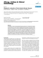
Consequences of targeting designated pro-cardiogenic components were investigated in isolated stem cells and differentiating embryoid bodies (Figure 4). While stimulating
pathways absent from the identified cardiopoietic network
had no effect on outcome (not illustrated), treatment of
embryonic stem cells with VEGF, IGF1 and IL6, to prioritize
charted signaling axes, increased expression of MEF2C (Figure 4a). Together with Nkx2-5 and GATA4 (data not shown),
these pro-cardiac transcription factors were upregulated after
growth factor supplementation, verifying association with
cardiomyogenesis. To investigate effects of treatment at later
developmental stages, stem cell-derived embryoid bodies
were assessed for beating areas, which reflect emergence of
electro-mechanical coupling (Figure 4b). BMP4, administered at day 9 of differentiation, increased the number of
beating areas compared to untreated embryoid bodies (Figure 4b, left panels). Conversely, treatment with the TGFβ signaling cascade inhibitor latency-associated peptide (LAP)
[30] significantly diminished the size and number of these
areas at day 9, while alternative inhibition with the BMP4
antagonist NOG [31] abrogated the development of contractile foci (Figure 4b, right panels). On average, there was an
approximately 20% increase in contractile regions within the
embryoid body following BMP4 treatment, while addition of
LAP decreased this number to <10% of the embryoid body.
NOG treatment precluded contractile foci generation (Figure
4c). Investigation of the JAK/STAT pathway on cardiopoiesis
was performed by adding leukemia inhibitory factor (LIF),
which promoted beating area formation (Figure 4d). Thus,
focused evaluation of individual network elements translated
into changes in cardiogenic yield, validating the functional
significance of the identified pro-cardiac scaffold.
Cluster analysis reveals defined functional
neighborhoods
Within the cardiopoietic network, integrin, Wnt/β-catenin,
VEGF and TGFβ anchor cascades all contain specific genes
used as foci for expression pattern segregation. Discrete correlated expression profiles within the transcriptome were
refined by Venn diagram analysis to yield shared signature
genes (Figure 5a and Additional data file 1). Bmp4 and Pitx2
are elements of the TGFβ cascade within the cardiopoietic
network that coordinated organization of 17 and 12 gene
profiles, respectively, into significantly correlated clusters
(Figure 5a). Multiple genes that comprise integrin signaling
within the network were queried separately and yielded
unique gene lists with distinct trends (Figure 5b). Tgfbr2, a
component of the Wnt pathway, distilled a core of 168
probesets (Figure 5c). Vcl integrates the VEGF cascade into
the cardiopoietic network and here extracted 235 associated
expression patterns (Figure 5d). Each cluster presented a sig-
Genome Biology 2008, 9:R6
/>
Genome Biology 2008,
Volume 9, Issue 1, Article R6
1
(a)
Faustino et al. R6.6
y = 0.1521x-0.4673
10-1
10-2
log P(k)
y = 0.9171x -0.4228
log C(k)
10-3
10-4
Fn1 (19)
Itga5,Itgb1(20)
10-5
log (k)
1
log (k)
10
100
(c) Integrin
signaling
pathway
Wnt/
beta-catenin
signaling
VEGF
signaling
pathway
TGFbeta
signaling
pathway
IGF, IL-6
and others
(b)
common node
- signaling pathway
Significance in CM (-log P-value)
10
Integrin
p38
CSF2
2.5
Wnt/
beta-catenin
Il-6
Jak/
STAT
2
Ca2+
handling
1.5
Hypoxia in
CV system
GSL
metabolism
Amyloid
processing
Cell
cycle
0.5
Death
receptor
CM
GAG degradation
TGFbeta
1
IGF-1
Ganglioside biosynthesis
Toll-like
receptor VEGF
Il-2
CP
N-glycan
biosynthesis
Apoptosis
ES
0
0
0.5
1
1.5
2
10
Significance in CP (-log P-value)
Figure 3 (see development
Cardiovascularprevious page) signaling network within cardiopoietic cells
Cardiovascular development signaling network within cardiopoietic cells. (a) Genes identified in Table 1 integrate into a network suggesting nonstochastic tendencies with emergent scale-free properties (top right). Examples of hubs, with number of first neighbor connections in parentheses, are
labeled on the clustering coefficient plot (top right, inset). (b) All upregulated genes in cardiopoietic cells analyzed for enriched functions were further
mined to identify top supporting signaling cascades. Individual signaling pathways (green circles) were distributed according to significance during stem cellderived cardiogenesis, indicating differences in pathway prioritization at discrete stages. The color scale at right indicates progression from embryonic
stem cells (ES) through the cardiopoietic stage (CP) to stem cell-derived cardiomyocytes (CM), shown in counterclockwise fashion. CSF, colony
stimulating factor; GAG, glycosaminoglycan; GSL, glycosphingolipid. (c) Cross-referencing the signaling cascades represented in (a) with all cardiopoietic
pathways identified in (b) converge on integrin, WNT/β-catenin, VEGF, TGFβ and other (IGF, IL6) signaling cascades anchoring the procardiogenic
network. A common node shared by these pathways, AKT, is outlined in (a).
Genome Biology 2008, 9:R6
/>
Genome Biology 2008,
Volume 9, Issue 1, Article R6
Table 1
Cardiopoietic cells demonstrate specific upregulation of genes involved in cardiovascular development
Gene name
GenBank ID
Fold change
*Actc
NM_009608
1.755
*Acvr1
NM_007394
2.531
Adam19
NM_009616
1.955
Akt1
M94335
2.518
*Amot
U80888
3.321
Anxa1
NM_010730
9.544
Anxa2
NM_007585
4.472
Anxa5
D63423
6.957
*Axl
AA500897
16.45
Bmp4
NM_007554
1.552
Casp8
BC006737
1.994
Cav1
AB029929
10.81
Cd47
NM_010581
2.265
Cd81
NM_133655
1.599
Cd151
U89772
1.597
Cdh2
BC022107
2.861
Col4a2
BC013560
16.98
Ctsb
M14222
4.591
Cyr61
NM_010516
6.394
*Ece1
AI551117
2.551
Egfr
AF277898
1.641
F3
BC024886
3.146
F2r
BQ173958
3.117
Fgfr1
M65053
1.807
Fn1
BC004724
2.536
Furin
NM_011046
1.858
*Has2
NM_008216
2.57
Hif1a
BB269715
2.586
Hmox1
NM_010442
1.728
Id3
NM_008321
1.75
Igf2
NM_010514
22.08
Igfbp4
NM_010517
10.46
ltga3
NM_013565
1.549
Genome Biology 2008, 9:R6
Faustino et al. R6.7
/>
Genome Biology 2008,
Volume 9, Issue 1, Article R6
Faustino et al. R6.8
Table 1 (Continued)
Cardiopoietic cells demonstrate specific upregulation of genes involved in cardiovascular development
ltga5
BB493533
2.908
Itgav
AK003416
2.012
Itgb1
BM120341
2.642
Itgb5
NM_010580
2.542
Lamc1
BG066605
3.632
Lmna
NM_019390
4.11
*Ltbr
NM_010736
5.021
*Mixl1
AF154573
1.647
Mmp2
NM_008610
5.483
Mmp14
NM_008608
5.742
*Nf1
BB526552
1.542
Nr3c1
NM_008173
1.836
Pitx2
U80011
2.062
*Pou6f1
AK009674
1.647
Ppap2b
NM_080555
2.42
*Ppp3r1
NM_024459
1.704
Reck
NM_016678
2.305
*Sema3c
AK004119
3.763
Serpinf1
NM_011340
8.628
*Smo
AW55532
1.652
Sparc
NM_009242
5.953
*Sptbn1
BM213516
1.628
Tgfbr2
BG793483
13.3
Tgm2
BC016492
2.979
Thbs1
AI385532
12.43
Timp1
BC008107
4.251
Timp2
BF168458
40.6
*Tnfrsf12a
NM_013749
3.322
Tnfrsf1a
L26349
4.763
Vcl
NM_009502
3.886
Vhl
NM_009507
1.523
Zfpm1
AA014267
3.272
A total of 65 genes were upregulated with transition from a pluripotent embryonic stem cell into the cardiopoietic phenotype, 49 of which
associated as an integrated network (Figure 3a). *Genes without curated interactions.
Genome Biology 2008, 9:R6
/>
(a)
Genome Biology 2008,
Volume 9, Issue 1, Article R6
+ VEGF
+ IGF-1
+ BMP4 (D0)
+ LAP (D0)
+ NOG (D0)
EB (D9)
Untreated ES
EB (D9)
Faustino et al. R6.9
+ IL-6
+ IL-6
EB (D9)
DAPI
MEF2C
(b)
Untreated
EB (D9)
50
Beating areas (%)
(c)
(d)
*
+ LIF (D5)
40
30
20
*
10
*
0
Untreated
BMP
LAP
NOG
Beating areas (%)
Beating
areas
40
30
20
10
0
EB (D9)
*
-
+
LIF
Figure 4
Biological validation of cardiogenic network
Biological validation of cardiogenic network. (a) LIF cultured stem cells were left untreated for 48 h after LIF withdrawal or were treated with VEGF, IGF1
or IL6. Changes in expression of the cardiac transcription factor MEF2C were revealed through immunocytochemistry. Nuclei were counterstained with
DAPI. Scale bar: 10 μm. (b) Stem cell-derived embryoid bodies were observed for the formation of beating areas (yellow circles) at day 9 (D9) in
untreated, BMP4, LAP and NOG supplemented conditions, with treatments beginning at day zero (D0). Scale bar: 1 μm. (c) Measurement of contractile
area activity using Metamorph software. Data reported as mean number of beating areas ± SEM, *P < 0.05, n = 40-50 embryoid bodies. (d) Visualization of
beating areas in embryoid bodies treated at day 5 (D5) with LIF, involved in JAK/STAT signaling (left). Evaluation of beating area as a percentage of total
area occupied by embryoid body (right). Data reported as mean number of beating areas ± SEM, *P < 0.05, n = 40-50 embryoid bodies.
nificant ontological function upon analysis, with cardiac specification as the first, rank-ordered tissue-specific
developmental process. Myoblast differentiation, regulation
of muscle contraction, cellular localization/assembly, and
vascular development were also prioritized within each cluster according to associated P values (Figure 5e). Therefore,
specific functional properties were ascribed to each network
node mapping respective contributions to the overall execution of cardiopoietic transformation of embryonic stem cells.
Discussion
Embryonic stem cells are developmentally malleable [32],
giving rise to highly specialized and discrete phenotypes crucial to the formative embryo. Specification through genomic
tailoring of stem cell pluripotency involves parsing the massive transcriptional background and deploying necessary
genetic instructions that drive commitment [33]. Since lineage segregation is governed by specific stimuli arising from a
rich transcriptional landscape, mapping pathways directing
distinct cellular fates is essential in identifying, engaging and
driving selected developmental routes [34]. The paradigm of
guided cardiogenesis used here provides a unique opportunity to dissect complex developmental processes underlying
cardiopoiesis, essential for cardiomyocyte derivation from
stem cells [18,35,36]. Using this paradigm in conjunction
with in silico bioinformatics approaches, comparative
genomic analyses uncovered a novel function-directed interactome connecting discrete genes that orchestrate cardiopoiesis. The identified multi-nodal transcriptome network
Genome Biology 2008, 9:R6
/>
Genome Biology 2008,
(a)
K-means
TGFβ
100
*
10
*
SOM
Pitx2 (12)
Bmp4 (17)
QT
1
0.1
Bmp4 (17)
ES
ES`
CP
CM
(b)
ItgaV (98)
Integrin
Itga5 (26)
Itgb5
(339)
(d)
Tgfbr2
(168)
VEGF
Wnt/β-catenin
(c)
Common Name
2310009N05Rik
3110001A13Rik
Acbd4
Actn1
Bcl2l1
Bmp4
Il6st
Itga3
Mfge8
Pcolce
Ppp3r1
Slc6a8
Stat3
Tbx3
Tgfbi
Tnpo2
Zfhx1a
Fn1 (25)
(e) Node
Bmp4
Pitx2
Itga5
ItgaV
Itgb5
Fn1
Lamc1
Tgfbr2
Vcl
Vcl
(235)
Volume 9, Issue 1, Article R6
Genbank
AK009256
BC021353
AK008243
BC003232
NM_009743
NM_007554
AA717838
NM_013565
NM_008594
BB250811
NM_024459
BG069516
AI325183
AA543734
BB532080
BI658203
NM_011546
Faustino et al. R6.10
Affymetrix ID
1430125_s_at
1416893_at
1428271_at
1452415_at
1420887_a_at
1422912_at
1460295_s_at
1460305_at
1420911_a_at
1437165_a_at
1450368_a_at
1448596_at
1426587_a_at
1448029_at
1437463_x_at
1425592_at
1418926_at
Lamc1 (38)
Cluster priority
heart development
pattern specification
regulation of muscle contraction
muscle contraction
cell adhesion
cell-substrate junction assembly
cellular localization
myoblast differentiation
blood vessel development
p
0.00127
0.00203
0.000512
0.00315
2.49 x 10⁻⁶
0.00328
0.0341
1.99 x 10⁻⁴
3.86 x 10⁻⁷
Nodal network anchors orchestrate coordinated recruitment of specialized ontological classes to secure a developmental theme
Figure 5
Nodal network anchors orchestrate coordinated recruitment of specialized ontological classes to secure a developmental theme. (a) Left: a five group Kmeans algorithm, 4 × 6 SOM, and QT filter were each used to independently dissect the transcriptome of embryonic stem cell (ES) derived cardiogenesis.
Cardiopoietic (CP) network nodes previously identified were then used to guide cluster extraction. Bmp4 and Pitx2, members of the TGFβ pathway, are
shown as examples. *Venn diagram of K-means, SOM and QT generated lists resolved clustered genes with correlated expression profiles (R = 0.95). For
each set, gene (node) identity used to extract associated profiles is indicated, along with number of probes per cluster given in parentheses. Right: genes
within the Bmp4 cluster. CM, cardiomyocyte. (b) Nodes belonging to the integrin cascade select discrete clusters with distinct trends. (c, d) Gene groups
associated with Tgfbr2 and Vcl nodes that represent WNT/β-catenin and VEGF signaling, respectively. (e) Gene clusters organized functional
neighborhoods with ontological priorities, with level of significance indicated on right.
establishes the cardiogenic gestalt, revealing targets for
manipulation of cardiac fate that will expedite translational
application [37-39].
Endogenous genetic flux through non-specific pluripotency
primes stem cells for a variety of phenotypes at the cost of elevated genetic noise [40,41]. Successful navigation of this
intricate genetic background is pivotal in developmental
specification, requiring non-stochastic gene activation to support emerging phenotypes [42]. Systems biology approaches
to analyze network randomness and propensity for hub for-
mation [43] suggest a robust topology framing the transcriptome that underlies cardiopoiesis.
Specifically, the current work demonstrates formation of an
integrated scaffold of genes activated during stem cellderived cardiomyocyte procurement that sculpts a resilient
cardiogenic transcriptome profile. The non-random presence
and distribution of hubs, that is, nodes with high connectivity
[44], indicates a switch where pluripotent stem cells are
directed and constrained to a cardiac fate. While
nonsignificantly changing genes represented heterogeneous
Genome Biology 2008, 9:R6
/>
Genome Biology 2008,
ontological annotation distributions without any functional
predispositions, inspection of downregulated transcripts
demonstrated a controlled diminution of transcriptional
chatter through reduction of genes associated with genetic
metabolism. Furthermore, diminished DNA metabolism that
accompanies differentiation of embryonic stem cells into cardiac precursors reflects the onset of specialization with loss of
genomic variability. Indeed, timing changes and restriction of
replication initiation has been reported for embryonic stem
cell differentiation [45]. Transcriptional narrowing has been
recently observed during stem cell differentiation for both
nuclear transport machinery [19] and metabolic energetic
remodeling [46]. Further streamlining, with specific upregulation of overrepresented pro-cardiogenic functions [35,47],
ultimately secures execution of the cardiac program.
nodes in vitro demonstrates discernible alterations in stem
cell-derived cardiomyocyte yields.
Gene or protein networks buttress pluripotency through integration of multiple pathways contributing to the final phenotype [48-51]. Here, distinct organization of signal pathways
secured the cardiopoietic network. Integrins are cytoskeletal
associated transmembrane glycoproteins that transduce
extracellular signals and prominently anchor the transcriptome. Within cardiogenesis, the integrin cascade dictates formation of terminal myocardial structure [52]. WNT/βcatenin signaling transduces extracellular cues during development [53] and is the second significant cascade identified
in the cardiopoietic network. Previous transcriptome analysis
identified upregulation of negative regulators of WNT signaling [11]. Here, in guided cardiopoiesis, distinct effectors that
feed into the WNT pathway were upregulated. Participation
of this cascade in cardiogenesis is bimodal [54-56], and concomitant expression variations of inhibitors and potentiators
may serve as a molecular rheostat indispensable for all types
of lineage specification. In this capacity, the WNT family has
been proposed to be transcriptional noise filters during differentiation [42]. The TGFβ cascade, connected to the cardiopoietic scaffold through BMP4, represents a source of potent
pro-cardiac stimuli [23,57-59]. Transgenic models deficient
in downstream signaling components of the TGFβ pathway,
such as SMAD3 insufficient mice, exhibit cardiac developmental defects [60]. Furthermore, cell-tracing studies
recently reported progenitors positive for the VEGF receptor
FLK1 that gave rise to cardiovascular, endothelial and smooth
muscle cell lineages [61,62], in line with the present identification of the VEGF signaling axis in the pro-cardiac transcriptome network. Cardiomyocyte development through IGF
[55], and cardiac hypertrophy mediated by the IL6 signaling
axis [63] are represented herein by single components
belonging to their respective pathways. The inclusion of other
cascades in the cardiopoietic network, also by single component representation, permits integration of lambent inputs
from other pathways, lessening the rigidity of the
transcriptome scaffold and allowing exogenous manipulability of the network without changing its fundamental architecture. Collectively, integration of discrete signaling pathways
secures overall network functionality and, indeed, targeting
Volume 9, Issue 1, Article R6
Faustino et al. R6.11
Targeted node validation by independent treatment with different growth factors significantly increased the number of
embryonic stem cells positive for cardiac transcription factors, indicating an engaged cardiogenic program. VEGF,
upon binding to its cognate receptor on the surface of embryonic stem cells [64], is transduced through focal adhesion
complexes containing vinculin [65], a significant node identified within the cardiopoietic network. This actin-binding,
cytoskeletal protein is essential to cardiac development, as
knockout models presented thin myocardial walls with compromised cardiac contractility along with diverse cardiac
defects [66]. Similarly, interaction of IGF1 with the IGF1
receptor expressed on the plasma membrane of embryonic
stem cells [67] increased the number of stem cells with an
engaged cardiac program. Both AKT and IGF binding protein
4, elements of the IGF1 pathway essential to the cardiopoietic
network, promote cell survival and proliferation, and affect
organismal growth [55,68,69]. AKT is critical for directing
hypertrophic myocardial responses to adaptive and maladaptive stimuli [70-73]. IL6 belongs to JAK/STAT/IL6 signal
transducer (IL6ST)-dependent cytokines, and here increased
cardiogenic engagement. This is supported by reports of
modulated cardiogenesis in embryoid bodies through the
JAK/STAT/IL6ST relay [74]. Conditional mutations of
IL6ST, a component of the IL6 receptor complex, manifest
cardiac defects, including ventricular thinning, right ventricular dilation, and significant size reductions in subpopulations of cardiomyocytes [63]. Furthermore, genetic ablation
of IL6ST demonstrates a definitive role for the IL6 signaling
axis in determination and maintenance of cardiac morphology [75]. Functionally, formation of contractile areas is a
definitive endpoint indicating syncytial integration of developed cardiomyocytes. Treatment with BMP4, a cardiopoietic
network ligand of the TGFβ cascade [76], distinctly increased
beating areas, whereas antagonism using LAP or NOG precluded beating. Together, these observations reveal that the
TGFβ signaling axis is embedded within the cardiopoietic
network, supported by well characterized effects on cardiogenesis [23,77]. LIF treatment increased contractile foci, and
exerts cardiogenic effects through the JAK/STAT/IL6ST signaling complex. Thus, the interactive transcriptome transduces pro-cardiac inputs, reflected through cardiogenic
engagement and subsequent functional cardiomyocyte
generation.
Network anchors within the emergent cardiovascular scaffold
are part of extant transcriptome gene clusters that collectively
foster distinct thematic climes [78]. As cellular identities
manifest from embryonic stem cell origins, developmental
programming is oriented through hubs that are part of an
ontological collective that defines specific transcriptome
neighborhoods and secures nascent phenotypes [79]. Furthermore, here collective ontological themes classifying hub-
Genome Biology 2008, 9:R6
/>
Genome Biology 2008,
organized gene clusters are complementary and non-stochastic, demonstrated in this paradigm of cardiogenesis. In this
way, the transcriptomic framework serves as a 'wireframe'
that co-ordinates and unifies discrete developmental elements to ultimately realize full specification.
using 3% FBS GMEM with 30 ng/ml of TNFα for 5 days and
20% FBS GMEM for 9 days, respectively [18]. Cells were subjected to confocal microscopy, assessing MEF2C, NKX2-5
and GATA4 nuclear translocation in cardiopoietic cells along
with expression of α-actinin or myosin heavy chain in cardiomyocytes both prior to and after purification of derived cells
using Percoll. The gradient was generated with dilution of a
Percoll stock (Sigma-Aldrich, St. Louis, Missouri, USA) to
densities of 1.09 and 1.07 g/ml, with 4 ml of the 1.07 density
overlaying 3 ml of the 1.09 density. The interface of these two
densities successfully yielded the cardiomyocyte population
[19]. For cardiopoietic cells, the previous densities used for
cardiomyocyte derivation were reduced by 0.02 g/ml [18].
Total RNA was harvested from ES-LIF(+), ES-LIF(-), cardiopoietic and cardiomyocyte samples for downstream microarray analysis.
Conclusion
Here, a manipulable, lineage-specifying genomic atlas was
extracted from the pluripotent content of an embryonic
source. Transcriptomic profile dissection of embryonic stem
cells undergoing cardiopoietic transition isolated a dynamic
intermolecular signaling scaffold unifying genetic crosstalk
critical to cardiogenic yield. Functional interrogation of this
focused network demonstrated treatment-dependent, bimodal responsiveness dictated by node and hub composition. A
demonstrable, refined control of guided cardiogenesis by in
vitro supplementation with exogenous growth factors efficiently accelerated the production of functional cardiomyocytes. In contrast, addition of network decelerants delayed
cardiogenesis. Thus, access and identification of nodes within
the cardiopoietic network is distinctly advantageous for procurement of an exogenous supply of cardiac cells. This circumvents limitations associated with a scarce endogenous
pool, and expedites translation of ex vivo stem cell-derived
cardiac-specified progeny as a regenerative therapeutic
modality. Consolidation of node-organized functional transcript clusters secured developmental attunement through
coordinated ontological neighborhoods that contained candidates promoting cardiac development. This paradigm of a
defined gene network architecture, supportive of the cardiac
progenitor phenotype, provides a diagnostic map to chart
susceptible nodes that conversely may promote cardiomyocyte attrition with resultant cardiac dysfunctions. Critical
rate-limiting hubs within such a framework can identify
unexplored molecular etiologies that impact cardiac precursor lifespan or capacity for self-renewal, defining individual
cardioprotective potential. Ultimately, this integrated
approach maps a dynamic and interactive transcriptomic grid
for definition, interrogation, and control of a discrete biological process.
Materials and methods
Stem cell culture and differentiation
Murine CGR8 embryonic stem (ES) cells were cultured without a feeder layer in 7.5% fetal bovine serum (FBS) in Glasgow's modified Eagle's medium (GMEM) as described [23].
Cells in the presence of LIF and after 48 h in a LIF-free environment were designated as ES-LIF(+) and ES-LIF(-),
respectively. Subsequently, embryonic stem cells were placed
in a cocktail containing 5 ng/ml BMP, 2.5 ng/ml TGFβ, 100
ng/ml IL-13, 100 ng/ml IL3, 50 ng/ml IGF1, 10 ng/ml VEGF,
2.5 ng/ml EGF, 10 ng/ml FGF and 100 ng/ml IL6. Cardiopoietic cells and cardiomyocytes derived from embryonic stem
cells stimulated in this cocktail were maintained in culture
Volume 9, Issue 1, Article R6
Faustino et al. R6.12
Scanning electron microscopy
Embryonic stem cells, cardiopoietic cells or derived cardiomyocytes were fixed with 1% glutaraldehyde and 4% formaldehyde in phosphate buffered saline (pH 7.2). Hypotonic
sarcolemmal stripping using a 1% Triton X-100 solution
exposed the nucleus prior to fixation [19]. Intact or stripped
fixed cells were rinsed in phosphate buffered saline with 1%
osmium, dehydrated with ethanol and dried in a critical point
dryer. Samples were examined on a 4700 field-emission scanning microscope (Hitachi, Tokyo, Japan) after coating with
platinum.
Stem cell immunocytochemistry and embryoid body
imaging
Embryonic stem cells, cardiopoietic cells and derived cardiomyocytes were fixed in 3% paraformaldehyde, permeabilized
with 0.5% Triton X-100, blocked with 100% Superblock
(Pierce, Rockford, Illinois, USA) and immunostained with
primary antibodies specific for the cardiac transcription factor MEF2C (Cell Signaling Technology, Boston, Massachusetts, USA) and/or sarcomeric α-actinin (Sigma-Aldrich, St
Louis, Missouri, USA), and corresponding ALEXA-labelled
secondary antibodies (Molecular Probes, Carlsbad, California, USA) along with nuclear-staining 4'-6-diamidino-2-phenylindole (DAPI; Molecular Probes) [19]. Imaging was
performed using a Zeiss laser scanning microscope 510 (Carl
Zeiss, Jena, Germany) microscope. Additionally, after 48 h
treatment of undifferentiated embryonic stem cells with 50
ng/ml IGF1, 10 ng/ml VEGF, or 100 ng/ml IL6 following LIF
withdrawal, images were obtained and stored in TIF format
with 10 distinct fields from at least 3 separate isolations for
each experimental condition. Image evaluation of fluorescent
intensity was performed using Metamorph (Sunnyvale, California, USA). Differentiated embryoid bodies, using the
established hanging drop method [80], were treated at day 0
(D0) with 5 ng/ml BMP4, 25 ng/ml LAP, or 25 ng/ml NOG.
Alternatively, 1,000 U/ml LIF was added at day 5 (D5) of differentiation. Prior to imaging at day 9 (D9), embryoid bodies
were plated on gelatin-coated six well dishes with sequential
Genome Biology 2008, 9:R6
/>
Genome Biology 2008,
timelapse images obtained at 5 Hz. Image sets were reconstituted in Metamorph to visualize beating areas, delineated for
area measurement.
Volume 9, Issue 1, Article R6
D=
1
n
Faustino et al. R6.13
n
∑( A − B )
i
i
2
(2)
i =1
Microarrays
To investigate transcriptome dynamics during guided cardiac
differentiation of murine embryonic stem cells, total RNA
was isolated at discrete timepoints using the Micro-to-Midi
Total RNA Purification System (Invitrogen, Carlsbad, California, USA) as described [46]. Each condition was independently sampled three times for a total of twelve biological
replicates. Double stranded complementary cDNA and
labeled complementary cRNA were obtained from isolated
total RNA, with the latter hybridized against the Mouse 430
2.0 GeneChip (Affymetrix, Santa Clara, California, USA).
Arrays were scanned using an argon-ion laser, and data visualized using MAS 5.0 Affymetrix software to assess quality of
hybridization. The dataset has been deposited at the Gene
Expression Omnibus [81] as an update to series GSE6689.
Expression analysis and gene/condition clustering
Gene expression data were analyzed using Genespring GX 7.3
(Agilent Technologies, Santa Clara, California, USA). All
probesets were initially quality filtered for the pluripotent
embryonic stem cell transcriptome (in the presence and
absence of LIF) according to an established flag value, with
values that are present (P), marginal (M) or absent (A)
assigned to the marker [46]. To ensure that transcriptional
changes were restricted to display gene profiles emerging
during cardiac differentiation, data were further restricted to
display genes demonstrating the present (P) and marginal
(M) flag values in all three replicates for the cardiopoietic
stage, and the present (P) flag value in all stem cell-derived
cardiomyocyte samples. Next, samples were filtered
according to background noise levels to remove genes
expressing signals below threshold. The final gene list was
delimited according to statistically relevant changes using
one-way ANOVA, P < 0.05 with the Benjamini and Hochberg
false discovery rate as multiple testing correction. Hierarchical dendrograms were used to establish the molecular fingerprints for each stage, and were generated using the Pearson
coefficient statistic (r) applied to determine correlation
between gene pairs in each condition as follows:
r=
n
∑ ( Ai − A)( B i − B )
i =1
⎛ n
⎞⎛ n
⎜ ∑ ( Ai − A) 2 ⎟ ⎜ ∑ ( B i − B ) 2
⎜
⎟⎜
⎝ i =1
⎠ ⎝ i =1
⎞
⎟
⎟
⎠
(1)
Above, (A) and (B) are respective sample means for genes Ai
and Bi for sample (i) out of the total number of samples (n),
with standard deviation terms for Ai and Bi used as denominator. Condition clustering was performed to determine sample
similarity using Euclidean distance as a measure of sample
'nearness'. The formula for calculating distance (D):
The square of the difference in expression levels between gene
A (Ai) and gene B (Bi) in sample (i) are divided by the total
number of samples (n), of which the square root is taken to
obtain distance (D). The clustering derived from distance calculation was further validated by bootstrapping, a conventional statistical resampling technique [24].
Taqman assays
RNA (1 μg) was reverse transcribed into cDNA using a high
capacity cDNA archive kit (Applied Biosystems, Foster City,
California, USA) and assayed using Taqman gene expression
assays for Pou5f1/Oct4 (Mm00658129_gH), Mybl2
(Mm00485340_m1), Mycn (Mm00476449_m1), Myocd
(Mm00455051_m1) and Lbh (Mm00522505_m1), prototypical markers of pluripotency, oncogenesis and cardiogenesis.
Samples were loaded onto an optical 96-well plate for
polymerase chain reactions performed using an ABI 7900HT
Fast Real Time System with cycling parameters set for a 15 s,
95°C duplex denaturing step followed by primer annealing/
extending for 1 minute at 60°C per cycle for 40 cycles. Relative fold change was determined using the 2-ΔΔCT method [82]
with pluripotent embryonic stem cells as baseline, normalized to Gapdh expression.
Enrichment analysis of functional categories
To examine overrepresented functions within the final upand downregulated filtered gene lists, Expression Analysis
Systematic Explorer (EASE version 2.0) [83] was used. Gene
lists were submitted as text files using GenBank accession
identifiers and ontology annotations for 'Molecular function'
were analyzed by linking, through EASE, to the online Database for Annotation, Visualization, and Integrated Discovery
[84]. For 'Molecular function', the population total (8,821) is
the group of annotations available for the Mouse 430 2.0
GeneChip. Population hits are defined as the genes for each
'Molecular function' sub-classification that are identifiable.
List totals indicate annotations (out of the population total)
that are available from user-submitted lists for 'Molecular
function', and list hits identify annotations belonging to specific groups within 'Molecular function' within the user-submitted list. Each category under 'Molecular function' had
specifically associated genes, and in some instances, genes
were assigned to more than one functional category. Significance was determined by Fisher's exact test and Bonferroni
correction for multiple category comparisons (P < 0.05) and
top functions were reported as a percentage of list totals, with
remaining functions classified as 'other' for both up- and
downregulated gene lists.
Genome Biology 2008, 9:R6
/>
Genome Biology 2008,
Network analysis
Authors' contributions
Using an established network analysis program, Ingenuity
Pathways Analysis [85], molecular interactions were examined in the cardiopoietic stage. One way ANOVA-delimited
gene lists used in enrichment analysis were studied using the
Ingenuity Pathways Knowledge base to identify, using a righttailed Fisher's exact test, overrepresented canonical functions
and signaling pathways at different timepoints during cardiogenic stem cell differentiation. The Institute for Systems
Biology Cytoscape 2.2 software [86] was applied to provide
data regarding network topology in addition to visualizing
relationships. Gene interactions identified by Ingenuity were
deconstructed according to pairwise interactions, and reformatted for use in Cytoscape 2.2. Basic network analyses,
including degree distribution and clustering coefficient distribution determination, were performed, providing statistical
measures of cardiopoietic network architecture.
Cluster analysis
Quality filtered genes were recursively and separately analyzed by K-means, self-organizing map (SOM) and quality
threshold (QT) clustering. Group size for K-means was set to
a maximum of 5 clusters, while a 4 × 6 array was specified for
SOM. For QT analysis, Pearson correlation was set at 0.95.
Each of the three analyses produced distinct transcript aggregates, and cross-reference by Venn diagram highlighted
genes consistently segregating with selected network nodes.
Discrete expression profile groups were bioinformatically
mined to uncover organized functional neighborhoods delimited by cluster oriented developmental themes. Hypergeometric P values for ontological assignations were calculated
as shown:
p=
⎛ m ⎞ ⎛ u −m ⎞
1
∑⎜ ⎟
⎛ u ⎞ i =k ⎝ i ⎠ ⎜ n −1 ⎟
⎝
⎠
⎜ ⎟
⎝m⎠
Additional data files
The following additional data are available with the online
version of this paper. Additional data file 1 is an Excel spreadsheet listing gene identities within node organized clusters.
Click few data yielded
detailed in Figure
script cluster file
with here anchors5.1 genes associated Furthermore, each
anchors that possessed prevalent ontological
Cluster analysis filesharing a single group.withspecification,
Geneaidentities within node organized clusters expression profiles,
Additionalfor fostered acorrelated and discrete network node tran-
Acknowledgements
We thank A-L Barabási (University of Notre Dame) for constructive comments during preparation of this manuscript. This work was supported by
grants from the National Institutes of Health, American Heart Association,
Marriott Heart Disease Research Program, Marriott Foundation, Ted Nash
Long Life Foundation, Ralph Wilson Medical Research Foundation, and
Asper Foundation. AB is supported by the Mayo Clinic Clinician-Investigator Program, and CPT by a Mayo Clinic FUTR Career Development Award.
References
1.
2.
3.
4.
(3)
6.
7.
8.
Abbreviations
BMP, bone morphogenic protein; DAPI, 4'-6-diamidino-2phenylindole; EASE, Expression Analysis Systematic
Explorer; EGF, epidermal growth factor; ES, embryonic
stem; FBS, fetal bovine serum; FGF, fibroblast growth factor;
GMEM, Glasgow's modified Eagle's medium; IGF1, insulinlike growth factor; IL, interleukin; IL6ST, IL-6 signal transducer; LAP, latency associated peptide; LIF, leukemia inhibitory factor; MEF2C, myocyte enhancer factor 2C; QT, quality
threshold; SOM, self organizing map; TGF, transforming
growth factor; TNF, tumor necrosis factor; VEGF, vascular
endothelial growth factor.
Faustino et al. R6.14
RSF, AB, CPT and AT contributed to the design of the study.
RSF performed bioinformatics involved in this study. AB carried out cell culture and immunocytochemistry. CPT did electron microscopy. RSF, AB, and CPT analyzed the data. RSF,
AB, CPT and AT prepared the manuscript. All authors have
read and approved the final version of this manuscript.
5.
The summation notation above yields the probability (p) of
overlap that corresponds to (k) or more genes that exist
between gene lists (m) and (n) when randomly sampled from
a universe containg (u) genes.
Volume 9, Issue 1, Article R6
9.
10.
11.
12.
13.
14.
Rhee H, Polak L, Fuchs E: Lhx2 maintains stem cell character in
hair follicles. Science 2006, 312:1946-1949.
Soen Y, Mori A, Palmer TD, Brown PO: Exploring the regulation
of human neural precursor cell differentiation using arrays of
signaling microenvironments. Mol Syst Biol 2006, 2:37.
Suzuki A, Raya Á, Kawakami Y, Morita M, Matsui T, Nakashima K,
Gage FH, Rodríguez-Esteban C, Belmonte JCI: Maintenance of
embryonic stem cell pluripotency by Nanog-mediated
reversal of mesoderm specification. Nat Clin Pract Cardiovasc
Med 2006, 3:S114-S122.
Pan G, Thomson JA: Nanog and transcriptional networks in
embryonic stem cell pluripotency. Cell Res 2007, 17:42-49.
Boyer LA, Lee TI, Cole MF, Johnstone SE, Levine SS, Zucker JP, Guenther MG, Kumar RM, Murray HL, Jenner RG, et al.: Core transcriptional regulatory circuitry in human embryonic stem cells.
Cell 2005, 122:947-956.
Loh Y-H, Wu Q, Chew J-L, Vega VB, Zhang W, Chen X, Bourque G,
George J, Leong B, Liu J, et al.: The Oct4 and Nanog transcription
network regulates pluripotency in mouse embryonic stem
cells. Nat Genet 2006, 38:431-440.
Palmqvist L, Glover CH, Hsu L, Lu M, Bossen B, Piret JM, Humphries
RK, Helgason CD: Correlation of murine embryonic stem cell
gene expression profiles with functional measures of
pluripotency. Stem Cells 2005, 23:663-680.
Armstrong L, Hughes O, Yung S, Hyslop L, Stewart R, Wappler I,
Peters H, Walter T, Stojkovic P, Evans J, et al.: The role of PI3K/
AKT, MAPK/ERK and NFκB signalling in the maintenance of
human embryonic stem cell pluripotency and viability highlighted by transcriptional profiling and functional analysis.
Hum Mol Genet 2006, 15:1894-1913.
Boiani M, Schöler HR: Regulatory networks in embryo-derived
pluripotent stem cells. Nat Rev Mol Cell Biol 2005, 6:872-884.
Olson EN: Gene regulatory networks in the evolution and
development of the heart. Science 2006, 313:1922-1927.
Doss MX, Winkler J, Chen S, Hippler-Altenburg R, Sotiriadou I, Halbach M, Pfannkuche K, Liang H, Schulz H, Hummel O, et al.: Global
transcriptome analysis of murine embryonic stem cellderived cardiomyocytes. Genome Biol 2007, 8:R56.
Uetz P, Stagljar I: The interactome of human EGF/ErbB
receptors. Mol Syst Biol 2006, 2:2006.0006.
Solter D: From teratocarcinomas to embryonic stem cells
and beyond: a history of embryonic stem cell research. Nat
Rev Genet 2006, 7:319-327.
Srivastava D, Ivey KN: Potential of stem-cell-based therapies
Genome Biology 2008, 9:R6
/>
15.
16.
17.
18.
19.
20.
21.
22.
23.
24.
25.
26.
27.
28.
29.
30.
31.
32.
33.
34.
35.
36.
37.
38.
39.
Genome Biology 2008,
for heart disease. Nature 2006, 441:1097-1099.
Foley A, Mercola M: Heart induction: embryology to cardiomyocyte regeneration. Trends Cardiovasc Med 2004, 14:121-125.
Behfar A, Hodgson DM, Zingman LV, Perez-Terzic C, Yamada S, Kane
GC, Alekseev AE, Puceat M, Terzic A: Administration of allogenic
stem cells dosed to secure cardiogenesis and sustained infarct repair. Ann NY Acad Sci 2005, 1049:189-198.
Laflamme MA, Murry CE: Regenerating the heart. Nat Biotechnol
2005, 23:845-856.
Behfar A, Perez-Terzic C, Faustino RS, Arrell DK, Hodgson DM,
Yamada S, Pucéat M, Niederländer N, Alekseev AE, Zingman LV, et
al.: Cardiopoietic programming of embryonic stem cells for
tumor-free heart repair. J Exp Med 2007, 204:405-420.
Perez-Terzic C, Faustino RS, Boorsma BJ, Arrell DK, Niederländer
NJ, Behfar A, Terzic A: Stem cells tranform into a cardiac phenotype with remodeling of the nuclear transport machinery.
Nat Clin Pract Cardiovasc Med 2007, 4:S68-S76.
Behfar A, Terzic A: Derivation of a cardiopoietic population
from human mesenchymal stem cells yields cardiac progeny.
Nat Clin Pract Cardiovasc Med 2006, 3:S78-S82.
Latif S, Masino A, Garry DJ: Transcriptional pathways direct cardiac development and regeneration. Trends Cardiovasc Med
2006, 16:234-240.
Thomson JA, Itskotz-Eldor J, Shapiro SS, Waknitz MA, Swiergiel JJ,
Marshall VS, Jones JM: Embryonic stem cell lines derived from
human blastocysts. Science 1998, 282:1145-1147.
Behfar A, Zingman LV, Hodgson DM, Rauzier J-M, Kane GC, Terzic
A, Pucéat M: Stem cell differentiation requires a paracrine
pathway in the heart. FASEB J 2002, 16:1558-1566.
Kerr MK, Churchill GA: Bootstrapping cluster analysis: assessing the reliability of conclusions from microarray
experiments. Proc Natl Acad Sci USA 2001, 98:8961-8965.
Morrison GM, Brickman JM: Conserved roles for Oct4 homologues in maintaining multipotency during early vertebrate
development. Development 2006, 133:2011-2022.
García P, Frampton J: The transcription factor B-Myb is essential for S-phase progression and genomic stability in diploid
and polyploid megakaryocytes. J Cell Sci 2006, 119:1483-1493.
Schwab M: MYCN in neuronal tumours. Cancer Lett 2004,
204:179-187.
Wang D-Z, Chang PS, Wang Z, Sutherland L, Richardson JA, Small E,
Krieg PA, Olson EN: Activation of cardiac gene expression by
myocardin, a transcriptional cofactor for serum response
factor. Cell 2001, 105:851-862.
Briegel KJ, Joyner AL: Identification and characterization of
Lbh, a novel conserved nuclear protein expressed during
early limb and heart development. Dev Biol 2001, 233:291-304.
Ge G, Greenspan DS: BMP1 controls TGFβ1 activation via
cleavage of latent TGFβ-binding protein. J Cell Biol 2006,
175:111-120.
Choi M, Stottmann RW, Yang Y-P, Meyers EN, Klingensmith J: The
bone morphogenetic protein antagonist noggin regulates
mammalian cardiac morphogenesis.
Circ Res 2007,
100:220-228.
Zhang J, Tam W-L, Tong GQ, Wu Q, Chan H-Y, Soh B-S, Lou Y, Yang
J, Ma Y, Chai L, et al.: Sall4 modulates embryonic stem cell
pluripotency and early embryonic development by the transcriptional regulation of Pou5f1. Nat Cell Biol 2006, 8:1114-1123.
Brandenburger R, Wei H, Zhang S, Lei S, Murage J, Fisk GJ, Li Y, Xu
C, Fang R, Guegler K, et al.: Transcriptome characterization elucidates signaling networks that control human ES cell
growth and differentiation. Nat Biotechnol 2004, 22:707-716.
Carninci P, Kasukawa T, Katayama S, Gough J, Frith M, Maeda N,
Oyama R, Ravasi T, Lenhard B, Wells C, et al.: The transcriptional
landscape of the mammalian genome.
Science 2005,
309:1559-1563.
Srivastava D: Making or breaking the heart: from lineage
determination to morphogenesis. Cell 2006, 126:1037-1048.
Buckingham M, Meilhac S, Zaffran S: Building the mammalian
heart from two sources of myocardial cells. Nat Rev Genet
2005, 6:826-835.
Anversa P, Nadal-Ginard B: Myocyte renewal and ventricular
remodelling. Nature 2002, 415:240-243.
Hodgson DM, Behfar A, Zingman LV, Kane GC, Perez-Terzic C, Alekseev AE, Pucéat M: Stable benefit of embryonic stem cell therapy in myocardial infarction. Am J Physiol Heart Circ Physiol 2004,
287:H471-H479.
Dimmeler S, Zeiher AM, Schneider MD: Unchain my heart: the
40.
41.
42.
43.
44.
45.
46.
47.
48.
49.
50.
51.
52.
53.
54.
55.
56.
57.
58.
59.
60.
61.
62.
63.
Volume 9, Issue 1, Article R6
Faustino et al. R6.15
scientific foundations of cardiac repair. J Clin Invest 2005,
115:572-583.
Niwa H: How is pluripotency determined and maintained?
Development 2007, 134:635-646.
Zipori D: The nature of stem cell: state rather than entity.
Nat Rev Genet 2004, 5:873-878.
Arias AM, Hayward P: Filtering transcriptional noise during
development: concepts and mechanisms. Nat Rev Genet 2006,
7:34-44.
Barabási A-L, Oltvai Z: Network biology: understanding the
cell's functional organization. Nat Rev Genet 2004, 5:101-113.
Albert R: Scale-free networks in cell biology. J Cell Sci 2005,
118:4947-4957.
Aladjem MI: Replication in context: dynamic regulation of
DNA replication patterns in metazoans. Nat Rev Genet 2007,
8:588-600.
Chung S, Dzeja PP, Faustino RS, Perez-Terzic C, Behfar A, Terzic A:
Mitochondrial oxidative metabolism is required for the cardiac differentiation of stem cells. Nat Clin Pract Cardiovasc Med
2007, 4:S60-S67.
Gottlieb PD, Pierce SA, III RJS, Yamagishi H, Weihe EK, Harriss JV,
Maika SD, Kuziel WA, King HL, Olson EN, et al.: Bop encodes a
muscle-restricted protein containing MYND and SET
domains and is essential for cardiac differentiation and
morphogenesis. Nat Genet 2002, 31:25-32.
Rao S, Orkin SH: Unraveling the transcriptional network controlling ES cell pluripotency. Genome Biol 2006, 7:230.
Chickarmane V, Troein C, Nuber UA, Sauro HM, Peterson C: Transcriptional dynamics of the embryonic stem cell switch. PLoS
Comput Biol 2006, 2:e123.
Wang J, Rao S, Chu J, Shen X, Levasseur DN, Theunissen TW, Orkin
SH: A protein interaction network for pluripotency of embryonic stem cells. Nature 2006, 444:364-368.
Arrell DK, Niederländer NJ, Faustino RS, Behfar A, Terzic A: Cardioinductive network guiding stem cell differentiation
revealed by proteomic cartography of TNF{alpha}-primed
endodermal secretome. Stem Cells 2008, 26:. doi: 10.1634/stemcells.2007-0599
Ross RS, Borg TK: Integrins and the myocardium. Circ Res 2001,
88:1112-1119.
Foley AC, Gupta RW, Guzzo RM, Korol O, Mercola M: Embryonic
heart induction. Ann NY Acad Sci 2006, 1080:85-96.
Naito AT, Shiojima I, Akazawa H, Hidaka K, Morisaki T, Kikuchi A,
Komuro I: Developmental stage-specific biphasic roles of
Wnt/β-catenin
signaling
in
cardiomyogenesis
and
hematopoiesis. Proc Natl Acad Sci USA 2006, 103:19812-19817.
Fraidenraich D, Stillwell E, Romero E, Wilkes D, Manova K, Basson
CT, Benezra R: Rescue of cardiac defects in Id knockout
embryos by injection of embryonic stem cells. Science 2004,
306:247-252.
vanGijn ME, Daemen MJAP, Smits JFM, Blankesteijn WM: The wntfrizzled cascade in cardiovascular disease. Cardiovasc Res 2002,
55:16-24.
Rivera-Feliciano J, Tabin CJ: Bmp2 instructs cardiac progenitors
to form the heart-valve-inducing field.
Dev Biol 2006,
295:580-588.
Kruithof BPT, vanWijk B, Somi S, Julio MK-d, Pomares JMP, Weesie
F, Wessels A, Moorman AFM, vandenHoff MJB: BMP and FGF regulate the differentiation of multipotential pericardial mesoderm into the myocardial or epicardial lineage. Dev Biol 2006,
295:507-522.
Zeineddine D, Papadimou E, Chebli K, Gineste M, Liu J, Grey C,
Thurig S, Behfar A, Wallace VA, Skerjanc IS, et al.: Oct-3/4 dose
dependently regulates specification of embryonic stem cells
toward a cardiac lineage and early heart development. Dev
Cell 2006, 11:535-546.
Vincent SD, Dunn NR, Hayashi S, Norris DP, Robertson EJ: Cell fate
decisions within the mouse organizer are governed by
graded Nodal signals. Genes Dev 2003, 17:1646-1662.
Kattman SJ, Huber TL, Keller GM: Multipotent Flk-1+ cardiovascular progenitor cells give rise to the cardiomyocyte,
endothelial, and vascular smooth muscle lineages. Dev Cell
2006, 11:723-732.
Garry DJ, Olson EN: A common progenitor at the heart of
development. Cell 2006, 127:1101-1104.
Betz UAK, Bloch W, vandenBroek M, Yoshida K, Taga T, Kishimoto
T, Addicks K, Rajewsky K, Miller W: Postnatally induced inactivation of gp130 in mice results in neurological, cardiac, hemot-
Genome Biology 2008, 9:R6
/>
64.
65.
66.
67.
68.
69.
70.
71.
72.
73.
74.
75.
76.
77.
78.
79.
80.
81.
82.
83.
84.
85.
86.
Genome Biology 2008,
poietic, immunological, hepatic, and pulmonary defects. J
Exp Med 1998, 188:1955-1965.
Chen Y, Amende I, Hampton TG, Yang Y, Ke Q, Min J-Y, Xiao Y-F,
Morgan JP: Vascular endothelial growth factor promotes cardiomyocyte differentiation of embryonic stem cells. Am J
Physiol Heart Circ Physiol 2006, 291:H1653-H1658.
LeBoeuf F, Houle F, Huot J: Regulation of vascular endothelial
growth factor receptor 2-mediated phosphorylation of focal
adhesion kinase by heat shock protein 90 and Src kinase
activities. J Biol Chem 2004, 279:39175-39185.
Hu W, Baribault H, Adamson ED: Vinculin knockout results in
heart and brain defects during embryonic development.
Development 1998, 125:327-337.
Wang L, Schulz TC, Sherrer ES, Dauphin DS, Shin S, Nelson AM,
Ware CB, Zhan M, Song C-Z, Chen X, et al.: Self-renewal of
human embryonic stem cells requires insulin-like growth
factor-1 receptor and Erbb2 receptor signaling. Blood 2007,
110:4111-4119.
Easton RM, Cho H, Roovers K, Shineman DW, Mizrahi M, Forman
MS, Lee VMY, Szabolcs M, deJong R, Oltersdorf T, et al.: Role for
Akt3/protein kinase Bγ in attainment of normal brain size.
Mol Cell Biol 2005, 25:1869-1878.
Ning Y, Schuller AGP, Bradshaw S, Rotwein P, Ludwig T, Frystyk J,
Pintar JE: Diminshed growth and enhanced glucose metabolism in triple knockout mice containing mutations of insulinlike growth factor binding protein-3, -4, and -5. Mol Endocrinol
2006, 20:2173-2186.
Noiseux N, Gnecchi M, Lopez-Ilasaca M, Zhang L, Solomon SD, Deb
A, Dzau VJ, Pratt RE: Mesenchymal stem cells overexpressing
Akt dramatically repair infarcted myocardium and improve
cardiac function despite infrequent cellular fusion or
differentiation. Mol Ther 2006, 14:840-850.
Gude N, Muraski J, Rubio M, Kajstura J, Schaefer E, Anversa P, Sussman MA: Aky promotes increased cardiomyocyte cycling and
expansion of the cardiac progenitor cell population. Circ Res
2006, 99:381-388.
Mangi AA, Noiseux N, Kong D, He H, Rezvani M, Ingwall JS, Dzau VJ:
Mesenchymal stem cells modified with Akt prevent remodeling and restore performance of infarcted hearts. Nat Med
2003, 9:1195-1201.
DeBosch B, Treskov I, Lupu TS, Weinheimer C, Kovacs A, Courtois
M, Muslin AJ: Akt1 is required for physiological cardiac
growth. Circulation 2006, 113:2097-2104.
Bader A, Al-Dubai H, Weitzer G: Leukemia inhibitory factor
modulates cardiogenesis in embryoid bodies in opposite
fashions. Circ Res 2000, 86:787-794.
Yoshida K, Taga T, Saito M, Suematsu S, Kumanogoh A, Tanaka T,
Fujiwara H, Hirata M, Yamagami T, Nakahata T, et al.: Targeted disruption of gp130, a common signal transducer for the interleukin 6 family of cytokines, leads to myocardial and
hematological disorders.
Proc Natl Acad Sci USA 1996,
93:407-411.
Xu R-H, Chen X, Li DS, Li R, Addicks GC, Glennon C, Zwaka TP,
Thomson JA: BMP4 initiates human embryonic stem cell differentiation to trophoblast. Nat Biotechnol 2002, 20:1261-1264.
Vandervelde S, vanLuyn MJA, Tio RA, Harmsen MC: Signaling factors in stem cell-mediated repair of infarcted myocardium.
J Mol Cell Cardiol 2005, 39:363-376.
Bookout AL, Jeong Y, Downes M, Yu RT, Evans RM, Mangelsdorf DJ:
Anatomical profiling of nuclear receptor expression reveals
a hierarchical transcriptional network. Cell 2006, 126:789-799.
Silva J, Chambers I, Pollard S, Smith A: Nanog promotes transfer
of pluripotency after cell fusion. Nature 2006, 441:997-1001.
Perez-Terzic C, Behfar A, Méry A, vanDeursen JMA, Terzic A, Pucéat
M: Structural adaptation of the nuclear pore complex in
stem cell-derived cardiomyocytes. Circ Res 2003, 92:444-452.
Gene Expression Omnibus [ />Livak KJ, Schmittgen TD: Analysis of relative gene expression
data using real-time quantitative PCR and the 2-ΔΔCT
method. Methods 2001, 25:402-408.
Expression Analysis Systematic Explorer (EASE version 2.0)
[ />Database for Annotation, Visualization, and Integrated
Discovery [ />Ingenuity Pathways Analysis []
Cytoscape []
Genome Biology 2008, 9:R6
Volume 9, Issue 1, Article R6
Faustino et al. R6.16