Báo cáo y học: " Programmed death-1 levels correlate with increased mortality, nosocomial infection and immune dysfunctions in septic shock patients" ppsx
Bạn đang xem bản rút gọn của tài liệu. Xem và tải ngay bản đầy đủ của tài liệu tại đây (739.06 KB, 11 trang )
RESEARCH Open Access
Programmed death-1 levels correlate with
increased mortality, nosocomial infection and
immune dysfunctions in septic shock patients
Caroline Guignant
1
, Alain Lepape
2
, Xin Huang
3
, Hakim Kherouf
1
, Laure Denis
4
, Françoise Poitevin
1
,
Christophe Malcus
1
, Aurélie Chéron
5
, Bernard Allaouchiche
5
, François Gueyffier
6
, Alfred Ayala
3
,
Guillaume Monneret
1*†
and Fabienne Venet
1†
Abstract
Introduction: Septic shock remains a major health care problem worldwide. Sepsis-induced immune alterations
are thought to play a major role in patients’ mortality and susceptibility to nosocomial infections. Programmed
death-1 (PD-1) receptor system constitutes a newly described immunoregulatory pathway that negatively controls
immune responses. It has recently been shown that PD-1 knock-out mice exhibited a lower mortality in response
to experimental sepsis. The objective of the present study was to investigate PD-1-related molecule expressions in
septic shock patients.
Methods: This prospective and observational study included 64 septic shock patients, 13 trauma patients and 49
healthy individuals. PD-1-related-molecule expressions were measured by flow cytometry on circulating leuko cytes.
Plasmatic interleukin (IL)-10 concentration as well as ex vivo mitogen-induced lymphocyte proliferation were
assessed.
Results: We observed that septic shock patients displayed increased PD-1, PD-Ligand1 (PD-L1) and PD-L2
monocyte expressions and enhanced PD-1 and PD-L1 CD4
+
T lymphocyte expressions at day 1-2 and 3-5 after the
onset of shock in comparison with patients with trauma and healthy volunteers. Importantly, increased expressions
were associated with increased occurrence of secondary nosocomial infections and mortality after septic shock as
well as with decreased mitogen-induced lymphocyte proliferation and increased circul ating IL-10 concentration.
Conclusions: These findings indicate that PD-1-related molecules may constitute a novel immunoregulatory
system involved in sepsis-induced immune alterations. Results should be confirmed in a larger coho rt of patients.
This may offer innovative therapeutic perspectives on the treatment of this hitherto deadly disease.
Introduction
Sepsis remains a major health-care problem worldwide
[1]. For example, during the last decade, its hospitaliza-
tion rate has almost doubled in the US [2]. This is asso-
ciated with a mortality rate approaching 50% in the case
of septic shock [3,4], despite the development of novel
treatments such as early appropriate antibiotherapy,
early goal-directed therapy, and activated protein C.
Therefore, a better understanding of pathophysiology of
severe sepsis is a necessity if we are to decrease the high
mortality rate of this condition.
Septic pathophysiology is a culmination of multiple
complex dynamic processes whose interactions are only
partially understood. However, it is now accepted that
after a rapid proinflammatory response, a counter-
regulatory phase characterized by immune alteratio ns
impacting both innate a nd adaptive responses develops
[1,5,6]. This second phase has been characterized by an
increased production of anti-inflammatory cytokines
(mainly interleukin-10 (IL-10) and transforming growth
factor-beta) [7], increased lymphocyte apoptosis [8],
increased proportion of circulating regulatory T cells
* Correspondence:
† Contributed equally
1
Hospices Civils de Lyon, Hôpital E. Herriot, Laboratoire d’Immunologie, 5
Place d’Arsonval, 69003 Lyon, France
Full list of author information is available at the end of the article
Guignant et al. Critical Care 2011, 15:R99
/>© 2011 Guignant et al.; licensee BioMed Central Ltd. This is an open access article distributed under the terms of the Creative
Commons Attribution License ( g/licenses/by/2.0), which permits unrestricted use, distribution, and
reproduction in any medium, provi ded the original work is properly cited.
[9], and a severe downregulation of monocyte HLA-DR
expression [10]. However, much remains to be under-
stood in order to clarify our vision of this complex and
multiparameter pathophysiologic process.
Programmed death-1 (PD-1)-related molecules consti-
tute a complex system of negative regulators involved in
controlling T-cell responses. This system is composed of
PD-1 (CD279) and its two ligands, PD-L1 (B7-H1,
CD274) and PD-L2 (B7-DC, CD273). These molecules
belong to the B7:CD28 family [11]. They are best under-
stood relative to their role in viral infections and oncol-
ogy [11-14]. It has been proposed that pathogens and
tumor cells may take advantage of this pathway to
escape the host’s immune defenses. Considering their
immunoregulatory properties, we postulated that the
PD-1 system could participate in sepsis-induced
immune dysfunctions. Indeed, it was recently shown
that PD-1 knockout mice exhibited not only a greater
capacity to clear bacteria but, more importantly, a lower
mortality in response to experimental sepsis [15]. There-
fore, the objective of this study was to investigate the
PD-1 system in patients with septic shock.
Materials and methods
Patients
After Hospices Civils de Lyon (Lyon, France) ethics
committee review and approval, we enrolled 64 patients
with septic shock in this observation al clinical study
(from2007to2009).Septicshockwasdiagnosed
according to the diagnostic criteria of the American
College of Chest Physicians/Society of Critical Care
Medicine [16]. Patients were admitted to one of the two
intensive care units (ICUs) (one medical, the other sur-
gical) of the Lyon-Sud University Hospital (France).
Septic shock was defined by an identifiable site of
infection, which was evidence of a systemic inf lamma-
tory response manifested by at least two of the following
criteria: (a) temperature of greater than 38°C or less
than 36°C, (b) heart rate of greater than 90 beats per
minute, (c) respiratory rate of greater than 20 breaths
per minute, and (d) white blood cell count of greater
than 12,000 or less than 4,000/mm
3
and hypotension
persisting despite fluid resuscitation and requiring vaso-
pressor therapy. The beginning of vasopressive therapy
was considered the time of diagnosis of septic shock.
Exclusion criteria were age of less than 18 years and the
absence of circulating leukocytes for f low cytometry
phenotyping. No patients with HIV were included.
Patients with cancer were excluded from our study if
they presented with an aplasia (defined by a polymor-
phonuclear neutrophil count of less than 0.5 G/L) or
were treated with a high dose of corticoids (estimated as
treatment superior to 10 mg equivalent prednisolone/
day or more than 700 mg equivalent prednisolone
accrued the first day of inclusion) or both.
The following clinical and biological data were collected:
demographic charact eris tics (age and gender), admission
category (elective or emergency surgery and medicine),
referral pattern (community-, hospital-, or ICU-acquired
septic shock), microbiological findings, clinical scores
(Simplified Acute Physiology Score II (SAPS II) and sep-
sis-related organ failure assessment (SOFA) score),
incidence of secondary nosocomial infections (defined
as microbiologically documented pulmonary infection,
urinary tra ct infection, bloodstream infection, and cathe-
ter-related infection that occurred 48 hours after ICU
admission and up to ICU discharge [17]), and the outcome
after 28 days (death or survival).
The proto col was reviewed by the institutional ethics
committee, which waived the need for informed consent
because the study was observational and involved sam-
pling of very small quantities of blood. The purpose of
the study was explained to the patients or members of
their families. Samples were collected from residual
blood after completion of routine follow-up. Ethylene-
diaminetetraacetic acid (EDTA)-anti-coagulated blood
was collected from patients at different time points: day
(D) 1-2, D3-5, and D6-10 after diagnosis of septic shock.
Additionally, 13 trauma patients were included in the
study within the first 48 hours of admission. Inclusion
criteria were trauma, age of at least 18 years, and an
initial injury severity score (ISS) of at l east 25. Finally,
49 healthy volunteers from laboratory staff of our hospi-
tal were included as controls.
Flow cytometry reagents
The following antibodies were used: PC5-labeled anti-
CD4, PC5-labeled anti-CD8, PC5-labeled anti-CD14,
PC5-labeled anti-CD25, PE-labeled anti-CD127, FITC-
labeled anti-CD14, ECD-labeled anti-CD4 (Beckman
Coulter, Miami, FL, USA), and PE-labeled anti-HLA-DR
or its isotype PE-labeled IgG2a (Becton-Dickinson Bios-
ciences, San Jose, CA, USA), PE-labeled anti-human
CD249 (PD-1, clone MIH4), FITC-labeled anti-human
CD274 (PD-L1, clone MIH1), or P E-labeled anti-human
CD273 (PD-L2, clone MIH18) (BD Biosciences). Red
blood cells were lysed using the automated TQ-Prep
(Beckman Coulter) or using FACS-lysing solution (BD
Biosciences). Samples were run on FC500 (Beckman
Coulter) and analyzed using CXP software (Beckman
Coulter).
Plasma cytokine measurements
IL-10 concentration in patients’ plasma samples was mea-
sured by Bio-Plex Pro Assays (Bio-Rad Laboratories, Inc.,
Hercules, CA, USA). Unknown sample values presented
Guignant et al. Critical Care 2011, 15:R99
/>Page 2 of 11
as picograms per milliliter were determined against
human standards as described by the manufacturer.
Cell isolation, culture conditions, and cell proliferation
assay
In brief, peripheral blood mononuclear cells (PBMCs)
were isolated by Ficoll density gradient centrifugation
(PAA Laboratories, Pasching, Austria). PBMCs were
washed three times in phosphate-buffered saline (bio-
Mérieux, Marcy-l’Etoi le, France) and resuspended in
complete medium - that is, RPMI supplemented with
HEPES (25 mM), sodium bicarbonate (2 g/L) (Eurobio
Laboratories, Les Ulis, France), 10% human serum
AB (obtained from a pool of healthy volunteers), 2 mM
L-glutamine (Lonza, Verviers, Belgium), 20 UI/mL peni-
cillin, 20 μg/mL streptomycin (Sigma- Aldrich, St. Louis,
MO, USA), and 2.5 μg/mL Amphoter icin B (Bristol-
Myers Squibb Company, Princeton, NJ, USA). Cells
were kept on ice until stainings or cell cultures were
performed.
PBMCs were seeded at a density of 1 × 10
6
cells/mL
(50,000 cells/well, 100 μL) in flat-bottom 96-well micro-
titer plates and were stimulated with 5 μg/mL phytohe-
magglutinin (PHA) (Remel, part of Thermo Fisher
Scientific, Lenexa, KS, USA). Cells were incubated
48 hours at 37°C in a humidified 5% CO
2
atmosphere.
[methyl-
3
H]-Thymidine (20 μCi/mL) (PerkinElmer,
Waltham, MA, USA) was added 24 hours before har-
vesting cells on fiberglass filters by means of an auto-
mated cell harvester (PerkinElmer). Incorporated
radioactivity was measured in a direct beta counter (Per-
kinElmer). Assays were carried out in triplicate.
Data analysis and statistics
Patients’ clinical and biological parameters were pre-
sented as frequencies, percentages, medians, and inter-
quartile ranges (IQRs). Differences in expression levels
were calculated using the Mann-Whitney U test or,
when multiple comparisons w ere performed, the Fried-
man test. Correlations were calculated using the Spear-
man rank test. P values of not more than 0.05 were
considered statistically significant; if necessary, correc-
tion for the number of tests was performed. Statistical
analysis was performed using SPSS software (version
12.0; SPSS Inc., Chicago, IL, USA).
Results
Clinical characteristics of the patient population
Sixty-four patients with septic shock (20 women and 44
men) were included in the study. Their clinical charac-
teristi cs are shown in Table 1. Median age at admission
was 63 years (IQR 54 to 73). Median values for SAPS II
and SOFA score at diagnosis of shock were 53 (IQR 39
to 64) an d 10 (IQR 8 to 12), respectively, indicating a
high level of severity. Approximately 30% of patients
developed secondary nosocomial infections, and 28-day
mortality was 17%.
Septic patients presented with typical features of sepsis-
induced immunosuppression and displayed a reduced
monocyte HLA-DR expression at D3-5 (median value
45.5%, IQR 29.5 to 69.5) in comparison with control
values (>90% [18]). Median CD4
+
T-cell count was also
decreased in patients in comparison with healthy
Table 1 Clinical characteristics of the patients with septic
shock
Parameters Patients with septic
shock
(n = 64)
Age at admission, years 63 (54-73)
Males, number (percentage) 44 (68.8)
SAPS II at diagnosis of shock 53 (39-64)
Main admission category, number
(percentage)
Medical 25 (39.1)
Surgery + trauma 39 (60.9)
Comorbidities, number (percentage) of
patients
None 35 (54.7)
One or more 29 (45.3)
SOFA score at diagnosis of shock 10 (8-12)
28-day non-survivors, number (percentage) 11 (17.2)
Infection, number (percentage)
Diagnosis
Radiology 10 (15.6)
Surgery 7 (10.9)
Microbiologically documented
Bacilli Gram-negative 26 (40.6)
Cocci Gram-positive 30 (46.9)
Fungi 8 (12.5)
Type of infection
Community-acquired 38 (59.4)
Nosocomial 26 (40.6)
Site of infection
Pulmonary 21 (32.8)
Abdominal 27 (42.2)
Others 16 (25)
Secondary nosocomial infections, number
(percentage)
19 (29.7)
Immunological parameters
Percentage mHLA-DR
a
45.5 (29.5-69.5)
CD4
+
T-cell counts, cells/μL
a
319 (226-681)
Percentage of regulatory T cells
a
8.5 (6.1-11.2)
Values are presented as median and interquartile range (IQR) for continuous
variables or as number of cases and percentage for categorical data.
a
Measured at day 3 to 5 after the onset of septic shock. CD4
+
T-cell counts
were measured in 41 patients with septic shock, and percentage of regulatory
T cells (CD4
+
CD25
+
CD127
-
) was measured in 42 patients. mHLA-DR, monocyte
HLA-DR; SAPS II, Simplified Acute Physiology Score II; SOFA, sepsis-related
organ failure assessment.
Guignant et al. Critical Care 2011, 15:R99
/>Page 3 of 11
volunteers (319 cells/μL (IQR 226 to 681) versus 822
cells/μL (IQR 679 to 1,075), respectively; P < 0.001 ),
whereas percentage of circulating regulatory T cells (CD4
+
CD25
+
CD127
-
T lymphocytes) was augmented (8.5%
(IQR 6.1% to 11.2%) versus 6.2% (IQR 5.2% to 7.6%),
respectively; P = 0.001).
Thirteen trauma patients (9 men and 4 women) were
also included in the study. Median age at admission wa s
34 years (IQR 24 to 56). In the first 24 hours of admis-
sion, t hey presented a median ISS of 32 (IQR 26 to 34)
and a median SAPS II of 39 (IQR 22 to 52).
PD-1-related molecule expression in patients with
septic shock
PD-1, PD-L1, and PD-L2 expressions were measured on
circulating CD4
+
lymphocytes, CD8
+
lymphocytes (PD-1
only), and monocytes at D1-2 and 3-5 after the onset of
septic shock. R esults for CD4
+
lymphocytes and m ono-
cytes are shown in Figure 1.
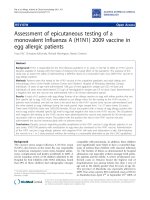
The percentages of circulating monocytes expressing
PD-1, PD-L1, or PD-L2 were markedly increased in
patients with septic shock in comparison with healthy
volunteers during the overall monitoring (Figure 1a).
This augmentation was present for PD-1 (median con-
trol values: 5.0% versus 18.6% (D1-2) and 17.8% (D3-5)
in patients; P < 0.001), for PD-L1 (control values: 10.2%
versus 46.6% (D1-2) and 34.9% (D3-5) in patients; P <
0.001), and for PD-L2 (control values: 2.6% versus 8.7%
(D1-2) and 8.5% (D3-5) in patients; P < 0.001). Similar
results were o bserved when flow cytometry data were
expressed as mean fluoresc ence intensity (MFI) (Table
2). In trauma patients, PD-1-related molecule expres-
sions on monocytes were significantly increased in com-
parison with healthy individuals (for PD-1: control
value: 5.0% versus 9.6%, P = 0.005; for PD-L1: control
value: 10.2% versus 40.1%, P < 0.001; and for PD-L2:
control value: 2.6% versus 7.2%, P < 0.001). However,
PD-1 expression on monocytes was significantly l ower
in trauma than in septic shock patients at D1-2 (9.6%
versus 18.6%, respectively; P = 0.008) (data not shown).
Likewise, the percentages of circulating CD4
+
lympho-
cytes e xpressing PD-1 or PD-L1 were notably increased
in patients with septic shock in comparison with healthy
volunteers during the overall monitoring (for PD-1: con-
trol values: 5.4% versus 15.0% (D1-2) and 13.6% (D3-5),
P < 0.001; for PD-L1: control values: 2.5% versus 3.9%
(D1-2; P = 0.002) and 3.6% (D3-5; P = 0.016) in
patients) (Figure 1b). Alternatively, no significant differ-
ences were observed between patients and healthy
volunteers for percentages of CD4
+
cells expressin g PD-
L2 (Figure 1b) or of CD8
+
lymphocytes positive for PD-
1 (Table 2). Once again, similar results w ere observed
when flow cytometry results were expressed as MFI
(Table 2). No difference in PD-1-related molecule
expressions was observed between trauma patients and
healthy individuals. However , the percentage of PD -1
expressing CD4
+
cells was significantly lower in trauma
than in septic shock patients at D1-2 (5.2% versus
15.0%, respectively; P < 0.001) (data not shown).
Of note, there was no variation of PD-1-related mole-
cule expressions in r egard to age or gender either in
healthy subjects or in patients with septic shock. Indeed,
we did no t observe significant correlations between PD-
1-related molecule expressions and the age of septic
shock patients (r =0.21,P = 0.12 for PD-1 expression
on CD4
+
lymphocytes; r = 0.04, P =0.78forPD-L1
expression on monocytes) or of healthy volunteers (r =
0.10, P = 0.49 for PD-1 expression on CD4
+
lympho-
cytes; r = -0.15, P = 0.30 for PD-L1 expression on
monocytes).
Finally, in 10 patients with septic shock, sequential
blood samples were obtained at D1-2, D3-5, and D6-10
after the onset of shock. During this period, no signifi-
cant variations over time in regard to PD-1 molecule
expressions either on monocytes or on lymphocytes
were observed (Figure 2).
Association between PD-1-related molecule expressions
and clinical parameters
To assess the clinical relevance of the increase in PD-1-
related molecule expressions after septic shock, flow
cytometric measurements were correlated with clinical
parameters and usual biomarkers of sepsis-induced
immunosuppression. No significant correlations were
found between PD-1-related molecule expressions and
percentages of HLA-DR expressing monocytes, CD4
+
lymphocyte count, percentage of c irculating regulatory
T cells, or severity scores calculated at the onset of
shock (SAPS II or SOFA score) (data not shown). How-
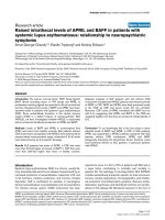
ever, at D1-2, we observed that PD-L1 expression on
monocytes was significantly higher in non-survivors in
comparison with survivors (Figure 3a). Moreover, at D3-
5, patients who went on to develop a secondary nosoco-
mial infection presented with higher PD-1 (Figure 3b)
and PD-L2 (Figure 3c) expressions on their blood
monocytes in comparison with those who remained free
of any secondary nosocomial episode.
Correlation between plasma IL-10 concentration and
PD-1-related molecule expression in patients with
septic shock
Increased circulating IL-10 concentration has been linked
with mortality after septic shock [19] and recently with
enhanced PD-1 expression in HIV-infected patients [20].
We thus measured circulating IL-10 levels in 29 septic
shock patients for whom plasma samples were available
and we correlated this parameter with leukocyte PD-1/
PD-L expressions. Not surprisingly, we observed that
Guignant et al. Critical Care 2011, 15:R99
/>Page 4 of 11
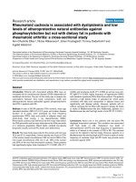
non-survivors exhibited higher plasma IL-10 concentra-
tion than surviv ors at D1-2 and D3-5 (P = 0.01 for both)
(Figure 4a). Interestingly, a significant positive correlation
was measured between PD-1 monocyte expression and
plasma IL-10 concentration in patients at D1-2 (r = 0.49;
P = 0.007) (Figure 4b) but no t at D3-5 (data not shown).
In addition, significant correlations were observed between
both PD-L1 or PD-L2 monocyte expressions and
increased plasma IL-10 concentration at D1-2 (r = 0.58;
P = 0.001 and r =0.45;P = 0.014, respectively) and D3-5
(r =0.45;P = 0.015 and r =0.53;P = 0.003, respectively)
(Figure 4c, d). Of note, no correlations were found
between PD-1/PD-L-re lated molecule expressions on
CD4
+
lymphocytes and changes in plasma IL-10 concen-
tration (data not shown). Also, for all of these observations
made for percentage of positive cells, similar correlations
were obtained when flow cytometry results were expressed
as MFI (data not shown).
CD4+ Lymphocytes
PD-1
0
10
20
30
40
50
Healthy
volunteers
Percent of positive cells
(77.1) (80.6)
PD-L1
Healthy
volunteers
0
5
10
15
20
(27.5)
(28.2)
**
**
**
*
Septic shock patients
D1-2 D3-5
Septic shock patients
D1-2 D3-5
B
0
1
2
3
4
5
6
(6.6)
(21.2)
(19.5)
Healthy
volunteers
Septic shock patients
D1-2 D3-5
PD-L2
0
20
40
60
80
PD-1
Healthy
volunteers
Percent of positive cells
**
**
0
20
40
60
80
100
PD-L1
Healthy
volunteers
PD-L2
0
10
20
30
40
(58.3)
(57.4)
(63.8)
Healthy
volunteers
Monocytes
**
**
**
**
Septic shock patients
D1-2 D3-5
Septic shock patients
D1-2 D3-5
Septic shock patients
D1-2 D3-5
A
Figure 1 PD-1, PD-L1, and PD-L2 measurements on circulating CD4
+
lymphocytes and monocytes in septic shock patients and healthy
volunteers. PD-1-related molecule expressions were measured on circulating monocytes (a) and CD4
+
lymphocytes (b) in whole blood from
healthy volunteers (n = 49) and septic shock patients at day 1 to 2 (D1-2) (n = 37) and at day 3 to 5 (D3-5) (n = 56) after the onset of shock.
Results are presented as percentages of positive cells among total population of monocytes or CD4
+
lymphocytes and as box-plots and
individual values. *P < 0.020, **P ≤ 0.002 (Mann-Whitney U test). A P value of less than 0.025 was considered statistically significant (with
correction for the number of tests). PD-1, programmed death-1; PD-L1, programmed death-ligand 1; PD-L2, programmed death-ligand 2.
Guignant et al. Critical Care 2011, 15:R99
/>Page 5 of 11
Decreased lymphocyte proliferation after septic shock
In an attempt to begin to address the biological signifi-
cance of these changes in PD -1 expression to the devel-
opment of sepsis-induced lymphocyte dysfunction,
freshly isolated PBMCs from septic shock patients and
healthy volunteers were assessed for their capacity to
respond to PHA. As expected, we observed that lym-
phocyte proliferation was significantly reduced in
patients in comparison with hea lthy volunteers (P <
0.001) (Figure 5a). Interestingly, in patients, a significant
negative correlation was observed between this reduced
proliferation and PD-1 (r = -0.81 with P = 0.003) (Figure
5b) or PD-L1 (r = -0.63 with P = 0.039) (data not
shown) overexpression on circulating CD4
+
lympho-
cytes. Similar results were obtained when PD-1 and PD-
L1 staining was expressed as MFI (r = -0.80 with P =
0.003 and r = -0.63 with P = 0.038, respectively).
Discussion
PD-1 and its ligands, PD-L1 and PD-L2, belong t o the
B7-CD28 family of molecules [11]. Co-ligation of T-cell
receptor with the PD-1 system is thought to induce an
inhibitory signal in T cells characterized by cell cycle
arrest, inability to proliferate, and reduced cytokine
synthesis (interferon-gamma (IFN-g)orIL-2orboth
[21-24]). The co-inhibitory PD-1 system has been stu-
died mainly in viral diseases and oncology. This system
may be used by viral pathogens or cancer cells to evade
the host’simmuneresponse[11].Ofnote,invirus-
infected patients, CD8
+
T cells overexpressing PD-1 (in
comparison with healthy volunteers) exhibit a so-called
‘exhaustion profile’ as they produced less IFN-g follow-
ing antigen stimulation, had reduced cytotoxic activity,
and had decreased proliferation in response to specific
antigens [25-27].
Table 2 PD-1-related molecule expressions as mean of fluorescence intensity on leukocytes in septic shock patients
and healthy volunteers
CD4
+
T cells CD8
+
T cells Monocytes
PD-1 PD-L1 PD-L2 PD-1 PD-1 PD-L1 PD-L2
Healthy volunteers Median 8.7 11.5 4.9 13.6 12.3 16.9 8.9
IQR (7.8-10.5) (10.1-12.0) (4.5-5.6) (11.1-20.4) (10.1-15.8) (15.3-18.2) (7.7-9.8)
Median 13.1 11.4 6.0 18.1 17.4 22.0 11.6
Day 1-2 IQR (11.4-19.7) (9.8-14.3) (4.8-7.1) (13.6-24.4) (14.6-24.0) (19.3-31.8) (9.9-13.6)
Septic shock patients P value <0.001 0.150 0.009 0.213 <0.001 <0.001 <0.001
Median 12.2 11.4 5.4 17.5 16.2 21.1 11.1
Day 3-5 IQR (10.8-15.7) (10.0-13.5) (4.4-7.1) (11.8-22.3) (13.0-20.4) (18.2-28.0) (9.6-13.3)
P value <0.001 0.289 0.232 0.306 <0.001 <0.001 <0.001
Programmed death-1 (PD-1)-related molecule expressions were measured on circulating CD4
+
and CD8
+
lymphocytes and monocytes in whole blood from
healthy volunteers (n = 49) and septic shock patients at day 1 to 2 (n = 37) and at day 3 to 5 (n = 56) after the onset of shock. Results are presented as mean
fluorescence intensity. A P value of less than 0.025 was considered statistically significant, and correction for the number of tests was performed (Mann-Whitney
U test). IQR, interquartile range.
PD-1
Percent of positive cells
PD-L1
0
10
20
30
40
D1-2 D3-5 D6-10
PD-L2
0
2
4
6
8
10
12
D1-2 D3-5 D6-10
10
15
20
25
30
35
D1-2 D3-5 D6-10
Figure 2 Sequenti al PD-1, PD-L1, and PD-L2 measurements on circulating CD4
+
lymphocytes and monocytes in patients with septic
shock. In 10 patients with septic shock, sequential blood samples were obtained at day 1 to 2 (D1-2), day 3 to 5 (D3-5), and day 6 to 10 (D6-
10) after the onset of shock, and percentages of PD-1-, PD-L1-, and PD-L2-positive CD4
+
lymphocytes (black diamonds) and monocytes (white
squares) were measured by flow cytometry. Results are expressed as mean ± standard error of the mean. The Friedman test was performed:
P values were greater than 0.05 for all of the analyses. PD-1, programmed death-1; PD-L1, programmed death-ligand 1; PD-L2, programmed
death-ligand 2.
Guignant et al. Critical Care 2011, 15:R99
/>Page 6 of 11
Interestingly, we demonstrated here f or the firs t time
that typical sepsis-immune dysfunctions such as
decreased monocyte HLA-DR expression, decreased cir-
culating CD4
+
T-cell count, and increased percentage of
regulatory T cells [6] were associated with an increased
PD-1 expressio n on CD4
+
lymphocytes (and PD-L1 to a
lesser extent) and increased PD-1, PD-L1, and PD-L2
expressions on monocytes. Of note, during the review of
Survivors Non survivors
10
20
30
40
Mean Fluorescence Intensity
(75)
p= 0.043
PD-L1 (monocytes. D1-2)
Percent of positive cells
Survivors
0
20
40
60
80
100
Non survivors
p= 0.036
PD-1 (monocytes. D3-5)
10
20
30
40
50
Mean Fluorescence Intensity
no NI
NI
p=0.036
A
B
0
20
40
60
80
Percent of positive cells
p=0.086
no NI
NI
5
10
15
20
25
Mean Fluorescence Intensity
p=0.038
no NI
NI
Percent of positive cells
p=0.021
0
10
20
30
40
(57.4)
(58.3)
no NI
NI
PD-L2 (monocytes. D3-5)
C
Figure 3 PD-1-related molecule expressions on monocytes and
clinical outcomes. (a) Monocyte PD-L1 expression was measured
on 26 survivors and 6 non-survivors at day 1 to 2 (D1-2) after the
onset of septic shock. Monocyte PD-1 (b) and PD-L2 (c) expressions
were measured at day 3 to 5 (D3-5) after the onset of shock on 15
patients who developed a secondary nosocomial infection during
their intensive care unit stay (NI) and 38 patients who remained free
of secondary infection (no NI). Flow cytometry data are expressed
as (left) mean fluorescence intensities and (right) percentages of
positive cells out of total circulating monocytes. Results are
presented as box-plots as well as individual values. The Mann-
Whitney U test was performed. PD-1, programmed death-1; PD-L1,
programmed death-ligand 1; PD-L2, programmed death-ligand 2.
% PD-1+ Monocytes
020406080
0
1
2
3
Log
10
IL-10 concentration
r=0.49
p=0.007
B
D1-2
0 20406080100
0
1
2
3
% PD-L1+ Monocytes
Log
10
IL-10 concentration
r=0.58
p=0.001
C
D1-2
0 20406080100
-0.5
0.0
0.5
1.0
1.5
2.0
2.5
% PD-L1+ Monocytes
Log
10
IL-10 concentration
r=0.45
p=0.015
D3-5
010203040506070
0
1
2
3
% PD-L2+ Monocytes
Log
10
IL-10 concentration
r=0.45
p=0.014
0102030405060
-0.5
0.0
0.5
1.0
1.5
2.0
2.5
% PD-L2+ Monocytes
Log
10
IL-10 concentration
r=0.53
p=0.003
D
D1-2
D3-5
A
D1-2
p=0.010
3
Survivors
Non survivors
Log
10
IL-10 concentration
0
1
2
p=0.012
Survivors
Non survivors
Log
10
IL-10 concentration
D3-5
-0.5
0.0
0.5
1.0
1.5
2.0
2.5
Figure 4 Plasma IL-10 concentration and PD- 1 expression in
patients with septic shock. (a) Plasma IL-10 concentration was
measured in survivors and non-survivors at day 1 to 2 (D1-2) (n =
23 and n = 6, respectively) and at day 3 to 5 (D3-5) (n = 24 and n =
5, respectively) after septic shock. Results are presented as box-plots
and as individual values, and horizontal lines represent medians. The
Mann-Whitney U test was performed. (b-d) Correlations between
increased plasma IL-10 concentration and increased PD-1 (b), PD-L1
(c), and PD-L2 (d) expressions on monocytes were calculated at D1-
2 and D3-5 in 29 patients with septic shock. The Spearman
correlation test was used to assess statistical significance. IL-10,
interleukin-10; PD-1, programmed death-1; PD-L1, programmed
death-ligand 1; PD-L2, programmed death-ligand 2.
Guignant et al. Critical Care 2011, 15:R99
/>Page 7 of 11
this article, a study including 19 patients w ith septic
shock confirmed that PD-1 expression on CD4
+
lym-
phocytes and PD-L1 expression on monocytes were ele-
vated in comparison with healthy volunteers [28].
Moreover, w e observed a significant inverse correlation
between increased PD-1 and PD-L1 CD4
+
lymphocyte
expressions and decreased PHA-induced lymphocyte
proliferation in patients with septic shock. Such inverse
correlations have been described in patients with hepati-
tis B [29] and in patients with HIV [14]. Additionally,
we observed a significant correlation between increased
plasma IL-10 concentration and increased PD-1-related
molecule expressions on monocytes from patients with
septic shock. Recently, in an HIV-infected patient
cohort, such a correlation was described and implicated
in the reduced CD4
+
T-cell proliferation observed in
these patients [20]. In accordance with these observa-
tions, we recently showed not only that the increased
septic blood levels of IL-10 are reduced but also that
the rise in lipopolysaccharide-induced IL-10 release by
septic mouse macrophages is lost in animals that are
gen etically deficient (knockou t) in functional PD-1 [15].
Overall, our results therefore suggest a link between
increased PD-1-related molecule expressions and the
development of sepsis-induced immune dysfunctions.
Surprisingly, we found no PD-1 overexpression on cir-
culating CD8
+
T cells in septic patients. This is diver-
gent from the observations made in patients w ith HIV,
hepatitis B virus, or hepatitis C virus [13,25,26,29]. One
explanation may be that CD8
+
cells, which play a promi-
nent role in viral infections, may be less central to the
response patients make to septic shock. This is because
thisresponseisthoughtmainlytobearesponsetoa
bacterial challenge. O f note, Zhang and c olleague s [28]
recently described an increased PD-1 expression on
CD8
+
lymphocytes in a small cohort of 19 septic shock
patients in comparison with healthy volunteers. Thus,
this observation deserves to be further examined in a
larger cohort of septic patients.
Of note, in our cohort, non-surv ivors displayed higher
monocyte PD-L1 expression in comparison with survi-
vors, and patients who went on to develop secondary
noso comial infections had significantly higher PD-1 and
PD-L2 monocyte expressions in comparison with
patients who remained free of secondary infection. This
is consistent with data observed in a murine model of
sepsis, in which after the induction of polymicrobial sep-
tic shock by cecal ligation and puncture (CLP), PD-1
knockout mice show ed a markedly improved capacity to
clear bacteria, both at the local (peritoneal lavage ) and
the systemic (blood) level, in comparison with wild-type
mice [15]. Moreover, PD-L1 blockade significantly
improved survival, prevented sepsis-induced depletion of
lymphocytes, increased tumor necrosis factor-alpha and
IL-6 productions, decreased IL-10 production, and
enhanced bacterial clearance in mice after CLP [30].
Similar data were recently observed ex vivo in patients
with septic shock [28]. Importantly, we show here that
the PD-1 system not only may play a role in immune
dysfunction but also may be an indicator of septic mor-
tality and subsequent infectious episodes in septic
patients.
Increased expressions of co-inhibitory as well as
decreased expressions of co-stimulatory members of the
% PD1+ CD4+ Ly
Proliferation ratio
r=-0.81
p=0.003
0
20
40
60
80
100
Proliferation ratio
Healthy
volunteers
Septic shock
patients
0
100
200
300
400
500
p < 0.001
(761)
A
B
510152025
Figure 5 Ly mphocyte proliferation and PD-1 expressi on in septic shock patients and healthy volunteers. (a) L ymphocyte proliferation
was measured in 16 healthy volunteers and 11 septic shock patients (at day 3 to 5, or D3-5) by
3
H-thymidine incorporation after stimulation
with phytohemagglutinin (5 μg/mL). The proliferation ratio was calculated as the ratio between the numbers of count per minute in the
stimulated wells, divided by non-stimulated wells. Results are presented as box-plots as well as individual values. Statistical significance was
calculated using the Mann-Whitney U test. (b) The correlation between percentages of PD-1
+
CD4
+
lymphocytes (Ly) and proliferation ratio was
assessed in 11 patients with septic shock at D3-5. The Spearman correlation test was performed. PD-1, programmed death-1.
Guignant et al. Critical Care 2011, 15:R99
/>Page 8 of 11
B7-CD28 family of molecules have been described in
ICU patients. In trauma patients, CTLA-4 and PD-1
expressions were elevated in anergic T cells [31]. Similar
results were observed at the mRNA level in trauma
patients with multiple organ dysfunction syndrome [32].
In mice, it was recently shown that B- and T-ly mpho-
cyte attenuator (BTL A) (another co-inhibitory molecule)
was induced at the early phase of Listeria monocytogenes
infection [33]. Moreover, CD3 expression on T lympho-
cytes was reduced in septic shock patients in compari-
son with healthy volunteers [34]. S imilar decreased
expression was observed at the mRNA level in patients
developing sepsis or severe sepsis postoperatively [35]
and in trauma patients [36]. Finally, CD28 expression
(delivering a positive co-signal after ligation to B7.1 or
B7.2) was depressed in trauma patients’ anergic T cells
and may contribute to incomplete activation of these
cells [36]. In total, these alterations may play a major
role in lymphocyte anergy that has been observed in
ICU patients and that has been associated with
increased mortality and risk of nosocomial infections.
They could thus represent potential therapeutic targets
and associated markers to guide future immunothera-
peutic decisions [37].
The present study has some limitations. We could not
address the involvement of the PD-1 system in sepsis-
induced apoptosis. Indeed, PD-1 was first described as
being implicated in programmed cell death [38]. It was
also recently described that PD-1
+
CD8
+
T cells were
more sensitive to both s pontaneous and Fas-induced
apoptosis in comparison with PD-1
-
CD8
+
T cells [14].
Most interestingly, it has recently been reported that
in vivo blockade of PD-1 could decrease T- and B-cell
apoptosis and improve survival in CLP-induced septic
mice [39]. However, given the technical difficulties
encountered in the measurement of apoptosis in clinical
samples, let alone in those of minimal-volume septic
shock patients’ whole blood samples that are already
dedicated to numerous assays [40], this aspect could not
be specifically addressed here and thus deserves to be
investigated in studies specifically dedicated to examin-
ing that process/index.
Conclusions
We describe here for the first time that PD-1/PD-L-
related molecule expressio n is markedly induced on cir-
culating cells of patients with septic shock. Moreover,
increased PD-1-related molecule expression appears to
be correlated with the development of immune dysfunc-
tions, secondary nosocomial infections, and death. We
believe that, although these findings need to be con-
firmed in a larger multicentered clinical study, our
results are in line with the recent commentary of
Hotchkiss and Opal [37], which propos es the use of
anti-PD-1 blocking antibodies in septic patients given
that these molecules are already being tested (and well
tolerated) in clinical trials in patients with cancer.
Although this hypothesis remains a speculation at the
moment and further functional studies are required to
understand the mechanism of action of PD-1-related
molecules in patients with septic shock, the PD-1 family
of receptor and ligands could represent a potential inno-
vative therapeutic strategy with which to restore
immune functions and may further alter morbidity/mor-
tality seen with sepsis, and this is in line with the con-
cept of tailored immunotherapy [41]. Through their
changing expression (alone or together with other mar-
kers), PD-1 molecules could give us insight i nto the
immune status of the septic individual as well as their
possible responsiveness to various establish ed or novel
the rapeutic approaches (or both) used in these critically
ill patients.
Key messages
• Programmed death-1 (PD-1)-related molecule
expressions are increased on circulating monocytes
and CD4
+
lymphocytes after septic shock in compar-
ison with healthy volunteers and trauma patients.
• Increased PD-1-related molecule expressions on
monocytes are significa ntly associated with increased
mortality and occurrence of secondary nosocomial
infections after septic shock.
• Augmented PD-1-relatedmoleculeexpressions
after septic shock are associate d with immune dys-
functions such as decreased mitogen-induced lym-
phocyte proliferation and increased circulating
interleukin-10 concentration.
Abbreviations
CLP: cecal ligation and puncture; D: day; ICU: intensive care unit; IFN-γ:
interferon-gamma; IL: interleukin; IQR: interquartile range; ISS: injury severity
score; MFI: mean fluorescence intensity; PBMC: peripheral blood
mononuclear cell; PD-1: programmed death-1; PD-L1: programmed death-
ligand 1; PD-L2: programmed death-ligand 2; PHA: phytohemagglutinin;
SAPS II: Simplified Acute Physiology Score II; SOFA: sepsis-related organ
failure assessment.
Acknowledgements
We would like to thank Hélène Thizy, Marion Provent, Carmen Fernandez,
and Anne Portier for technical assistance and Nicolas Voirin for his fruitful
advice on statistical analysis.
This research was supported by funds from the Hospices Civils de Lyon, by
DHOS-Inserm ‘Recherche Clinique Translationnelle 2009’ (to GM and FG), by
Fondation Innovation en Infectiologie (FINOVI) (to GM and FV), by the
French Ministry of Health (PHRC 2008) (to GM and AL), and by US National
Institutes of Health grants R01s GM46354 and GM53209 (to AA).
Author details
1
Hospices Civils de Lyon, Hôpital E. Herriot, Laboratoire d’Immunologie, 5
Place d’Arsonval, 69003 Lyon, France.
2
Hospices Civils de Lyon, CH Lyon-Sud,
Service de Réanimation, Chemin du Grand Revoyet, 69495 Pierre-Bénite,
France.
3
Division of Surgical Research, Department of Surgery, Brown
University School of Medicine/Rhode Island Hospital, 593 Eddy Street,
Guignant et al. Critical Care 2011, 15:R99
/>Page 9 of 11
Providence, RI 02903, USA.
4
Hospices Civils de Lyon, CH Lyon-Sud,
Laboratoire d’Immunologie, Chemin du Grand Revoyet, 69495 Pierre-Bénite,
France.
5
Hospices Civils de Lyon, Hôpital E. Herriot, Service de Réanimation, 5
Place d’Arsonval, 69003 Lyon, France.
6
Hospices Civils de Lyon/INSERM,
Centre d’Investigation Clinique (CIC 0201), 52, Boulevard Pinel, 69003 Lyon,
France.
Authors’ contributions
CG, FV, GM, and AL designed the study, collected clinical information,
analyzed raw data, performed statistical analysis, and contributed to writing
the paper. HK, FP, CM, and LD performed the immunological monitoring.
AA, FG, and XH designed the study and contributed to writing the paper.
AC and BA collected clinical information about trauma patients. All the
authors read and approved the final version of the manuscript.
Competing interests
The authors declare that they have no competing interests.
Received: 26 January 2011 Revised: 3 March 2011
Accepted: 21 March 2011 Published: 21 March 2011
References
1. Hotchkiss RS, Karl IE: The pathophysiology and treatment of sepsis. N Engl
J Med 2003, 348:138-150.
2. Dombrovskiy VY, Martin AA, Sunderram J, Paz HL: Rapid increase in
hospitalization and mortality rates for severe sepsis in the United States:
a trend analysis from 1993 to 2003. Crit Care Med 2007, 35:1244-1250.
3. Angus DC, Linde-Zwirble WT, Lidicker J, Clermont G, Carcillo J, Pinsky MR:
Epidemiology of severe sepsis in the United States: analysis of
incidence, outcome, and associated costs of care. Crit Care Med 2001,
29:1303-1310.
4. Vincent JL, Sakr Y, Sprung CL, Ranieri VM, Reinhart K, Gerlach H, Moreno R,
Carlet J, Le Gall JR, Payen D: Sepsis in European intensive care units:
results of the SOAP study. Crit Care Med 2006, 34:344-353.
5. Munford RS, Pugin J: Normal responses to injury prevent systemic
inflammation and can be immunosuppressive. Am J Respir Crit Care Med
2001, 163:316-321.
6. Monneret G, Venet F, Pachot A, Lepape A: Monitoring immune
dysfunctions in the septic patient: a new skin for the old ceremony. Mol
Med 2008, 14:64-78.
7. Cavaillon JM, Adib-Conquy M: Bench-to-bedside review: endotoxin
tolerance as a model of leukocyte reprogramming in sepsis. Crit Care
2006, 10:233.
8. Hotchkiss RS, Tinsley KW, Swanson PE, Schmieg RE Jr, Hui JJ, Chang KC,
Osborne DF, Freeman BD, Cobb JP, Buchman TG, Karl IE: Sepsis-induced
apoptosis causes progressive profound depletion of B and CD4+ T
lymphocytes in humans. J Immunol 2001, 166:6952-6963.
9. Venet F, Chung CS, Kherouf H, Geeraert A, Malcus C, Poitevin F, Bohe J,
Lepape A, Ayala A, Monneret G: Increased circulating regulatory T cells
(CD4(+)CD25 (+)CD127 (-)) contribute to lymphocyte anergy in septic
shock patients. Intensive Care Med 2009, 35:678-686.
10. Monneret G, Lepape A, Voirin N, Bohe J, Venet F, Debard AL, Thizy H,
Bienvenu J, Gueyffier F, Vanhems P: Persisting low monocyte human
leukocyte antigen-DR expression predicts mortality in septic shock.
Intensive Care Med 2006, 32:1175-1183.
11. Keir ME, Butte MJ, Freeman GJ, Sharpe AH: PD-1 and its ligands in
tolerance and immunity. Annu Rev Immunol 2008, 26:677-704.
12. Day CL, Kaufmann DE, Kiepiela P, Brown JA, Moodley ES, Reddy S,
Mackey EW, Miller JD, Leslie AJ, DePierres C, Mncube Z, Duraiswamy J,
Zhu B, Eichbaum Q, Altfeld M, Wherry EJ, Coovadia HM, Goulder PJ,
Klenerman P, Ahmed R, Freeman GJ, Walker BD: PD-1 expression on HIV-
specific T cells is associated with T-cell exhaustion and disease
progression. Nature 2006, 443:350-354.
13. Trautmann L, Janbazian L, Chomont N, Said EA, Gimmig S, Bessette B,
Boulassel MR, Delwart E, Sepulveda H, Balderas RS, Routy JP, Haddad EK,
Sekaly RP: Upregulation of PD-1 expression on HIV-specific CD8+ T cells
leads to reversible immune dysfunction. Nat Med 2006, 12:1198-1202.
14. Petrovas C, Casazza JP, Brenchley JM, Price DA, Gostick E, Adams WC,
Precopio ML, Schacker T, Roederer M, Douek DC, Koup RA: PD-1
is a
regulator of virus-specific CD8+ T cell survival in HIV infection. J Exp Med
2006, 203:2281-2292.
15. Huang X, Venet F, Wang YL, Lepape A, Yuan Z, Chen Y, Swan R, Kherouf H,
Monneret G, Chung CS, Ayala A: PD-1 expression by macrophages plays a
pathologic role in altering microbial clearance and the innate
inflammatory response to sepsis. Proc Natl Acad Sci USA 2009,
106:6303-6308.
16. Bone RC: Toward an epidemiology and natural history of SIRS (systemic
inflammatory response syndrome). JAMA 1992, 268:3452-3455.
17. Landelle C, Lepape A, Francais A, Tognet E, Thizy H, Voirin N, Timsit JF,
Monneret G, Vanhems P: Nosocomial infection after septic shock among
intensive care unit patients. Infect Control Hosp Epidemiol 2008,
29:1054-1065.
18. Monneret G, Elmenkouri N, Bohe J, Debard AL, Gutowski MC, Bienvenu J,
Lepape A: Analytical requirements for measuring monocytic human
lymphocyte antigen DR by flow cytometry: application to the
monitoring of patients with septic shock. Clin Chem 2002, 48:1589-1592.
19. Monneret G, Finck ME, Venet F, Debard AL, Bohe J, Bienvenu J, Lepape A:
The anti-inflammatory response dominates after septic shock:
association of low monocyte HLA-DR expression and high interleukin-10
concentration. Immunol Lett 2004, 95:193-198.
20. Said EA, Dupuy FP, Trautmann L, Zhang Y, Shi Y, El-Far M, Hill BJ, Noto A,
Ancuta P, Peretz Y, Fonseca SG, Van Grevenynghe J, Boulassel MR,
Bruneau J, Shoukry NH, Routy JP, Douek DC, Haddad EK, Sekaly RP:
Programmed death-1-induced interleukin-10 production by monocytes
impairs CD4+ T cell activation during HIV infection. Nat Med 2010,
16:452-459.
21. Latchman Y, Wood CR, Chernova T, Chaudhary D, Borde M, Chernova I,
Iwai Y, Long AJ, Brown JA, Nunes R, Greenfield EA, Bourque K,
Boussiotis VA, Carter LL, Carreno BM, Malenkovich N, Nishimura H,
Okazaki T, Honjo T, Sharpe AH, Freeman GJ: PD-L2 is a second ligand for
PD-1 and inhibits T cell activation. Nat Immunol 2001, 2:261-268.
22. Carter L, Fouser LA, Jussif J, Fitz L, Deng B, Wood CR, Collins M, Honjo T,
Freeman GJ, Carreno BM: PD-1:PD-L inhibitory pathway affects both CD4
(+) and CD8(+) T cells and is overcome by IL-2. Eur J Immunol 2002,
32:634-643.
23. Freeman GJ, Long AJ, Iwai Y, Bourque K, Chernova T, Nishimura H, Fitz LJ,
Malenkovich N, Okazaki T, Byrne MC, Horton HF, Fouser L, Carter L, Ling V,
Bowman MR, Carreno BM, Collins M, Wood CR, Honjo T: Engagement of
the PD-1 immunoinhibitory receptor by a novel B7 family member leads
to negative regulation of lymphocyte activation. J Exp Med 2000,
192:1027-1034.
24. Chemnitz JM, Parry RV, Nichols KE, June CH, Riley JL: SHP-1 and SHP-2
associate with immunoreceptor tyrosine-based switch motif of
programmed death 1 upon primary human T cell stimulation, but only
receptor ligation prevents T cell activation. J Immunol 2004, 173:945-954.
25. Golden-Mason L, Palmer B, Klarquist J, Mengshol JA, Castelblanco N,
Rosen HR: Upregulation of PD-1 expression on circulating and
intrahepatic hepatitis C virus-specific CD8+ T cells associated with
reversible immune dysfunction. J Virol 2007, 81:9249-9258.
26. Nakamoto N, Kaplan DE, Coleclough J, Li Y, Valiga ME, Kaminski M,
Shaked A, Olthoff K, Gostick E, Price DA, Freeman GJ, Wherry EJ, Chang KM:
Functional restoration of HCV-specific CD8 T cells by PD-1 blockade is
defined by PD-1 expression and compartmentalization. Gastroenterology
2008, 134:1927-1937, 1937 e1921-1922.
27. Zhang JY, Zhang Z, Wang X, Fu JL, Yao J, Jiao Y, Chen L, Zhang H, Wei J,
Jin
L, Shi M, Gao GF, Wu H, Wang FS: PD-1 up-regulation is correlated
with HIV-specific memory CD8+ T-cell exhaustion in typical progressors
but not in long-term nonprogressors. Blood 2007, 109:4671-4678.
28. Zhang Y, Li J, Lou J, Zhou Y, Bo L, Zhu J, Zhu K, Wan X, Cai Z, Deng X:
Upregulation of programmed death-1 on T cells and programmed
death ligand-1 on monocytes in septic shock patients. Crit Care 2011, 15:
R70.
29. Peng G, Li S, Wu W, Tan X, Chen Y, Chen Z: PD-1 upregulation is
associated with HBV-specific T cell dysfunction in chronic hepatitis B
patients. Mol Immunol 2008, 45:963-970.
30. Zhang Y, Zhou Y, Lou J, Li J, Bo L, Zhu K, Wan X, Deng X, Cai Z: PD-L1
blockade improves survival in experimental sepsis by inhibiting
lymphocyte apoptosis and reversing monocyte dysfunction. Crit Care
2010, 14:R220.
31. Bandyopadhyay G, De A, Laudanski K, Li F, Lentz C, Bankey P, Miller-
Graziano C: Negative signaling contributes to T-cell anergy in trauma
patients. Crit Care Med 2007, 35:794-801.
Guignant et al. Critical Care 2011, 15:R99
/>Page 10 of 11
32. Laudanski K, Miller-Graziano C, Xiao W, Mindrinos MN, Richards DR, De A,
Moldawer LL, Maier RV, Bankey P, Baker HV, Brownstein BH, Cobb JP,
Calvano SE, Davis RW, Tompkins RG: Cell-specific expression and pathway
analyses reveal alterations in trauma-related human T cell and
monocyte pathways. Proc Natl Acad Sci USA 2006, 103:15564-15569.
33. Sun Y, Brown NK, Ruddy MJ, Miller ML, Lee Y, Wang Y, Murphy KM,
Pfeffer K, Chen L, Kaye J, Fu YX: B and T lymphocyte attenuator tempers
early infection immunity. J Immunol 2009, 183:1946-1951.
34. Venet F, Bohe J, Debard AL, Bienvenu J, Lepape A, Monneret G: Both
percentage of gammadelta T lymphocytes and CD3 expression are
reduced during septic shock. Crit Care Med 2005, 33:2836-2840.
35. Hinrichs C, Kotsch K, Buchwald S, Habicher M, Saak N, Gerlach H, Volk HD,
Keh D: Perioperative gene expression analysis for prediction of
postoperative sepsis. Clin Chem 2010, 56:613-622.
36. De AK, Kodys KM, Pellegrini J, Yeh B, Furse RK, Bankey P, Miller-Graziano CL:
Induction of global anergy rather than inhibitory Th2 lymphokines
mediates posttrauma T cell immunodepression. Clin Immunol 2000,
96:52-66.
37. Hotchkiss RS, Opal S: Immunotherapy for sepsis–a new approach against
an ancient foe. N Engl J Med 2010, 363:87-89.
38. Ishida Y, Agata Y, Shibahara K, Honjo T: Induced expression of PD-1, a
novel member of the immunoglobulin gene superfamily, upon
programmed cell death. EMBO J 1992, 11:3887-3895.
39. Brahmamdam P, Inoue S, Unsinger J, Chang KC, McDunn JE, Hotchkiss RS:
Delayed administration of anti-PD-1 antibody reverses immune
dysfunction and improves survival during sepsis. J Leukoc Biol 2010,
88:233-240.
40. Turrel-Davin F, Guignant C, Lepape A, Mougin B, Monneret G, Venet F: Up
regulation of the pro-apoptotic genes BID and FAS in septic shock
patients. Crit Care 2010, 14:R133.
41. Goyert SM, Silver J: Editorial: PD-1, a new target for sepsis treatment:
better late than never. J Leukoc Biol 2010, 88:225-226.
doi:10.1186/cc10112
Cite this article as: Guignant et al.: Programmed death-1 levels correlate
with increased mortality, nosocomial infection and immune
dysfunctions in septic shock patients. Critical Care 2011 15:R99.
Submit your next manuscript to BioMed Central
and take full advantage of:
• Convenient online submission
• Thorough peer review
• No space constraints or color figure charges
• Immediate publication on acceptance
• Inclusion in PubMed, CAS, Scopus and Google Scholar
• Research which is freely available for redistribution
Submit your manuscript at
www.biomedcentral.com/submit
Guignant et al. Critical Care 2011, 15:R99
/>Page 11 of 11