Báo cáo y học: "New tricks for old NODs" ppsx
Bạn đang xem bản rút gọn của tài liệu. Xem và tải ngay bản đầy đủ của tài liệu tại đây (145.38 KB, 4 trang )
Genome
BBiioollooggyy
2008,
99::
217
Minireview
NNeeww ttrriicckkss ffoorr oolldd NNOODDss
Eric M Pietras* and Genhong Cheng*
†‡
Addresses: *Department of Microbiology, Immunology and Molecular Genetics,
†
Molecular Biology Institute,
‡
Jonsson Comprehensive
Cancer Center, University of California Los Angeles, Los Angeles, CA 90095, USA.
Correspondence: Genhong Cheng. Email:
AAbbssttrraacctt
Recent work has identified the human NOD-like receptor NLRX1 as a negative regulator of
intracellular signaling leading to type I interferon production. Here we discuss these findings and
the questions and implications they raise regarding the function of NOD-like receptors in the
antiviral response.
Published: 25 April 2008
Genome
BBiioollooggyy
2008,
99::
217 (doi:10.1186/gb-2008-9-4-217)
The electronic version of this article is the complete one and can be
found online at />© 2008 BioMed Central Ltd
Upon infection with a pathogen, the host cell must recognize
its presence, communicate this to neighboring cells and
tissues and initiate a biological response to limit the spread
of infection and clear the pathogen. Recognition of invading
microbes proceeds via specialized intracellular and extra-
cellular proteins termed pattern recognition receptors (PRRs),
which recognize conserved molecular motifs found on patho-
gens, known as pathogen-associated molecular patterns
(PAMPs). Recognition of PAMPs by PRRs leads to the
activation of downstream transcription factors, resulting in
induction of programs of host defense gene expression
designed to effect immunity to the pathogen. In the innate
immune response to viruses, the genes activated include
those for the type I interferons - the primary cytokines
mediating the innate response to viral infection. In
mammals, these comprise IFNβ, 13 IFNαs and the more
recently discovered IFNω. Type I interferons signal via the
IFNα/β receptor to induce further sets of genes that regulate
cellular metabolic processes, intracellular nutrient availa-
bility, apoptotic responses and direct elimination of the
pathogen [1].
The recognition of single-stranded RNA viruses in the intra-
cellular space is based on the processing of their genomes by
one of at least two cellular RNA helicases - RIG-I/DDX58
and MDA5/Helicard [2,3]. This processing generates a
conformational change in the helicases, allowing their twin
caspase-recruitment domains (CARDs) to interact directly
with a single amino-terminal CARD in the adaptor protein
MAVS (also known as IPS-1, VISA or Cardif), which is
anchored to the outer mitochondrial membrane [4-7]. MAVS
complexes with the adaptor protein TRAF3, recruiting the
scaffold protein TANK and the IκB kinases (IKKs) TANK-
binding kinase 1 (TBK1) and IKKε, which activate the trans-
cription factor IRF3. IRF3 activation leads to the trans-
criptional activation of a number of antiviral genes, includ-
ing that for IFNβ (Figure 1) [8-11]. MAVS also acts as a
bifurcation point for a second signaling pathway that can be
triggered by RIG-I and some other PRRs. In this pathway
the transcription factor NF-κB is activated, resulting in the
activation of NF-κB-responsive genes (Figure 1) [4-7,10,12].
In a paper recently published in Nature, Moore et al. [13]
have shown that these MAVS-mediated pathways can be
inhibited by the action of an intracellular NOD-like receptor
(NLR), the protein NLRX1, indicating that members of this
ancient family of pathogen sensors can evolve to acquire new
regulatory roles in mammalian host defense.
NNOODD lliikkee rreecceeppttoorrss aanndd tthhee aannttiivviirraall rreessppoonnssee
The NLR proteins generally act as intracellular sensors of
infection, analogous to the cell-surface Toll-like receptors
(TLRs), and their role in responses to bacterial and viral
pathogens is of considerable current interest. These
proteins are components of an evolutionarily ancient
immune mechanism that appears to have evolved before the
divergence of the plant and animal kingdoms - in plants,
NLRs function as sensors of infection or physiological
‘danger’ signals that trigger cell-death processes to limit the
spread of disease [14]. NLRs contain a central nucleotide-
binding domain (NBD) and a series of leucine-rich repeats
(LRRs), the latter appearing to constitute a regulatory sensor
region that enables activation of the protein [15]. Most NLRs
also contain an effector domain such as a CARD or pyrin
domain, with which activated NLRs can interact with
proteins such as the CARD- and pyrin-containing adaptor
protein ASC, which links pyrin-containing NLRs with the
CARD domain of the protease caspase-1 [15,16]. Whereas
their plant-based relatives primarily mediate cell-death
processes, some mammalian NLRs have been suggested to
regulate genetic responses directly, as in the case of NOD1
and NOD2, or indirectly by mediating the proteolytic
activation of cytokines that in turn activate pathways leading
to expression of host-defense genes [17].
Of the latter NLRs, one of the best characterized in
responses to viral infection is NLRP3/NALP3/CIAS, which
mediates caspase-1 activation via aggregation with ASC and
caspase-1 into ‘inflammasomes’. These inflammasomes
mediate the autoproteolytic cleavage of caspase-1 into its
active form, which in turn cleaves the pro-inflammatory
cytokines IL-1β and IL-18, enabling them to be secreted [16].
The demonstration that NALP3 is involved in caspase-1
activation and the secretion of IL-1β and IL-18 in macro-
phages in response to RNA and DNA viruses helped to
clarify the role of NLRs in antiviral responses [18,19]. These
findings suggested that in mammals NLR proteins retained
their classical role as soluble activators of caspases in
response to viral infection, much as they do in plants. But
was it possible that NLRs could also have a quite different
role in regulating host-defense pathways?
The recent study by Moore et al. [13] suggests that old NODs
can indeed learn new tricks. These authors used bio-
informatics approaches to predict a mitochondrial localiza-
tion for NLRX1 (also known as CLR11.3 or NOD9), one of 22
NLRs found in humans. After verifying its localization in the
outer mitochondrial membrane, the group assessed whether
NLRX1 might be involved in MAVS-mediated antiviral
responses, given that MAVS is also anchored on the mito-
chondrial surface. Indeed, their biochemical data suggest
that the NBD of NLRX1 interacts with the CARD domain of
MAVS, even in the absence of viral infection. Interestingly,
they found that NLRX1 overexpression seems to strongly
repress MAVS or RIG-I-driven IFNβ and NFκB reporter
activity and IRF3 dimerization. Furthermore, the authors
show that knockdown of NLRX1 by small interfering RNAs
leads to increased interferon production in response to
MAVS overexpression or viral infection. Taken collectively,
their data suggest that NLRX1 attenuates MAVS-mediated
activation of NFκB and IRF3, possibly by interfering with
the interaction of RIG-I with MAVS. These findings suggest
that NLRX1 functions to negatively regulate interferon
responses activated via RIG-I, highlighting the malleability
of the evolutionarily ancient NLR family in its capacity to
carry out numerous immunological functions in distinct
cellular compartments.
FFuurrtthheerr qquueessttiioonnss aabboouutt NNLLRRXX11
This study leaves a number of interesting questions still
open. In particular, the precise mechanism by which NLRX1
inhibits MAVS-mediated signaling is not clear. The data of
Moore et al. [13] suggest that MAVS and NLRX1 may
interact constitutively, and that NLRX1 can inhibit the inter-
action between RIG-I and MAVS. While this suggests that
NLRX1 interferes with the interaction between RIG-I and
MAVS, it follows that this interference must be overcome to
allow for proper interferon signaling. Perhaps activated
RIG-I has a higher affinity for the CARD domain of MAVS
than does NLRX1, thus titrating out the NLRX1-MAVS
/>Genome
BBiioollooggyy
2008, Volume 9, Issue 4, Article 217 Pietras and Cheng 217.2
Genome
BBiioollooggyy
2008,
99::
217
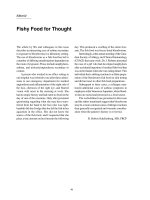
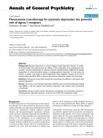
FFiigguurree 11
Activation of the transcription factors IRF3 and NF-κB in response to
infection with a single-stranded RNA virus. On viral infection, RIG-I
activated by viral RNA interacts with the adaptor protein MAVS, which
represents a bifurcation point for the activation of IRF3 and NF-κB via
activation of distinct IKK family members. Activation of NF-κB involves
phosphorylation of its cytoplasmic inhibitor IκBκ, which tags that protein
for destruction with the consequent release of NF-κB. IRF3 and NF-κB in
turn activate a number of genes important in the antiviral response,
including that for IFNβ. NLRX1 has been recently shown to inhibit this
pathway, possibly by blocking the interaction of RIG-I with MAVS.
NLRX1
IFNβ
P
P
P
RIG-I
MAVS
TRAF3
TBK1 IKKε
IRF3
Mitochondrion
IKKα IKKβ
NEMO
NFκB
P
P
IκBκ
ssRNA virus
FADD
TANK
RIP1
Casp 8/10
NFκB
IRF3
P
CARD domains
Nucleus
interaction and allowing interferon signaling. Alternatively,
the LRR domain of NLRX1 might pick up ‘danger’ signals
generated by viral infection in a fashion similar to NALP3,
thus releasing inhibition by making the NLRX1-MAVS inter-
action less favorable. Furthermore, as NLRX1 can inhibit
interferon signaling induced by overexpressed MAVS in the
absence of virus, the role of NLRX1 in blocking the inter-
action between MAVS and downstream interferon signaling
components should be addressed.
An interesting, although elusive, aspect of the MAVS-NLRX1
story is the function of mitochondrial localization for these
proteins. In both cases, loss of mitochondrial localization as
a result of experimental manipulation or cleavage from the
membrane by viral proteases, as in the case of deactivation
of MAVS by the hepatitis C virus protease NS3/4A,
completely destroys the function of these proteins [5,6,13]. It
may be, as Moore et al. [13] suggest, that the mitochondrion
provides a useful platform on which sufficient concentra-
tions of signaling elements can be marshaled to effect down-
stream signaling processes. Given the key role of mito-
chondria in apoptotic and metabolic functions and the inti-
mate relationship of these processes with viral infection, it is
no small leap to reason that MAVS and NLRX1 may serve as
an interface between them. In addition, as with MAVS,
cleavage of NLRX1 from the mitochondrial surface by endo-
genous or viral proteases might serve as a mechanism for
damping NLRX1-mediated inhibition of interferon production.
It was previously shown by the same group that Monarch-1/
NLRP12, a soluble NLR family member, can inhibit activation
of the noncanonical NF-κB pathway in response to CD40
stimulation [15,20]. Thus, Monarch-1, and now NLRX1,
represent what is probably a recent evolutionary retooling of
some NLRs from inflammatory or cell-death mediators to
checkpoint proteins designed to regulate immunological
signaling processes. Given the fact that NLRs essentially act as
molecular switches in response to stimuli sensed via their
LRRs, it seems logical that they might be adapted to act as
negative regulators that can be inducibly released or activated
in the appropriate conditions. Indeed, the concept of such
switches is recapitulated in many other biological systems: the
Ras family of GTPases is but one example.
A persistent question and the genesis of significant debate
within the innate immunity field is the mechanism by which
these NLR switches are activated. Taking a precedent from
the study of Toll-like receptors, some of whose LRR domains
have been shown to physically interact with ligands, the
conventional wisdom has been that NLRs also respond to
specific PAMPs. Indeed, NOD1 and NOD2 have been shown
to respond via their LRRs to bacterial peptidoglycans,
although convincing biochemical evidence showing a direct
interaction is lacking [16,17]. However, several studies
showing that NALP3-mediated inflammasome formation is
induced by a wide range of stimuli, from uric acid crystals to
double-stranded RNA to ionophore stimulation, has thrown
this conventional wisdom into disfavor [18,19,21-23]. The
prevailing alternative hypothesis is that NLRs respond to
nonspecific cellular perturbations or danger signals rather
than discrete ligands. Thus, it will be important to determine
what, if any, signal might be sensed via the LRRs of NLRX1.
Given that NALP3 also responds to viral infection, it will be
interesting to determine whether these two NLRs might
respond to the same signal upon viral infection.
It is clear that there are numerous unanswered questions on
the biology of NLRX1 in the interferon response as well as on
the biology of NLRs in general. Although the interferon
response might be considered an evolutionary contemporary
of NLRs, the findings of Moore et al. [13] clearly suggest that
the members of this family of proteins, and NLRX1 in
particular, have evolved to play significant roles in directly
regulating pathways that control more modern biological
functions.
RReeffeerreenncceess
1. Theofilopoulos AN, Baccala R, Beutler B, Kono DH:
TTyyppee II iinntteerrffeerr
oonnss ((aallpphhaa//bbeettaa)) iinn iimmmmuunniittyy aanndd aauuttooiimmmmuunniittyy
Annu Rev Immunol
2005,
2233::
307-336.
2. Kato H, Takeuchi O, Sato S, Yoneyama M, Yamamoto M, Matsui K,
Uematsu S, Jung A, Kawai T, Ishii KJ, Yamaguchi O, Otsu K, Tsu-
jimura T, Koh CS, Reis e Sousa C, Matsuura Y, Fujita T, Akira S:
DDiiff
ffeerreennttiiaall rroolleess ooff MMDDAA55 aanndd RRIIGG II hheelliiccaasseess iinn tthhee rreeccooggnniittiioonn ooff
RRNNAA vviirruusseess
Nature
2006,
444411::
101-105.
3. Yoneyama M, Kikuchi M, Natsukawa T, Shinobu N, Imaizumi T,
Miyagishi M, Taira K, Akira S, Fujita T:
TThhee RRNNAA hheelliiccaassee RRIIGG II hhaass
aann eesssseennttiiaall ffuunnccttiioonn iinn ddoouubbllee ssttrraannddeedd RRNNAA iinndduucceedd iinnnnaattee aannttiivvii
rraall rreessppoonnsseess
Nat Immunol
2004,
55::
730-737.
4. Kawai T, Takahashi K, Sato S, Coban C, Kumar H, Kato H, Ishii KJ,
Takeuchi O, Akira S:
IIPPSS 11,, aann aaddaappttoorr ttrriiggggeerriinngg RRIIGG II aanndd MMddaa55
mmeeddiiaatteedd ttyyppee II iinntteerrffeerroonn iinndduuccttiioonn
Nat Immunol
2005,
66::
981-988.
5. Meylan E, Curran J, Hofmann K, Moradpour D, Binder M, Barten-
schlager R, Tschopp J:
CCaarrddiiff iiss aann aaddaappttoorr pprrootteeiinn iinn tthhee RRIIGG II
aannttiivviirraall ppaatthhwwaayy aanndd iiss ttaarrggeetteedd bbyy hheeppaattiittiiss CC vviirruuss
Nature
2005,
443377::
1167-1172.
6. Seth RB, Sun L, Ea CK, Chen ZJ:
IIddeennttiiffiiccaattiioonn aanndd cchhaarraacctteerriizzaattiioonn
ooff MMAAVVSS,, aa mmiittoocchhoonnddrriiaall aannttiivviirraall ssiiggnnaalliinngg pprrootteeiinn tthha
att aaccttiivvaatteess
NNFF kkaappppaaBB aanndd IIRRFF 33
Cell
2005,
112222::
669-682.
7. Xu LG, Wang YY, Han KJ, Li LY, Zhai Z, Shu HB:
VVIISSAA iiss aann aaddaapptteerr
pprrootteeiinn rreeqquuiirreedd ffoorr vviirruuss ttrriiggggeerreedd IIFFNN bbeettaa ssiiggnnaalliinngg
Mol Cell
2005,
1199::
727-740.
8. Hiscott J, Grandvaux N, Sharma S, Tenoever BR, Servant MJ, Lin R:
CCoonnvveerrggeennccee ooff tthhee NNFF kkaappppaaBB aanndd iinntteerrffeerroonn ssiiggnnaalliinngg ppaatthhwwaayyss iinn
tthhee rreegguullaattiioonn ooff aannttiivviirraall ddeeffeennssee aanndd aappooppttoossiiss
Ann NY Acad Sci
2003,
11001100::
237-248.
9. Honda K, Takaoka A, Taniguchi T:
TTyyppee II iinntteerrffeerroonn [[ccoorrrreecctteedd]]
ggeennee iinndduuccttiioonn bbyy tthhee iinntteerrffeerroonn rreegguullaattoorryy ffaaccttoorr ffaammiillyy ooff ttrraannssccrriipp
ttiioonn ffaaccttoorrss
Immunity
2006,
2255::
349-360.
10. Saha SK, Pietras EM, He JQ, Kang JR, Liu SY, Oganesyan G, Sha-
hangian A, Zarnegar B, Shiba TL, Wang Y, Cheng G:
RReegguullaattiioonn ooff
aannttiivviirraall rreessppoonnsseess bbyy aa ddiirreecctt aanndd ssppeecciiffiicc iinntteerraaccttiioonn bbeettwweeeenn
TTRRAAFF33 aanndd CCaarrddiiff
EMBO J
2006,
2255::
3257-3263.
11. Sato S, Sugiyama M, Yamamoto M, Watanabe Y, Kawai T, Takeda K,
Akira S:
TToollll//IILL 11 rreecceeppttoorr ddoommaaiinn ccoonnttaaiinniinngg aaddaappttoorr iinndduucciinngg IIFFNN
bbeettaa ((TTRRIIFF)) aassssoocciiaatteess wwiitthh TTNNFF rreecceeppttoorr aassssoocciiaatteedd ffaaccttoorr 66 aanndd
TTAANNKK bbiinnddiinngg kkiinnaassee 11,, aanndd aaccttiivvaatteess ttwwoo ddiissttiinncctt ttrraannssccrriippttiioonn
ffaaccttoorrss,, NNFF kkaappppaa BB aanndd IIFFNN rreegguullaattoorryy ffaaccttoorr 33,, iinn tthhee TToollll lliikkee
rreecceeppttoorr ssiiggnnaalliinngg
J Immunol
2003,
117711::
4304-4310.
12. Takahashi K, Kawai T, Kumar H, Sato S, Yonehara S, Akira S:
RRoolleess
ooff ccaassppaassee 88 aanndd ccaassppaassee 1100 iinn iinnnnaattee iimmmmuunnee rreessppoonnsseess ttoo ddoouubbllee
ssttrraannddeedd RRNNAA
J Immunol
2006,
117766::
4520-4524.
13. Moore CB, Bergstralh DT, Duncan JA, Lei Y, Morrison TE, Zimmer-
mann AG, Accavitti-Loper MA, Madden VJ, Sun L, Ye Z, Lich JD,
/>Genome
BBiioollooggyy
2008, Volume 9, Issue 4, Article 217 Pietras and Cheng 217.3
Genome
BBiioollooggyy
2008,
99::
217
Heise MT, Chen Z, Ting JP:
NNLLRRXX11 iiss aa rreegguullaattoorr ooff mmiittoocchhoonnddrriiaall
aannttiivviirraall iimmmmuunniittyy
Nature
2008,
445511::
573-577.
14. DeYoung BJ, Innes RW:
PPllaanntt NNBBSS LLRRRR pprrootteeiinnss iinn ppaatthhooggeenn sseennssiinngg
aanndd hhoosstt ddeeffeennssee
Nat Immunol
2006,
77::
1243-1249.
15. Lich JD, Ting JP:
CCAATTEERRPPIILLLLEERR ((NNLLRR)) ffaammiillyy mmeemmbbeerrss aass ppoossiittiivvee
aanndd nneeggaattiivvee rreegguullaattoorrss ooff iinnffllaammmmaattoorryy rreessppoonnsseess
Proc Am Thorac
Soc
2007,
44::
263-266.
16. Petrilli V, Dostert C, Muruve DA, Tschopp J:
TThhee iinnffllaammmmaassoommee:: aa
ddaannggeerr sseennssiinngg ccoommpplleexx ttrriiggggeerriinngg iinnnnaattee iimmmmuunniittyy
Curr Opin
Immunol
2007,
1199::
615-622.
17. Franchi L, McDonald C, Kanneganti TD, Amer A, Núñez G:
NNuucclleeoottiiddee bbiinnddiinngg oolliiggoommeerriizzaattiioonn ddoommaaiinn lliikkee rreecceeppttoorrss:: iinnttrraacceelllluu
llaarr ppaatttteerrnn rreeccooggnniittiioonn mmoolleeccuulleess ffoorr ppaatthhooggeenn ddeetteeccttiioonn aanndd hhoosstt
ddeeffeennssee
J Immunol
2006,
117777::
3507-3513.
18. Kanneganti TD, Body-Malapel M, Amer A, Park JH, Whitfield J,
Franchi L, Taraporewala ZF, Miller D, Patton JT, Inohara N, Núñez
G:
CCrriittiiccaall rroollee ffoorr CCrryyooppyyrriinn//NNaallpp33 iinn aaccttiivvaattiioonn ooff ccaassppaassee 11 iinn
rreessppoonnssee ttoo vviirraall iinnffeeccttiioonn aanndd ddoouubbllee ssttrraannddeedd RRNNAA
J Biol Chem
2006,
228811::
36560-36568.
19. Muruve DA, Petrilli V, Zaiss AK, White LR, Clark SA, Ross PJ, Parks
RJ, Tschopp J:
TThhee iinnffllaammmmaassoommee rreeccooggnniizzeess ccyyttoossoolliicc mmiiccrroobbiiaall aanndd
hhoosstt DDNNAA aanndd ttrriiggggeerrss aann iinnnnaattee iimmmmuunnee rreessppoonnssee
Nature
2008,
445522::
103-107.
20. Lich JD, Williams KL, Moore CB, Arthur JC, Davis BK, Taxman DJ,
Ting JP:
MMoonnaarrcchh 11 ssuupppprreesssseess nnoonn ccaannoonniiccaall NNFF kkaappppaaBB aaccttiivvaattiioonn
aanndd pp5522 ddeeppeennddeenntt cchheemmookkiinnee eexxpprreessssiioonn iinn mmoonnooccyytteess
J Immunol
2007,
117788::
1256-1260.
21. Martinon F:
DDeetteeccttiioonn ooff iimmmmuunnee ddaannggeerr ssiiggnnaallss bbyy NNAALLPP33
J Leukoc
Biol
2008,
8833::
507-511.
22. Kanneganti TD, Ozören N, Body-Malapel M, Amer A, Park JH,
Franchi L, Whitfield J, Barchet W, Colonna M, Vandenabeele P,
Bertin J, Coyle A, Grant EP, Akira S, Núñez G:
BBaacctteerriiaall RRNNAA aanndd
ssmmaallll aannttiivviirraall ccoommppoouunnddss aaccttiivvaattee ccaassppaassee 11 tthhrroouugghh
ccrryyooppyyrriinn//NNaallpp33
Nature
2006,
444400::
233-236.
23. Mariathasan S, Weiss DS, Newton K, McBride J, O’Rourke K, Roose-
Girma M, Lee WP, Weinrauch Y, Monack DM, Dixit VM:
CCrryyooppyyrriinn
aaccttiivvaatteess tthhee iinnffllaammmmaassoommee iinn rreessppoonnssee ttoo ttooxxiinnss aanndd AATTPP
Nature
2006,
444400::
228-232.
/>Genome
BBiioollooggyy
2008, Volume 9, Issue 4, Article 217 Pietras and Cheng 217.4
Genome
BBiioollooggyy
2008,
99::
217