Báo cáo y học: "A non-random walk through the genome" pdf
Bạn đang xem bản rút gọn của tài liệu. Xem và tải ngay bản đầy đủ của tài liệu tại đây (271.44 KB, 6 trang )
Genome Biology 2005, 6:214
comment
reviews
reports deposited research
interactions
information
refereed research
Review
A non-random walk through the genome
Brian Oliver* and Tom Misteli
†
Addresses: *National Institute of Diabetes and Digestive and Kidney Diseases, National Institutes of Health, Department of Health and
Human Services, Bethesda, MD 20892, USA.
†
National Cancer Institute, National Institutes of Health, Department of Health and Human
Services, Bethesda, MD 20892, USA.
Correspondence: Brian Oliver. E-mail: Tom Misteli. E-mail:
Abstract
Recent publications on a wide range of eukaryotes indicate that genes showing particular
expression patterns are not randomly distributed in the genome but are clustered into
contiguous regions that we call neighborhoods. It seems probable that this organization is related
to chromatin and the structure of the nucleus.
Published: 31 March 2005
Genome Biology 2005, 6:214 (doi:10.1186/gb-2005-6-4-214)
The electronic version of this article is the complete one and can be
found online at />© 2005 BioMed Central Ltd
Consideration of gene regulation as an interaction between the
basal transcription machinery, regulators, and a segment of
naked DNA containing a gene has been an extremely successful
model for understanding how genetic information is read [1]. It
is impossible to separate this model from the great success of
modern molecular genetics. Naked DNA does not exist in
nature, however. Packing DNA into the nucleus, segregating it
during cell division, and making sure that it is readily available
when needed for transcription is an enormously complex task
for the cell and needs extensive interactions of proteins with
DNA. Structural considerations have dominated our views of
nuclear architecture but have not greatly influenced our con-
cepts of transcription, other than by the broad assumption that
such structure can hinder transcription. As with a building,
however, the structure of the nucleus has a significant, albeit
possibly subtle, influence on the work performed within it.
Recent work in many different eukaryotes has suggested that
genes with particular expression patterns are sometimes
found in contiguous regions of the genome. We call these
regions gene-expression neighborhoods; we avoid using the
term ‘cluster’ because this usually refers to potentially co-
regulated genes, regardless of their genomic position. The
human eye is exceedingly adept at finding patterns and does
so even in randomness (the constellations of stars are an
example of patterns that humans have imposed on a random
distribution). Here, we review the evidence that gene-
expression neighborhoods are real. Although there are some
lingering questions about the best methods for finding them
and about how to avoid being tricked by spurious or artifac-
tual patterns, the body of available evidence leaves little
doubt that they exist. With a few exceptions from classical
molecular biology, the regulatory mechanisms underlying
gene-expression neighborhoods are not understood. Investi-
gating these mechanisms will be a major challenge, but we
believe that some will involve the structure of the nucleus. We
review the evidence for non-random positioning of chromo-
somes and genes in the nucleus and suggest ways in which
the regulation of gene-expression neighborhoods can be
studied.
Evidence for gene-expression neighborhoods
It has long been known that prokaryotic genes are organized
into operons [2]. Even in eukaryotes, it has been recognized
that some genes encoding related functions are near neighbors
in the genome: the clusters of genes encoding histones and
tRNAs and the arrays of ribosomal RNA (rRNA) genes were
early examples [3]. Although the lists of known gene neighbor-
hoods like these have grown and now include important regu-
latory genes such as the Hox and microRNA genes [4,5], most
researchers have considered gene neighborhoods oddities.
There have been hints that gene-expression neighborhoods are
widespread. For example, direct examination of Drosophila
polytene chromosomes showed nascent transcripts being
generated from many genes in some multigene segments [6],
and the systematic analysis of the effect of position on trans-
gene expression supported the idea that there are regions,
both euchromatic and heterochromatic, that are broadly
repressive [7]. Functional assays like these, in conjunction
with structural studies, raise the possibility that loop
domains within chromosomes (the domains between
matrix-attachment regions) are units of transcriptional regu-
lation [8]. Genome-scale analyses of gene expression using
microarrays have resulted in an explosion of papers describing
gene-expression neighborhoods. Non-random expression of
pairs of adjacent genes has been reported in yeast [9] and
plants [10], and among highly expressed human genes [11],
genes expressed in Caenorhabditis elegans muscle [12], genes
differentially expressed in embryonic versus adult Drosophila
[13,14], genes differing in expression between the sexes [15,16],
and genes expressed in cancerous cells [17].
There are a number of pitfalls to the analysis of neighbor-
hoods of contiguous genes, calling for great analytical rigor.
For example, the position of the elements on the array must
be taken into account, as non-random hybridization across
the array landscape is often significant: for example, some
early arrays had probes printed in the same order as they
appear in the genome [18]. But expression data generated
using arrays with probes printed randomly with respect to
chromosome position, analyzed under very stringent criteria
comparing real data to 100,000 random sets, still show
gene-expression neighborhoods (an example from our own
analyses on Drosophila [16] is shown in Figure 1a). Findings
of such non-random patterns are also independent of the
technique used. Sequencing-based assays such as expressed
sequence tags (ESTs) and serial analysis of gene expression
(SAGE) are not subject to array artifacts of any kind; experi-
ments using these techniques have shown in mammals that
highly expressed genes [19] and genes with testis-biased
[20] or organ-biased [21] expression are organized into
neighborhoods. Finally, a computational analysis of the gene
correlation between gene location and inferred gene func-
tion in many eukaryotes shows neighborhood structure [19].
The fact that genes can be moved to new locations in the
genome and often behave more or less as expected in the
new location suggests that the effects of neighborhoods on
gene expression are subtle. But subtle is not synonymous
with unimportant. Reverse-genetic studies have shown that
many genes have little overt phenotype when mutated or
deleted - usually because of redundancy with other genes -
but even such apparently redundant duplicated genes cannot
last through evolutionary timescales without evolving func-
tions that differentiate them from other genes [22]. Our
inability to assay small differences in fitness limits the identi-
fication of subtle effects. Conservation of neighborhood struc-
ture over a long evolutionary history, as seen for the Hox
genes [4] and in prokaryotic genomes [23], is a very powerful
indicator that neighborhoods are functional. Gene expression
within the blocks of the Drosophila melanogaster genome
that are conserved with Drosophila pseudoobscura is highly
correlated, suggesting that neighborhood structure is gener-
ally conserved [15].
Non-random positioning of chromosomes in
the nucleus
In the interphase nucleus of virtually all eukaryotes, the
genetic material of each chromosome occupies a spatially
limited, roughly spherical volume, with a diameter about a
tenth of that of the nucleus, referred to as a chromosome ter-
ritory [24,25] (Figure 1b). Despite the homogeneous dense
appearance of these territories when visualized by in situ
hybridization methods, their interiors are accessible to regu-
latory factors, and they are open enough to allow transport
of mRNA and proteins through the nucleus. Chromosome
214.2 Genome Biology 2005, Volume 6, Issue 4, Article 214 Oliver and Misteli />Genome Biology 2005, 6:214
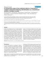
Figure 1
An illustration of gene neighborhoods and chromosome territories.
(a) A heat diagram showing normalized hybridization intensities along a
segment of a Drosophila chromosome. Samples are arrayed from left to
right, and samples from testis and males (including testis) are indicated;
genes that are adjacent on chromosome 3R are listed from top to
bottom. Four contiguous genes, including the don juan gene (dj) that
encodes a sperm tail protein, show testis-biased and male-biased
expression. Figure generated using data from [16]. (b) A micrograph of
liver cells, showing the positions that two chromosomes preferentially
occupy within the nucleus. Chromosome 12 (green) is frequently found
towards the periphery, whereas chromosome 15 (red) tends to localize
towards the center of the nucleus. Blue indicates total DNA staining.
CG1162 BG:DS00276.8
CG1034 bcd
CG2198 Ama
CG2189 Dfd
CG1030 Scr
CG2047 ftz
CG1028 Antp
CG1982 Sodh-1
CG1979 BG:DS00464.1
CG1980 dj
CG1984 CG1984
CG1988 CG1988
CG1105 CG1105
CG1965 CG1965
CG1104 CG1104
CG1034 bcd
CG1943 CG1943
CG1943 CG1943
CG1101 Aly
CG1939 CG1939
Testis
Samples
Males
High Low
Genes in order along the chromosome
CG ID Name
(a)
(b)
Chr 12
Chr 15
DNA
territories are arranged non-randomly within the volume of
the nucleus. In plants, flies and yeast, chromosomes are
often arranged with their telomeres clustered at one end of
the nucleus and their centromeres associated with the other
[26-29]. An extreme example is the polarized nuclei of
Drosophila embryos, where chromosomes are aligned in
apical-basal orientation with each gene localized according
to its chromosomal position [30]. In mammalian cells, chro-
mosomes are not aligned in this way, but they do occupy
non-random positions. Analysis of human lymphocytes and
fibroblasts suggests preferential localization of chromo-
somes relative to the center of the nucleus [31,32]: in this
radial arrangement, gene-dense chromosomes tend to local-
ize towards the center of the nucleus, whereas chromosomes
with low gene density tend to associate with the nuclear
periphery [32]. Other studies provide evidence for a correla-
tion between chromosome size and radial position, with
small chromosomes clustering towards the center of the
nucleus and larger chromosomes towards the periphery
[33]. We are largely ignorant about the rules that determine
this organization.
Although the molecular mechanisms determining chromo-
some position are unknown, it seems unlikely that the posi-
tioning of entire chromosomes is controlled by a precise
positioning mechanism involving dedicated machinery
because chromosome position differs between cell types and
even varies widely within a cell population [31,34]. It seems
more probable that preferential radial chromosome posi-
tions are a reflection of the global physical properties of a
chromosome, such as size, the amount of chromatin conden-
sation, and levels of gene expression. For example, the corre-
lation between radial position and gene density agrees with
findings that gene-poor chromosome regions are generally
more condensed than gene-rich regions. This is consistent
with the idea that the physical nature of a chromosome con-
tributes to its position [35]. A report that highly transcribed
genes are in neighborhoods on human chromosomes [11]
suggests that the transcription of these genes might drive the
positioning of host chromosomes within the nucleus, or
certain positions might enable higher expression.
Chromosomes are also non-randomly positioned with
respect to each other within the nuclear space [36]. The
classic example is the clustering of chromosomes bearing the
genes encoding rRNAs. In mammalian cells, these chromo-
somes congregate to form a nucleolus where ribosomal RNA
is transcribed from the tandemly repeated rRNA genes [37].
Although it can be argued that this is a special case, other
non-rDNA-bearing chromosomes associate near the nucleo-
lus. For example, in mouse cells, the rDNA-bearing chromo-
somes 12 and 15 form a triplet cluster with the
non-rDNA-bearing chromosome 14 at high frequency [25].
This kind of higher-order arrangement could link gene-
expression neighborhoods that are distant in linear terms
along the genome.
Is the chromosome a unit of expression, such that all genes on
a chromosome share some aspect of their regulation? While
this might seem unlikely, the sex chromosomes of many
organisms show an unusual expression pattern. The mam-
malian Y chromosome is highly heterochromatic and has
gene-expression neighborhoods that are required for testis
function [38]. In mammalian females, X chromosomes
undergo inactivation [39], and the inactive X is characteristi-
cally positioned at the nuclear periphery. In Drosophila males,
the X chromosome associates with chromosome-specific chro-
matin-remodeling machines that upregulate expression [40].
More recently, it has been shown that the X chromosome has
fewer genes with testis-biased expression than other chromo-
somes [41-43]. Although it seems likely that sex chromosomes
are the exceptions in this respect, the small chromosome 4 in
Drosophila is decorated with a specific chromatin-associated
protein of unknown function [44], which might regulate
expression and/or positioning.
Non-random positioning of genes
In contrast to entire chromosomes, a gene’s position rela-
tive to various nuclear landmarks is emerging as an impor-
tant contributor to its function [45]. Association of genes
with the nuclear periphery is a hallmark of silencing. Tran-
scriptionally silent heterochromatin is enriched at the
edges of the nucleus in many organisms; for example,
silenced Saccharomyces cerevisiae telomeres are always at
the nuclear periphery [46]. But the story is more compli-
cated. It is now clear that silencing of telomere regions
does not require association with the periphery but occurs
throughout the nucleus [47]. In addition, a genome-wide
survey shows that a large number of S. cerevisiae genes
appear to translocate towards the nuclear periphery upon
activation and associate with a nuclear pore complex in
their active state, suggesting that the nuclear periphery is
not a silencing compartment per se but rather a general
gene-regulatory environment [45,48].
It is not clear how closely these observations in yeast can be
applied to mammalian cells, considering that a typical mam-
malian nucleus is 50-100 times larger than a yeast nucleus.
Given that it is known that a gene locus in both yeast and
mammals has a similar random motion, exploring a sphere
about 1 m in diameter, the probability that a locus will
encounter the periphery is significantly lower in mammalian
cells than in yeast cells. Perhaps association with the periph-
ery has different functional meaning in yeast from that in
higher eukaryotes [49,50]. There is evidence, however, that
radial position is a regulatory mechanism in mammals [51].
In cell types where the locus encoding the cystic fibrosis
transmembrane regulator (CFTR) is silent, it is generally
closely associated with peripheral heterochromatin, but in
cell types where CFTR is expressed it dissociates from the
periphery. Importantly, this behavior appears to be a prop-
erty of the locus itself, as gene neighbors within 50 kilobases
comment
reviews
reports deposited research
interactions
information
refereed research
Genome Biology 2005, Volume 6, Issue 4, Article 214 Oliver and Misteli 214.3
Genome Biology 2005, 6:214
show a different association behavior, correlating with their
own transcriptional activity [51]. Additional evidence for a role
of peripheral localization in gene function comes from the cor-
relation between gene silencing and preferential peripheral-
ization of several marker genes for B-cell and T-cell
differentiation. Several differentiation-specific genes have
been found nearer the periphery in their inactive state [52-55].
A clear correlation between gene activity and positioning has
been established for the association of loci with heterochro-
matin domains [56]. Inactive genes are frequently found
associated with centromeric heterochromatin regions and
upon activation dissociate from them. Well characterized
examples of such positioning effects are several genes specific
to certain stages of differentiation in B-cell and T-cell devel-
opment [57,58]. In Drosophila the insertion of a heterochro-
matin block near the brown locus leads to the association of
this normally euchromatic region with heterochromatin and
its consequent silencing (an example of the position effect);
this makes it clear that heterochromatin regions can silence
loci in trans [59]. It is not known whether the dissociation of
genes from heterochromatin regions upon their reactivation
occurs prior to reactivation or is a consequence of new tran-
scriptional activity [45].
Similarly to the situation for chromosomes, it can be asked
whether the arrangement of gene loci with respect to each
other relates to their function. The clearest example for such
functional spatial grouping is the previously mentioned
organization of rDNA at nucleoli. Similar spatial association
has also been observed for tRNA genes in S. cerevisiae,
where the loci congregate near the nucleolus [60]. Such
neighborhood structure presumably arises because the con-
centration of loci with similar requirements for transcrip-
tional regulators facilitates their coordinated and efficient
expression. This model is attractive, but there is limited
experimental evidence for spatial positioning of genes tran-
scribed by RNA polymerase II. The -globin-like gene Hbb-
b1 and the gene encoding the ␣-hemoglobin-stabilizing
protein Eraf are examples. These genes are separated by
more than 20 megabases on the same chromosome, but they
converge onto a shared transcription site upon their activa-
tion in erythroid progenitor cells [61]. How generally applic-
able this finding is, and whether it also applies to genes
located on distinct chromosomes, remain to be seen.
New methods for getting the complete picture
It is increasingly clear that genes in neighborhoods are co-
regulated. The broad correlation between gene activity and
spatial positioning suggests that the spatial position of a
gene in the nucleus is important for its function and regu-
lation. To move towards a better understanding of how
gene neighborhoods are regulated, we will need to map
chromatin status and nuclear structure onto the genome,
in addition to expression data [62]. Scaffold-attachment
sites, origins of replication, and RNA polymerase will need
to be mapped in addition to histone codes and transcription
factors. These efforts are underway.
In order to codify the rules that link positioning with genome
function, systematic analysis of whole genomes must be
extended to three dimensions. As a first step, the positions of
all chromosomes must be analyzed simultaneously in cells
whose expression profiles have been determined and for
which the chromatin status of the whole genome has been
carefully mapped. Systematic positional analysis of gene-
expression neighborhoods and individual genes will be
required, especially under various physiological conditions,
such as differentiation, development and disease progres-
sion. Such visualization of the whole genome using multi-
color microscopy methods has been recently accomplished
and will provide an invaluable tool to comparatively deter-
mine the precise higher-order arrangement of genomes.
Unfortunately, state-of-the-art spatial mapping of a single
locus is highly labor-intensive and involves the acquisition
and analysis of imaging data from several hundred cells.
Clearly, spatial mapping of neighborhoods and genes will
require the development of automated microscopy systems
and image-analysis methods - a revolution in scale analo-
gous to the development of the microarray. We have become
accustomed to image-analysis packages that can find spots
on a microarray, but these pattern-recognition methodolo-
gies are primitive compared with what will be required for
the three-dimensional analysis of the genome. At present
only simple spatial relationships such as pairing, clustering
or association of a chromosome or a gene with a cellular
structure can be visualized, and the more complex patterns
involving multiple genes, each present in two alleles that are
by definition indistinguishable, are currently not amenable
to analysis.
Reciprocal mapping of expression, structure and position
onto the genome sequence and the interphase nucleus will
undoubtedly be complicated by biological realities. Genome
expression and position will be cell-type specific, so the work
performed by multiple research groups will need to be coor-
dinated. Indeed, this is an important aspect of new efforts to
map all kinds of DNA elements onto the genome [63]. More
importantly, none of the positioning patterns is absolute;
they are probabilistic, most likely reflecting the stochastic
nature of genome expression programs. Gene expression
may be probabilistic as well. The probabilistic nature of such
events highlights the increasing need for statistical analysis.
But none of the limitations is insurmountable. The advent of
full genome sequences and the capacity to probe expression
of whole genomes using microarray analysis, together with
the ongoing development of fully automated imaging
systems, has laid the foundation to map the genome in space
and time. Although this is a colossal challenge, the promise
of understanding how genomes are organized and function
214.4 Genome Biology 2005, Volume 6, Issue 4, Article 214 Oliver and Misteli />Genome Biology 2005, 6:214
in their natural environment, the cell nucleus, is worth the
persistent pursuit. It will be a fun ride.
Acknowledgements
We thank Jamileh Jemison, Michael Parisi, Ann Dean, and our colleagues in
the Misteli and Oliver laboratories for discussion and comments on the
manuscript.
References
1. Ptashne M: A Genetic Switch: Phage Lambda And Higher Organisms.
Cambridge MA: Cell Press; 1992.
2. Miller J, Reznikoff W (Eds): The Operon. Cold Spring Harbor: Cold
Spring Harbor Laboratory Press; 1978.
3. Brown DD: Some genes were isolated and their structure
studied before the recombinant DNA era. BioEssays 1994,
16:139-143.
4. Lewis EB: The bithorax complex: the first fifty years. Int J Dev
Biol 1998, 42:403-415.
5. Bartel DP: MicroRNAs: genomics, biogenesis, mechanism, and
function. Cell 2004, 116:281-297.
6. Hager EJ, Miller OL Jr: Ultrastructural analysis of polytene chro-
matin of Drosophila melanogaster reveals clusters of tightly
linked co-expressed genes. Chromosoma 1991, 100:173-186.
7. Pirrotta V, Rastelli L: White gene expression, repressive chro-
matin domains and homeotic gene regulation in Drosophila.
BioEssays 1994, 16:549-556.
8. Dillon N, Grosveld F: Chromatin domains as potential units of
eukaryotic gene function. Curr Opin Genet Dev 1994, 4:260-264.
9. Cohen BA, Mitra RD, Hughes JD, Church GM: A computational
analysis of whole-genome expression data reveals chromoso-
mal domains of gene expression. Nat Genet 2000, 26:183-186.
10. Williams EJ, Bowles DJ: Coexpression of neighboring genes in
the genome of Arabidopsis thaliana. Genome Res 2004, 14:1060-
1067.
11. Caron H, van Schaik B, van der Mee M, Baas F, Riggins G, van Sluis P,
Hermus MC, van Asperen R, Boon K, Voute PA, et al.: The human
transcriptome map: clustering of highly expressed genes in
chromosomal domains. Science 2001, 291:1289-1292.
12. Roy PJ, Stuart JM, Lund J, Kim SK: Chromosomal clustering of
muscle-expressed genes in Caenorhabditis elegans. Nature
2002, 418:975-979.
13. Spellman PT, Rubin GM: Evidence for large domains of similarly
expressed genes in the Drosophila genome. J Biol 2002, 1:5.
14. Thygesen HH, Zwinderman AH: Modelling the correlation
between the activities of adjacent genes in Drosophila. BMC
Bioinformatics 2005, 6:10.
15. Stolc V, Gauhar Z, Mason C, Halasz G, van Batenburg MF, Rifkin SA,
Hua S, Herreman T, Tongprasit W, Barbano PE, et al.: A gene
expression map for the euchromatic genome of Drosophila
melanogaster. Science 2004, 306:655-660.
16. Parisi M, Nuttall R, Edwards P, Minor J, Naiman D, Lu J, Doctolero M,
Vainer M, Chan C, Malley J, et al.: A survey of ovary-, testis-, and
soma-biased gene expression in Drosophila melanogaster
adults. Genome Biol 2004, 5:R40.
17. Reyal F, Stransky N, Bernard-Pierrot I, Vincent-Salomon A, de Rycke
Y, Elvin P, Cassidy A, Graham A, Spraggon C, Desille Y, et al.: Visual-
izing chromosomes as transcriptome correlation maps: evi-
dence of chromosomal domains containing co-expressed
genes a study of 130 invasive ductal breast carcinomas.
Cancer Res 2005, 65:1376-1383.
18. Kluger Y, Yu H, Qian J, Gerstein M: Relationship between gene
co-expression and probe localization on microarray slides.
BMC Genomics 2003, 4:49.
19. Lee JM, Sonnhammer EL: Genomic gene clustering analysis of
pathways in eukaryotes. Genome Res 2003, 13:875-882.
20. Li Q, Lee BT, Zhang L: Genome-scale analysis of positional clus-
tering of mouse testis-specific genes. BMC Genomics 2005, 6:7.
21. Yamashita T, Honda M, Takatori H, Nishino R, Hoshino N, Kaneko S:
Genome-wide transcriptome mapping analysis identifies
organ-specific gene expression patterns along human chro-
mosomes. Genomics 2004, 84:867-875.
22. Krakauer DC, Nowak MA: Evolutionary preservation of redun-
dant duplicated genes. Semin Cell Dev Biol 1999, 10:555-559.
23. Rogozin IB, Makarova KS, Murvai J, Czabarka E, Wolf YI, Tatusov RL,
Szekely LA, Koonin EV: Connected gene neighborhoods in
prokaryotic genomes. Nucleic Acids Res 2002, 30:2212-2223.
24. Cremer T, Cremer C: Chromosome territories, nuclear archi-
tecture and gene regulation in mammalian cells. Nat Rev Genet
2001, 2:292-301.
25. Parada L, Misteli T: Chromosome positioning in the interphase
nucleus. Trends Cell Biol 2002, 12:425-432.
26. Hochstrasser M, Mathog D, Gruenbaum Y, Saumweber H, Sedat JW:
Spatial organization of chromosomes in the salivary gland
nuclei of Drosophila melanogaster. J Cell Biol 1986, 102:112-123.
27. Jin QW, Fuchs J, Loidl J: Centromere clustering is a major
determinant of yeast interphase nuclear organization. J Cell
Sci 2000, 113:1903-1912.
28. Rabl C: Ûber Zellteilung. In: Morphologisches Jahrbuch. Volume 10.
Edited by Gegenbauer C; 1885:214-258.
29. Abranches R, Beven AF, Aragon-Alcaide L, Shaw PJ: Transcription
sites are not correlated with chromosome territories in
wheat nuclei. J Cell Biol 1998, 143:5-12.
30. Wilkie GS, Shermoen AW, O’Farrell PH, Davis I: Transcribed
genes are localized according to chromosomal position
within polarized Drosophila embryonic nuclei. Curr Biol 1999,
9:1263-1266.
31. Cremer M, Kupper K, Wagler B, Wizelman L, von Hase J, Weiland
Y, Kreja L, Diebold J, Speicher MR, Cremer T: Inheritance of gene
density-related higher order chromatin arrangements in
normal and tumor cell nuclei. J Cell Biol 2003, 162:809-820.
32. Croft JA, Bridger JM, Boyle S, Perry P, Teague P, Bickmore WA: Dif-
ferences in the localization and morphology of chromo-
somes in the human nucleus. J Cell Biol 1999, 145:1119-1131.
33. Sun HB, Shen J, Yokota H: Size-dependent positioning of
human chromosomes in interphase nuclei. Biophys J 2000,
79:184-190.
34. Parada L, McQueen P, Misteli T: Tissue-specific spatial organiza-
tion of genomes. Genome Biol 2004, 7:R44.
35. Gilbert N, Boyle S, Fiegler H, Woodfine K, Carter NP, Bickmore
WA: Chromatin architecture of the human genome: gene-
rich domains are enriched in open chromatin fibers. Cell
2004, 118:555-566.
36. Parada L, McQueen P, Munson P, Misteli T: Conservation of rela-
tive chromosome positioning in normal and cancer cells.
Curr Biol 2002, 12:1692-1697.
37. McClintock B: The relation of a particular chromosomal
element to the development of the nucleoli in Zea mays. Z
Zellforsch Mikrosk Anat 1934, 21:294-328.
38. Skaletsky H, Kuroda-Kawaguchi T, Minx PJ, Cordum HS, Hillier L,
Brown LG, Repping S, Pyntikova T, Ali J, Bieri T et al.: The male-
specific region of the human Y chromosome is a mosaic of
discrete sequence classes. Nature 2003, 423:825-837.
39. Heard E, Clerc P, Avner P: X-chromosome inactivation in
mammals. Annu Rev Genet 1997, 31:571-610.
40. Baker BS, Gorman M, Marin I: Dosage compensation in
Drosophila. Annu Rev Genet 1994, 28:491-521.
41. Wu CI, Xu EY: Sexual antagonism and X inactivation-the
SAXI hypothesis. Trends Genet 2003, 19:243-247.
42. Khil PP, Oliver B, Camerini-Ortero RD: X for intersection: retro-
transposition both on and off the X chromosome is more
frequent. Trends Genet 2005, 21:3-7.
43. Oliver B, Parisi M: Battle of the Xs. Bioessays 2004, 26:543-548.
44. Larsson J, Svensson MJ, Stenberg P, Makitalo M: Painting of fourth
in genus Drosophila suggests autosome-specific gene regula-
tion. Proc Natl Acad Sci USA 2004, 101:9728-9733.
45. Misteli T: Spatial positioning; a new dimension in genome
function. Cell 2004, 119:153-156.
46. Gasser SM: Positions of potential: nuclear organization and
gene expression. Cell 2001, 104:639-642.
47. Gartenberg MR, Neumann FR, Laroche T, Blaszczyk M, Gasser SM:
Sir-mediated repression can occur independently of chro-
mosomal and subnuclear contexts. Cell 2004, 119:955-967.
48. Casolari JM, Brown CR, Komili S, West J, Hieronymus H, Silver PA:
Genome-wide localization of the nuclear transport machinery
couples transcriptional status and nuclear organization. Cell
2004, 117:427-439.
49. Heun P, Laroche T, Shimada K, Furrer P, Gasser SM: Chromosome
dynamics in the yeast interphase nucleus. Science 2001,
294:2181-2186.
comment
reviews
reports deposited research
interactions
information
refereed research
Genome Biology 2005, Volume 6, Issue 4, Article 214 Oliver and Misteli 214.5
Genome Biology 2005, 6:214
50. Vazquez J, Belmont AS, Sedat JW: Multiple regimes of con-
strained chromosome motion are regulated in the inter-
phase Drosophila nucleus. Curr Biol 2001, 11:1227-1239.
51. Zink D, Amaral MD, Englmann A, Land S, Clarke LA, Rudolph C, Alt
F, Luther K, Braz C, Sadoni N, et al.: Transcription-dependent
spatial arrangement of CFTR and adjacent genes in human
cell nuclei. J Cell Biol 2004, I166:815-825.
52. Kim SH, McQueen PG, Lichtman MK, Shevach EM, Parada LA, Misteli
T: Spatial genome organization during T-cell differentiation.
Cytogenet Genome Res 2004, 105:292-301.
53. Kosak ST, Skok JA, Medina KL, Riblet R, Le Beau MM, Fisher AG,
Singh H: Subnuclear compartmentalization of immunoglobulin
loci during lymphocyte development. Science 2002, 296:158-
162.
54. Skok JA, Brown KE, Azuara V, Caparros ML, Baxter J, Takacs K,
Dillon N, Gray D, Perry RP, Merkenschlager M, et al.: Nonequiva-
lent nuclear location of immunoglobulin alleles in B lympho-
cytes. Nat Immunol 2001, 2:848-854.
55. Brown KE, Baxter J, Graf D, Merkenschlager M, Fisher AG:
Dynamic repositioning of genes in the nucleus of lympho-
cytes preparing for cell division. Mol Cell 1999, 3:207-217.
56. Francastel C, Schubeler D, Martin DI, Groudine M: Nuclear com-
partmentalization and gene activity. Nat Rev Mol Cell Biol 2000,
1:137-143.
57. Brown KE, Guest SS, Smale ST, Hahm K, Merkenschlager M, Fisher
AG: Association of transcriptionally silent genes with Ikaros
complexes at centromeric heterochromatin. Cell 1997,
91:845-854.
58. Francastel C, Magis W, Groudine M: Nuclear relocation of a
transactivator subunit precedes target gene activation. Proc
Natl Acad Sci USA 2001, 98:12120-12125.
59. Dernburg AF, Broman KW, Fung JC, Marshall WF, Philips J, Agard
DA, Sedat JW: Perturbation of nuclear architecture by long-
distance chromosome interactions. Cell 1996, 85:745-759.
60. Thompson M, Haeusler RA, Good PD, Engelke DR: Nucleolar
clustering of dispersed tRNA genes. Science 2003, 302:1399-
1401.
61. Osborne CS, Chakalova L, Brown KE, Carter D, Horton A, Debrand
E, Goyenechea B, Mitchell JA, Lopes S, Reik W, et al.: Active genes
dynamically colocalize to shared sites of ongoing transcrip-
tion. Nat Genet 2004, 36:1065-1071.
62. Robert F, Pokholok DK, Hannett NM, Rinaldi NJ, Chandy M, Rolfe
A, Workman JL, Gifford DK, Young RA: Global position and
recruitment of HATs and HDACs in the yeast genome. Mol
Cell 2004, 16:199-209.
63. ENCODE Project Consortium: The ENCODE (ENCyclopedia
Of DNA Elements) Project. Science 2004, 306:636-640.
214.6 Genome Biology 2005, Volume 6, Issue 4, Article 214 Oliver and Misteli />Genome Biology 2005, 6:214