Báo cáo y học: "Utrecht, The Netherlands. ¶Department of Integrated Biosciences, The University of Tokyo" docx
Bạn đang xem bản rút gọn của tài liệu. Xem và tải ngay bản đầy đủ của tài liệu tại đây (1.86 MB, 14 trang )
Genome Biology 2006, 7:R116
comment reviews reports deposited research refereed research interactions information
Open Access
2006Taniguchiet al.Volume 7, Issue 12, Article R116
Method
Generation of medaka gene knockout models by target-selected
mutagenesis
Yoshihito Taniguchi
*
, Shunichi Takeda
*
, Makoto Furutani-Seiki
†
,
Yasuhiro Kamei
‡
, Takeshi Todo
‡
, Takao Sasado
†
, Tomonori Deguchi
†
,
Hisato Kondoh
†
, Josine Mudde
§
, Mitsuyoshi Yamazoe
*
, Masayuki Hidaka
¶
,
Hiroshi Mitani
¶
, Atsushi Toyoda
¥
, Yoshiyuki Sakaki
¥
, Ronald HA Plasterk
§
and Edwin Cuppen
§
Addresses:
*
Department of Radiation Genetics, CREST, Japan Science and Technology Laboratory, Kyoto University, Yoshida Konoe, Sakyo-
ku, Kyoto 606-8501, Japan.
†
Kondoh Differentiation Signaling Project, Exploratory Research for Advanced Technology (ERATO), Japan
Science and Technology Corporation, Yoshida-kawaramachi, Sakyo-ku, Kyoto, 606-8305, Japan.
‡
Department of Mutagenesis, Radiation
Biology Center, Kyoto University, Yoshida Konoe, Sakyoku, Kyoto 606-8501, Japan.
§
Hubrecht Laboratory, Uppsalalaan, Utrecht, The
Netherlands.
¶
Department of Integrated Biosciences, The University of Tokyo, 5-1-5 Kashiwa-no-ha, Kashiwa, Chiba 277-8562, Japan.
¥
The
Institute of Physical and Chemical Research Genomic Sciences Center, RIKEN Yokohama Institute, 1-7-22 Suehiro, Tsurumi-ku, Yokohama,
Kanagawa 230-0045, Japan.
Correspondence: Ronald HA Plasterk. Email:
© 2006 Taniguchi et al.; licensee BioMed Central Ltd.
This is an open access article distributed under the terms of the Creative Commons Attribution License ( which
permits unrestricted use, distribution, and reproduction in any medium, provided the original work is properly cited.
Abstract
We have established a reverse genetics approach for the routine generation of medaka (Oryzias
latipes) gene knockouts. A cryopreserved library of N-ethyl-N-nitrosourea (ENU) mutagenized fish
was screened by high-throughput resequencing for induced point mutations. Nonsense and splice
site mutations were retrieved for the Blm, Sirt1, Parkin and p53 genes and functional
characterization of p53 mutants indicated a complete knockout of p53 function. The current
cryopreserved resource is expected to contain knockouts for most medaka genes.
Background
Small laboratory fish such as zebrafish and medaka, the Jap-
anese killifish, are attractive vertebrate animal models that
are easy to handle and are ideally suited for genetic studies
because of their large numbers of progeny per generation [1].
Furthermore, fish models are being embraced because of
their extended similarity in mutagenesis and carcinogenesis
processes with rodent models and possibly humans [2]. The
development of fish mutants will provide additional tools to
explore the mechanisms of these processes.
In forward genetics, the mutated gene that underlies a certain
phenotype is identified, while in reverse genetics, the pheno-
type that results from mutating a given gene is determined.
To date, the majority of large-scale genetic studies have been
confined to forward genetics [3-5]. Although these studies are
very powerful and have been very successful, only conspicu-
ous gene functions can be detected within the limits of the
very labor-intensive phenotype-driven assays. Furthermore,
biological pathways are often characterized by two or more
parallel pathways that support a single biological process
(genetic redundancy; reviewed by Tautz [6]). In particular,
Published: 8 December 2006
Genome Biology 2006, 7:R116 (doi:10.1186/gb-2006-7-12-r116)
Received: 15 August 2006
Revised: 1 November 2006
Accepted: 8 December 2006
The electronic version of this article is the complete one and can be
found online at />R116.2 Genome Biology 2006, Volume 7, Issue 12, Article R116 Taniguchi et al. />Genome Biology 2006, 7:R116
teleosts underwent a lineage-specific partial or whole genome
duplication [7], making it possible that phenotypic conse-
quences of the inactivation of a single gene, as is the case in
forward genetic screens, are masked by the action of a paral-
ogous gene(s) with (partial) overlapping functions. Reverse
genetics or knockout approaches are well-suited not only to
address these issues via the generation of double mutants but
also for assigning biological function to uncharacterized
genes in a genome. Draft genome sequences for both
zebrafish and medaka are already available and many genes
with unknown function have been annotated [8].
Morpholino-modified oligonucleotides can be used to inacti-
vate genes in both zebrafish and medaka [9], but there are
also some important drawbacks to this approach: first, the
knockout effect is transient and diminishes a few days after
the injection; second, therefore, there is only very limited
application to adult phenotypes; third, morpholinos must be
injected into eggs in each individual experiment, over and
over again; and fourth, extensive amounts of controls have to
be included in every experiment to control for specificity. Per-
manent gene inactivation by genetic modification would
overcome these issues. Although conventional gene targeting
in zebrafish embryonic stem (ES) cells using homologous
recombination has recently been established in vitro [10], no
transgenic knockout fish have been generated yet using this
approach. Instead, all existing zebrafish knockouts have been
generated using a more general target-selected mutagenesis
approach [11,12]. The germline of male founder fish was ran-
domly mutagenized using the supermutagen ENU (N-ethyl-
N-nitrosourea) and induced mutations were retrieved from a
large library of F1 progeny using PCR-based amplification of
target genes of interest, followed by mutation discovery by
dideoxy resequencing.
Here, we report the establishment of an efficient target-
selected gene inactivation approach for medaka, and demon-
strate that the mutations that were retrieved in the p53 gene
result in a complete loss-of-function phenotype.
Results and discussion
Medaka mutant library generation and screening
The mutant medaka library was generated and screened as
schematically outlined in Figure 1. Founder fish were repeat-
edly mutagenized with ENU, crossed with wild-type females,
and the progeny were used to establish a permanent cryopre-
served resource of 5,771 F1 males (Table 1). To get an indica-
tion about the induced mutation frequency, we performed a
specific locus test using the albino mutant [4]. The appear-
ance of a white-eyed embryo at a rate of 1 in 272 (Table 1) is in
line with previously observed frequencies [4], suggesting that
the mutagenesis was very effective.
The mutant library was screened for genes involved in tumor
biology (p53, and Blm, encoding Bloom helicase), neurode-
generation (Parkin, encoding ubiquitin ligase), aging (Sirt1,
encoding deacetylase), and miRNA metabolism (Dcr-1,
encoding Dicer). Although a variety of mutation discovery
technologies have been established for targeted retrieval of
induced mutations [11-14], we chose to use dideoxy rese-
quencing of PCR-amplified target sequences for routine
mutation discovery [15], as this technology is robust and can
be automated very well at both the experimental and data
interpretation levels [16]. Most importantly, it provides
highly informative data about the exact location and nature of
the mutation.
We screened the complete library for 10 different amplicons
covering 20 exons in 5 different genes (Table 2). In total,
about 22 Mbp were screened and 64 independent mutations
were identified (Table 3). The average ENU-induced muta-
tion frequency for the library was found to be 1 mutation per
345,000 bp, similar to what was found for reverse genetic
screens in zebrafish [12]. We retrieved highly likely loss-of-
function mutations for four out of five genes screened by the
identification of four nonsense and two splice site mutations.
Although a full loss-of-function has to be demonstrated for
each mutant individually, we refer to these mutants as knock-
outs in this paper. Furthermore, 38 missense mutations were
found in the different genes (Tables 2 and 3), some of which
could potentially result in a partial or complete loss-of-func-
tion or gain-of-function phenotype.
All nonsense and splice site mutants were recovered from the
frozen sperm archive by in vitro fertilization (Table 4). A very
high fertilization rate of more than 90% was consistently
obtained following standard in vitro fertilization procedures,
with only 7% to 33% of the fertilized eggs failing to develop
and hatch. Genotyping tail fin tissue from a portion of F2 off-
spring revealed that the ratio of wild-type fish to mutant het-
erozygotes was about one-to-one, as expected (data not
shown).
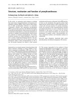
Schematic outline of the mutant medaka library generation and screeningFigure 1 (see following page)
Schematic outline of the mutant medaka library generation and screening. Male G0 fish were ENU-mutagenized and crossed with wild-type (WT) females.
Male F1 progeny were used for sperm cryopreservation and parallel DNA isolation. The library was screened for induced mutations in target genes of
interest by dideoxy resequencing. Interesting mutants were retrieved from the cryopreserved archive by in vitro fertilization and incrossed to
homozygosity for phenotypic analysis.
Genome Biology 2006, Volume 7, Issue 12, Article R116 Taniguchi et al. R116.3
comment reviews reports refereed researchdeposited research interactions information
Genome Biology 2006, 7:R116
Figure 1 (see legend on previous page)
R116.4 Genome Biology 2006, Volume 7, Issue 12, Article R116 Taniguchi et al. />Genome Biology 2006, 7:R116
p53
E241X
mutant characterization
We identified seven induced mutations in the medaka p53
gene [17], including three missense mutations, one splice site,
and two nonsense mutations (Figure 2). The p53
E241X
allele is
a G to T substitution that results in the alteration of Glu241 to
a stop codon, whereas the p53
Y186X
allele is a T to A substitu-
tion that alters Tyr186 to a stop codon. Both were presumed
to result in a truncated protein that terminates prematurely
in the midst of a DNA-binding domain. These proteins retain
the amino-terminal transactivation domain but lack the
nuclear localization signal and tetramerization domain
required for full activity. Furthermore, no alternative splicing
variants involving these mutation-containing exons are
known in any species, indicating that these nonsense muta-
tions are most likely to result in a null phenotype. All three
missense mutations are at highly conserved residues within
the DNA-binding region, but more detailed characterization
will be needed to conclude anything about their effect on pro-
tein function.
Impaired target gene induction upon DNA damage is one of
the phenotypes that is expected in a p53 knockout animal
[18]. p53
E241X/E241X
embryos were γ-irradiated and the induc-
tion of p21, Mdm2 and Bax genes was examined by RT-PCR.
As expected, no increase of these target genes was observed in
p53
E241X/E241X
homozygous fish, while control fish clearly
showed upregulation of p21 and Mdm2 transcription level in
response to ionizing radiation (IR), (Figure 3a). Interestingly,
the basal level of the p53 transcript was decreased in p53
E241X/
E241X
fish. This could be due to nonsense-mediated decay [19]
of mutant RNA, a phenomenon that is frequently observed in
ENU-induced nonsense mutants (E Cuppen, unpublished
observations), although an autoregulatory mechanism can-
not be excluded. The same results were obtained for the sec-
ond nonsense allele (p53
Y186X/Y186X
; data not shown). Next,
we investigated whether IR-induced apoptosis was affected in
p53
E241X/E241X
mutants. Primary cell cultures were derived
from wild-type and p53
E241X/E241X
fish, γ-irradiated, and
observed by time-lapse video microscopy for apoptosis. While
13.2% (15 out of 142 cells counted) of p53
+/+
cells underwent
apoptosis, none of the p53
E241X/E241X
cells (0 out of 121 cells)
showed fragmentation of the nucleus (Figure 3b). These
results are consistent with a complete loss-of-function pheno-
type of p53 in these medaka mutants.
To monitor for spontaneous tumorigenesis, p53 knockout
(p53
E241X/E241X
, n = 21), heterozygote (p53
+/E241X
, n = 26), and
wild-type (p53
+/+
, n = 10) littermates were raised to adult-
hood to monitor for spontaneous tumorigenesis. Only a single
p53
+/+
fish died within 10 months after birth with no obvious
signs of cancer (Figure 4). Heterozygous fish developed some
tumors during the course of observation (two out of the five
fish that died during the first ten months had clear tumors),
but the mortality rate was relatively low. In contrast, a dra-
matic tumor predisposition was observed in the homozy-
Table 1
Statistics on the mutant medaka library generation
Library generation Specific locus test
G0 87 9*
Fertilized eggs
†
26,226 1,360
F1 5,771 mature males 5 albino mutants
*The fish used for specific locus test were eventually mated to wild-type females and overlap with 87 fish that were used for library generation.
†
The
number of fertilized eggs includes those that died during embryogenesis.
Table 2
Medaka mutant library* screening statistics
Gene Exons Exons screened Amplicons
†
Base-pairs screened
‡
Exonic Intronic Total Mutation rate
Stop Missense Silent Intron Splice
Blm 23 2 2 3,129,006 1 4 0 1 0 6 1/521,501
p53 11 3 1 1,854,603 2 3 0 1 1 7 1/264,943
Sirt1 9 5 2 5,767,496 0 12 0 2 1 15 1/384,500
Dcr-1 27 7 4 7,879,290 0 16 4 7 0 27 1/291,826
Parkin 11 3 1 3,461,661 1 3 3 2 0 9 1/384,629
Total 81 20 10 22,092,056 4 38 7 13 2 64 1/345,188
*The mutant library consists of 5,771 cryopreserved male progeny from ENU-mutagenized fish.
†
Due to the compact medaka genome architecture,
multiple exons can often be amplified and sequenced from a single amplicon.
‡
Determined by counting all bases in the resequencing reads that were
read with phred quality >20.
Genome Biology 2006, Volume 7, Issue 12, Article R116 Taniguchi et al. R116.5
comment reviews reports refereed researchdeposited research interactions information
Genome Biology 2006, 7:R116
Table 3
Detailed overview of the induced mutations retrieved from the mutant medaka library
Number Exon Sequence context Amino acid change Type of mutation
Dicer (Dcr-1)
1 10_11 5'-GATCCTTAGG (A>G) ACAAATGCTC-3' N578D Substitution
2 10_11 5'-GTGGTTGACG (A>G) TGACAACATC-3' D597G Substitution
3 10_11 5'-ACCGTCAACA (C>A) AGCCATCGGT-3' T619K Substitution
4 10_11 5'-CGTCAACACA (G>A) CCATCGGTCA-3' A620T Silent
5 10_11 5'-CAGGTACCTG (C>T) CCTGCTTTGA-3' Intron
6 10_11 5'-CCTGCCCTGC (T>A) TTGATGTGGA-3' Intron
7 10_11 5'-AGAATTAACT (T>A) CAACTCAACA-3' Intron
8 16_17 5'-ATTTTTGACT (T>A) GAATAGTTGG-3' Intron
9 16_17 5'-GAGGCTCGCA (C>T) TGGCATTCCT-3' T897I Substitution
10 16_17 5'-CGCACTGGCA (T>G) TCCTACCACT-3' I899S Substitution
11 16_17 5'-ACTACCAGGA (C>A) GCTGTCATCA-3' D919E Substitution
12 16_17 5'-GCTCCTTCAG (T>A) GAAACTCTTG-3' Intron
13 16_17 5'-TCTCCATAGA (T>A) ATCGTAACTT-3' Y926N Substitution
14 16_17 5'-CCATAGATAT (C>T) GTAACTTTGA-3' R927C Substitution
15 16_17 5'-GCCACTCAGC (A>G) AGTTTCCTTC-3' K949E Substitution
16 16_17 5'-TTCCTTCACC (A>T) GAATACGAGA-3' P953P Silent
17 16_17 5'-ACCTGTCAAA (T>A) CTGAACCAGC-3' N972K Substitution
18 20a 5'-GGTTTTTGTG (T>C) CAGATATCCA-3' Intron
19 20a 5'-CCATTGACAA (C>A) AAAGCTTACA-3' N1094K Substitution
20 20a 5'-AAGCTTACAG (T>A) TCTTGCTCCG-3' S1098R Substitution
21 20a 5'-TTGCTCCGAG (T>C) CCTGCAGCGA-3' S1103P Substitution
22 20a 5'-GCTCAGAACC (T>G) GCCCTCTCAG-3' P1120P Silent
23 20a 5'-CCTTCACCAA (C>T) CTGACAGCTG-3' P1168S Substitution
24 22b 5'-AATAAGGCCT (A>G) CCTGCTGCAA-3' Y1635C Substitution
25 25_26 5'-AGGAAGAGGA (C>T) ATTGAGGTCC-3' D1754D Silent
26 25_26 5'-TTCATCACTG (T>A) TGTTGGAGAT-3' Intron
27 25_26 5'-CTGCTGGAGA (T>A) GGAGCCGGAA-3' M1813K Substitution
p53
1 5_6_7 5'-TCCCTTTTCT (C>T) CATCGACTGT-3' Intron
2 5_6_7 5'-TGGCCCAGTA (T>A) TTTGAAGACC-3' Y186X Truncation
3 5_6_7 5'-CTACATGTGT (A>G) ACAGCTCGTG-3' N220D Substitution
4 5_6_7 5'-TACATGTGTA (A>G) CAGCTCGTGC-3' N220S Substitution
5 5_6_7 5'-GTGTAACAGC (T>C) CGTGCATGGG-3' S222P Substitution
6 5_6_7 5'-TCTGGAAACC (G>T) AGTAAGTTTA-3' E241X Truncation
7 5_6_7 5'-GGAAACCGAG(T>C)AAGTTTAGTC-3' Splice
Sirt1
1 2_3_4 5'-CGATGACGGA (T>A) CCTCTCATGC-3' S138T Substitution
2 2_3_4 5'-CTAGTTCCAG (C>G) GACTGGACTC-3' S144R Substitution
3 2_3_4 5'-AGTTCCAGCG (A>G) CTGGACTCCG-3' D145G Substitution
4 2_3_4 5'-AGCGACTGGA (C>T) TCCGCAGCCC-3' T147I Substitution
5 2_3_4 5'-CAGCCCCAGA (T>A) CGGTCAGAAT-3' I152N Substitution
6 2_3_4 5'-AAGCCGTTGT (G>T) AGCTCAGGTG-3' Intron
7 2_3_4 5'-CCCGAGACCA (T>C) ACTCCCACCC-3' I179T Substitution
8 2_3_4 5'-CTGTGGCAGA (T>C) CATCATCAAC-3' I192T Substitution
9 2_3_4 5'-ATCATGGTTC (T>C) GACCGGTGCA-3' L227P Substitution
10 2_3_4 5'-CGGTGCAGGT (G>T) TAGGTGTTTC-3' Splice
11 2_3_4 5'-TAAAGAAACG (G>A) TAAACACCGG-3' Intron
12 2_3_4 5'-CGGCTTGCTG (T>C) CGACTTTCCC-3' V253A Substitution
13 5_6 5'-AACATCGACA (C>A) GCTGGAACAA-3' T317K Substitution
14 5_6 5'-TGCGACGGCT (T>C) CCTGTCTCGT-3' S338P Substitution
15 5_6 5'-CGTTTGTAAA (C>A) ACAAAGTGGA-3' H344N Substitution
R116.6 Genome Biology 2006, Volume 7, Issue 12, Article R116 Taniguchi et al. />Genome Biology 2006, 7:R116
gotes, with the first incidence of tumorigenesis observed
already at 2.5 months of age. The frequency of tumor forma-
tion increased after 5 months of age, resulting in a median
lifespan of 228 days. All homozygous fish died within 10
months and 11 out of the 21 animals had clear tumors. The
real tumor rate is most likely higher, as a significant part of
the dead fish could unfortunately not be examined properly,
due to rapid decomposition. It should be mentioned that at
least 2 out of the 21 p53
E241X/E241X
fish died without any mac-
roscopic signs of tumors. The p53
Y186X/Y186X
fish developed
tumors as well but at a lower rate compared to the p53
E241X/
E241X
mutant. The median lifespan was also slightly increased
(311 days), but was still much shorter than for wild-type fish
(Figure 4). The difference in tumorigenesis between the two
different nonsense alleles is not clear at this moment. We can-
not exclude the possibility that co-segregating ENU muta-
tions affect the predisposition to develop tumors in the
p53
E241X
background. The analysis of heteroallelic p53
E241X/
Y186X
fish and/or analysis of further outcrossed lines should
resolve this issue.
Stereoscopic as well as histological characterization of tumor-
bearing p53
E241X
mutant fish revealed a wide variety of tumor
types in kidney, eye, brain, intestine, gill, thymus and testis
(Figures 5 and 6). In one case, where kidney is the primary
origin, lymphoid cells spread throughout the interstitial
space, destroying the normal architecture of renal tubules
and glomeruli (Figure 5). This is consistent with the
observation that the teleost kidney is developmentally a mes-
onephros, which is the site for hematopoiesis in adult fish and
is thought to function analogously to the bone marrow in
mammals [20]. Considering a very low natural occurrence of
tumors in young medaka (<0.01%) and the propensity of
medaka to liver tumors [21], the diversity in tumor types and
the high incidence of tumors observed in p53-deficient fish
implicate that the p53 knockout medaka are highly suscepti-
ble to spontaneous tumorigenesis compared to their p53-pro-
Blm
1 5_6 5'-AGCAGTAGGG (C>T) AATCTGTGTG-3' A477V Substitution
2 5_6 5'-TGTGACTCTC (T>G) ATCAACTCCC-3' L489R Substitution
3 5_6 5'-ACTTCTAAAA (C>T) AACCTTGTTT-3' Q497X Truncation
4 5_6 5'-TTTCTCAGAG (A>G) GCACAAGTCG-3' S503G Substitution
5 7 5'-TATTTTCTAT (C>T) TTCATTCAGA-3' Intron
6 7 5'-CTTGATGCCC (A>G) CAGGTTGGTG-3' T670A Substitution
Parkin (Park2)
1 9_10_11 5'-ATGCACGGTA (C>G) CAGCAATATG-3' Y314X Truncation
2 9_10_11 5'-GACTCATGTG (T>C) CCGGCACCTG-3' C331C Silent
3 9_10_11 5'-AGGGTGGAGT (G>T) TGAGAGACAG-3' C351F Substitution
4 9_10_11 5'-GCTGTGGCTT (T>A) GTCTTCTGTA-3' F359L Substitution
5 9_10_11 5'-TTTTGTGATG (A>T) CATTGCCGTG-3' Intron
6 9_10_11 5'-GTCTTATTCA (G>A) GAGATGACCA-3' Q410Q Silent
7 9_10_11 5'-TCTCCACCTG (C>T) AGGTGGCTGC-3' Intron
8 9_10_11 5'-TGCACATGCA (T>C) TGTGCTCTGT-3' H433H Silent
9 9_10_11 5'-AGGGAGTGCA (T>A) GGGAAACCAC-3' M454K Substitution
Table 3 (Continued)
Detailed overview of the induced mutations retrieved from the mutant medaka library
Table 4
In vitro fertilization statistics
Mutants Eggs used Fertilized* Hatched*
p53
Y186X
101 100 (99) 88 (87)
p53
E241X
106 105 (99) 81 (76)
p53
splice
101 99 (98) 86 (85)
Sirt1
splice
99 93 (94) 84 (85)
Blm
Q497X
103 98 (95) 66 (64)
Parkin
Y314X
98 88 (90) 82 (84)
*The number in parentheses indicates the percentage of fertilized/hatched embryos.
Genome Biology 2006, Volume 7, Issue 12, Article R116 Taniguchi et al. R116.7
comment reviews reports refereed researchdeposited research interactions information
Genome Biology 2006, 7:R116
ficient littermates, even though the number of fish examined
in this study was relatively small.
In p53-deficient zebrafish, peripheral nerve sheath tumors
were found to predominate [22]. The difference in tumor
spectrum may be caused by the type of mutation introduced
in the genome, namely a missense mutation at a conserved
residue in zebrafish versus a nonsense mutation in medaka,
or by the presence of organism-specific secondary genes that
are differentially involved in tumor susceptibility. This tissue
specific tumor development in different species is of great
interest as this phenomenon is also found in mammals: in Li-
Fraumeni syndrome patients, caused by mutations in the
human p53 gene, breast cancer and sarcomas are most
common, whereas p53 knockout mice develop T cell lympho-
mas [23,24]. Such differences strengthen the need for parallel
studies in multiple model organisms.
We identified a nonsense mutation that results in a truncated
Parkin protein at Tyr314, eliminating the inbetween RING
domain (IBR) and the second RING domain (RING2), which
are critical for its ubiquitin ligase activity [25]. Interestingly,
a similar mutation, which results in Parkin protein truncation
at Glu311, has been found in a human juvenile parkinsonism
patient [26]. For the Blm gene, the premature stop codon was
introduced at position Glu497, which removes the entire crit-
ical helicase domain. Again, a similar 515 amino acid-long
truncated protein has been reported in a human disease case
that results from a 1 bp insertion prior to the helicase domain
[27]. It should be noted that the complete knockout of the Blm
gene results in embryonic lethality in mice [28], while Blm
mutant medaka fish are viable, similar to human. We expect
that the medaka mutants of the Parkinsonism and Bloom
syndrome genes may serve as valuable disease models, and
are currently characterizing their phenotypes in detail.
Conclusion
The estimated evolutionary distance of 110 to 200 million
years between medaka and zebrafish, and the partial or whole
genome duplication that occurred in the common ancestor of
teleosts with subsequent diversification events in the differ-
ent lineages make medaka a suitable animal for comparative
approaches [1,29]. The establishment of knockout technology
for medaka, as described here, adds significantly to the exper-
imental possibilities in this emerging model organism. A
compact genome that lacks the complex repetitive elements
observed in zebrafish, and the availability of several inbred
strains [30] make the medaka fish model especially suited for
genome-based analyses. Furthermore, in contrast to
zebrafish, which inhabit tropical areas, medaka passes the
winter in Japan, surviving water temperatures as low as 4°C
[1]. This opens the possibility for heat- or cold shock-based
experiments. Considering this, the missense mutations
retrieved by our target-selected mutagenesis approach could
be very interesting as some of them may represent tempera-
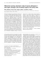
Target-selected mutagenesis of Oryzias latipes p53 geneFigure 2
Target-selected mutagenesis of Oryzias latipes p53 gene. Genomic organization and protein structure of the medaka p53 gene. The region analyzed by PCR
and dideoxy resequencing is indicated by bidirectional arrows. The ENU mutations are shown by solid arrows. Basic, basic regulatory region; DBD, DNA-
binding domain; NLS, nuclear localization signal; Pro-rich, proline-rich domain; TAD, transactivation domain; TET, tetramerization domain.
1234 567 8910 11
1 kb
ATG stop
TAD Pro-rich DBD NLS TET Basic
Y186X
N220D
S222P
N220S
Induced
mutations
E241X
1234 567 8910 11
1 kb
ATG stop
TAD Pro-rich DBD NLS TET Basic
Y186X
N220D
S222P
N220S
Induced
mutations
E241X
Genome
Protein
R116.8 Genome Biology 2006, Volume 7, Issue 12, Article R116 Taniguchi et al. />Genome Biology 2006, 7:R116
Figure 3 (see legend on next page)
p53
β-actin
mdm2
p21
bax
–––+++IR
+/+
+/E241X E241X/E241X
p53
+/+
6 h
p53
+/+
0 h
p53
E241X/E241X
6 h0 h
p53
E241X/E241X
A
B
(a)
(b)
Genome Biology 2006, Volume 7, Issue 12, Article R116 Taniguchi et al. R116.9
comment reviews reports refereed researchdeposited research interactions information
Genome Biology 2006, 7:R116
ture sensitive alleles. Among the mutants we recovered,
N220S and N220D of p53 are of particular interest, because
Asn220 is located next to the Zn-binding cysteine in loop 3,
which is important for stabilization of p53 folding [31]. In
fact, the change in the thermostability of human p53 protein
has been observed for the mutation in Asn239 (equivalent to
Asn220 of medaka p53) [32,33]. It would be interesting to
examine the thermodynamics and temperature sensitive
effect on the animal carrying these mutations.
Fish, like medaka and zebrafish, are becoming increasingly
important models in biomedical research [1,29]. In relation to
tumor biology, transgenic approaches have been shown to be
valuable to induce cancers and leukemia in both zebrafish
and medaka [34,35]. The p53-deficient medaka reported here
and two other recently described target-selected knockouts in
zebrafish [22,36] are unique in that the disease is caused by
the loss of a tumor suppressor rather than overexpression or
activation of an oncogene. The role of p53 in fish cancer has
been questioned because mutations in the p53 gene have only
rarely been found in naturally occurring or induced tumors in
teleosts [37], but our results and the work by Berghmans et al.
[22] clearly show that p53 also plays a general role in
tumorigenesis in fish as 'a guardian of the genome'. Since it is
known that tumor formation with oncogene or chemical
mutagens is accelerated by p53 mutations [38], p53-deficient
medaka fish are likely to become an important tool to under-
stand the mechanisms underlying oncogenesis in general.
Taken together, the high ENU-induced mutation frequency
and efficient mutation discovery, combined with the compact
medaka genome and efficient cryopreservation and rederiva-
tion protocols, have resulted in the development of a highly
effective approach for the routine generation of knockouts in
medaka. More detailed phenotypic characterization of the
retrieved mutants will undoubtedly provide valuable insight
into the molecular mechanisms in which these genes are
involved, and add to the versatility of the medaka animal
model in general. Finally, the cryopreserved mutant library
described here is expected to contain knockouts for most
medaka genes, providing a valuable resource for the research
community.
Radiation-induced p53 target gene induction and apoptosisFigure 3 (see previous page)
Radiation-induced p53 target gene induction and apoptosis. (a) Impaired IR-induced transactivation of target genes. Using semi-quantitative RT-PCR,
induction of Mdm2 and p21 upon γ-irradiation can readily be observed in wild-type and heterozygous embryos, but is absent in animals homozygous for
the p53 mutant allele. (b) Suppression of apoptosis in primary cultured cells. Primary cells derived from p53
E241X/E241X
and p53
+/+
embryos were irradiated
with 10 Gy of ionizing radiation and observed by time-lapse microscopy. The apoptotic cells from homozygous embryos with fragmented nuclei are
indicated with arrows.
Survival curve of p53 mutant medakaFigure 4
Survival curve of p53 mutant medaka. The viability of wild-type (dotted lines), heterozygote (dashed lines), and homozygote (solid lines) littermates of the
p53
E241X
(black) and p53
Y186X/Y186X
(grey) fish was monitored for 10 months.
0
20
40
60
80
100
0 50 100 150 200 250 300
Days after birth
p53+/+ (n=10, E241X littermate
p53E241X/+ (n=26)
p53E241X/E241X (n=21)
p53+/+ (n=15, Y186X littermate
p53Y186X/+ (n=25)
p53Y186X/Y186X (n=15)
R116.10 Genome Biology 2006, Volume 7, Issue 12, Article R116 Taniguchi et al. />Genome Biology 2006, 7:R116
Materials and methods
Mutagenesis
Kyoto-Cab, a substrain of Cab, was mutagenized as described
previously with slight modifications [4]. Males (102; G0)
were treated weekly with 3 mM ENU (Sigma-Aldrich, St.
Louis, MO USA) in 10 mM sodium phosphate buffer (pH 6.3)
at 26°C for 1 h. After the third treatment with ENU, the G0
were crossed with wild-type females to monitor the recovery
of fecundity. A month after the last ENU treatment, crosses
with wild-type females were set up and fertilized eggs were
left to develop to full term, resulting in the mutant F1 library
(only males were kept). The number of offspring produced
from a single mutagenized male founder varied from 1 to 239,
presumably reflecting variability in ENU-induced damage to
the testis. Ten mutagenized male founders were crossed with
albino fish (Heino) to monitor the mutagenesis efficiency
using a single locus test.
Cryopreservation of sperm
The sperm from each F1 medaka was cryopreserved as
described in Section 3.3.1 of the medaka protocols book [39].
The sperm was suspended in 60 μl of freezing medium (10%
dimethylformamide in fetal calf serum) and was divided into
6 glass capillaries. The amount of sperm held in each capillary
was enough to fertilize more than 100 eggs.
Typical kidney tumor as found in p53
E241X/E241X
homozygous fishFigure 5
Typical kidney tumor as found in p53
E241X/E241X
homozygous fish. (a) A stereoscopic view of the kidney tumor identified in a 2.5 month old homozygous
p53
E241X/E241X
fish. (b-d) Hematoxylin-eosin staining of normal (b) and neoplastic (c) kidney of medaka. Note that the interstitial tissue is infiltrated with
numerous hematopoietic cells destroying the normal architecture of renal tubules. The higher magnification shows the mixture of small lymphocytes with
little cytoplasm and the plasmacyte-like cells with large basophilic cytoplasm (d).
AB
CD
(a) (b)
(c) (d)
Genome Biology 2006, Volume 7, Issue 12, Article R116 Taniguchi et al. R116.11
comment reviews reports refereed researchdeposited research interactions information
Genome Biology 2006, 7:R116
Genomic DNA extraction
After removing the testis, fish were cut in two halves and kept
at -80°C until DNA was extracted. The tail side of the fish was
incubated overnight at 55°C in 500 μl of lysis buffer contain-
ing 10 mM Tris-HCl (pH 7.5), 1 mM EDTA (pH 8.0), 120 mM
sodium chloride, 12 mM sodium citrate (pH7.0), 1% SDS, and
200 μg/ml of proteinase K (Sigma-Aldrich). The lysate was
phenol extracted and precipitated with isopropanol. The DNA
pellet was dissolved in 1 ml of TE (10 mM Tris pH 7.5, 1 mM
EDTA, pH8.0). The concentration was adjusted to 40 ng/μl,
aliquoted into 96 deep well plates, and stored at -20°C.
PCR assay design
For p53, both the cDNA and genomic sequence available
(NCBI accession AF212997) have been published [17,37]. For
Blm, Sirt1 and Parkin the medaka cDNA sequences were
determined using a combination of RT-PCR and rapid ampli-
fication of cDNA ends (RACE). Overall, the similarity of the
encoded medaka proteins to their human counterparts was
80%, 80% and 90%, respectively, and the identity was 42%,
50% and 56%, respectively. The cDNA sequences were used
to retrieve the genomic sequence from the draft medaka
genome assembly [8]. For Dcr-1, the zebrafish protein
sequence was used in combination with gene prediction algo-
rithms to retrieve the genomic sequences required for ampli-
con design. For all genes, only a single ortholog of the human
gene was identified in the medaka draft genome. All genes
were imported in the laboratory information management
system for the identification of mutations by sequencing or
TILLING: LIMSTILL [40] to facilitate amplicon design for
screening and mutation annotation. Oligonucleotides for the
amplification of exons of interest by nested PCR were
designed in LIMSTILL. Details on oligonucleotides used can
be obtained from the authors upon request. For universal
processing at the sequencing stage, the primers for the second
PCR reaction contained M13 tails. The sequence of the uni-
versal tails and M13 oligonucleotides used for sequencing
were: M13-Forward, TGTAAAACGACGGCCAGT; and M13-
Reverse, AGGAAACAGCTATGACCAT.
Discovery of induced point mutations by dideoxy
resequencing of PCR amplicons
Genomic DNA stocks were diluted 25-fold and gridded out as
5 μl aliquots into 384 well plates. The first PCR (PCR1) was
carried out using a touchdown thermocycling program (92°C
for 60 s; 12 cycles of 92°C for 20 s, 65°C for 20 s with a decre-
ment of 0.6°C per cycle, 72°C for 30 s; followed by 20 cycles
of 92°C for 20 s, 58°C for 20 s and 72°C for 30 s; 72°C for 180
s; GeneAmp9700, Applied Biosystems, Foster City, CA, USA).
This reaction contained 5 μl genomic DNA, 0.2 μM forward
primer and 0.2 μM reverse primer, 400 μM of each dNTP, 25
mM tricine, 7.0% (w/v) glycerol, 1.6% (w/v) DMSO, 2 mM
MgCl
2
, 85 mM ammonium acetate pH 8.7 and 0.2 U Taq
polymerase in a total volume of 10 μl. After thermocycling,
the PCR1 reactions were diluted with 25 μl water, mixed by
pipetting, and 1 μl was used as template for the second round
of PCR. The second PCR (PCR2) was done using a standard
thermocycling program (92°C for 60 s; 30 cycles of 92°C for
20 s, 58°C for 20 s and 72°C for 30 s; 72°C for 180 s;
GeneAmp9700, Applied Biosystems). PCR2 mixes contained
1 μl diluted PCR1 template, 0.1 μM forward primer, 0.1 μM
reverse primer, 100 μM of each dNTP, 25 mM tricine, 7.0%
(w/v) glycerol, 1.6% (w/v) DMSO, 2 mM MgCl
2
, 85 mM
ammonium acetate pH 8.7 and 0.1 U Taq polymerase in a
total volume of 5 μl. Several samples of each amplicon were
tested on a 1% agarose gel containing ethidium bromide for
the presence of the proper PCR fragment.
PCR2 products were diluted with 20 μl water and 1 μl was
directly used as template for the sequencing reactions.
Sequencing reactions, containing 0.12 μl BigDYE (v3.1;
Applied Biosystems), 1.88 μl 2.5× dilution buffer (Applied
Biosystems) and 0.4 μM universal M13 primer in a total vol-
ume of 5 μl, were performed using cycling conditions recom-
mended by the manufacturer (40 cycles of 92°C for 10 s, 50°C
for 5 s and 60°C for 120 s). Sequencing products were purified
by ethanol precipitation in the presence of 40 mM sodium
acetate and analyzed on a 96-capillary 3730XL DNA analyzer
(Applied Biosystems), using the standard RapidSeq protocol
on a 36 cm array. Sequences were analyzed for the presence
of heterozygous mutations using PolyPhred [41] and manual
inspection of the mutated positions. Every candidate muta-
tion was verified by an independent PCR and resequencing
read. Nucleotide variations that were present in more than
two F1 fish were considered to be single nucleotide polymor-
phisms (SNPs) and, therefore, excluded from further
analysis, while mutations found in only two animals were
included, as examination of the breeding records revealed
that, in most cases, these originated from the same muta-
genized parent and are thus most likely to be derived from the
same spermatogonial stem cell. These mutations are counted
as a single mutation.
In vitro fertilization
About 100 unfertilized eggs were squeezed out from the wild-
type cab females. A single glass capillary containing 10 μl of
sperm from the F1 fish identified in the screening was
removed from the liquid nitrogen and thawed by placing at
ambient temperature. Immediately after the sperm was
thawed, the content was pushed out in balanced salt solution
(BSS; 0.65% sodium chloride, 0.04% potassium chloride,
0.02% magnesium sulfate heptahydrate, 0.02% calcium chlo-
ride dihydrate, 0.00005% phenol red, 0.01% sodium hydro-
gen carbonate, pH 7.3) and incubated with the eggs for 20
minutes with occasional pipetting. The eggs that were not fer-
tilized were removed 3 h later, and BSS was replaced with
0.03% Red Sea salt water. The eggs were incubated at 28°C
until they hatched. The quality of thawed sperm and the
fertilization rate was checked under the microscope. Only a
single cryopreserved straw (out of six straws frozen in total)
was needed for successful recovery of each mutation of inter-
est. For each in vitro fertilization, between 66 and 88 fish
R116.12 Genome Biology 2006, Volume 7, Issue 12, Article R116 Taniguchi et al. />Genome Biology 2006, 7:R116
Figure 6 (see legend on next page)
(a) (b)
(c) (d)
(e) (f)
(g) (h)
Genome Biology 2006, Volume 7, Issue 12, Article R116 Taniguchi et al. R116.13
comment reviews reports refereed researchdeposited research interactions information
Genome Biology 2006, 7:R116
were obtained that developed to adulthood. Progeny were
genotyped by sequencing and heterozygous fish carrying the
mutation of interest were incrossed to obtain homozygous
fish.
p53 target gene induction
For most of the studies, the p53
E241X
allele was used. F2 p53
+/
E241X
heterozygous fish resulting from the in vitro fertilization
were incrossed to produce F3 progeny of all genotypes. Four
days post-fertilization, F3 embryos were irradiated with 20
Gy of ionizing radiation using
137
Cs (0.02 Gy/s, Gammacell
40, Atomic Energy of Canada Limited Industrial Products,
Ontario). Six hours later, the embryos were frozen in liquid
nitrogen and RNA was extracted by Trizol (Invitrogen,
Carlsbad, CA, USA) according to the manufacturer's instruc-
tion. The embryos were genotyped by PCR and resequencing
of the simultaneously extracted genomic DNA. cDNA was
synthesized from each genotype using SuperScript III
(Invitrogen). The mRNA expression levels were determined
by PCR reactions (94°C for 1 minute; predetermined cycles of
94°C for 30 s, 55°C for 20 s, 72°C for 30 s). The numbers of
cycles used were:
β
-actin, 15; Mdm2, 24; and p53, p21 and
Bax, 26. Details on oligonucleotides used can be obtained
from the authors upon request.
Apoptosis assay
Primary cell cultures derived from p53
E241X/E241X
and p53
+/+
embryos were obtained as described previously [42]. Cells
(1.5 × 10
5
) were inoculated in a 35 mm dish and irradiated
with 10 Gy of γ-rays. The cells were monitored for
fragmentation using a IX81 inverted microscope (Olympus,
Tokyo, Japan) controlled by IPLab software (BD Biosciences,
Rockville, MD, USA) from zero to eight hours after γ-irradia-
tion.
Histology
After tumors were observed under the stereomicroscope, fish
were fixed in 4% paraformaldehyde for 24 hours and
embedded in paraffin. Tissue sections were stained by stand-
ard hematoxylin-eosin staining. Photographs of the slides
were obtained by a VC4500G digital camera (Omron, Kyoto,
Japan) mounted on an ECLIPSE E800 microscope (Nikon,
Tokyo, Japan).
Acknowledgements
We thank R Ohta, R Hamaguchi, Y Yoshiura, S Yonezawa, H Miyamoto, N
Matsuo and all technical staff of Sequencing Technology Team RIKEN GSC
for technical assistance. We thank the medaka genome sequencing consor-
tium for sharing sequence information prior to publication. This work was
supported by grants-in-aid from the Ministry of Education, Sports and Cul-
ture of Japan and an investment grant from the Netherlands Organization
for Scientific Research (NWO) to RHAP and EC.
References
1. Wittbrodt J, Shima A, Schartl M: Medaka - a model organism
from the far East. Nat Rev Genet 2002, 3:53-64.
2. Hawkins WE, Walker WW, Fournie JW, Manning CS, Krol RM: Use
of the Japanese medaka (Oryzias latipes) and guppy (Poecilia
reticulata) in carcinogenesis testing under national toxicol-
ogy program protocols. Toxicol Pathol 2003, 31(Suppl):88-91.
3. Driever W, Solnica-Krezel L, Schier AF, Neuhauss SC, Malicki J, Stem-
ple DL, Stainier DY, Zwartkruis F, Abdelilah S, Rangini Z, et al.: A
genetic screen for mutations affecting embryogenesis in
zebrafish. Development 1996, 123:37-46.
4. Furutani-Seiki M, Sasado T, Morinaga C, Suwa H, Niwa K, Yoda H,
Deguchi T, Hirose Y, Yasuoka A, Henrich T, et al.: A systematic
genome-wide screen for mutations affecting organogenesis
in Medaka, Oryzias latipes. Mech Dev 2004, 121:647-658.
5. Haffter P, Granato M, Brand M, Mullins MC, Hammerschmidt M, Kane
DA, Odenthal J, van Eeden FJ, Jiang YJ, Heisenberg CP, et al.: The
identification of genes with unique and essential functions in
the development of the zebrafish, Danio rerio. Development
1996, 123:1-36.
6. Tautz D: Redundancies, development and the flow of
information. Bioessays 1992, 14:263-266.
7. Jaillon O, Aury JM, Brunet F, Petit JL, Stange-Thomann N, Mauceli E,
Bouneau L, Fischer C, Ozouf-Costaz C, Bernot A, et al.: Genome
duplication in the teleost fish Tetraodon nigroviridis reveals
the early vertebrate proto-karyotype. Nature 2004,
431:946-957.
8. Medaka Genome Project [ />9. Nasevicius A, Ekker SC: Effective targeted gene 'knockdown' in
zebrafish. Nat Genet 2000,
26:216-220.
10. Fan L, Moon J, Crodian J, Collodi P: Homologous recombination
in zebrafish ES cells. Transgenic Res 2006, 15:21-30.
11. Wienholds E, Schulte-Merker S, Walderich B, Plasterk RH: Target-
selected inactivation of the zebrafish rag1 gene. Science 2002,
297:99-102.
12. Wienholds E, van Eeden F, Kosters M, Mudde J, Plasterk RH, Cuppen
E: Efficient target-selected mutagenesis in zebrafish. Genome
Res 2003, 13:2700-2707.
13. Li Q, Liu Z, Monroe H, Culiat CT: Integrated platform for detec-
tion of DNA sequence variants using capillary array
electrophoresis. Electrophoresis 2002, 23:1499-1511.
14. McCallum CM, Comai L, Greene EA, Henikoff S: Targeted screen-
ing for induced mutations. Nat Biotechnol 2000, 18:455-457.
15. Smits BM, Mudde JB, van de Belt J, Verheul M, Olivier J, Homberg J,
Guryev V, Cools AR, Ellenbroek BA, Plasterk RH, Cuppen E: Gener-
ation of gene knockouts and mutant models in the labora-
tory rat by ENU-driven target-selected mutagenesis.
Pharmacogenet Genomics 2006, 16:159-169.
16. Stephens M, Sloan JS, Robertson PD, Scheet P, Nickerson DA: Auto-
mating sequence-based detection and genotyping of SNPs
from diploid samples. Nat Genet 2006, 38:375-381.
17. Chen S, Hong Y, Scherer SJ, Schartl M: Lack of ultraviolet-light
inducibility of the medakafish (Oryzias latipes) tumor sup-
pressor gene p53. Gene 2001, 264:197-203.
Various tumors that developed spontaneously in p53 medaka knockoutsFigure 6 (see previous page)
Various tumors that developed spontaneously in p53 medaka knockouts. (a,b) The tumor that arose in the left gill of a p53
E241X/+
fish with the
lymphomatous infiltrate, consistent with the diagnosis of thymic lymphoma. (c,d) Adenocarcinoma found in the right gill of a p53
E241X/E241X
homozygous
fish. (e,f) Retinoblastoma in the right eye of a p53
E241X/E241X
homozygous fish. Note the rosette-like structures throughout the tumor. (g,h) A germ cell
tumor found in the anterior upper part of the peritoneal cavity of a p53
E241X/E241X
homozygous fish. All fish presented here died or were sacrificed at
around 8 months of age. Arrowheads indicate tumors. Hematoxylin-eosin staining, original magnification: (b,d) 100×; (f,h) 10×.
R116.14 Genome Biology 2006, Volume 7, Issue 12, Article R116 Taniguchi et al. />Genome Biology 2006, 7:R116
18. Yu J, Zhang L, Hwang PM, Rago C, Kinzler KW, Vogelstein B: Identi-
fication and classification of p53-regulated genes. Proc Natl
Acad Sci USA 1999, 96:14517-14522.
19. Maquat LE: Nonsense-mediated mRNA decay: splicing, trans-
lation and mRNP dynamics. Nat Rev Mol Cell Biol 2004, 5:89-99.
20. Amatruda JF, Zon LI: Dissecting hematopoiesis and disease
using the zebrafish. Dev Biol 1999, 216:1-15.
21. Masahito P, Aoki K, Egami N, Ishikawa T, Sugano H: Life-span stud-
ies on spontaneous tumor development in the medaka
(Oryzias latipes). Jpn J Cancer Res 1989, 80:1058-1065.
22. Berghmans S, Murphey RD, Wienholds E, Neuberg D, Kutok JL,
Fletcher CD, Morris JP, Liu TX, Schulte-Merker S, Kanki JP, et al.:
tp53 mutant zebrafish develop malignant peripheral nerve
sheath tumors. Proc Natl Acad Sci USA 2005, 102:407-412.
23. Donehower LA, Harvey M, Slagle BL, McArthur MJ, Montgomery CA
Jr, Butel JS, Bradley A: Mice deficient for p53 are developmen-
tally normal but susceptible to spontaneous tumours. Nature
1992, 356:215-221.
24. Li FP, Fraumeni JF Jr, Mulvihill JJ, Blattner WA, Dreyfus MG, Tucker
MA, Miller RW: A cancer family syndrome in twenty-four
kindreds. Cancer Res 1988, 48:5358-5362.
25. Giasson BI, Lee VM: Parkin and the molecular pathways of Par-
kinson's disease. Neuron 2001, 31:885-888.
26. Hattori N, Matsumine H, Asakawa S, Kitada T, Yoshino H, Elibol B,
Brookes AJ, Yamamura Y, Kobayashi T, Wang M, et al.: Point muta-
tions (Thr240Arg and Gln311Stop) [correction of
Thr240Arg and Ala311Stop] in the Parkin gene. Biochem Bio-
phys Res Commun 1998, 249:754-758.
27. Ellis NA, Groden J, Ye TZ, Straughen J, Lennon DJ, Ciocci S, Proytch-
eva M, German J: The Bloom's syndrome gene product is
homologous to RecQ helicases. Cell 1995, 83:655-666.
28. Chester N, Kuo F, Kozak C, O'Hara CD, Leder P: Stage-specific
apoptosis, developmental delay, and embryonic lethality in
mice homozygous for a targeted disruption in the murine
Bloom's syndrome gene. Genes Dev 1998, 12:3382-3393.
29. Furutani-Seiki M, Wittbrodt J: Medaka and zebrafish, an evolu-
tionary twin study. Mech Dev 2004, 121:629-637.
30. Hyodo-Taguchi Y: Inbred strains of the medaka (Oryzias
latipes). Fish Biol J Medaka 1996, 8:29-30.
31. Cho Y, Gorina S, Jeffrey PD, Pavletich NP: Crystal structure of a
p53 tumor suppressor-DNA complex: understanding tumor-
igenic mutations. Science 1994, 265:346-355.
32. Joerger AC, Allen MD, Fersht AR: Crystal structure of a super-
stable mutant of human p53 core domain. Insights into the
mechanism of rescuing oncogenic mutations. J Biol Chem 2004,
279:1291-1296.
33. Rolley N, Butcher S, Milner J: Specific DNA binding by different
classes of human p53 mutants. Oncogene 1995, 11:763-770.
34. Dimitrijevic N, Winkler C, Wellbrock C, Gomez A, Duschl J, Alt-
schmied J, Schartl M: Activation of the Xmrk proto-oncogene of
Xiphophorus by overexpression and mutational alterations.
Oncogene 1998, 16:1681-1690.
35. Langenau DM, Traver D, Ferrando AA, Kutok JL, Aster JC, Kanki JP,
Lin S, Prochownik E, Trede NS, Zon LI, Look AT: Myc-induced T
cell leukemia in transgenic zebrafish. Science 2003,
299:887-890.
36. Haramis AP, Hurlstone A, van der Velden Y, Begthel H, van den Born
M, Offerhaus GJ, Clevers HC: Adenomatous polyposis coli-defi-
cient zebrafish are susceptible to digestive tract neoplasia.
EMBO Rep 2006, 7:444-449.
37. Krause MK, Rhodes LD, Van Beneden RJ: Cloning of the p53
tumor suppressor gene from the Japanese medaka (Oryzias
latipes) and evaluation of mutational hotspots in MNNG-
exposed fish. Gene 1997, 189:101-106.
38. Patton EE, Widlund HR, Kutok JL, Kopani KR, Amatruda JF, Murphey
RD, Berghmans S, Mayhall EA, Traver D, Fletcher CD, et al.: BRAF
mutations are sufficient to promote nevi formation and
cooperate with p53 in the genesis of melanoma. Curr Biol 2005,
15:249-254.
39. Medaka Book 3.3.1 Cryo-preservation of Medaka sperm
[ />index.php?3.3.1%20Cryo-preservation%20of%20Medaka%20sperm]
40. LIMSTILL: Laboratory Information System for the Identifi-
cation of Mutations by sequencing and TILLing [http://lim
still.niob.knaw.nl]
41. Nickerson DA, Tobe VO, Taylor SL: PolyPhred: automating the
detection and genotyping of single nucleotide substitutions
using fluorescence-based resequencing. Nucleic Acids Res 1997,
25:2745-2751.
42. Komura J, Mitani H, Shima A: Fish cell culture: Establishment of
two fibroblast-like cell lines (OL-17 and OL-32) from fin of
the Medaka, Oryzias latipes. In Vitro Cell Dev Biol 1998,
24:294-298.