Báo cáo y học: "RNA-editing-mediated exon evolution" ppsx
Bạn đang xem bản rút gọn của tài liệu. Xem và tải ngay bản đầy đủ của tài liệu tại đây (1.16 MB, 12 trang )
Genome Biology 2007, 8:R29
comment reviews reports deposited research refereed research interactions information
Open Access
2007Lev-Maoret al.Volume 8, Issue 2, Article R29
Research
RNA-editing-mediated exon evolution
Galit Lev-Maor
¤
*
, Rotem Sorek
¤
*†
, Erez Y Levanon
‡§
, Nurit Paz
¶
,
Eli Eisenberg
¥
and Gil Ast
*
Addresses:
*
Department of Human Molecular Genetics and Biochemistry, Sackler Faculty of Medicine, Tel Aviv University, Ramat Aviv 69978,
Israel.
†
Genomics Division, Lawrence Berkeley National Laboratory, Berkeley, CA 94720, USA.
‡
Compugen Ltd, Pinchas Rosen St, Tel-Aviv
69512, Israel.
§
Department of Genetics, Harvard Medical School, Avenue Louis Pasteur, Boston, Massachusetts 02115, USA.
¶
Department of
Pediatric Hemato-Oncology, Chaim Sheba Medical Center, Sackler Faculty of Medicine, Tel Aviv University, Ramat Aviv 69978, Israel.
¥
School
of Physics and Astronomy, Raymond and Beverly Sackler Faculty of Exact Sciences, Tel-Aviv University, Ramat Aviv 69978, Israel.
¤ These authors contributed equally to this work.
Correspondence: Gil Ast. Email:
© 2007 Lev-Maor et al.; licensee BioMed Central Ltd.
This is an open access article distributed under the terms of the Creative Commons Attribution License ( which
permits unrestricted use, distribution, and reproduction in any medium, provided the original work is properly cited.
RNA-editing-mediated exon evolution<p>A primate-specific exon is found to be dependent on RNA editing for its exonization.</p>
Abstract
Background: Alu retroelements are specific to primates and abundant in the human genome.
Through mutations that create functional splice sites within intronic Alus, these elements can
become new exons in a process denoted exonization. It was recently shown that Alu elements are
also heavily changed by RNA editing in the human genome.
Results: Here we show that the human nuclear prelamin A recognition factor contains a primate-
specific Alu-exon that exclusively depends on RNA editing for its exonization. We demonstrate that
RNA editing regulates the exonization in a tissue-dependent manner, through both the creation of
a functional AG 3' splice site, and alteration of functional exonic splicing enhancers within the exon.
Furthermore, a premature stop codon within the Alu-exon is eliminated by an exceptionally
efficient RNA editing event. The sequence surrounding this editing site is important not only for
editing of that site but also for editing in other neighboring sites as well.
Conclusion: Our results show that the abundant RNA editing of Alu sequences can be recruited
as a mechanism supporting the birth of new exons in the human genome.
Background
Analysis of the sequenced human genome has revealed that it
contains about 200,000 exons [1]. However, the exon content
in mammalian genes is far from static. Rather, it is constantly
changing through a dynamic evolutionary process in which
exons are newly created and deleted. New exons can arise
from gene duplication [2] and exon duplication [3], but per-
haps the most intriguing process by which exons can be born
is exonization by exaptation, where genomic sequences that
did not originally function as exons are adopted into exonic
sequences [4].
We have recently shown that Alu elements use this exoniza-
tion mechanism to give rise to hundreds of novel internal
exons in the human genome [5]. Alu elements are unique pri-
mate-specific retrotransposons that occur in over one million
Published: 27 February 2007
Genome Biology 2007, 8:R29 (doi:10.1186/gb-2007-8-2-r29)
Received: 25 September 2006
Revised: 2 January 2007
Accepted: 27 February 2007
The electronic version of this article is the complete one and can be
found online at />R29.2 Genome Biology 2007, Volume 8, Issue 2, Article R29 Lev-Maor et al. />Genome Biology 2007, 8:R29
copies in the human genome [2,6,7]. Their 300 bases-long
consensus sequence contains motifs that resemble 5' and 3'
potential splice sites (5' ss and 3' ss, respectively). Random
mutations can turn these motifs into functional splice sites
that can be recognized by the splicing machinery [5,8,9]. Alu-
derived internal exons are almost always alternatively
spliced, allowing the original isoform to coexist with the new
one and preventing the deleterious effects of introducing a
new protein at the expense of the original one [5,8]. Thus, Alu
elements can increase the coding capacity of human genes
while maintaining the original protein repertoire.
Recently, Alus were reported to contribute to human tran-
scriptome diversity by an additional mechanism, involving
adenosine-to-inosine (A-to-I) RNA editing. A-to-I RNA edit-
ing refers to the deamination of selected adenosine residues,
altering the nucleotide sequence of RNA transcripts from that
encoded by genomic DNA. It is catalyzed by enzymes from the
ADAR (adenosine deaminase acting on RNA) family. Editing
targets are typically located within double stranded RNA
(dsRNA), which is recognized by the ADAR enzymes [10].
RNA editing can cause non-synonymous codon changes when
occurring inside the coding sequence or occur in the non-cod-
ing parts of the pre-RNA molecule. It was lately reported that
human transcripts contain excess editing over mouse, rat,
chicken and fly transcripts [5,11,12]. The majority of editing
sites in human (approximately 96%) were found to occur
within Alu sequences. Due to the abundance of Alu elements
in the human genome, two Alus in opposite orientation are
frequently found near each other. When transcribed in the
pre-mRNA, these two Alus can presumably fold to create a
dsRNA; this substrate is recognized and edited by the ADAR
enzymes. Edited Alus typically do not contribute to the pro-
tein repertoire, but rather reside in non-coding parts of the
pre-RNA molecule - untranslated regions (UTRs) and introns
[11,13-18].
The observation that Alu elements are both extensively edited
and can give rise to novel alternatively spliced exons in pri-
mate genomes raises the question of whether RNA editing can
be involved in the birth of new Alu-exons. RNA editing was
previously shown to regulate alternative splicing by creating a
splice site [19]. The most studied event is the auto-editing of
the ADAR2 gene, in which intronic AA dinucleotide turns into
a functional AG 3' ss following RNA editing [19]. Indeed, it
was recently suggested that such editing events may create
functional splice sites in silent intronic Alu elements, thus
promoting their exonization [13]. In this study we detected a
novel primate-specific Alu-exon that exclusively depends on
RNA editing for its exonization. We show that RNA editing
regulates the exonization in a tissue-dependent manner, both
through creation of a functional AG 3' ss, elimination of a pre-
mature stop codon, and regulation of the inclusion/skipping
level through alteration of exonic splicing enhancers and
silencers within the exon. We also demonstrated that the
sequence around an editing site is important not only for the
editing in that site but also editing in neighboring sites
located along this Alu-exon. Our results show that RNA edit-
ing can be recruited as a mechanism supporting the birth of
new exons in the human genome.
Results
RNA editing enables exonization of a nuclear prelamin
A recognition factor Alu-exon
To check the possibility that Alu-derived exons were fixed
into protein-coding genes through an RNA-editing-mediated
process, we first used expressed sequence tags (ESTs) and
cDNAs from GenBank version 136 aligned to the human
genome (version hg16) to identify internal human exons that
contain Alu elements, as described in [5]. We looked for Alu-
containing exons flanked by either AA at the 3' ss or AT at the
5'ss. These non-canonical splice sites will normally not be
selected by the splicing machinery; however, each of these
splice sites is theoretically capable of becoming a bona fide
splice site through A-to-I RNA editing, because inosine is rec-
ognized by the splicing apparatus as a guanosine [19]. We
demanded that the Alu-exon will be supported by more than
one EST/cDNA, and that it neither induced frame-shift nor
contained a stop codon, as these parameters were shown to be
indicative of functional alternatively spliced exons [5].
We were able to detect one such Alu-derived exon, having an
AA 3' ss, that conformed to the above conditions (Figure 1).
This exon is the eighth exon in the nuclear prelamin A recog-
nition factor (NARF), a protein that interacts with the car-
boxyl-terminal tail of prelamin A and localizes to the nuclear
lamina [20]. As with all internal Alu-containing exons
described to date, it is alternatively spliced, and exon-inclu-
sion is supported by seven cDNAs, including the full-length
cDNA (GenBank:BC000438
; UCSC March 2006 version).
The exon inserts 46 in-frame additional amino acids into the
coding sequence of NARF.
A-to-I RNA editing takes place in the context of dsRNA, to
which the ADAR proteins bind through a dsRNA-binding
domain (reviewed in [21,22]). It was therefore shown that, for
the vast majority of edited Alus in human exons, there is a
nearby (up to 2,000 base-pairs apart) intronic Alu counter-
part in the opposite orientation, which presumably serves as
the template for dsRNA formation [13,14]. Indeed, we were
able to find an oppositely oriented Alu sequence 25 bases
upstream of the exonized Alu. The antisense Alu has 81%
identity when aligned to the exon-producing Alu, suggesting
that these two Alus might form a stable intramolecular
dsRNA formation following transcription (Figure 1b). This, in
addition to the non-canonical AA splice site, implies that RNA
editing participates in the exonization of that Alu.
Editing within Alu elements frequently occurs in more than
one position, due to the long RNA duplex usually formed by
two oppositely oriented nearby Alus [11,13-15,18,23]. Indeed,
Genome Biology 2007, Volume 8, Issue 2, Article R29 Lev-Maor et al. R29.3
comment reviews reports refereed researchdeposited research interactions information
Genome Biology 2007, 8:R29
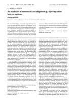
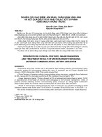
The birth of an Alu-exon through RNA editingFigure 1
The birth of an Alu-exon through RNA editing. Editing prediction was inferred from alignment of cDNAs to human genomic DNA. (a) Schematic
illustration of exons 7 to 9 of the NARF gene. Exons are depicted as blue boxes; the Alu-exon, derived from AluSx (AEx; purple box), is in a sense
orientation and is shown in the middle. The intronic, antisense-orientation Alu sequence (AluS) is 25 base-pairs upstream of the exonized Alu. Sense and
antisense Alus are expected to create a dsRNA secondary structure, thus allowing RNA editing. RNA editing changes an AA dinucleotide into a functional
AG 3' splice site (lower panel). RNA editing also occurs in five positions in the Alu-exon itself (E1, E2, E3, E4 and E5). In the first position (E1), editing
changes a UAG stop codon into a UGG Trp codon. (b) Predicted folding between the sense and antisense Alu sequences (upper and lower lines,
respectively). Adenosines that undergo editing are marked by red. Splice sites utilized for Alu exonization are marked as 5' ss and 3' ss on the alignment.
exon 7
>
A
G
>
GUA
G
>
>
E1 E2 E4
>
aa
G
gu
3’ss 5’ss
A
G
>
E3
A
G
>
E5
A
G
exon 9
AluS
AEx
exon 7 exon 9
AluS
25 nt
>
>
NARF gene
(b)
3’ss
(a)
AEx
AEx
AEx
AEx
AluS
V
V
E1 E2 E3E4 E5
5’ss
R29.4 Genome Biology 2007, Volume 8, Issue 2, Article R29 Lev-Maor et al. />Genome Biology 2007, 8:R29
by searching for A→G discrepancies in the alignments of
cDNAs to the human genome we detected five additional
potential editing sites in the Alu-exon (Figure 1b, E1-E5). The
first of these, found in position 19 of the exon, is of particular
interest, because it has the potential to change a TAG codon
(termination of translation) to a TGG codon (coding for tryp-
tophane). In the absence of RNA editing in the E1 position,
the insertion of this Alu-exon would have caused a premature
termination. It is important to note that the editing in the
exonic E1-E5 sites is directly recognizable from the ESTs or
cDNAs in comparison to human genomic DNA, whereas edit-
ing in the potential 3'ss is postulated based only on the
genomic sequence.
Different levels of exonization among human tissues
To check whether this putative Alu-exon is indeed spliced into
the mature mRNA of NARF, we tested the existence of the
exon-inclusion and exon-skipping forms in endogenous
mRNA from various normal human tissues and cell-lines. As
shown in Figure 2, the inclusion form was detected in all
cDNAs generated from normal human tissues as well as from
different human cell lines. This indicates that the exonization
of the NARF Alu is evolutionarily fixed in the human tran-
scriptome. Moreover, exon inclusion levels in different tis-
sues followed expected levels of RNA editing in those tissues.
For example, brain, kidney and spleen showed the highest
levels of exon inclusion while skeletal muscle showed the low-
est levels of exon inclusion (Figure 2a; Additional data file 1).
These results are in line with genome wide analysis of edited
RNA in different tissues [5,11,13,15] and to the amount of ino-
sine detected in RNA in various tissues [24]. The above
results further suggest that RNA editing is involved in the reg-
ulation of alternative splicing of the exonized Alu in the gene
encoding NARF. Interestingly, we note high levels of exon
inclusion in MCF7 and 293T cell-lines, but not in HeLa,
SKOV3 and MDAH cell-lines (human cancer cell lines origi-
nated from breast, kidney, cervix and ovaries, respectively)
(Figure 2a), although the global editing level in cell-lines is
expected to be relatively low [11]. This demonstrates that the
amplitude of editing level in various cell line types is of a var-
iable nature.
We sequenced all of the exon inclusion PCR products and
analyzed the editing frequencies at the five editing sites
(named E1-E5, Figure 1b) using the Discovery Studio (DS)
Gene 1.5 program (Accelrys Inc., San Diego, CA, USA).
Importantly, the first exonic editing site, E1 (at position 19 of
the exon), was edited at nearly 100% efficiency in all tested
tissues and cell lines, whereas the editing levels of the other
sites varied (E2 being edited in an average of 53.6% of RNAs,
E3 in an average of 26.1%, E4 in an average of 7.9% and E5 in
an average of 37.5%) (Figure 2b). Notably, editing sites E1, E3
and E5 are mistakenly annotated as single nucleotide poly-
morphisms (SNPs) in dbSNP [25] (rs17855348, rs17849311
and rs17855349, respectively) based on the variance in cDNA
data. Many similar examples of RNA editing sites erroneously
deposited in dbSNP have been recently reported [26].
Usually, editing efficiency is much lower than 100% per site,
depending on the expression levels of the ADAR enzymes in
the given tissue, the secondary structure of the substrate, or
the surrounding sequence. As shown above, position E1 in the
NARF Alu-exon is edited in nearly all RNA molecules con-
taining this exon. Inactivation of the nonsense-mediated
mRNA decay (NMD) by adding puromycine (see Materials
and methods) to 293T cell line did not affect the >97% editing
efficiency in site E1 (data not shown). This indicates that the
high level of editing in site E1 is not due to elimination of
unedited, stop-codon containing mRNAs, but rather is indic-
ative of a high efficiency of editing in that site. Apart from the
Q/R site of gluR-B [27], which is restricted to brain, this is the
highest editing efficiency documented in human, though it
has a much broader tissue expression spectrum. This result
suggests that additional regulatory mechanisms have evolved
to ensure that the stop codon is edited to a Trp codon in all
mRNAs containing the Alu-exon. It further implies that the
exonization of the NARF Alu-exon is functional in the human
transcriptome.
Alu-Alu dsRNA directs exonization
To substantiate the possibility that exon 8 in NARF was
exonized through an RNA-editing-mediated process, we con-
structed a minigene containing the human genomic sequence
of the gene encoding NARF from exon 7 to 9, including the
two introns in between and the alternative Alu-exon. Follow-
ing transfection of this minigene into 293T cells, total RNA
was collected, and the splicing pattern of the NARF minigene
was examined by RT-PCR analysis using primers specific to
the plasmid cDNA and not the endogenous one (see Materials
and methods). We then tested the effect of serial intronic and
exonic mutations on the splicing of the Alu-exon (Figure 3a).
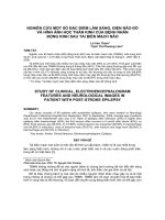
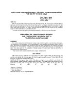
Levels of Alu-exon inclusion and RNA editing in the endogenous human NARF geneFigure 2 (see following page)
Levels of Alu-exon inclusion and RNA editing in the endogenous human NARF gene. (a) cDNAs from various normal human tissues or cDNAs from
various cell-lines were PCR amplified using primers specific for the two exons flanking the exonized Alu (upper and lower panels, respectively). The
inclusion level of the Alu-exon is indicated at the top of the panel, and represents the total percentage of the Alu-containing mRNA isoform, where 100%
corresponds to the total of both mRNA isoforms (inferred by the ImageJ program). Each PCR product was confirmed by sequencing. Schemata of the two
mRNA products are shown on the right. (b) Editing efficiency in the five exonic sites (E1, E2, E3, E4 and E5; see Figure 1 for site positions) in different
tissues and cell lines. The editing frequencies in each of the five edited sites, derived from sequence results obtained from an average of three independent
amplifications, were quantified using the Discovery Studio Gene 1.5 program.
Genome Biology 2007, Volume 8, Issue 2, Article R29 Lev-Maor et al. R29.5
comment reviews reports refereed researchdeposited research interactions information
Genome Biology 2007, 8:R29
Figure 2 (see legend on previous page)
E1
E2
E3
Sample
spleen
97% 18.4%12.5%
pancreas
97.7% 25.1%19%
lung
97.5% 30.4%16.8%
skeletal mus.
99% 27.7%5.7%
kidney 100% 65.4%
17.6%
heart
99.7%
35.7%
9.5%
liver
99%
21.3%
12.3%
brain
99.7%
56.1%
23.1%
293T
100%
56%
16.2%
MCF7
99%
28.9%8%
SKOV3
99%
34.5%
12.4%
MDAH
99%
28.7%9.2%
99%
HeLa
31.3%
19%
E5
21.3%
28.6%
25.1%
27.9%
51.6%
41.4%
20.5%
64.3%
68.1%
40%
39.5%
46%
38.6%
HeLa
293T
MCF7
SKOV3
MDAH
13 45 28 20 9
1
2
4
5
6
inclusion level
spleen
pancreas
lung
skeletal muscle
kidney
heart
liver
brain
15
7
14
4
24
9
11
32
1
2
3
4
5
6
7
8
inclusion level
editing frequencies:
E4
6.7%
5.1%
5.9%
0.6%
8.3%
3.5%
5.5%
9.7%
7.8%
4.6%
7.2%
3.1%
3.3%
(a)
(b)
R29.6 Genome Biology 2007, Volume 8, Issue 2, Article R29 Lev-Maor et al. />Genome Biology 2007, 8:R29
When the wild-type minigene was transfected into 293T cells,
23% of the mature mRNAs derived from this minigene repre-
sented the exon-inclusion form (Figure 3b, lane 1). However,
deletion of the antisense Alu element upstream of the
exonized Alu resulted in total abrogation of exon inclusion
(Figure 3b, lane 2), indicating that these two adjacent Alus
probably pair to create the dsRNA that is required for RNA
editing. Without this dsRNA, editing does not occur, and
functional AG in the 3'ss cannot be created. The effect of the
antisense Alu deletion was reversed when the AA splice site
near the Alu-exon was mutated to AG, indicating that a single
AA→AG change is sufficient for exonization of this Alu (Fig-
ure 3b, lane 3). Interestingly, the AA→AG mutation increased
exon-inclusion two-fold over the wild type, suggesting that, in
293T cells, about one-third of the AA pairs in the 3'ss are
edited into a functional AG 3' ss. Also, a single AA→AT muta-
tion at the 3'ss, created on the wild-type plasmid, resulted in
full exon skipping, indicating the importance of editing at that
site for exonization. Whereas, a single AA→AG mutation
resulted in approximately 30% exonization (Figure 3b, lanes
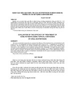
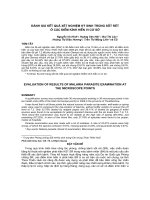
The antisense Alu is essential for exonizationFigure 3
The antisense Alu is essential for exonization. (a) An illustration of the NARF minigene that was constructed, containing the genomic sequence of the
human NARF gene from exon 7 to 9. The sites that were mutated in (b) are shown. (b) The minigene was transfected to human 293T cells, and total RNA
was collected and examined by RT-PCR analysis using specific primers to mRNA products of the plasmid minigene. The first lane is the wild-type (WT)
pattern. Lanes 2 and 3 represent a deletion of the antisense intronic Alu. Lane 3 also represents an AA→AG mutation at the 3'ss. Lanes 4 and 5 represent
an AA→AT and AA→AG mutation at the 3'ss (without deletion of the antisense Alu), respectively. The inclusion level of the Alu-exon is indicated at the
top of the gel, and represents the total percentage of the edited-Alu-containing mRNA isoform, where 100% corresponds to the total of both mRNA
isoforms (inferred using the ImageJ program). Schemata of the two mRNA products are shown on the right.
1234
Δ Alu antisense
WT
3'ss AA>AT
23 0 48 0
3'ss AA>
A
G
5
30
3'ss AA>AG
exon 7 exon 9
AE
NARF minigene
Δ Alu antisense
aa
>
g/t
(a)
(b)
AEx
AEx
AluS
Inclusion level
Genome Biology 2007, Volume 8, Issue 2, Article R29 Lev-Maor et al. R29.7
comment reviews reports refereed researchdeposited research interactions information
Genome Biology 2007, 8:R29
4 and 5). The higher level of exonization after a single
AA→AG mutation at the 3'ss without and with the antisense
Alu presumably suggests that although the antisense Alu is
essential for exonization, it also reduces the level of maximum
exonization by interfering with spliceosome accessibility to
the Alu-exon due to dsRNA formation (compare lanes 3 and
5). Combined together, these results demonstrate that the
exonization of the Alu-exon 8 in NARF is mediated by RNA
editing, and that this mechanism also controls the level of
inclusion of this exon in different tissues.
Editing in one site affects the level of editing in other
sites and the surrounding sequence and the opposite
nucleotide are important for editing
To test the possibility that specific sequences within the Alu-
Alu duplex are involved in the regulation of high efficiency
editing at the E1 site, we mutated the two nucleotides sur-
rounding the edited site, as well as the nucleotide in the anti-
sense Alu that is postulated to be opposite to the edited
nucleotide within the dsRNA (see Figure 4a for the mutated
nucleotides). All these mutations substantially reduced the
editing in the E1 site (Figure 4b,c), indicating the importance
of the surrounding sequence and the postulated opposite
nucleotide in the antisense Alu for editing at that site. More-
over, mutations M2 and M3 also resulted in a significant
reduction of RNA editing in the other exonic sites - the most
significant effect was on site E2 (Figure 4b,c). This might sug-
gest that the edited position is part of a sequence motif that
directs high efficiency RNA editing at the other sites as well.
Our results indicate that RNA editing not only enables the
exonization of the NARF Alu-exon, but also regulates its
inclusion levels in different tissues (Figure 2). This regulation
is probably attributed to the efficiency by which the AA splice
site is edited to AG. However, another possible mechanism by
which RNA editing can control exon inclusion levels is by
altering exonic splicing enhancers and silencers (ESEs and
ESSs, also denoted exonic splicing regulatory sequences
(ESRs)) within the Alu-exon (Table 1). Indeed, editing of the
first exonic site (E1) is predicted to eliminate a putative ESR
([28]; see also ESRsearch [29]. It also exchanges a putative
ESS (GGTA
GT) with another putative ESS (TGGTGG), as
predicted by RescueESE [30]. In addition, the second exonic
edited site (E2; position 30 in the exon) is part of four puta-
tive SR binding sites (Serine/Argenine-rich domain); editing
reduces the score of the SF2/ASF binding site, eliminates a
putative SRp40 ESR, creates a SRp55 ESR and also elimi-
nates a putative ESR (as predicted by ESEfinder and ESR-
search [28,31]). In site E3, editing creates a putative high-
scoring recognition site for the splicing factor SC35, as pre-
dicted by ESEfinder. Editing of E4 creates a putative recogni-
tion site for the splicing factor SC35, as predicted by
ESEfinder. Editing of site E5 is predicted to have an effect on
multiple ESRs (Table 1).
RNA editing regulates the inclusion level of the NARF
Alu-exon
To test the possibility that RNA editing regulates the inclu-
sion levels of the NARF Alu-exon by altering ESRs within the
exon, we serially mutated each of the exonic edited sites from
A-to-G, simulating 100% editing efficiency. To examine the
effect of the exonic sites only we used a minigene in which an
A-to-G mutation mimics 100% editing in the 3'ss, and we also
deleted the antisense Alu that affects editing of the exonic
sites. As shown in Figure 5, an A-to-G mutation in E1 and E3,
but not in E2 and E5, resulted in a significant increase in exon
inclusion levels (Figure 5, compare lanes 2 and 4 with lanes 3
and 6). However, editing in position E4 significantly reduced
the inclusion level, suggesting the creation of a putative SC35
site that functions as an ESS (Figure 5, lane 5). These results
indicate that editing of three out of the five exonic edited sites
affects alternative splicing levels. However, it is unlikely that
alternative splicing is regulated through editing in the E1 site,
because it is uniformly edited at high levels in all tissues
tested (Figure 2b).
Discussion
We have demonstrated that the NARF Alu-exon 8 is exonized
via RNA editing and that RNA editing is also involved in its
tissue-dependent regulation. Previously, RNA editing was
implicated in both anti-viral protection and transcript diver-
sity regulation; we now show that editing can also support
evolutionary processes such as the birth of new exons. In a
recent study, Athanasiadis et al. [13] presented computa-
tional predictions of several Alu exonization events (not
including the NARF Alu-exon) that were hypothesized to be
regulated by RNA editing; our results provide exemplary con-
firmation of the validity of these predictions.
It has been shown that a few hundred Alu elements become
exonized through single base-pair mutations that create func-
tional splice sites within their sequences. Yet in the case of the
NARF Alu-exon, exonization strictly depends on RNA edit-
ing. This situation provides a simple, yet powerful, way to reg-
ulate the levels of exon inclusion in a tissue/developmental
stage-specific manner. Since editing levels control the level of
Alu-exon inclusion, exon inclusion rates would follow the var-
ying editing levels in different tissues (Figure 2). Usually,
many regulatory sequence elements are needed to regulate
alternative splicing in a tissue-specific manner. These
sequence elements presumably can extend up to 150 bases
from each side of the regulated exon [32]. It is unlikely that a
recently retroposed Alu element will carry all needed splicing
regulatory elements; however, the RNA-editing-dependent
exonization does not rely on such extensive sequence ele-
ments, and mainly depends on the expression level of the
editing enzymes (ADARs) in the specific tissue. Moreover, we
show that editing of two out of the five exonic edited sites
affects alternative splicing levels (Figure 5). This provides an
R29.8 Genome Biology 2007, Volume 8, Issue 2, Article R29 Lev-Maor et al. />Genome Biology 2007, 8:R29
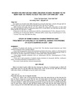
Editing is directed by a specific sequence surrounding the editing nucleotideFigure 4
Editing is directed by a specific sequence surrounding the editing nucleotide. (a) An illustration showing the positions that were mutated in the Alu-exon
(AEx) and the antisense intronic AluS (AluS): the flanking nucleotides of the edited E1 site, and the position in the antisense Alu that is predicted to be
opposite to the E1 in the dsRNA formation. (b) Chromas sequences of the Alu-exon editing of the wild-type (WT) and three mutants from (a). WT and
mutant plasmids were introduced into 293T cells by transfection, total RNA was extracted, and splicing products were separated on 1.5% agarose gel
following RT-PCR analysis. The Alu-exon inclusion of the WT and mutants is highly similar (not shown). The edited positions are highlighted in black. (c)
Rounded editing frequencies of each of the five edited sites, from three separate experiments, were quantified using the Discovery Studio Gene 1.5
program.
E1 E2 E3 E4 E5
WT
M1
M2
M3
68% 16% 8% 56%100%
10% 71% 17% 1% 41%
12% 38% 5% 1% 33%
10% 34% 4% 0 40%
mut.
site
(a)
(c)
GGTAGTG
CCACCCC
G
C
C
M2
M3
M1
AEx-
AluS-
(b)
Genome Biology 2007, Volume 8, Issue 2, Article R29 Lev-Maor et al. R29.9
comment reviews reports refereed researchdeposited research interactions information
Genome Biology 2007, 8:R29
additional layer of regulation of alternative splicing through
RNA editing.
Interestingly, the E1 as well as the E5 editing sites in the rhe-
sus macaque (but not in chimpanzee) genome encode 'G',
thus presenting only the edited version of the gene in those
sites. However, there are differences between the genomic
sequence of human and chimpanzee and that of rhesus. The
Alu-exon (AluSx) and the sequence upstream and down-
stream of it are highly conserved between human and chim-
panzee. But in the rhesus macaque there was an insertion of
AluY (in the sense orientation) immediately upstream of
AluSx (the one that exonized in human), leading the antisense
AluSg (the one that forms the dsRNA) to be located 344
nucleotides upstream of the sense AluSx (and not 25 nucle-
otides upstream as it is in human). In addition, there was an
insertion of 8 nucleotides in the sense AluSx in the rhesus
macaque as well as a deletion of 44 nucleotides that includes
the site used in human as 3'ss (Additional data file 2). These
differences raise the question of whether AluSx in the rhesus
macaque exonized at all.
The observed exonization of the NARF Alu-exon in all tested
tissues and cell lines indicates that this exon is a bona fide,
fixed functional exon in the human genome that originated
from an exapted Alu (that is, an Alu that adopted a new func-
tion that was not its original function) [4]. An additional
example for such exaptation is exon 8 of the ADAR2 gene,
which is an Alu-exon of 120 nucleotides (inserts 40 amino
acids). The Alu-exon inclusion isoform does not change the
specificity of ADAR2 activity compared to the original iso-
form (exon skipping) but rather changes the rate of the enzy-
matic activity [33].
Few mammalian ADAR substrates in which editing causes
amino acid substitutions have been found so far; the first (and
most studied ones) encode receptors that are all expressed in
the central nervous system, including subunits of the gluta-
mate receptor superfamily [27], the serotonin 5-HT2C-recep-
tor [34] and the potassium channel KCNA1 [35]. In all these
examples, the amino acid substitutions due to editing have
been shown to have a major impact on protein properties, and
altered editing patterns in the genes encoding them have been
found to be associated with several diseases, such as epilepsy
[36], depression [37], ALS (Amyotrophic Lateral Sclerosis)
[38], and malignant gliomas [39]. Lately, additional evolu-
tionarily conserved RNA editing sites that lead to a codon
exchange have been discovered in another four genes [15,40]
- the functional importance of these sites was deduced by
their extreme evolutionary conservation. The editing in the
NARF Alu-exon is the only experimentally verified editing
site in the coding region that is primate-specific. It would be
interesting, therefore, to understand the function of the Alu-
containing NARF isoform in the human transcriptome (as it
might be responsible for a primate-specific trait); however, as
the function of NARF itself is currently not clear, this must
await future studies. A Pfam analysis indicates that the Alu-
exon is inserted in NARF within a domain defined as 'Iron
only hydrogenase large subunit, carboxy-terminal domain',
and hence can presumably affect the substrate binding affin-
ity/specificity, or the catalytic activity, of this domain.
The effect of editing in exonic sites on exon inclusion levelsFigure 5
The effect of editing in exonic sites on exon inclusion levels. Lane 1 represents a deletion of the Alu antisense and also a mutation that creates an AG at the
3'ss. This plasmid was used to generate an A-to-G mutation in each of the exonic edited sites (lanes 2-6). This is a similar analysis to that shown in Figure 3.
WT
Δ
E1
E3
E5
48
85
78
59
234
inclusion level
56
E
4
28
E2
62
1
Alu antisense + AG-3’ss
R29.10 Genome Biology 2007, Volume 8, Issue 2, Article R29 Lev-Maor et al. />Genome Biology 2007, 8:R29
It is worth noting that several other editing targets that cause
predicted amino acid changes were detected in a genome-
wide search for editing in Alu [13,15], but most of them were
located in predicted genes or in aberrantly spliced RNAs.
Thus, the actual expression of proteins from these transcripts
and the possible functional implications of these sites remain
to be verified.
Our study provides additional verification for the close rela-
tionship between splicing and editing, which was demon-
strated when physical association between spliceosomal
components and ADAR proteins was reported [41]. The
actual mechanism that controls the interconnection of splic-
ing and editing is still largely unknown, but it was shown that
additional nuclear machineries are involved, such as the car-
boxy-terminal domain of RNA polymerase II in the auto-edit-
ing of ADAR2 [42,43]. This auto-editing is so far the most
studied demonstration of the feedback loop between editing
and splicing, where editing-mediated inclusion of an exon
fragment in the rat ADAR2 gene changes, in turn, the editing
capacitates of the ADAR protein itself [19]. Editing-mediated
selection of splice sites has also been observed in other genes
[39,44]. ADAR2 knockout mice provide another example of
the tight connection between editing and splicing, since the
absence of editing in the Q/R site prevents proper splicing of
the nearby intron [45]. Our results show that this splicing-
editing interconnection can also have evolutionary
significance.
Although several thousand Alu sequences have the potential
to undergo exonization [5], we were able to detect only one
reliable event of a coding Alu-exon that seemed to be
exonized through RNA editing, indicating that such a combi-
nation of evolutionary events is relatively rare in the human
genome. However, this evolutionary mechanism for the birth
of new exons might recur in other genomes. Moreover, this
mechanism might allow additional Alu exonizations in the
evolutionary future of Homo sapiens and other primate spe-
cies. As some Alus are still active in the human genome (at a
rate of 1 transposition every 200 births [46]), a novel Alu ret-
roposition in the opposite orientation from a nearby
preexisting Alu might lead to dsRNA formation and Alu-
exonization even if this Alu does not contain a canonical
splice site.
Conclusion
We have shown that RNA editing can lead to the creation of a
new exon in the human genome. Similarly to Alu retroposi-
tion and alternative splicing, RNA editing was not originally
'designed' to serve evolutionary purposes; it was rather
recruited for this, probably serendipitously. This demon-
strates that the creation of genomic novelty can be assisted by
numerous molecular biological mechanisms, most of which
were originally designed to function in other processes. The
dynamism of our genome can, therefore, arise through sur-
prising paths.
Materials and methods
Computational search for candidate edited Alus
ESTs and cDNAs from GenBank version 136 were aligned to
the human genome (version hg16) to identify internal human
exons that contain Alu elements, as described in [5]Alu-con-
taining exons were identified using blastn analysis against the
Alu consensus with a threshold of 1E-10. Alus having AA/GT
or AG/AT 3' ss/5' ss, which flank exons in the protein-coding
region of the genes, were taken for further analysis. Only Alu-
exons supported by multiple cDNAs and not containing stop
codons (in the ESTs and cDNA) were further considered.
Exons were manually screened to remove false computational
predictions.
Table 1
Exonic regulatory sequences predicted to be changed following editing in the five exonic sites
Site e1* Site e2* Site e3* Site e4* Site e5*
SF2/ASF
†
CACACCT (3.2/3.6) CTCAGGA (4.8/NA)
SC35
†
AATCACAG (NA/4) AATCACAG (NA/2.6)
SRp40
†
GCACACC (2.6/NA) CTACTCA (NA/5.1)
ACTCAGG (4.2/NA)
SRp55
†
TGCACA (NA/3.1)
Ast
‡
AGTGCA
§
TCAGGA
Burge
¶¥
TGGTGG GGGAGG
GGTAGT TCAGGA
hnRNP F GGGA
The sequence of the exonic splicing regulatory sequence is shown. The edited 'A' nucleotide is in bold (unless otherwise indicated). The numbers in
parentheses indicate the binding score before/after editing; NA indicates no available score. *Edited sites: E1, position 19 in the Alu-exon; E2, position
24; E3, position 32; E4, position 33; E5, position 46.
†
Predicted by ESEfinder [31].
‡
Predicted by Goren et al. [28].
§
This unedited ESR overlaps with
both E1 and E2 sites.
¶
Predicted by RESCU-ESE [30].
¥
Sites E1 and E5 created two different hexamers for the edited and unedited position, according
to RESCU-ESE [30].
Genome Biology 2007, Volume 8, Issue 2, Article R29 Lev-Maor et al. R29.11
comment reviews reports refereed researchdeposited research interactions information
Genome Biology 2007, 8:R29
Plasmid construction
Oligonucleotide primers were designed to amplify (from
human genomic DNA) a minigene that contains exons 7, 8,
and 9 (and the introns in between) of NARF (GenBank:
NM_012336
). Each primer contained an additional exten-
sion encoding a restriction enzyme sequence. The PCR prod-
uct of NARF (2.8 kb) was restriction digested and inserted
between the KpnI/XhoI sites in the pEGFP-C3 vector, which
contains green fluorescent protein (GFP; Clontech, Palo Alto,
CA, USA).
Site directed mutagenesis
Overlapping oligonucleotide primers containing the desired
mutations were used to amplify a mutation-containing rep-
lica of the wild-type minigene plasmid, using PfuTurbo DNA
polymerase (Stratagene, La Jolla, CA, USA) (Additional data
file 3). After PCR amplification the reaction was digested with
DpnI restriction enzyme (New England Biolabs, Ipswich, MA,
USA) for 1 h at 37°C, 1-3 μl of the reaction was transformed
into the Escherichia coli XL-1 strain, and colonies were
picked for Mini-prep extraction (Qiagen, Valencia, CA, USA).
All plasmids were confirmed by sequencing.
Transfection, RNA isolation and RT-PCR amplification
293T cells were cultured in Dulbecco's modified Eagle's
medium, supplemented with 4.5 g/ml glucose (Biological
Industries, Bait-Haemek, Israel), 10% fetal calf serum, 100
U/ml penicillin, 0.1 mg/ml streptomycin, and 1 U/ml nystatin
(Biological Industries, Bait-Haemek, Israel). Cells were
grown on 6-well plates under standard conditions at 37°C
with 5% CO
2
. Cells were grown to 60% confluence, and trans-
fection was performed using FuGENE6 (Roche Diagnostics,
Basel, Switzerland) with 1 μg of plasmid DNA. Cells were har-
vested after 48 h. Total RNA was extracted using TRIzol Rea-
gent (Sigma-Aldrich, St. Louis, MO, USA), followed by
treatment with 1 U of RNase-free DNase (Ambion, Austin,
TX, USA). Reverse transcription (RT) was preformed on 2 μg
total RNA for 1 h at 42°C using Oligo-dT reverse primer and 2
U of reverse transcriptase avian myeloblastosis virus (A-
AMV; Roche Diagnostics, Basel, Switzerland).
The spliced cDNA products derived from the expressed mini-
genes were detected by PCR using the pEGFP-C3-specific
reverse primer and an exon 7 forward primer (Additional data
file 3). Amplification was performed for 30 cycles, consisting
of 1 minute at 94°C, 45 s at 61°C, and 1.5 minutes at 72°C. The
products were resolved on 1.5% agarose gel and confirmed by
sequencing. Band quantification was performed by densitom-
etry scanning of ethidium bromide stained gels, using ImageJ
software [47].
For the NMD treatment, cells 48 h post-transfection were
subjected to 100 μg/ml puromycin (Sigma-Aldrich, St. Louis,
MO, USA) for 4 h before RNA extraction.
Analysis of RNA editing
Products from RT-PCR or from PCR obtained from commer-
cial cDNAs (BioChain, Hayward, CA, USA) were separated by
electrophoresis on 1.5% agarose gels. The appropriate PCR
product was excised and the DNA was extracted and purified
(Promega, Madison, WI, USA). Direct sequencing from both
ends was done using the ABI PRISM (Applied Biosystems,
Foster-City, CA, USA). The editing percentage from direct
sequencing was calculated as for the forward
primer and for the reverse primer; the presented
percentages represent an average of three separated experi-
ments or three independent amplifications. The nucleotides
were quantified by the Discovery Studio (DS) Gene 1.5 pro-
gram (Accelrys Inc., San Diego, CA, USA).
Additional data files
The following additional data are available with the online
version of this paper. Additional data file 1 contains two fig-
ures showing semi-quantitative PCR analysis. Additional
data file 2 contains alignment of AluSx between human,
chimpanzee and rhesus macaque, and also the rhesus
macaque sequence of AluSx and its upstream surrounding
AluY sequence. Additional data file 3 is a table listing the
primer sequences used in this research.
Additional data file 1Semi-quantitative PCR analysisTwo figures showing semi-quantitative PCR analysisClick here for fileAdditional data file 2Alignment of AluSx between human, chimpanzee and rhesus macaque, and the rhesus macaque sequence of AluSx and its upstream surrounding AluY sequenceAlignment of AluSx between human, chimpanzee and rhesus macaque, and the rhesus macaque sequence of AluSx and its upstream surrounding AluY sequenceClick here for fileAdditional data file 3Primer sequences used in this researchPrimer sequences used in this researchClick here for file
Acknowledgements
We thank Sergey Nemzer for his dedicated assistance. This work was sup-
ported by a grant from the Israel Science Foundation (1449/04 and 40/05),
MOP Germany-Israel, GIF, DIP, and EURASNET. EE was supported by an
Alon Fellowship at Tel Aviv University.
References
1. Waterston RH, Lindblad-Toh K, Birney E, Rogers J, Abril JF, Agarwal
P, Agarwala R, Ainscough R, Alexandersson M, An P, et al.: Initial
sequencing and comparative analysis of the mouse genome.
Nature 2002, 420:520-562.
2. Wang W, Zheng H, Yang S, Yu H, Li J, Jiang H, Su J, Yang L, Zhang J,
McDermott J, et al.: Origin and evolution of new exons in
rodents. Genome Res 2005, 15:1258-1264.
3. Kondrashov FA, Koonin EV: Origin of alternative splicing by tan-
dem exon duplication. Hum Mol Genet 2001, 10:2661-2669.
4. Brosius J, Gould SJ: On "genomenclature": a comprehensive
(and respectful) taxonomy for pseudogenes and other "junk
DNA". Proc Natl Acad Sci USA 1992, 89:10706-10710.
5. Eisenberg E, Nemzer S, Kinar Y, Sorek R, Rechavi G, Levanon EY: Is
abundant A-to-I RNA editing primate-specific? Trends Genet
2005, 21:77-81.
6. Lander ES, Linton LM, Birren B, Nusbaum C, Zody MC, Baldwin J,
Devon K, Dewar K, Doyle M, FitzHugh W, et al.: Initial sequencing
and analysis of the human genome. Nature 2001, 409:860-921.
7. Zhang XH, Chasin LA: Comparison of multiple vertebrate
genomes reveals the birth and evolution of human exons.
Proc Natl Acad Sci USA 2006, 103:13427-13432.
8. Lev-Maor G, Sorek R, Shomron N, Ast G: The birth of an alterna-
tively spliced exon: 3' splice-site selection in Alu exons. Sci-
ence 2003, 300:1288-1291.
9. Krull M, Brosius J, Schmitz J: Alu-SINE exonization: en route to
G
AG+
× 100
C
TC+
× 100
R29.12 Genome Biology 2007, Volume 8, Issue 2, Article R29 Lev-Maor et al. />Genome Biology 2007, 8:R29
protein-coding function. Mol Biol Evol 2005, 22:1702-1711.
10. Gallo A, Keegan LP, Ring GM, O'Connell MA: An ADAR that edits
transcripts encoding ion channel subunits functions as a
dimer. EMBO J 2003, 22:3421-3430.
11. Kim DD, Kim TT, Walsh T, Kobayashi Y, Matise TC, Buyske S,
Gabriel A: Widespread RNA editing of embedded alu ele-
ments in the human transcriptome. Genome Res 2004,
14:1719-1725.
12. Neeman Y, Levanon EY, Jantsch MF, Eisenberg E: RNA editing level
in the mouse is determined by the genomic repeat
repertoire. Rna 2006, 12:1802-1809.
13. Athanasiadis A, Rich A, Maas S: Widespread A-to-I RNA editing
of Alu-containing mRNAs in the human transcriptome. PLoS
Biol 2004, 2:e391.
14. Blow M, Futreal PA, Wooster R, Stratton MR: A survey of RNA
editing in human brain. Genome Res 2004, 14:2379-2387.
15. Levanon EY, Eisenberg E, Yelin R, Nemzer S, Hallegger M, Shemesh R,
Fligelman ZY, Shoshan A, Pollock SR, Sztybel D, et al.: Systematic
identification of abundant A-to-I editing sites in the human
transcriptome. Nat Biotechnol 2004, 22:1001-1005.
16. Mattick JS, Makunin IV: Small regulatory RNAs in mammals.
Hum Mol Genet 2005, 14(Spec No 1):R121-132.
17. Prasanth KV, Prasanth SG, Xuan Z, Hearn S, Freier SM, Bennett CF,
Zhang MQ, Spector DL: Regulating gene expression through
RNA nuclear retention. Cell 2005, 123:249-263.
18. DeCerbo J, Carmichael GG: SINEs point to abundant editing in
the human genome. Genome Biol 2005, 6:216.
19. Rueter SM, Dawson TR, Emeson RB: Regulation of alternative
splicing by RNA editing.
Nature 1999, 399:75-80.
20. Barton RM, Worman HJ: Prenylated prelamin A interacts with
Narf, a novel nuclear protein. J Biol Chem 1999,
274:30008-30018.
21. Bass BL: RNA editing by adenosine deaminases that act on
RNA. Annu Rev Biochem 2002, 71:817-846.
22. Keegan LP, Gallo A, O'Connell MA: The many roles of an RNA
editor. Nat Rev Genet 2001, 2:869-878.
23. Morse DP, Aruscavage PJ, Bass BL: RNA hairpins in noncoding
regions of human brain and Caenorhabditis elegans mRNA
are edited by adenosine deaminases that act on RNA. Proc
Natl Acad Sci USA 2002, 99:7906-7911.
24. Paul MS, Bass BL: Inosine exists in mRNA at tissue-specific lev-
els and is most abundant in brain mRNA. EMBO J 1998,
17:1120-1127.
25. Single Nucleotide Polymorphism [ />projects/SNP/]
26. Eisenberg E, Adamsky K, Cohen L, Amariglio N, Hirshberg A, Rechavi
G, Levanon EY: Identification of RNA editing sites in the SNP
database. Nucleic Acids Res 2005, 33:4612-4617.
27. Sommer B, Kohler M, Sprengel R, Seeburg PH: RNA editing in
brain controls a determinant of ion flow in glutamate-gated
channels. Cell 1991, 67:11-19.
28. Goren A, Ram O, Amit M, Keren H, Lev-Maor G, Vig I, Pupko T, Ast
G: Comparative analysis identifies exonic splicing regulatory
sequences - the complex definition of enhancers and
silencers. Mol Cell 2006, 22:769-781.
29. ESRsearch Tool [ />30. Fairbrother WG, Yeo GW, Yeh R, Goldstein P, Mawson M, Sharp PA,
Burge CB: RESCUE-ESE identifies candidate exonic splicing
enhancers in vertebrate exons. Nucleic Acids Res 2004,
32:W187-190.
31. Cartegni L, Wang J, Zhu Z, Zhang MQ, Krainer AR: ESEfinder: A
web resource to identify exonic splicing enhancers. Nucleic
Acids Res 2003, 31:3568-3571.
32. Blencowe BJ: Exonic splicing enhancers: mechanism of action,
diversity and role in human genetic diseases. Trends Biochem
Sci 2000, 25:106-110.
33. Gerber A, O'Connell MA, Keller W: Two forms of human dou-
ble-stranded RNA-specific editase 1 (hRED1) generated by
the insertion of an Alu cassette. Rna 1997, 3:453-463.
34. Burns CM, Chu H, Rueter SM, Hutchinson LK, Canton H, Sanders-
Bush E, Emeson RB: Regulation of serotonin-2C receptor G-
protein coupling by RNA editing. Nature 1997, 387:303-308.
35. Hoopengardner B, Bhalla T, Staber C, Reenan R: Nervous system
targets of RNA editing identified by comparative genomics.
Science 2003, 301:832-836.
36. Brusa R, Zimmermann F, Koh DS, Feldmeyer D, Gass P, Seeburg PH,
Sprengel R: Early-onset epilepsy and postnatal lethality associ-
ated with an editing-deficient GluR-B allele in mice. Science
1995, 270:1677-1680.
37. Gurevich I, Tamir H, Arango V, Dwork AJ, Mann JJ, Schmauss C:
Altered editing of serotonin 2C receptor pre-mRNA in the
prefrontal cortex of depressed suicide victims. Neuron 2002,
34:349-356.
38. Kawahara Y, Ito K, Sun H, Aizawa H, Kanazawa I, Kwak S: Gluta-
mate receptors: RNA editing and death of motor neurons.
Nature 2004, 427:801.
39. Maas S, Patt S, Schrey M, Rich A: Underediting of glutamate
receptor GluR-B mRNA in malignant gliomas. Proc Natl Acad
Sci USA 2001, 98:14687-14692.
40. Clutterbuck DR, Leroy A, O'Connell MA, Semple CA: A bioinfor-
matic screen for novel A-I RNA editing sites reveals recoding
editing in BC10. Bioinformatics 2005, 21:2590-2595.
41. Raitskin O, Cho DS, Sperling J, Nishikura K, Sperling R: RNA editing
activity is associated with splicing factors in lnRNP particles:
The nuclear pre-mRNA processing machinery. Proc Natl Acad
Sci USA 2001, 98:6571-6576.
42. Laurencikiene J, Kallman AM, Fong N, Bentley DL, Ohman M: RNA
editing and alternative splicing: the importance of co-tran-
scriptional coordination. EMBO Rep 2006, 7:303-307.
43. Riedmann EM, Jantsch MF: An editor controlled by
transcription. EMBO Rep 2006, 7:269-270.
44. Agrawal R, Stormo GD: Editing efficiency of a Drosophila gene
correlates with a distant splice site selection. Rna 2005,
11:563-566.
45. Higuchi M, Maas S, Single FN, Hartner J, Rozov A, Burnashev N, Feld-
meyer D, Sprengel R, Seeburg PH: Point mutation in an AMPA
receptor gene rescues lethality in mice deficient in the RNA-
editing enzyme ADAR2. Nature 2000, 406:78-81.
46. Deininger PL, Batzer MA: Mammalian retroelements. Genome
Res 2002, 12:1455-1465.
47. ImageJ [ />