Báo cáo sinh học: "Novel non-viral method for transfection of primary leukemia cells and cell lines" docx
Bạn đang xem bản rút gọn của tài liệu. Xem và tải ngay bản đầy đủ của tài liệu tại đây (483.92 KB, 11 trang )
BioMed Central
Page 1 of 11
(page number not for citation purposes)
Genetic Vaccines and Therapy
Open Access
Research
Novel non-viral method for transfection of primary leukemia cells
and cell lines
Frank Schakowski
1
, Peter Buttgereit
1
, Martin Mazur
1
, Angela Märten
2
,
Björn Schöttker
3
, Marcus Gorschlüter
1
and Ingo GH Schmidt-Wolf*
1
Address:
1
Medizinische Klinik und Poliklinik I, Rheinische Friedrich-Wilhelms-Universität, Bonn, Germany,
2
Present address: Chirurgische Klinik,
Universität Heidelberg, Germany and
3
Present address: Med. Klinik, Universität Würzburg, Germany
Email: Frank Schakowski - ; Peter Buttgereit - ; Martin Mazur - ;
Angela Märten - ; Björn Schöttker - ;
Marcus Gorschlüter - ; Ingo GH Schmidt-Wolf* -
* Corresponding author
leukemiagene transfergreen fluorescent proteinnucleofectiongene gun
Abstract
Background: Tumor cells such as leukemia and lymphoma cells are possible targets for gene
therapy. However, previously leukemia and lymphoma cells have been demonstrated to be
resistant to most of non-viral gene transfer methods.
Methods: The aim of this study was to analyze various methods for transfection of primary
leukemia cells and leukemia cell lines and to improve the efficiency of gene delivery. Here, we
evaluated a novel electroporation based technique called nucleofection. This novel technique uses
a combination of special electrical parameters and specific solutions to deliver the DNA directly to
the cell nucleus under mild conditions.
Results: Using this technique for gene transfer up to 75% of primary cells derived from three acute
myeloid leukemia (AML) patients and K562 cells were transfected with the green flourescent
protein (GFP) reporter gene with low cytotoxicity. In addition, 49(+/- 9.7%) of HL60 leukemia cells
showed expression of GFP.
Conclusion: The non-viral transfection method described here may have an impact on the use of
primary leukemia cells and leukemia cell lines in cancer gene therapy.
Background
Leukemia cells are obvious and attractive targets for gene
transfer since these cells are potentially susceptible to
immunotherapeutic strategies. Recently, cytokine gene
transfer and expression of immunomodulatory genes in
various kinds of tumor cells have been shown to mediate
tumor regression and antimetastatic effects in several ani-
mal models [1]. Many leukemic entities respond to a treat-
ment with interferon-alpha [2]. Therefore, gene transfer of
various cytokine genes such as interleukin-2 (IL-2), IL-7
and IL-12 has been envisaged [3,4]. Despite of expressing
MHC molecules, leukemia cells are ineffective antigen
presenting cells (APC) [5]. Often leukemic cells are unable
to stimulate T cells because they lack expression of impor-
tant co-stimulatory molecules [5]. The use of vectors
expressing co-stimulatory molecules or cytokines and the
Published: 12 January 2004
Genetic Vaccines and Therapy 2004, 2:1
Received: 26 September 2003
Accepted: 12 January 2004
This article is available from: />© 2004 Schakowski et al; licensee BioMed Central Ltd. This is an Open Access article: verbatim copying and redistribution of this article are permitted in
all media for any purpose, provided this notice is preserved along with the article's original URL.
Genetic Vaccines and Therapy 2004, 2 />Page 2 of 11
(page number not for citation purposes)
use of genetically modified cells for therapeutic purposes
are likely to have a significant role for patients with leuke-
mia in the future [6,7]. To date, only a few strategies appli-
cable to the therapy of these diseases have reached the
point of clinical trial [8,9].
The availability of molecular genetic technology has
opened up a large range of potential strategies for the
treatment of leukemia. Hematological malignancies have
several features that make them particularly amenable to
gene transfer approaches. The neoplastic cells circulate in
the blood, so that large numbers of tumor cells can be har-
vested and sorted for ex vivo manipulation. The efficiency
of transduction can easily be monitored in vitro and sim-
ple blood tests can be used to monitor expression of the
transgene or changes in bystander effects following gene
transfer. Finally, normal host cells that infiltrate the tumor
can be found in the blood, making them accessible for
isolation and analysis. Therapeutic approaches using ex
vivo immunological modification of malignant cells have
not been widely investigated in hematological malignan-
cies. One of the prerequesites to these applications is an
appropriate vector that can achieve high efficiency gene
transfer in leukemic cells, without major cytotoxicity. Ade-
noviral vectors are able to transduce a wide range of cells
[10], however there are only little data concerning their
ability to transduce hematopoetic cells [11-13]. Recent
reports have described successful gene transfer with aden-
oviral vectors into chronic myeloid leukemia cells (CML)
after preactivation of the target cells [14]. With the use of
an adenoviral vector, containing a modified fiber protein,
an increased gene transfer in acute myeloid leukemic
(AML) cells could be shown [15]. Although virus based
systems enhance delivery efficiency, recombinant viral
based treatments have been associated with complica-
tions that result from highly evolved and complex viral
biology and / or host parasites interactions [16]. The
future of gene therapy requires the development of effi-
cient and nontoxic delivery mechanisms. However, at the
present non-viral methods concerning the transfection of
hematopoetic cells [17-19] remain poorly efficient.
Oliver Zelphati et al. tested seven commercially available
transfection reagents, and he found out that all tested rea-
gents were inefficient for delivery charged molecules into
hematopoetic cell lines and primary AML blasts [20]. Very
little data on cell viability and transfection efficiency of
primary leukemic cells could be provided, most of the
researchers using enzymatic bulk assays or a PCR analysis
to detect reporter gene expression.
Studies in our laboratory aimed developing an efficient
non-viral DNA delivery system for transfection of leuke-
mia cells. We have analyzed several methods of gene
delivery into this cell type. To compare non-viral and viral
techniques, leukemia cells were transfected with an aden-
oviral vector expressing the reporter gene green fluores-
cent protein (GFP). Electroporation as general approach
to the introduction of macromolecules into cells was used
as a non-viral method. In addition, gene gun, a helium gas
pressure-driven device, that delivers gold microparticles
coated with plasmid DNA directly into cells, and a novel
electroporation based technique called nucleofection [21]
were used.
Here, we describe a new transfection protocol accom-
plishing highly efficient gene transfer to human chronic
and acute myeloid leukemia (AML) cell lines and into pri-
mary AML cells derived from three patients.
Methods
Primary leukemia cells
Three untreated AML patients were included in the
present study. AML cells were isolated from peripheral
blood by Ficoll-Paque density centrifugation (Lympho-
prep, Nycomed, Oslo, Norway). Cells were cultured in
complete RPMI 1640 with Glutamax (GIBCO, Berlin, Ger-
many) supplemented with 10% heat-inactivated fetal calf
serum (FCS) (PAA, Cölbe, Germany), 100 U/ml penicillin
and 100 µg/ml streptomycin (Seromed, Berlin, Ger-
many), and 25 mM Hepes (hydroxyethylpiperazine
ethane sulfonic acid, GIBCO).
Patient 1 was a 71-year-old man with FAB M1 classifica-
tion (acute myelocytic leukemia). Immunophenotyping
for this patient was not done. Patient 2 was a 43-year-old
woman with TdT-positive FAB M5b classification (acute
monoblastic leukemia) with the following immunophe-
notype: CD13 (78%), CD14 (66%), CD15 (72%), CD33
(73%), and CD64 (77%). Patient 3 was a 63-year-old
woman with FAB M4 EO classification (acute myelo-
monocytic leukemia) with the following immunopheno-
type: CD13 (89%), CD14 (15%), CD15 (13%), CD33
(24%) and CD64 (12%).
Cell lines
The following cell lines were analyzed: K562 (human
chronic myeloid leukemia cell line) and HL60 (human
acute myeloid leukemia cell line), both obtained from
Deutsche Sammlung von Mikroorganismen und Zellkul-
turen (DSMZ, Braunschweig, Germany). The cell lines
were grown in complete RPMI 1640 with Glutamax sup-
plemented with 10% heat-inactivated fetal calf serum,
100 U/ml penicillin and 100 µg/ml streptomycin, and
were kept in a humid incubator with 5% CO
2
at 37°C.
Virus propagation was performed in the Ad5 E1-trans-
formed human embryonic retina cell line 911 [22]. This
cell line was grown in Dulbecco's modified eagle medium
(DMEM, GIBCO) supplemented with 10% FCS, 100 U/
ml penicillin and 100 µg/ml streptomycin.
Genetic Vaccines and Therapy 2004, 2 />Page 3 of 11
(page number not for citation purposes)
Adenovirus preparation and infection of leukemic cells
Transfection efficacy was determined with the GFP
expressing adenovirus pQB-AdBM5GFP (E1 and E3
deleted replication defective Adenovirus type 5, Quantum
Biotechnologies INC., Montreal, Canada). This adenovi-
rus contains a cytomegalovirus (CMV) promotor. Viral
stocks were generated as described before [11] and puri-
fied by CsCl
2
centrifugation [22]. Plaque assays were
essentially performed as described by Graham and Prevec
[23]. The titer of the Ad-GFP was 5 × 10
9
plaque forming
units (pfu)/ml. For the adenoviral transfection we used
our protocol for transfection of lymphoma cells pub-
lished recently [11]. In brief, adenoviral transfections with
double CsCl
2
purified Ad-GFP of K562 cells were carried
out in 24-well plates with 5 × 10
5
cells in 50 µl phosphate-
buffered saline (PBS) with 1 mM MgCl
2
/1% horse serum
(HS), at an MOI of 200. After 2 hours of incubation at
37°C, 5% CO
2
, 1 ml of complete culture medium was
added to the cells. Because no visible toxic effect in com-
parison to the controls (only PBS +1 mM MgCl
2
/1% HS)
were observed, it was not necessary to remove the virus.
Expression plasmid for eGFP
The plasmid pMGV was described before [24] and was
obtained from Mologen (Berlin, Germany). The vector
contains a CMV enhancer promotor sequence from the
immediate early gene of the human cytomegalovirus, the
GFP open reading frame from A. victoria, a simian virus
(SV)-40 polyadenylation signal, a self-replication-origin
(ori p) and a gene for ampicillin resistance.
Plasmid preparation
The plasmid used for transfection was prepared with the
Qiagen EndoFree plasmid kit following the manufactures
instructions (Qiagen, Hilden, Germany). This kit removes
more than 99% of contaminating endotoxin.
Electroporation of leukemia cells
K562 and HL60 cells were transfected by electroporation
at various conditions using the electroporation system
easyject plus (Eurogentec, Seraing, Belgium). In brief, 5 ×
10
6
cells were suspended in 500 µl complete RPMI
medium, mixed with 30 µg of pMGV-plasmid in a 4 mm
electroporation cuvette, and incubated on ice for 10 min.
After electroporation with a single pulse (electrical param-
eters for K562 between 270 volt, 1050 µF and 300 volt,
1800 µF, 99 Ω, and for HL60 electroporation condition
differs between 1050 and 1800 µF, 200 – 450 V (in 50 V
steps), 99 Ω.) the cells were transferred into complete
RPMI medium at a density of 1 × 10
6
cells per ml.
Nucleofection of cells
Primary AML cells, K562 and HL60 cells were transfected
by nucleofection with the optimised conditions by using
the Nucleofector system from amaxa GmbH (Cologne,
Germany). The Nucleofector technology is a highly effi-
cient non-viral gene transfer method for most primary
cells and for hard-to-transfect cell lines [25-27]. This tech-
nology is based on the long-known method of electropo-
ration, which has now been significantly improved. Cell-
type specific combinations of electrical current and solu-
tions make the technology unique in its ability to transfer
polyanionic macromolecules directly into the nucleus.
Thus, cells with limited potential to divide, like many
medically highly relevant primary cells, are made accessi-
ble for efficient gene transfer. The condition for each cell
type have been optimised by using the manufactures'
guidelines.
After centrifugation 5 × 10
5
(cell lines) or 1 × 10
6
(primary
cells) cells were suspended in 100 µl prewarmed Nucleo-
fector Solution Kit R (K562, HL60) or Nucleofector Solu-
tion Kit T (primary AML cells), containing 10 µg of pMGV-
plasmid in a 2 mm electroporation cuvette (amaxa
GmbH, Cologne; Germany). The Nucleofector Kits are
cell type specific solutions and commercial available for
different cell types (amaxa GmbH). The samples were
kept in the cuvette only for the time of the pulse. The
leukemic cell line K562 was transfected with the electrical
setting P-13 and T-02, the HL60 cell line with electrical
setting T-01 and S-11. For primary cells we used the elec-
trical setting U-15 and S-04. After nucleofection with opti-
mized programs the cells were transferred immediately
into prewarmed complete RPMI medium.
Particle bombardement of leukemic cells (Gene gun)
Here we used a Biolistic PDS-1000/He unit (BioRad;
Munich, Germany). Gold particles (1.0 µm, ABCR) were
washed twice in 70% ethanol followed by washing twice
in aqua dest. and were concentrated at 60 mg/ml. 6.3 mg
gold particles (0.9 mg/macrocarrier) and 42 µg plasmid-
DNA were mixed by pipetting (total volume: 56 µl). After-
wards, 504 µl isopropanol was added drop by drop during
vortexing the gold/DNA suspension. 7 macrocarriers (Bio-
Rad) were overlayed with 80 µl gold/DNA/isopropanol
suspension and air-dried. Transfection was performed by
20 Hg below atmospheric pressure, 2200 or 1550 psi
(rupture disks) at different positions.
1 × 10
8
cells were transferred to transwell dishes (Corning
costar, 3.0 µm pore size). Short before transfection super-
natant was removed by pipetting. After transfection cells
was harvest, resuspended in medium and cultured in cell
culture flask. 24 hours after transfection expression of
transgene was measured by flow cytometry.
Growth curves after transfection
Cell viability was determinated by trypan blue exclusion.
PBS was used for control transfection. Non-transfected
and GFP transfected cells were counted after adenoviral
Genetic Vaccines and Therapy 2004, 2 />Page 4 of 11
(page number not for citation purposes)
infection, nucleofection, and particle bombardment 24 –
72 hours after transfection.
Immunofluorescence and flow cytometric studies
Leukemia cells (5 × 10
5
cells) were washed with PBS and
stained with 10 µl monoclonal CD80-FITC and CD86-PE
antibody (Pharmingen, Heidelberg, Germany) in a total
volume of 50 µl for 15 minutes. Isotype-matched antibod-
ies were used as controls. Stained cells were washed with
PBS/1% BSA and subsequently analyzed using an Epics XL
flow cytometry system (Coulter-Immunotech, Hamburg,
Germany). Background staining using irrelevant antibod-
ies was less than 2%. 10
5
cells were analyzed for each
sample.
To analyze the percentage of GFP positive cells we meas-
ured 5 × 10
5
cells. Cells were washed with PBS, resus-
pended in 1 ml PBS and 10 µg/ml propidiumiodid (PI)
was added immediately before flow cytometric analysis.
Lymphocytes were gated based on their scatter profile and
cells were evaluated for GFP expression. The transfection
efficiency was determined 24 hours up to 72 hours post-
transfection. Nucleofected primary cells were also meas-
ured after four hours.
Results
Expression of co-stimulatory molecules
In order to elucidate the possibility of co-stimulatory mol-
ecules-mediated gene therapy for leukemia cells we ana-
lyzed the expression of CD80 and CD86. We determined
the expression of these receptors on the cell surface of pri-
mary AML cells and leukemic cell lines. The primary cells
derived from three AML patients did not express CD80
receptors and only a low level of CD86 (4.5 +/- 1.6 %).
The leukemic cell lines K562 and HL60 did not express
CD80 receptors as determined by immunophenotyping
and FACS analysis. In contrast expression of CD86 was
found on the cell surface of K562 (72.4+/-5.1%) and
HL60 (76.5+/- 0.5%) cells.
Adenoviral gene transfer
In preliminary experiments using the leukemic cell line
K562, we observed that these cells could be successfully
transfected with an adenoviral vector expressing the
reporter gene GFP [28]. At an MOI of 200, 49+/-4% of the
cells showed a positive GFP signal after 72 hours in flow
cytometric analysis (Figure 1a). These results were in
accordance with previous reports [14,15,29], which
showed that leukemic cells could be efficiently transfected
with adenoviral vectors. Roddie and coworkers showed
high adenoviral transduction efficiency in three of four
leukemia cell lines but not in HL60 [30].
Transfection of leukemia cells using electroporation
To compare the new nucleofection technology (nucleo-
fector, amaxa) with the standard electroporation tech-
niques (easyject plus, BioRad), we transfected K562 cells
with pMGV with various electrical parameters as
described in the literature [17,18,31]. Transfection effi-
ciency was determined 24, 48, and 72 hours after electro-
poration by flow cytometric and fluorescence
microscopical analysis. For K562 cells the transfection
efficiency was 15.5+/-3.5% (Table 1). Cell viability deter-
mined by trypan blue exclusion, was markedly impaired
by electroporation (data not shown). These results are in
accordance with previous reports [17,18]. For HL60 cells
maximum transfection efficiency was 30.2+/-5.6% (1050
µF, 450 V, 99Ω) with high toxicity 66.2+/-6.8% (Table 1).
Transfection of leukemia cells using gene gun
We transfected HL60 and K562 cells by gene gun tech-
nique. Various parameters were examined. Here we used
2200 or 1550 psi (rupture disks) at different positions
(second, third, and fourth position). The transfection effi-
ciency was determined and quantified by the expression
of the encoding plasmid pMGV after 24 and 48 hours
posttransfection. In summary, the transfection rates were
3.0+/-1% for HL60 and 1.5+/-0.5% for K562 cells (Table
1).
Transfection of leukemic cells by nucleofection
With the aim of developing a gene therapy protocol for
leukemia, we were interested in identifying the most effec-
tive non-viral method of DNA delivery into primary
leukemia cells and leukemia cell lines. Several approaches
have been developed to enhance the efficiency of non-
viral gene transfer via naked DNA including gene gun and
electroporation. Here, we tested a novel electroporation
based technique called nucleofection. This electropora-
tion based technique combines cell type specific solutions
with mild conditions which guarantee high efficiency and
low cell death rates. Physical approach allow DNA to pen-
etrate directly the cell membrane and bypass endosomes /
lysosomes, thus avoiding enzymatic degradation. The
DNA may also be directly delivered to the nucleus by
nucleofection. Transfection efficiency was determined 24
hours after nucleofection by flow cytometric assays. Figure
2 shows the flow cytometry analysis of the three primary
AML cells 24 hours after nucleofection. Cells were pulsed
with two different programs (U15, S04) and show trans-
fection relative efficiencies up to 71,5% (+/- 1.8) (pro-
gram S04) with low toxicity (5,4% +/-0.2). Flow
cytometric analysis four hours after nucleofection showed
same transfection efficiencies like the 24 hours measure-
ment (data not shown) The transfection data of the pri-
mary AML cells of three patients and flow cytometric
analysis are shown in figure 2a and 2b. Figures 1b (K562
cells) and 1c (HL60 cells) show the primary data of one
Genetic Vaccines and Therapy 2004, 2 />Page 5 of 11
(page number not for citation purposes)
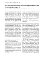
Primary data of GFP and eGFP transfected leukemic cell linesFigure 1
Primary data of GFP and eGFP transfected leukemic cell lines. Lymphocytes were gated based on their scatter profile (figures
on the right) and cells were evaluated for transgene expression. a) GFP-expression of K562 cells. Cells were transfected with
Ad-GFP at an MOI of 200 and assayed by flow cytometry 72 hours posttransfection. The overlay of the shaded histogram rep-
resents the background fluorescence of untreated cells. Positive transfected cells expressed intracellular green fluorescent pro-
tein. b) eGFP-expression of K562 cells 24 hours after nucleofection. c) eGFP-expression of HL60 cells 24 hours after
nucleofection. Data are shown from one representative experiment.
Genetic Vaccines and Therapy 2004, 2 />Page 6 of 11
(page number not for citation purposes)
Table 1: Comparison of various methods in the transfection efficiency of leukemic cells. Transfections were performed with an
expression plasmid for eGFP or adenoviral GFP expressing vector as described in materials and methods. 24 hours after transfections
cells were harvested and assayed by flow cytometry. Ad-GFP transfected cells were analyzed 72 hours after transduction. Results of
three separate experiments are presented (ND, not done).
Cell line Adenoviral gene transfer
(MOI 200) GFP-pos. cells
[%]
Electroporation GFP-pos.
cells [%]
Gene gun GFP-pos. cells
[%]
Nucleofection GFP-pos.
cells [%]
primary AML cells ND ND ND 60.3 +/- 9.7
K562 49 +/- 4 15.5 +/- 3.5 1.5 +/- 0.5 74.7 +/- 8.0
HL60 ND 30.2 +/- 5.6 3.0 +/- 1 49.0 +/- 9.7
Nucleofection mediated gene transfer in primary leukemic cellsFigure 2
Nucleofection mediated gene transfer in primary leukemic cells. eGFP expression in AML cells after exposure to optimized
pulses. After 24 hours cells were harvested and analyzed by flow cytometric analysis. a) Representative flow cytomeric analysis
for each of the three patients. Control cells were pulsed without DNA and showed no eGFP expression (left side); percentage
of positive transfected cells is shown on the right. b) Gated and ungated transfection efficiencies of the primary AML cells. Per-
centage of dead cells was determined by PI staining. The figure represents data from two experiments, respectively. Data are
presented as mean +/- standard error of the mean.
Genetic Vaccines and Therapy 2004, 2 />Page 7 of 11
(page number not for citation purposes)
representative experiment with the optimized electrical
parameters. The shaded histogram represents the back-
ground fluorescence of transfected cells without DNA,
positive transfected cells express intracellular green fluo-
rescent protein. Various electrical parameters were assayed
to optimize the transfection efficiency of the leukemic cell
lines K562 and HL60. Figure 3 demonstrates the two opti-
mized programs for each cell line (K562, HL60) and
shows the absolute percentage of GFP positive cells and
the relative efficiency of the transfected gated cells. Viabil-
ity was determined by PI staining. By balancing survival
rate and transfection efficiency optimal program for K562
resulted in a population of 76.1 +/- 2.3% positive viable
cells in the gate. The program T02 showed low toxicity
with 34.0 +/- 14.6% dead cells 24 hours after nucleofec-
tion. Gene transfer into leukemic HL60 cell line showed
relative efficiencies ranging from 33.4 +/-14.9 to 49.0 +/-
9.7%. Due to the higher percentage of dead cells (49.6 +/
- 21.5 up to 63.0 +/- 16%) the relative efficiency was lower
in comparison with K562 cells. These results demon-
strated a high efficient gene transfer with the nucleofec-
tion technique. The time course of GFP transgene
expression after 72 hours showed a constant expression of
GFP in the cell line K562 (relative efficiency: 74.7 +/- 8%,
program T02). The transgene expression in HL60 cells
decreased to 24.3 +/- 9.1% after 72 hours (program T01).
Fourteen days after transfection the percentage of trans-
gene expressing cells decreased to 3%. These results indi-
cate that nucleofection mediated gene expression in cells
was transient.
Growth curves of transfected leukemia cell lines
Cell counts were determined 24, 48 and 72 hours after
transfection. Following transfection with various parame-
ters cell numbers differed from untransfected and control
transfected leukemia cells (Fig. 4). In the case of HL60
cells the viability of transfected cells decreased rapidly
(Fig. 4b). As shown in figure 4a nucleofection of K562
cells with different electrical parameters revealed a small
reduction of cell number over a three day period. Control
transfected cells (without DNA) showed only a reduction
of cell number in comparison to non-transfected cells (1.3
× 10
6
cells/ml versus 9.8 × 10
5
cells/ml). The cell prolifer-
ation of transfected HL60 cells was strongly retarded
Nucleofection mediated gene transfer in leukemia cell linesFigure 3
Nucleofection mediated gene transfer in leukemia cell lines. eGFP expression in K562 and HL60 cells after exposure to the
optimized pulses. After 24 hours cells were harvested and analyzed by flow cytometric analysis. GFP positivity was assayed in
gated as well as in ungated cell populations. Control cells were pulsed without DNA and showed no eGFP expression. Per-
centage of dead cells was determined by PI staining. The figure represents data from five separate experiments, respectively.
Data are presented as mean +/- standard error of the mean.
Genetic Vaccines and Therapy 2004, 2 />Page 8 of 11
(page number not for citation purposes)
Growth curves of K562 (a) and HL60 (b) cells, transfected by nucleofection techniqueFigure 4
Growth curves of K562 (a) and HL60 (b) cells, transfected by nucleofection technique. Cell proliferation was measured by
trypan blue staining and cell count. The figure represents data from five separate experiments. Data are presented as mean +/-
standard error of the mean.
Genetic Vaccines and Therapy 2004, 2 />Page 9 of 11
(page number not for citation purposes)
(untransfected cells after 72 hours 1.9 × 10
6
cells/ml ver-
sus transfected cells 4 × 10
4
cells/ml), the non-transfected
control cells proliferated continuously. Transfection of
the HL60 cells lines with nucleofection resulted in a stag-
nation of cell growth. After 72 hours cell lines continued
to proliferate however at a slower rate than the control
(data not shown).
Discussion
Chemotherapy and allogeneic bone marrow transplanta-
tion (BMT) are the conventional treatment strategies for
acute myelogenous leukemia (AML) [32-35]. Complete
remissions can be achieved in the majority of patients, but
disease recurrence remains a frequent subsequent of treat-
ment failure. For example, in patients whose AML blasts
bear complex chromosomal mutations the risk of leuke-
mia relapse is very high, and in AML patients over 60 years
of age the five year survival rate after established treatment
regimens is below 20% [36]. Unfortunately, therapeutic
options for patients with recurrent leukemia are still lim-
ited and the prognosis is poor [37]. Second marrow trans-
plants from the same donor may be considered for
patients with disease relapse after BMT, but the mortality,
treatment-related morbidity and risk of further relapse are
high [38,39].
Alternative or additional treatment strategies are provided
by immunotherapeutic approaches. Successful employ-
ment of donor lymphocyte infusions (DLI) in patients
with relapsed chronic myelogenous leukemia (CML) after
allogeneic BMT gave reason for the application to acute
leukemia patients, but the treatment turned out to be far
less effective [40-43]. In other cases of refractory or
relapsed AML, infusions of anti-CD33 antibody-conju-
gated antitumor agents have been successfully used [44].
Furthermore, gene therapy has emerged as a promising
approach to provide new treatment options.
Leukemia cells are considered as suitable targets for gene
therapy. Cytogenetic studies of leukemia cells have identi-
fied mutations, chromosomal aberrations the failure of
expression of co-stimulatory molecules [1,5,9]. Co-stimu-
latory molecules such as CD80 (B7.1) and CD86 (B7.2)
that are necessary to bind CD28 on T-cells, to maintain
production of IL-2 after initial T cells activation, have been
shown to be lacking in acute leukemic cells, resulting in T-
cell anergy [5]. The lack of expression of CD80 could also
be detected in the leukemic cell lines used here. Vectors
expressing co-stimulatory molecules or cytokines have
been suggested for gene therapy strategies [1,3]. Adenovi-
rus based vectors can be used for targeted gene transfer to
AML [15] and after stimulation of the target cells CML and
B-CLL could be efficiently transfected by adenoviral vec-
tors. Similar results could be obtained by the use of pri-
mary cells [14]. We previously demonstrated efficient
gene transfer in Burkitt lymphoma (BL) cell lines and pri-
mary lymphoma cells after transfection with adenoviral
vectors [11] and could here show similar results in trans-
fection efficiency of the leukemic cell line K562 by use of
adenoviral vectors. Recent reports have described the suc-
cessful gene transfer in the cell line HL60 and primary
cells derived from AML patients up to 100% of positive
cells after adenoviral gene transfer [15]. Although viral
vectors induce long term, high gene expression, phenom-
ena such as the possibility of creating recombination
competent adenovirus (RCA) induction of the host
immune response are cutting back the use of viral
delivery.
Since the efficiency of non-viral gene transfer by naked
DNA is lower than that of viral delivery, both chemical
and physical techniques have been used to increase the
efficiency of DNA uptake and expression. The physical
gene transfer approaches allow DNA to penetrate directly
the cell membrane and bypass endosomes / lysosomes,
thus avoiding enzymatic degradation. The DNA may also
be delivered directly to the nucleus by gene gun, electro-
poration and novel electroporation based technique
called nucleofection. Physical gene transfer methods,
unlike viral vectors, do not require cell type specific recep-
tors, are safe, highly reproducible and time saving. Due to
low transfection efficiency most investigations were made
with established cell lines after stable transfection and
selection [19]. Even many transfection reagents which
show high gene transfer efficiency in common adherent
cell lines are not suitable to transfect establish blood cell
lines or primary leukemia cells from patients. All samples
showed a transfection rate of below 5% positive cells [20].
There is no data concerning efficient non viral gene trans-
fer into primary leukemic cells with gene gun or standard
electroporation.
The use of electrotransfer for DNA delivery to eukaryotic
cells in vitro has been well known and widely used in
basic research. However, it is only recently that electric
fields have been used to enhance DNA transfer to animal
cells in vivo, and this is known as DNA electrotransfer or
in vivo DNA electroporation. This is especially useful to
transfect whole tissues or tumors. As well as exciting appli-
cations in developmental biology, in vivo DNA electro-
transfer is also being used to transfer genes to skeletal
muscle and drive expression of therapeutically active pro-
teins and to examine exogenous gene and protein func-
tion in normal adult cells situated within the complex
environment of a tissue and organ system in vivo [45].
However, the use of in vivo electroporation has just begun
and so far nothing has been published of in vivo transfec-
tion of cells of the blood system.
Genetic Vaccines and Therapy 2004, 2 />Page 10 of 11
(page number not for citation purposes)
Here, we established an optimized non-viral gene delivery
into leukemic cells as a first step towards a gene therapy
approach. We compared the efficiency of adenoviral
mediated gene transfer with the efficiency obtained by
electroporation, particle bombardment and nucleofection
into leukemic cells in vitro. Using established human
leukemic cell lines we have analyzed the standard tech-
niques with the novel nucleofection technique. We have
examined different electrical programs with the new
nucleofector advice to determine the effects on the trans-
fection efficiency and viability of the cells.
In this study we have shown that the novel non-viral
transfection technique called nucleofection is an efficient
way to transfect not only AML cells lines but also primary
AML cells. Here, we achieved transfection efficiency for
primary cells from three AML patients up to 75 % with
low toxicity after 24 hours (< 20 %). After 72 hours the
toxicity inside the lymphocyte gate increased up to 40–45
% (non-nucleofected primary cells 25 %). However, the
ability of nucleofection to mediate gene transfer into non-
dividing cells and the feasibility to transfect a high range
of cells makes it attractive for gene delivery in vitro. Nucle-
ofection does only very rarely result in nuclear integration
of the transgene. Loss of gene expression during propaga-
tion of cells is most likely to be due to loss of the transgene
rather than due to loss of transgene expression. In terms
of cell numbers, K562 and HL60 cells cease proliferation
72 hrs after nucleofection. It is expected that these cells
finally enter cell death. This, however, makes the proce-
dure less suitable for biochemical or pharmacological
studies due to severe cell damage during nucleoporation.
However, in immunotherapeutic approaches, sustained
gene transfer is not essential. Thus, the transient gene
expression achieved by use of nucleofection is an availa-
ble tool for gene therapy.
Conclusions
The ability to efficiently manipulate gene expression in
leukemia using non-viral methods should facilitate the
functional characterization of pathways affecting lym-
phocytes physiology. In conclusion, we present a protocol
of a new gene transfer method leading to highly efficient
gene transfer in primary leukemic cells and established
cell lines without major toxicity and low risk of inser-
tional mutagenesis or induction of the host immune
response. This protocol should have an important impact
on the use of hematopoetic cells in cancer gene therapy
protocols.
List of abbreviations
AML, acute myeloid leukemia; APC, antigen presenting
cell; BMT, bone marrow transplantation; CML, chronic
myelogenous leukemia; DLI, donor lymphocyte infusion;
FCS, fetal calf serum; GFP, green fluorescent protein; HS,
horse serum; IL-2, interleukin-2; ND, not done.
Competing interests
PB is presently working at Amaxa, he was not when per-
forming the experiments described herein.
Authors contributions
FS, PB, MM and AM carried out the studies, BS was clini-
cally involved and helped in carrying out the studies, ISW
participated in the design of the study and its coordina-
tion. All authors read and approved the final manuscript.
Acknowledgments
This work was kindly supported by a generous grant from the H. W. & J.
Hector-Stiftung, Weinheim, Germany and from the Deutsche José Carre-
ras Leukaemie-Stiftung e.V., Munich, Germany.
References
1. Adams SW, Emerson SG: Gene therapy for leukemia and
lymphoma. Hematol Oncol Clin North Am 1998, 12:631-648.
2. Einhorn S, Strander H: Interferon treatment of human malig-
nancies – a short review. Med Oncol Tumor Pharmacother 1993,
10:25-29.
3. Schmidt-Wolf GD, Schmidt-Wolf IGH: Cytokines and gene
therapy. Immunol Today 1995, 16:173-175.
4. Finke S, Trojaneck B, Lefterova P, Csipai M, Wagner E, Neubauer A,
Huhn D, Wittig B, Schmidt-Wolf IGH: Increase of proliferation
rate and enhancement of antitumor cytotoxicity of human
CD3+CD56+ immunologic effector cells by receptor-medi-
ated transfection with the interleukin-7 gene. Gene Therapy
1998, 5:31-39.
5. Zheng Z, Takahashi M, Aoki S, Toba K, Liu A, Osman Y, Takahashi H,
Tsukada N, Suzuki N, Nikkuni K, Furukawa T, Koike T, Aizawa Y:
Expression patterns of costimulatory molecules on cells
derived from human hematological malignancies. J Exp Clin
Cancer Res 1998, 17:251-258.
6. Stripecke R, Cardoso AA, Pepper KA, Skelton DC, Yu XJ, Mascaren-
has L, Weinberg KI, Nadler LM, Kohn DB: Lentiviral vectors for
efficient delivery of CD80 and granulocyte-macrophage-col-
ony-stimulating factor in human acute lymphoblastic leuke-
mia and acute myeloid leukemia cells to induce antileukemic
immune responses. Blood 2000, 96:1317-1326.
7. Notter M, Willinger T, Erben U, Thiel E: Targeting of a B7-1
(CD80) immunoglobulin G fusion protein to acute myeloid
leukemia blasts increases their costimulatory activity for
autologous remission T cells. Blood 2001, 97:3138-3145.
8. Kato K, Cantwell MJ, Sharma S, Kipps TJ: Gene transfer of CD40-
ligand induces autologous immune recognition of chronic
lymphocytic leukemia B cells. J Clin Invest 1998, 101:1133-1141.
9. Wierda WG, Kipps TJ: Gene therapy of hematologic
malignancies. Semin Oncol 2000, 27:502-511.
10. Prince HM, Dessureault S, Gallinger S, Krajden M, Sutherland DR,
Addison C, Zhang Y, Graham FL, Stewart AK: Efficient adenovirus-
mediated gene expression in malignant human plasma cells:
relative lymphoid cell resistance. Exp Hematol 1998, 26:27-36.
11. Buttgereit P, Weineck S, Ropke G, Marten A, Brand K, Heinicke T,
Caselmann WH, Huhn D, Schmidt-Wolf IGH: Efficient gene trans-
fer into lymphoma cells using adenoviral vectors combined
with lipofection. Cancer Gene Ther 2000, 7:1145-1155.
12. Meeker TC, Lay LT, Wroblewski JM, Turturro F, Li Z, Seth P: Aden-
oviral vectors efficiently target cell lines derived from
selected lymphocytic malignancies, including anaplastic
large cell lymphoma and Hodgkin's disease. Clin Cancer Res
1997, 3:357-364.
13. Turturro F, Heineke HL, Drevyanko TF, Link CJ, Seth P Jr: Adenovi-
rus-p53-mediated gene therapy of anaplastic large cell lym-
phoma with t(2;5) in a nude mouse model. Gene Ther 2000,
7:930-933.
Publish with BioMed Central and every
scientist can read your work free of charge
"BioMed Central will be the most significant development for
disseminating the results of biomedical research in our lifetime."
Sir Paul Nurse, Cancer Research UK
Your research papers will be:
available free of charge to the entire biomedical community
peer reviewed and published immediately upon acceptance
cited in PubMed and archived on PubMed Central
yours — you keep the copyright
Submit your manuscript here:
/>BioMedcentral
Genetic Vaccines and Therapy 2004, 2 />Page 11 of 11
(page number not for citation purposes)
14. Huang MR, Olsson M, Kallin A, Pettersson U, Totterman TH: Effi-
cient adenovirus-mediated gene transduction of normal and
leukemic hematopoietic cells. Gene Ther 1997, 4:1093-1099.
15. Gonzalez R, Vereecque R, Wickham TJ, Vanrumbeke M, Kovesdi I,
Bauters F, Fenaux P, Quesnel B: Increased gene transfer in acute
myeloid leukemic cells by an adenovirus vector containing a
modified fiber protein. Gene Ther 1999, 6:314-320.
16. Haddada H, Cordier L, Perricaudet M: Gene therapy using aden-
ovirus vectors. Curr Top Microbiol Immunol 1995, 199:297-306.
17. Migliaccio AR, Bengra C, Ling J, Pi W, Li C, Zeng S, Keskintepe M,
Whitney B, Sanchez M, Migliaccio G, Tuan D: Stable and unstable
transgene integration sites in the human genome: extinction
of the Green Fluorescent Protein transgene in K562 cells.
Gene 2000, 256:197-214.
18. Baum C, Forster P, Hegewisch-Becker S, Harbers K: An optimized
electroporation protocol applicable to a wide range of cell
lines. Biotechniques 1994, 17:1058-1062.
19. Brielmeier M, Bechet JM, Falk MH, Pawlita M, Polack A, Bornkamm
GW: Improving stable transfection efficiency: antioxidants
dramatically improve the outgrowth of clones under domi-
nant marker selection. Nucleic Acids Res 1998, 26:2082-2085.
20. Zelphati O, Wang Y, Kitada S, Reed JC, Felgner PL, Corbeil J: Intra-
cellular Delivery of Proteins with a new lipid-mediated deliv-
ery system. J Biol Chem 2001, 276:35103-35110.
21. Lenz P, Bacot SM, Frazier-Jessen MR, Feldman GM.: Nucleopora-
tion of dendritic cells: efficient gene transfer by electropora-
tion into human monocyte-derived dendritic cells. FEBS Lett
2003, 538:149-154.
22. Fallaux FJ, Kranenburg O, Cramer SJ, Houweling A, Van Ormondt H,
Hoeben RC, Van Der Eb AJ: Characterization of 911: a new
helper cell line for the titration and propagation of early
region 1-deleted adenoviral vectors. Hum Gene Ther 1996,
7:215-222.
23. Graham FL, Prevec L: Methods for construction of adenovirus
vectors. Mol Biotechnol 1995, 3:207-220.
24. Schakowski F, Gorschluter M, Junghans C, Schroff M, Buttgereit P,
Ziske C, Schottker B, Konig-Merediz SA, Sauerbruch T, Wittig B,
Schmidt-Wolf IGH: A novel minimal-size vector (midge)
improves transgene expression in colon carcinoma cells and
avoids transfection of undesired DNA. Mol Ther 2001,
3:793-800.
25. Hamm A, Krott N, Breibach I, Blindt R, Bosserhoff AK.: Efficient
transfection method for primary cells. Tissue Eng 2002,
8(2):235-245.
26. Harriague J, Bismuth G: Imaging antigen-induced PI3K activa-
tion in T cells. Nat Immunol 2002, 3:1090-1096.
27. Zernecke A, Erl W, Fraemohs L, Lietz M, Weber C: Suppression of
endothelial adhesion molecule up-regulation with cyclopen-
tenone prostaglandins is dissociated from IkappaB-alpha
kinase inhibition and cell death induction. Faseb J 2003,
17:1099-1101.
28. Buttgereit P, Schakowski F, Märten A, Brand K, Renoth S, Ziske C,
Schöttker B, Ebert O, Schroers T, Schmidt-Wolf IGH: Effects of
adenoviral wild-type p53 gene transfer in p53 mutated lym-
phoma cells. Cancer gene Ther 2001, 8(6):430-439.
29. Wills KN, Maneval DC, Menzel P, Harris MP, Sutjipto S, Vaillancourt
MT, Huang WM, Johnson DE, Anderson SC, Wen SF et al.: Devel-
opment and characterization of recombinant adenoviruses
encoding human p53 for gene therapy of cancer. Hum Gene
Ther 1994, 5:1079-1088.
30. Roddie PH, Paterson T, Turner ML: Gene transfer to primary
acute myeloid leukaemia blasts and myeloid leukaemia cell
lines. Cytokines Cell Mol Ther 2000, 6:127-134.
31. Van Tendeloo VF, Willems R, Ponsaerts P, Lenjou M, Nijs G, Vanhove
M, Muylaert P, Van Cauwelaert P, Van Broeckhoven C, Van Bocks-
taele DR, Berneman ZN: High-level transgene expression in pri-
mary human T lymphocytes and adult bone marrow CD34+
cells via electroporation-mediated gene delivery. Gene Ther
2000, 7:1431-1437.
32. Estey EH: Treatment of acute myelogenous leukemia and
myelodysplastic syndromes. Semin Hematol 1995, 32:132-151.
33. Horowitz MM, Gale RP, Sondel PM, Goldman JM, Kersey J, Kolb HJ,
Rimm AA, Ringden O, Rozman C, Speck B et al.: Graft-versus-
leukemia reactions after bone marrow transplantation. Blood
1990, 75:555-562.
34. O'Reilly RJ: Allogeneic bone marrow transplantation: current
status and future directions. Blood 1983, 62:941-964.
35. Mayer RJ, Davis RB, Schiffer CA, Berg DT, Powell BL, Schulman P,
Omura GA, Moore JO, McIntyre OR, Frei E 3rd: Intensive pos-
tremission chemotherapy in adults with acute myeloid
leukemia. Cancer and Leukemia Group B. N Engl J Med 1994,
331:896-903.
36. Bauduer F, Ducout L, Dastugue N, Capdupuy C, Renoux M: De novo
and secondary acute myeloid leukemia in patients over the
age of 65: a review of fifty-six successive and unselected cases
from a general hospital. Leuk Lymphoma 1999, 35:289-296.
37. Ringden O, Sundberg B, Lonnqvist B, Tollemar J, Gahrton G, Nilsson
B: Allogeneic bone marrow transplantation for leukemia:
factors of importance for long-term survival and relapse.
Bone Marrow Transplant 1988, 3:281-290.
38. Barrett AJ, Locatelli F, Treleaven JG, Gratwohl A, Szydlo R, Zwaan FE:
Second transplants for leukaemic relapse after bone marrow
transplantation: high early mortality but favourable effect of
chronic GVHD on continued remission. A report by the
EBMT Leukaemia Working Party. Br J Haematol 1991,
79:567-574.
39. Mrsic M, Horowitz MM, Atkinson K, Biggs JC, Champlin RE, Ehninger
G, Gajewski JL, Gale RP, Herzig RH, Prentice HG et al.: Second
HLA-identical sibling transplants for leukemia recurrence.
Bone MarrowTransplant 1992, 9:269-275.
40. Gratwohl A, Hermans J, Apperley J, Arcese W, Bacigalupo A, Bandini
G, di Bartolomeo P, Boogaerts M, Bosi A, Carreras E et al.: Acute
graft-versus-host disease: grade and outcome in patients
with chronic myelogenous leukemia. Working Party
Chronic Leukemia of the European Group for Blood and
Marrow Transplantation. Blood 1995, 86:813-818.
41. Kolb HJ, Schattenberg A, Goldman JM, Hertenstein B, Jacobsen N,
Arcese W, Ljungman P, Ferrant A, Verdonck L, Niederwieser D et
al.: Graft-versus-leukemia effect of donor lymphocyte trans-
fusions in marrow grafted patients. European Group for
Blood and Marrow Transplantation Working Party Chronic
Leukemia. Blood 1995, 86:2041-2050.
42. Kolb HJ, Holler E: Adoptive immunotherapy with donor lym-
phocyte transfusions. Curr Opin Oncol 1997, 9:139-145.
43. Szer J, Grigg AP, Phillips GL, Sheridan WP: Donor leucocyte infu-
sions after chemotherapy for patients relapsing with acute
leukaemia following allogeneic BMT. Bone Marrow Transplant
1993, 11:109-111.
44. Sievers EL, Appelbaum FR, Spielberger RT, Forman SJ, Flowers D,
Smith FO, Shannon-Dorcy K, Berger MS, Bernstein ID: Selective
ablation of acute myeloid leukemia using antibody-targeted
chemotherapy: a phase I study of an anti-CD33 calicheam-
icin immunoconjugate. Blood 1999, 93:3678-3684.
45. Trezise AE: In vivo DNA electrotransfer. DNA Cell Biol 2002,
21(12):869-877.