Báo cáo sinh học: "Tissue distribution of a plasmid DNA encoding Hsp65 gene is dependent on the dose administered through intramuscular delivery" docx
Bạn đang xem bản rút gọn của tài liệu. Xem và tải ngay bản đầy đủ của tài liệu tại đây (600.94 KB, 10 trang )
BioMed Central
Page 1 of 10
(page number not for citation purposes)
Genetic Vaccines and Therapy
Open Access
Research
Tissue distribution of a plasmid DNA encoding Hsp65 gene is
dependent on the dose administered through intramuscular
delivery
AAM Coelho-Castelo
1,2
, AP Trombone
1,2
, RS Rosada
1,2
, RR Santos Jr
1,2
,
VLD Bonato
1,2
, A Sartori
3
and CL Silva*
1,2
Address:
1
Departamento de Bioquímica e Imunologia, Faculdade de Medicina Universidade de São Paulo, Ribeirão Preto, SP, Brazil,
2
REDE-TB:
Rede Brasileira de combate à tuberculose, USP, Riberiao Preto, São Paulo, Brasil and
3
Instituto de Biociências, UNESP, Botucatu, São Paulo, Brasil
Email: AAM Coelho-Castelo - ; AP Trombone - ; RS Rosada - ;
RR Santos - ; VLD Bonato - ; A Sartori - ; CL Silva* -
* Corresponding author
Abstract
In order to assess a new strategy of DNA vaccine for a more complete understanding of its action
in immune response, it is important to determine the in vivo biodistribution fate and antigen
expression. In previous studies, our group focused on the prophylactic and therapeutic use of a
plasmid DNA encoding the Mycobacterium leprae 65-kDa heat shock protein (Hsp65) and achieved
an efficient immune response induction as well as protection against virulent M. tuberculosis
challenge. In the present study, we examined in vivo tissue distribution of naked DNA-Hsp65
vaccine, the Hsp65 message, genome integration and methylation status of plasmid DNA. The
DNA-Hsp65 was detectable in several tissue types, indicating that DNA-Hsp65 disseminates widely
throughout the body. The biodistribution was dose-dependent. In contrast, RT-PCR detected the
Hsp65 message for at least 15 days in muscle or liver tissue from immunized mice. We also analyzed
the methylation status and integration of the injected plasmid DNA into the host cellular genome.
The bacterial methylation pattern persisted for at least 6 months, indicating that the plasmid DNA-
Hsp65 does not replicate in mammalian tissue, and Southern blot analysis showed that plasmid
DNA was not integrated. These results have important implications for the use of DNA-Hsp65
vaccine in a clinical setting and open new perspectives for DNA vaccines and new considerations
about the inoculation site and delivery system.
Introduction
It had been discovered that plasmid DNA encoding a pro-
tein antigen could serve as an effective immunogen. Since
then, DNA-based vaccines have garnered attention for
their potential as alternative treatments for various dis-
eases [1]. For vaccinologists, the main advantages of this
approach are the adjuvant effects provided by unmethyl-
ated CpG motifs in the plasmid backbone and by CD8 T
cell activation. However, despite the efficacy of naked
DNA vaccines, different results concerning the biodistri-
bution, the kind of cells involved in the uptake process, as
well as the in vivo genome integration and the time of anti-
gen expression have been demonstrated for each DNA
construct and delivery method (2–8). The analysis of such
factors could advance the understanding of this vaccina-
tion strategy and lead to methodological improvements,
Published: 30 January 2006
Genetic Vaccines and Therapy 2006, 4:1 doi:10.1186/1479-0556-4-1
Received: 14 October 2005
Accepted: 30 January 2006
This article is available from: />© 2006 Coelho-Castelo et al; licensee BioMed Central Ltd.
This is an Open Access article distributed under the terms of the Creative Commons Attribution License ( />),
which permits unrestricted use, distribution, and reproduction in any medium, provided the original work is properly cited.
Genetic Vaccines and Therapy 2006, 4:1 />Page 2 of 10
(page number not for citation purposes)
such as the use of lower amounts of plasmid without alter-
ing the immune response.
Our group has focused on intramuscular administration
of naked plasmid DNA encoding the Mycobacterium leprae
65-kDa heat shock protein (HSP65) and has demon-
strated that this form of plasmid administration results in
a good immune induction, as well as provides protection
against virulent M. tuberculosis challenge [9]. The protec-
tion was attributed to the induction of a cellular immune
response dominated by antigen-specific T lymphocytes
that not only produced interferon-γ but also were cyto-
toxic to infected cells [10]. In addition, in heavily infected
mice, vaccination with Hsp65-encoding DNA resulted in
a pronounced therapeutic effect altering the relatively
inefficient immune response which produces bacterial
stasis, into an efficient response that was able to kill the
bacteria [9]. This vaccine also showed good results in stud-
ies employing the prime-boost strategy against both
experimental [11] and bovine tuberculosis [12].
As previously mentioned, the plasmid biodistribution
and genome integration, as well as its in vivo persistence
and antigen expression, have been little explored in these
DNA vaccination models. Nevertheless, these aspects are
very important for the design of new delivery strategies
and biosafety.
Integration into the host cell genome could produce inser-
tional mutagenesis, which would have the potential of
activating or inactivating genes. In addition, the plasmids
used in Hsp65 DNA vaccine have, in their nucleotide
sequence, the SV40 virus origin of replication, which
could permit the in vivo replication of the plasmid. These
phenomena can be verified by distinguishing between
prokaryotic methylation patterns present in plasmid DNA
and the eukaryotic genome of the host, which can be con-
sidered another safety measure.
Some aspects of biodistribution have been analyzed with
other delivery systems and doses, such as vector models or
naked DNA encapsulated in a delivery vehicle [13-17].
The results have shown that a widespread biodistribution
of the vector occurs in all systems. However, the time ana-
lyzed after immunization was variable and it is difficult to
compare the results.
In the present study, we observed tissue distribution of
naked plasmid DNA-Hsp65 vaccine and RNA expression
by 6 months following intramuscular administration in
mice. Additionally, we investigated whether the plasmid
DNA replicates or integrates into the mammalian genome
when residing in tissues over the long term.
Materials and methods
Plasmid DNA construction and purification
The construction of a pcDNA3 plasmid containing the
cytomegalovirus (CMV) promoter and a cDNA encoding
the HSP65 gene for M. leprae (pcDNA3-HSP65) has been
previously described [9]. Plasmid DNA was purified as
described in the EndoFree plasmid purification handbook
(Qiagen, Ltd., Crawley, UK). Spectrophotometric analysis
revealed the 260/280 nm ratios to be ≥ 1.80. The purity of
DNA preparations was confirmed on a 1% agarose gel.
Immunization
BALB/c mice (three animals for each time point) evalu-
ated in the study were 6–8 weeks old and were obtained
from the Animal Facilities of the University of Sao Paulo,
School of Medicine at Ribeirão Preto. The mice were
maintained under standard laboratory conditions. The
naked plasmid DNA doses used were 4, 20 and 100 µg/
mouse (w/v) in 25% PBS-sucrose (100 µl total volume)
and were administered by intramuscular injection into
the right quadriceps muscle at two separate sites in the
same muscle. As negative control mice were immunized
with control vector in PBS-sucrose (three mice per group).
Isolation of DNA and RNA
At various time points following the administration of
naked pcDNA3-Hsp65 or pcDNA3 DNA vector (data not
shown), samples of several tissue types, including muscle,
draining lymph node, spleen, lung, liver, kidney and thy-
mus, as well as a single-cell suspension of bone marrow,
were obtained. The samples were treated with Trizol rea-
gent (Invitrogen, Carlsbad, CA, USA) and total RNA and
DNA were isolated according to the manufacturer proto-
cols. Subsequently, RNA was extracted with chloroform
and precipitated with isopropyl alcohol. The DNA was
isolated by ethanol precipitation of the interphase and
phenol phase. The precipitated DNA was washed with 0.1
M sodium citrate followed by 75% ethanol. The total
extracted RNA and DNA were dissolved in nuclease-free
water (Invitrogen). Total DNA of E. coli was isolated by
the same protocol.
RT-PCR
Total cellular RNA (10 µg/ml) was reverse transcribed
using oligo(dT) primers and reverse transcriptase (Invitro-
gen) according to the manufacturer instructions. The con-
taminating plasmid DNA was removed by treatment with
DNAse I, amplification-grade (Invitrogen). The cDNA (2
µg) was amplified for 35 cycles at 94°C for 30 seconds,
60°C for 45 seconds and 72°C for 1.5 minutes, using the
primer pairs 5'- ACC AAC GAT GGC GTG TCC AT-3' and
5'- TAG AAG GCA CAG TCG AGG-3', resulting in a 400-
bp cDNA encoding Hsp65, or the primer pairs 5'- GTG
GGC CGC TCT AGG CAC CAA-3'and 5'- CTC TTT GAT
GTC ACG CAC GAT TTC-3', resulting in a 450-bp cDNA
Genetic Vaccines and Therapy 2006, 4:1 />Page 3 of 10
(page number not for citation purposes)
encoding β-actin. In order to avoid cross-contamination,
all procedures, including the PCR, were performed in sep-
arate laminar flow hoods.
Plasmid rescue procedure
Total DNA was extracted from all tissues using Trizol rea-
gent according to the manufacturer instructions. From the
total DNA isolated, 1 µg was transformed into competent
E. coli DH 5α as previously described [3,5,19] and plated
on Luria-Bertani agar plates using an ampicillin selection
for the plasmids (100 µg/ml). Rescued plasmids were ana-
lyzed by restriction mapping (data not shown), insert
release and by PCR using primers (5'- ATG GCC AAC ACA
ATT GCG TAC-3' and 5'- TTG AGC AGG TCC TCG TCG
TAC TCA C-3') that amplified a 1500-bp fragment, under
the same conditions as for the PCR reaction describe
above. The nucleotide sequencing of rescued plasmid was
carried out with a DNA sequencing kit (Big Dye Termina-
tor Cycle Sequencing Kit; Perkin-Elmer, Norwalk, CT,
USA) and an ABI PRISM 3100 Genetic analyzer (Applied
Biosystems, Foster City, CA, USA) according to manufac-
turer instructions. The primers used were T7 or BGH.
Sequence homologies were obtained by using the Basic
Local Alignment Search Tool (National Center for Bio-
technology Information, Bethesda, MD, USA). Transfor-
mation of E. coli with 1 µg of wild type pcDNA3-hsp65
was used as positive control of transformation.
Methylation status of plasmid DNA
Of the total cellular DNA obtained from muscle, 1 µg was
digested for 4 hours with 10 units of Nde I and the dam
methylation pattern was then analyzed [18] with Mbo I or
Dpn I (Invitrogen) overnight at 37°C. Ten µl of the reac-
tion mixture were used to perform a PCR for 35 cycles at
94°C for 30 seconds, 60°C for 45 seconds and 72°C for
1.5 minutes, using T7 and BGH primer pairs (5'-TAA TAC
GAC TCA CTA TAG GG- 3' and 5'-TAG AAG GCA CAG
TCG AGG- 3'). Amplified DNA was analyzed by ethidium
bromide staining after 1% agarose gel electrophoresis. The
methylation pattern of E. coli was used as a positive con-
trol.
Southern blot
After one month of inoculation with naked plasmid
pcDNA3-Hsp65, genomic DNA was isolated from the liv-
ers of immunized mice, as well as from those of nonim-
munized mice, using Trizol reagent (Invitrogen). The
samples were digested with Nde I overnight or were left
undigested. From each, 10 µg of total cellular DNA were
subjected to electrophoresis on a 0.8% agarose gel. The
Southern blot analysis was carried out using Gene
Images™ (Amersham Pharmacia Biotech, Uppsala, Swe-
den), and hybridization bands were revealed using naked
pcDNA3 vector (1 µg/ml) labeled with the random prime
labeling module (Amersham).
Results
Biodistribution of plasmid pcDNA3-Hsp65 and detection
of message of Hsp65
To identify plasmid DNA-Hsp65 and expression of the
encoding protein at remote sites after intramuscular injec-
tion, we used RT-PCR and bacterial transformation
approaches. Animals received intramuscular injections of
naked pcDNA3-Hsp65 or the control vector (pcDNA3)
and then were sacrificed at various time points. Multiple
tissue samples were collected for detection of plasmid
DNA and Hsp65 message. On day 2 after inoculation, RT-
PCR tissue analysis demonstrated the presence of Hsp65
transcripts in nearly all the tissue samples examined, with
the exception of kidney and lung (Fig. 1; Table 1). On day
7, the Hsp65 message was still present in liver, muscle,
bone marrow, draining lymph node and spleen. However,
Table 1: Biodistribution of pcDNA3-Hsp65 and detection of hsp65 message in vivo.
Days after intramuscular inoculation
RNAm (message) Plasmid DNA
Tissue
types
2 7 15 30 180 2 7 15 30 180
Muscle + + + - - + + + + +
DLN ++ +++++
BM ++ +++++
Spleen++ +++++
Liver +++ - -+++++
Lung +++++
Kidney +++++
Thymus+ +++++
Detection of plasmid and hsp65 mRNA from tissues isolated at indicate time point following intramuscular inoculation of 100 µg pcDNA3hsp65.
DNA and total RNA were isolated from the tissues using Trizol reagent per manufacturer's protocol. The presence of plasmid DNA was done by
bacterial transformation and the hsp65 message by RT-PCR. DLN: draining lymph nodes; BM: Bone marrow
Genetic Vaccines and Therapy 2006, 4:1 />Page 4 of 10
(page number not for citation purposes)
by day 15, the Hsp65 message could be detected only in
muscle and liver tissue (Fig. 1; Table 1). The Hsp65 tran-
scripts were not detected in tissues from animals immu-
nized with the plasmid DNA vector (data not shown).
However, plasmid DNA-hsp65 was widespread when the
injection was done with the higher dose but not with the
lower doses (Table 2). Moreover, it was possible to rescue
plasmid DNA from mice injected with the higher plasmid
dose in all tissue analyzed until 6 months (table 1). Inter-
estingly the number of plasmid rescues increased over 30
days following immunization in nearly all tissues (Table
2).
Identification of the rescued plasmid
In order to clarify that the rescued plasmids, after bacteria
transformation, were pcDNA3-Hsp65, we digested the
plasmids from different tissues (muscle, bone marrow,
liver and spleen), or wild type plasmid (as a positive con-
trol) using restriction enzymes that release the insert. The
digestion pattern indicated that ampicilin-resistant colo-
nies obtained from these tissues, contained the pcDNA3-
Hsp65 plasmid (Fig. 2A). The identity of pcDNA3-HSP65
was also confirmed by PCR, using Hsp65-specific primers
and plasmid DNA sequence analysis (Figure 2B and 2C).
These results confirmed that the plasmids rescued in our
analyses were pcDNA3-Hsp65 demonstrating the specifi-
city of the methodology.
Methylation status of plasmid DNA in muscle
In order to determine whether pcDNA3-Hsp65 replicated
in tissue over the long-term, muscle DNA preparations
were digested with Nde I and then with Dpn I (Fig. 3A,
lane a) or Mbo I (Fig. 3A, lane b) prior to PCR analysis. All
DNA samples were digested with Nde I because PCR
amplification with linear plasmid yielded more product
than circular plasmid DNA that was undigested (data not
shown). Samples of muscle DNA obtained six months
after inoculation were subjected to the above procedure.
Amplified fragments appeared only in those samples
digested with Nde I alone (Fig. 3A, lane c) or with Mbo I
alone (Fig. 3A, lane b). No amplified fragments were evi-
dent in samples digested with Dpn I prior to PCR (Fig. 3A,
lane a). We obtained similar results using total cellular
DNA from liver and spleen samples (data not shown). The
positive control of dam methylation in Escherichia coli is
indicated in the Figure 3B.
Genomic integration
To exclude the possibility that pcDNA3-Hsp65 was inte-
grated into the host cell genome, we performed Southern
blot analyses in liver tissue samples from immunized and
nonimmunized mice. The pcDNA3-Hsp65 from the liver
tissue samples was digested with Nde I to become a linear
plasmid, which was evidenced by a single 9000-kb band
(Fig 4, lane e). The undigested plasmid (Fig. 4, lane f) pre-
sented the characteristic bands corresponding to mul-
timeric forms of nonintegrated plasmid DNA. This same
band pattern was observed in the genomic DNA of immu-
nized mice (Fig. 4, lanes a and b), showing that pcDNA3-
Hsp65 was not integrated. The liver tissue from nonimmu-
nized animals presented no bands (Fig. 4, lanes c and d).
Discussion
It has been proposed that the long-term expression of a
foreign gene is one of the principal indicators of gene ther-
apy success. From the standpoint of vaccines, however,
short-term expression may prevent the systemic tolerance
induced by repeated exposure to antigen [20]. There are a
variety of factors that can potentially affect plasmid gene
expression, including antigen-specific immune response
and cytokine-regulated promoter function [reviewed
[21]]. Using only one dose of 100 µg pcDNA3-Hsp65 by
Table 2: Number of bacterial colonies obtained after transformation of tissue DNA after immunization with different doses.
pcDNA3-
hsp65 doses
Time after
immunization
Number of colonies/µg total DNA
a
Muscle DLN BM Liver Spleen Lung Kidney Thymus
100 µg 2 51 45 113.4 120.7 94.7 34.3 19.7 23.3
7 45.6 92.7 96.7 102.7 110.7 13.7 14 16.7
15 60.3 106.4 93 93.3 101.3 14 9.7 19.3
30 64.7 98.4 109 98.7 101 6.3 1.3 12.7
180 9.4 11.4 15.7 93.3 17.7 3 1 9.7
20 µg2 23103000
30 00000000
180 00000000
a
This represents the total number of colonies obtained using 1 µg of total tissue DNA when the transformation efficiency was about 10
4
colonies/
µg plasmid DNA (1 µg pcDNA3-Hsp65 wild type, data not shown). The total number of colonies is given as the median of values obtained in three
independents transformation assay (three mice).
Genetic Vaccines and Therapy 2006, 4:1 />Page 5 of 10
(page number not for citation purposes)
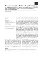
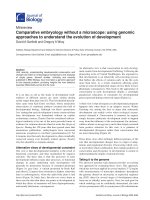
Tissue distribution of Hsp65 messageFigure 1
Tissue distribution of Hsp65 message. The presence of Hsp65 message in various tissue samples obtained from BALB/c mice
after intramuscular immunization with 100 µg of pcDNA3-HSP65 in 25% sucrose. Total RNA (10 µg) isolated from each tissue
was treated with DNase I and subjected to RT-PCR amplification with HSP65 or β-actin primers (RT+). As an RNA quality
control, β-actin was amplified. No products (HSP65/β-actin) were seen when total RNA in the absence of reverse transcrip-
tion was subjected to PCR amplification (RT-). All RT-PCR products were analyzed by agarose gel electrophoresis and visual-
ized by ethidium bromide staining. The results were obtained from one mouse and are representative of three independent
experiments. RT-PCR from the material obtained of mice (three animals for each point) immunized with the vector (pcDNA3)
was negative in all analysis.
Genetic Vaccines and Therapy 2006, 4:1 />Page 6 of 10
(page number not for citation purposes)
intramuscular delivery, we observed the presence of
Hsp65 message in several tissue samples until 7 days after
injection, including secondary lymphoid organs. After
this period the Hsp65 message was detected only in mus-
cle and liver (Figure 1). Since our assay was not quantita-
tive, we did not determine the amount of Hsp65 message
in each tissue; but its presence in each material for a lim-
ited time was shown. However we believe that the success
of our vaccine against experimental tuberculosis could be
due to another two doses of plasmid at 15 days. This
schedule of immunization could preserve the Hsp65 mes-
sage at lymphoid organs resulting in the induction of spe-
cific and efficient immune response [9].
Even though the Hsp65 message was not detected after fif-
teen days, the plasmid DNA could still be found in all ana-
lyzed tissues including lung, DLN, spleen, liver, bone
marrow, kidney, muscle and thymus for at least six
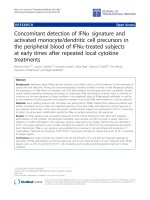
Identification of plasmid DNA rescuedFigure 2
Identification of plasmid DNA rescued. Nature of plasmid DNA obtained after transformation of cellular DNA from tissues of
mice 2 days after i.m. immunization with pcDNA3-Hsp65. Escherichia coli DH5-α was transformed with 1 µg of total DNA from
tissues of mice previously immunized with pcDNA3-Hsp65. Plasmid DNA was recovered from ampicillin-resistant colonies.
(A) Agarose gel showing plasmid DNA digested overnight with Hind III and Xba I: wild-type pcDNA3-Hsp65 (lane a); plasmid
DNA recovered from muscle (lane b); plasmid DNA from bone marrow (lane c) plasmid DNA from liver (lane d); plasmid
DNA from spleen (lane e). (B) PCR analysis of rescued plasmid using HSP65 primers: Wild type pcDNA3-Hsp65 (lane a); plas-
mid rescued from muscle of immunized mice (lane b). The mobility of DNA size standards (λ DNA cut with Hind III) are
shown on the left. (C) Identification of nucleotide sequence of plasmid DNA rescued from muscle. Sequence analyses were
performed using the blastn program from BLAST.
Genetic Vaccines and Therapy 2006, 4:1 />Page 7 of 10
(page number not for citation purposes)
months post-inoculation. These findings indicate that
plasmid DNA-Hsp65 disseminated widely throughout the
body and persisted as a plasmid DNA form or produced
lower doses of message not detectable in our RT-PCR
assay (Table 1 and 2). After two days of immunization,
about 100 ampicillin resistant colonies/µg genomic DNA
could be obtained from different tissues. However, the
number of the ampicillin-resistant colonies decreased at
later time points, suggesting that the levels of plasmid
DNA was also reduced (Table 2). At six months a lower
frequency of recovered plasmid DNA was observed in kid-
ney, lung and thymus (Table 2). The presence of plasmid
DNA in the thymus could be a concern, since the expres-
sion of antigen in this tissue could induce tolerance by
deletion of Hsp65-specific T cells altering the induction of
Hsp65 immune response after intramuscular immuniza-
tion schedule. However, the message was detected only
two days after immunization in almost all tissues ana-
lyzed and the number of plasmids rescued decreased after
six months, suggesting that the plasmids could have been
damaged or digested by endonucleases. Interestingly, the
presence of plasmid DNA was observed in different tissues
for longer time points, even in the presence of lower num-
bers of colonies. These results are important because they
show that even after widespread biodistribution of plas-
mid, the detection was reduced after 6 months. Further-
more, there are substantial data provided by our
laboratory demonstrating that the presence of naked DNA
in different organs does not change the histological pat-
tern, suggesting the absence of inflammatory response in
these tissues (manuscript in preparation, Deison Soares
personal communication).
To assure that the plasmid rescued from different tissues
was pcDNA3-Hsp65, we analyzed several plasmids by
three different methods: restriction pattern (data not
shown) and insert released, PCR and nucleotide sequence.
Figure 2 illustrates the results of the same samples
obtained from different tissues. These experiments were
done to determine the identity of the plasmid DNA and to
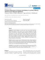
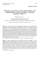
Analysis of the pcDNA3-Hsp65 genome integrationFigure 4
Analysis of the pcDNA3-Hsp65 genome integration. Samples of
liver tissue from mice immunized with 100 µg of pcDNA3-
HSP65 (lanes a and b) and from nonimmunized mice (lanes c
and d) (negative control) were submitted to Southern blot
after Nde I digestion. Lanes e and f correspond to wild-type
plasmid digested with Nde I or undigested, respectively. The
bands were detected using pcDNA3 labeled with chemilumi-
nescent reagent. The multiple forms of plasmid DNA are
indicated in the figure. The samples were loaded in a same
gel and the lanes not used were removed.
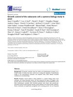
Persistence of DNA adenine methylase site methylations (dam) of pcDNA3-HSP65 in muscle at 6 months after immu-nizationFigure 3
Persistence of DNA adenine methylase site methylations (dam) of
pcDNA3-HSP65 in muscle at 6 months after immunization. (A)
Approximately 1 µg cellular DNA obtained from muscle of
immunized mouse were digested with Nde I and Dpn I (lane
a), Nde I and Mbo I (lane b), or with Nde I alone (lane c) and
amplified by PCR using Hsp65 primers. The samples were
submitted to electrophoresis on a 1% agarose gel (B) The
positive control was done using E.coli DNA digested with
Dpn I, Mbo I or non-digested to show the dam methylation
pattern. The mobility of DNA size standards (l DNA cut with
Hind III) are shown on the left.
Genetic Vaccines and Therapy 2006, 4:1 />Page 8 of 10
(page number not for citation purposes)
avoid false-positive results. The results confirmed that the
plasmid rescued was the pcDNA3-Hsp65.
The widespread distribution of plasmid DNA throughout
the body, regardless of the method and route of adminis-
tration, has been previously reported [4,6,8,15,16,22].
Currently, the mechanism involved in this widespread
biodistribution of plasmid DNA is not completely
defined. It has been speculated that this may occur as a
result of the transport of free plasmid DNA as well as its
transport by transfected cells [22]. Nevertheless, the wide-
spread biodistribution cannot be restricted to a particular
mechanism or cell type, since after immunization with
naked DNA it can be taken up by different cells like
CD11b+ (2), CD11c (23,24), CD11c and CD19 (3),
reaching distant sites. However, we cannot exclude the
transport of plasmid DNA by serum or lymph.
Based on our results we suggest that the widespread bio-
distribution can also be correlated with the plasmid dose
injected. To evaluate this possibility, some mice were
immunized with lower doses of plasmid DNA-Hsp65 (4
and 20 µg/mouse). The results presented in Table 2
showed that in mice receiving the 20 µg dose, the plasmid
remained in some tissues such as muscle, lymph nodes
and bone marrow and was recovered only after 48 h after
inoculation, whereas in mice that received the lower dose
of 4 µg, the plasmid DNA was not recovered from any of
the tissues analyzed (data not shown).
The limited biodistribution in mice that received lower
doses cannot be attributed to the sensitivity of the method
used, since by using plasmid DNA labeled with fluores-
cent dye (10–12 µg/mouse) we showed a similar pattern
of biodistribution [3]. Consequently, we suggest that the
widespread biodistribution is also dose correlated. These
results are important for the development of new vectors,
enabling the use of lower doses and thereby reducing the
risks in the clinical application of DNA vaccines. Nowa-
days, one of the alternatives to reduce the amount of the
plasmid administered is the delivery of it by micropheres
[14,16,17] or liposomes [7]. However, the plasmid DNA
is not easily released from these complexes and this can
arrest the induction of an efficient immune response.
Therefore, new approaches, such as that described in [25]
can be an alternative to reduce the plasmid DNA dose
without altering the immune response.
Interestingly, the Hsp65 message was consistently
detected in muscle and liver tissue samples at day 15. In
addition, the number of colonies in the liver was quite
reduced (Table 2) which could, in part, explain the detec-
tion of Hsp65 message in such tissue fifteen days after
DNA injection. The long-term persistence of plasmids in
liver and muscle tissues has also been observed by other
authors using different plasmid constructs [5]. However,
other authors obtained different results after using alter-
native routes of plasmid administration, such as the intra-
nasal [26]. As suggested by Wolff et al. (1992), the long-
term antigen expression by muscle cells can be related to
structural features, such as multinucleated cells. It is pos-
sible that these tissue characteristics are responsible for
the prolonged presence of the message to Hsp65 in the
muscle tissue. However, there is no satisfactory explana-
tion for the Hsp65 long-term message expression in liver
tissue yet, but we must be clear that the backbone of plas-
mid DNA can also take part in message expression.
The recovery of plasmid DNA from eukaryotic cells using
bacterial transformation is a simple, fast and sensitive
method, which was suitable for our objective of verifying
the presence or absence of plasmid DNA in different tis-
sues independent of the copy number in the tissues. On
the other hand we also detected the plasmid in some tis-
sues by PCR indicating that both methods showed the
same results (data not shown).
In general, plasmid DNA used in genetic vaccines pos-
sesses an SV40 sequence of replication and could replicate
in vivo. Replicative synthesis of plasmid DNA in mamma-
lian cells can be evidenced by the appearance of molecules
lacking the DNA adenine methylase-dependent (DAM)
adenosine methylation [[27]. Plasmid DNA from bacteria
contains a methylated adenosine within the GATC recog-
nition sites for Dpn I and Mbo I. The Dpn I cleaves the site
more efficiently if the adenine is methylated, whereas
Mbo I cleaves the site more efficiently if the adenine is
unmethylated [18,27]. If the plasmid DNA replicates in
mammalian cells, then the bacterial methylation pattern
is lost. The pattern of methylation in E.coli was shown in
Figure 3B, as a positive control. Prokaryotic DNA was
cleaved by DpnI as expected, but not by Mbo I (Fig. 3B),
to assure the dam methylation pattern. The methylation of
plasmid rescued from muscle is shown in Figure 3A. This
result revealed that the bacterial methylation pattern of
the injected pcDNA3-Hsp65 DNA was unchanged after
having resided in muscle for at least six months, indicat-
ing that this plasmid DNA did not replicate in vivo. These
results are in agreement with those from previous studies
using different plasmid DNA vaccines [5,28]. In addition,
the lack of plasmid replication in vivo provides a higher
degree of safety for gene therapy vaccination.
The recovery of ampicillin-resistant colonies from the tis-
sues of pcDNA3-Hsp65-immunized mice after the trans-
formation of total cellular DNA suggests that some
extrachromosomal plasmid DNA was maintained for at
least 6 months. Southern blot analysis was done with liver
and muscle (data not shown) samples at 30 days after
immunization due the higher number of plasmid rescued
Genetic Vaccines and Therapy 2006, 4:1 />Page 9 of 10
(page number not for citation purposes)
by bacterial transformation in liver sample. Furthermore,
the number of plasmid rescued at 180 days was compara-
ble to the number of plasmids obtained at 30 days. The
results displayed in figure 4 showed that the plasmid did
not integrate into the BALB/c genome. In general, plasmid
integration into genomic DNA occurs by tandem repeats
[30], and when released from the genome the plasmid
shows a linear form in Southern blot. On the other hand,
when the plasmid DNA is not integrated it has the blue-
print of undigested DNA. The pcDNA3-Hsp65 showed a
pattern similar to undigested plasmid DNA when com-
pared to wild type plasmid DNA used in immunization
(Figure 4, lane a and f). These results indicated that
pcDNA3-Hsp65 was not integrated into the mouse
genome at the time point analyzed.
The frequency of integration into the cellular genome
could be affected by several factors, such as the plasmid
sequence, the presence of chi-like elements [29], Alu seg-
ments [30] and minisatellite regions [31]. However, the
integration of bacterial plasmid DNA is not quite so sim-
plistic. The mammalian genome appears to possess a
mechanism to protect its integrity [32]. In addition, the
results provided by Ledwith et al. (2000), using different
plasmid constructs suggest that the risk of integration of
plasmid DNA vaccines following intramuscular inocula-
tion is negligible. Therefore, the use of plasmid DNA in
gene therapy can be safer than vector systems. At the
present time, the mechanisms involved in the non-inte-
gration of pcDNA3-Hsp65 could not be definitively char-
acterized. However, our results suggest that this vaccine is
safe for clinical use and indicate that the use of a plasmid
containing the Hsp65 gene is reliable for gene therapy
purposes as well as for vaccination in a clinical setting. In
addition, the results of long time biodistribution/dose
after intramuscular delivery were observed and described
for the first time herein. Accordingly, we consider that our
findings open not only new perspectives for DNA vaccines
but also lead to new considerations about the inoculation
site and delivery systems.
Acknowledgements
The authors are grateful to Izaíra T. Brandão for her technical assistance.
This study was supported by grants from the Fundação de Amparo à
Pesquisa do Estado de São Paulo (FAPESP, Foundation for the Support of
Research in the State of São Paulo) and from the Conselho Nacional de
Desenvolvimento Científico e Tecnológico (CNPq, Brazilian Council for
Scientific and Technological Development).
References
1. Liu MA: DNA vaccines: a review. J Int Med 2003, 253:402-410.
2. La Cava A, Billetta R, Gaietta G, Bonnin DB, Baird SM, Albani S: Cell-
mediated DNA transport between distant inflammatory
sites following intradermal DNA immunization in the pres-
ence of adjuvant. J Immunol 2000, 164:1340-5.
3. Coelho-Castelo AAM, Santos-Junior RR, Bonato VL, et al.: B-lym-
phocytes in bone marrow or lymph nodes can take up plas-
mid DNA after intramuscular delivery. Hum Gene Ther 2003,
14:1279-1285.
4. Tuomela M, Malm M, Wallen M, et al.: Biodistribution and general
safety of naked DNA plasmid GTU
®
– multiHIV, in a rat,
using a quantitative PCR method. Vaccine 2005, 23:890-896.
5. Wolff JA, Ludtke JJ, Acsadi G, et al.: Long-term persistence of
plasmid DNA and foreign gene expression in mouse muscle.
Hum Mol Gene 1992, 1:363-369.
6. Parker SE, Borellini F, Wenk ML, et al.: J.A. Plasmid DNA malaria
vaccine: tissue distribution and safety studies in mice and
rabbits. Hum Gene Ther 1999, 10:741-758.
7. Klavinskis LS, Barnfield C, Gao L, et al.: Intranasal immunization
with plasmid DNA-lipid complexes elicits mucosal immunity
in the female genital and rectal tracts. J Immunol 1999,
162:254-262.
8. Koshkina NV, Agoulnik SL, Densmore CL, et al.: Biodistribution
and Pharmacokinetics of Aerosol and intravenously adminis-
tered DNA-polyethyleneimine complexes: Optimization of
Pulmonary Delivery and Retention. Mol Ther 2003, 8:249-254.
9. Lowrie DB, Tascon RE, Bonato VLD: Therapy of tuberculosis in
mice by DNA vaccination. Nature 1999, 400:269-271.
10. Bonato VL, Lima VM, Tascon RE, et al.: Identification and charac-
terization of protective T cells in HSP65 DNA-vaccinated
and Mycobacterium tuberculosis-infected mice. Infect Immun
1988, 66:169-175.
11. Ruberti M, De Melo LK, Dos Santos SA, et al.: Prime-boost vacci-
nation based on DNA and protein-loaded microspheres for
tuberculosis prevention. J Drug Target 2004, 12:195-203.
12. Vordermeier HM, Lowrie DB, Hewinson RG: Improved immuno-
genicity of DNA vaccination with mycobacterial HSP65
against bovine tuberculosis by protein boosting. Vet Microbiol
2003, 93:349-359.
13. Chun S, Daheshia M, Lee S, et al.: Distribution fate and mecha-
nism of immune modulation following mucosal delivery of
plasmid DNA encoding IL-10. J Immunol 1999, 163:2393-2402.
14. Lunsdford L, Mc Keever U, Eckstein V, et al.: Tissue biodistribution
and persistence in mice of plasmid DNA encapsulated on a
PLGA-based microsphere delivery vehicle. J Drug Target 2000,
8:39-50.
15. Tadokoro K, Koizumi Y, Miayagi Y, et al.: Rapid and wide-reaching
delivery of HIV-1 env DNA vaccine intranasal administra-
tion. Viral Immunol 2001, 14:159-167.
16. Oh YK, Kim J-P, Yoon H, et al.: Prolonged organ retention and
safety of plasmid DNA administered in polyethylenimine
complexes. Gene Ther 2001, 8:1587-1592.
17. Oh YK, Kim J-P, Hwang TS, et al.: Nasal absorption and biodistri-
bution of plasmid DNA: an alternative route of DNA vaccine
delivery. Vaccine 2001, 19:4519-4525.
18. Maniatis T, Fritsch EF, Sambrook J: Molecular cloning: a laboratory man-
ual 2nd edition. Cold Spring Harbor Laboratory Press; 1989.
19. Mearini G, Nielsen PE, Fackelmayer FO: Localization and dynam-
ics of small circular DNA in live mammalian nuclei. Nucleic
Acids Res 2004, 32:2642-651.
20. Sedgwick JD, Holt PG: Induction of IgE-secreting cells and IgE
isotype specific suppressor T cells in the respiratory lymph
nodes of rats in response to antigen. Cell Immunol 1985,
94:182-194.
21. Liu MA: Overview of DNA vaccines. Ann N Y Acad Sci 1995,
772:15-20.
22. Dai Y, Roman M, Naviaux RK, et al.: Gene Therapy via primary
myoblasts: long-term expression of factor IX protein follow-
ing transplantation in vivo. Proc Natl Acad Sci USA 1992,
89:10892-10895.
23. Dupuis M, Denis-Mize K, Woo C, et al.: Distribution of DNA vac-
cines determines their immunogenicity after intramuscular
injection in mice. J Immunol 2000, 165:2850-2858.
24. Chattergoon MA, Robinson TM, Boyer JD, Weiner DB: Specific
immune induction following DNA-based immunization
through in vivo transfection and activation of macrophages/
antigen-presenting cells. J Immunol 1998, 160:5707-18.
25. Ribeiro SC, Monteiro GA, Prazeres DM: The role of polyadenyla-
tion signal secondary structures on the resistence of plas-
mids vectors to nucleases. J Gene Med 2004, 5:563-573.
26. Arruda VR, Fields PA, Milner R, et al.: Lack of germline transmis-
sion of vector sequences following systemic administration
Publish with BioMed Central and every
scientist can read your work free of charge
"BioMed Central will be the most significant development for
disseminating the results of biomedical research in our lifetime."
Sir Paul Nurse, Cancer Research UK
Your research papers will be:
available free of charge to the entire biomedical community
peer reviewed and published immediately upon acceptance
cited in PubMed and archived on PubMed Central
yours — you keep the copyright
Submit your manuscript here:
/>BioMedcentral
Genetic Vaccines and Therapy 2006, 4:1 />Page 10 of 10
(page number not for citation purposes)
of recombinant AAV-2 vector in males. Mol Ther 2001,
4:586-592.
27. Ringquist S, Smith CL: The Escherichia coli chromosome con-
tains specific, unmethylated dam and dcm sites. Proc Natl Acad
Sci 1992, 89:4539-4543.
28. Shiroishi T, Koide T, Yoshino M, et al.: Hotspots of homologous
recombination in mouse meiosis. Adv Biophys 1995, 31:119-132.
29. Manam S, Ledwith BJ, Barmum AB, et al.: Plasmid DNA vaccine:
Tissue distribution and effects of DNA sequence, adjuvants
and delivery method on integration into host DNA. Intervirol-
ogy 2000, 43:273-281.
30. Rudiger NS, Gregersen N, Kielland-Brandt MC: One short well
conserved region of Alu-sequences is involved in human
gene rearrangements and has homology with prokaryotic
chi. Nucleic Acids Res 1995, 23:256-260.
31. Krowczynska AM, Rudders RA, Krontiris TG: The human minisat-
ellite consensus at break-points of oncogene translocations.
Nucleic Acids Res 1990, 18:1121-1127.
32. Scrable H, Stambrook PJ: A genetic program for deletion of for-
eign DNA from the mammalian genome. Mutat Res 1999,
429:225-237.
33. Ledwith BJ, Manam S, Troilo PJ, et al.: Plasmid DNA vaccines:
Investigation of integration into host cellular DNA. Intervirol-
ogy 2000, 43:258-272.