Báo cáo sinh học: "Careful adjustment of Epo non-viral gene therapy for β-thalassemic anaemia treatment" pot
Bạn đang xem bản rút gọn của tài liệu. Xem và tải ngay bản đầy đủ của tài liệu tại đây (367.85 KB, 6 trang )
BioMed Central
Page 1 of 6
(page number not for citation purposes)
Genetic Vaccines and Therapy
Open Access
Research
Careful adjustment of Epo non-viral gene therapy for β-thalassemic
anaemia treatment
Emmanuelle E Fabre
1,2,3,4
, Pascal Bigey*
1,2,3,4
, Yves Beuzard
5
,
Daniel Scherman
1,2,3,4
and Emmanuel Payen
5
Address:
1
Unité de Pharmacologie Chimique et Génétique, INSERM U640, Faculté de Pharmacie, 4 avenue de l'observatoire, 75006 Paris, France,
2
Unité de Pharmacologie Chimique et Génétique, CNRS UMR 8151, Faculté de Pharmacie, 4 avenue de l'observatoire, 75006 Paris, France,
3
Unité
de Pharmacologie Chimique et Génétique, Université Paris Descartes, Faculté de Pharmacie, 4 avenue de l'observatoire, 75006 Paris, France,
4
Unité
de Pharmacologie Chimique et Génétique, Ecole Nationale Supérieure de Chimie de Paris, 11 rue Pierre et Marie Curie, 75005 Paris, France and
5
Laboratoire de Thérapie Génique Hématopoïétique, Institut d'Hématologie (IUH), INSERM U733, Hôpital Saint-Louis, 75011 Paris, France
Email: Emmanuelle E Fabre - ; Pascal Bigey* - ; Yves Beuzard - ;
Daniel Scherman - ; Emmanuel Payen -
* Corresponding author
Abstract
Background: In situ production of a secreted therapeutic protein is one of the major gene therapy
applications. Nevertheless, the plasmatic secretion peak of transgenic protein may be deleterious
in many gene therapy applications including Epo gene therapy. Epo gene transfer appears to be a
promising alternative to recombinant Epo therapy for severe anaemia treatment despite
polycythemia was reached in many previous studies. Therefore, an accurate level of transgene
expression is required for Epo application safety. The aim of this study was to adapt posology and
administration schedule of a chosen therapeutic gene to avoid this potentially toxic plasmatic peak
and maintain treatment efficiency. The therapeutic potential of repeated muscular electrotransfer
of light Epo-plasmid doses was evaluated for anaemia treatment in β-thalassemic mice.
Methods: Muscular electrotransfer of 1 μg, 1.5 μg, 2 μg 4 μg or 6 μg of Epo-plasmid was
performed in β-thalassemic mice. Electrotransfer was repeated first after 3.5 or 5 weeks first as a
initiating dose and then according to hematocrit evolution.
Results: Muscular electrotransfer of the 1.5 μg Epo-plasmid dose repeated first after 5 weeks and
then every 3 months was sufficient to restore a subnormal hematrocrit in β-thalassemic mice for
more than 9 months.
Conclusion: This strategy led to efficient, long-lasting and non-toxic treatment of β-thalassemic
mouse anaemia avoiding the deleterious initial hematocrit peak and maintaining a normal
hematocrit with small fluctuation amplitude. This repeat delivery protocol of light doses of
therapeutic gene could be applied to a wide variety of candidate genes as it leads to therapeutic
effect reiterations and increases safety by allowing careful therapeutic adjustments.
Published: 11 March 2008
Genetic Vaccines and Therapy 2008, 6:10 doi:10.1186/1479-0556-6-10
Received: 12 September 2007
Accepted: 11 March 2008
This article is available from: />© 2008 Fabre et al; licensee BioMed Central Ltd.
This is an Open Access article distributed under the terms of the Creative Commons Attribution License ( />),
which permits unrestricted use, distribution, and reproduction in any medium, provided the original work is properly cited.
Genetic Vaccines and Therapy 2008, 6:10 />Page 2 of 6
(page number not for citation purposes)
Background
Therapeutic protein secretion by an in vivo transfected
organ is one of the major gene therapy applications. One
drawback to be avoided in such therapeutic strategy is the
potentially deleterious secretion peak of therapeutic pro-
tein following DNA administration. The aim of this study
was to adapt dosage and administration schedule of a
chosen therapeutic gene to avoid this potentially toxic
plasmatic peak.
Recombinant erythropoietin (rhEpo) injections are com-
monly used to treat anaemia linked to cancer treatment or
chronic renal failure. However, rhEpo injections remain
an expensive treatment which requires frequent delivery
injection repeats and which can lead to anti-Epo antibod-
ies production by the patient [1]. Therefore, erythropoie-
tin (Epo) gene transfer appears to be a promising
alternative for severe anaemia treatment since it requires
less frequent treatment repeat and may allow sustained
Epo secretion and constant patient coverage. Epo gene
transfer has already been tested on normal animals and
on anaemia animal models such as β-thalassemia and
chronic renal failure models. To this end, various gene
transfer strategies have been used such as ex-vivo strategies
using engrafted transduced myoblasts or other cell types
[2-4], viral strategies using adenovirus [5] adeno-associ-
ated virus [6,7], helper-dependent adenovirus [8], or non-
viral strategies using naked DNA injection [9], poloxamer/
DNA formulations [10] or naked DNA injection associ-
ated to electrotransfer [9,11-13]. In several of these stud-
ies, the gene dose transferred led to a maximum
hematocrit value between 70 and 80% [6,9-13] which cor-
responds to potentially lethal polycythemia [6]. There-
fore, in the particular case of Epo, an accurate level of
transgene expression is required for safety reasons.
Temporal control systems of transgene expression have
already been used in gene therapy preclinical experiments,
including for Epo gene use [6,10,14,15]. These systems
could avoid deleterious Epo secretion peak, but unsolved
problems such as host immune response against the trans-
activator [10] or inducing agents adverse effects, are still
restricting their use.
In order to avoid the toxic Epo plasmatic peak and to
reduce plasmatic fluctuation amplitude, we decided to
test different doses and administration schedules of an
Epo encoding plasmid in anaemia treatment of β-tha-
lassemic mice. Considering electrotransfer advantages in
terms of safety, efficiency and cost, we chose this well-
handled gene transfer method. Our previous experiment
with β-thalassemic mice using intramuscular electrotrans-
fer of an Epo encoding plasmid [9] led to a first estimation
of transgene product kinetics and physiologic effects. Epo
plasmatic level was found to reach a peak value within
two weeks after gene therapy treatment and then to
decrease approximately of 40%, 20% and 15% of this
peak after 1, 2 and 3 months, respectively. This plasmatic
Epo kinetics was roughly confirmed in normal mice by
other studies with a secretion peak one week after electro-
transfer [11,13]. However, Epo main physiologic effect on
erythropoiesis which can be evaluated through hemat-
ocrit measurement remained intense for several months
because of red blood cell half-life. Indeed, β-thalassemic
mice hematocrit was still at the polycythemic value of
60% four months after 20 μg Epo-plasmid electrotransfer
[9].
Considering those results, we have presently tested the
therapeutic potential of repeated electrotransfer of subop-
timal low Epo-plasmid doses in the β-thalassemic mouse
model to restore and maintain a normal hematocrit with-
out reaching toxicity.
Methods
Plasmid
The pCMV-Epo plasmid used for experiments was a pCOR
plasmid [16] containing the mouse erythropoietin cDNA
under the regulatory control of the hCMV E/P [17]. Plas-
mid large-scale production and double caesium chloride
gradient ultracentrifugation used as purification method,
were realised according to traditional molecular biology
methods [18]. Plasmid construct was checked by restric-
tion fragment length profile and sequencing.
Animal experiments
Animal experiments were conducted following NIH rec-
ommendations. The β-thalassemic Hbb-thal1 mice [19]
from the laboratory of Haematopoietic Gene Therapy
(Saint Louis Hospital, Paris, France) were used for experi-
ments. Two to four months female mice were separated
into 6 groups: six Hbb-thal1 mice per group were used for
the higher plasmid dose experiment, and eight Hbb-thal1
mice per group were used for the lower plasmid dose
experiment. Mice were first anaesthetised by intra-perito-
neal injection of 250 μl of a ketamine-xylazine solution
(respectively 8.66 mg/ml and 0.31 mg/ml in 150 mM
NaCl). Left rear legs were shaved and the Epo-plasmid
solution was injected in the tibialis-cranialis muscle. The
DNA solutions were diluted in 150 mM NaCl to contain
the desired plasmid quantity in 30 μl: 1 μg, 1.5 μg, 2 μg, 4
μg and 6 μg, respectively, for the corresponding groups
(meaning 50, 75, 100, 200 or 300 ng of plasmid per
mouse gram, respectively). The DNA injection was imme-
diately followed by application of eight electric pulses of
200 V/cm intensity, 20 ms duration and delivered at a fre-
quency of 1 Hz, using plate electrodes and generator BTX
ECM 830 (Genetronics™), as previously described [20].
Genetic Vaccines and Therapy 2008, 6:10 />Page 3 of 6
(page number not for citation purposes)
Sample collection, measurement and assay
Blood samples were collected by retro-orbital puncture of
anaesthetised mice at desired time after plasmid electro-
transfer. Hematocrits were measured using a standard
micro-hematocrit method [21]. Mouse Epo assay was real-
ised on serum samples using the EPO ELISA Medac
®
kit
(Medac™) based on cross-reaction with human Epo.
Statistical analysis
Analysis of variance (ANOVA) and Fisher PLSD were used.
Results and discussion
Our previous study of β-thalassemic mice demonstrated
that electrotransfer of 1–10 μg Epo-plasmid doses were
sufficient to induce a significant hematocrit increase.
However, after a hematocrit burst depending on the dose
of injected DNA during the first month after treatment,
the hematocrit of treated mice started to decrease, and
finally stabilised two months after electrotransfer. Surpris-
ingly, this plateau was the same whatever the DNA dose
used for gene transfer, and hematocrit still remained dif-
ferent from controls for at least 4 months [9]. Moreover,
the 5 μg Epo-plasmid dose seemed to be the most appro-
priate since it led to normal hematocrit at peak value
(approximately 45%). This hematocrit profile resulted
from a shorter Epo plasmatic kinetics with peak of expres-
sion reached in less than 2 weeks and an expression level
relative to this peak value of 40%, 20% and 15% respec-
tively 1, 2 and 3 months after electrotransfer. Higher doses
of Epo-plasmid led to hazardous unsafe hematocrit peak
(60 to 80%). This study is then designed to slowly reach
and maintain the hematocrit plateau and to avoid the ini-
tial hemarocrit burst.
To avoid a possible hematocrit busrt following the elec-
trotransfer treatment, we decided to raise the hematocrit
step by step by repetitive treatments with small doses of
plasmid DNA. In our mind, the first treatment should be
performed with a small dose of the plasmid that would be
insufficient to reach a normal hematocrit value, but which
should just raise it a little. The purpose of this first dose
was to initiate the treatment. The following treatments
would then performed to assess the possibility to raise the
hematocrit a little bit more, closer to a normal value, and
to maintain it to an almost constant value. To assess the
DNA dose appropriate to this aim, we first evaluated Epo
plasmid doses of 2, 4 and 6 μg per mouse which were elec-
trotransfered at days 0 and 25 (fig 1). Maximum hemat-
ocrit values of 56.2% ± 3.2%, 74.5% ± 2.5% and 73.7% ±
2.4% respectively for the 2 μg, the 4 μg and the 6 μg
groups, were reached two months after the first electro-
transfer (fig 1). Therefore each dose led to polycythemia
which was stronger for the 4 μg and 6 μg groups. Four
months after the first electrotransfer, the hematocrit levels
became equivalent between the three plasmid doses (no
statistical difference), and kinetics showed similar slow
decrease. Moreover, hematocrit level of each treated group
remained significantly different from the control group up
to 7.5 months (p < 0.05).
Regarding those results, we decided to decrease plasmid
doses down to 1 μg and 1.5 μg and to increase the time
interval between electrotransfer treatments (fig 2). Elec-
trotransfer of those plasmid doses was first repeated at day
34 and then according to hematocrit value. For additional
treatments, we decided to use in each group the same dose
used for the first treatment (i.e. 1 μg or 1.5 μg, respec-
tively, for the two treated groups); treatments were per-
formed when the mean hematocrit of the highest dose
(1.5 μg) decreased around 40%. An additional treatment
(day 80) was performed with the 1 μg group because we
estimated that the hematocrit was too low. Following
treatments were then performed at the same time points
than for the 1.5 μg group.
A hematocrit decrease of approximately 3% was observed
in the control group between the beginning and the end
of the experiment (fig 2-A) (p < 0.0001). As the study pro-
ceeded over 17 months, this is to be linked with anaemia
escalation coming along with ageing in our β-thalassemic
context, which as already been described [22]. The 1 μg
dose delivered at day 0, 34, 77, 112 and day 215, led to
significant hematocrit increase which was maintained
between 35.4% and 38.7% during 10 months (fig 2-A and
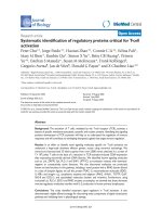
Hematocrit of β-thalassemic mice electrotransfered twice with 2, 4 and 6 μg of Epo plasmidFigure 1
Hematocrit of β-thalassemic mice electrotransfered
twice with 2, 4 and 6 μg of Epo plasmid. Hematocrit
kinetics of β-thalassemic mice electrotransfered at day 0 and
day 25 with 2 μg (cross), 4 μg (empty square) and 6 μg (solid
square) Epo plasmid doses. The negative control (solid dia-
mond) was realised by intramuscular injection of NaCl (150
mM) followed by electric pulse application. Error bars show
standard error of mean (SEM). Arrows indicate electrotrans-
fer applications.
25
30
35
40
45
50
55
60
65
70
75
80
0 30 60 90 120 150 180 210 240 270 300 330 360
Days
Hematocrit (%)
25
30
35
40
45
50
55
60
65
70
75
80
0 30 60 90 120 150 180 210 240 270 300 330 360
Days
Hematocrit (%)
Genetic Vaccines and Therapy 2008, 6:10 />Page 4 of 6
(page number not for citation purposes)
2-B). The mean hematocrit value was significantly higher
for this group than for the control group from day 69 (p <
0.05) to day 493 (p < 0.05). As compared to the β-tha-
lassemic mice control group, the 1 μg administration
schedule led to a progressive delta hematocrit increase
during 3 months and then reached a 4–6% plateau value
which was maintained until the end of the experiment.
However, it appeared that with this dose we could not get
any better than 39% (Fig 2). This dose is then definitely
not sufficient for our goal to approach normal value. The
administration schedule corresponding to 1.5 μg Epo-
plasmid deliveries at day 0, 34, 112 and day 215 gave
more promising results. An improved hematocrit value,
between 38.4% and 42.3%, was steadily maintained for
more than 9 months (fig 2-A and 2-C). The delta hemat-
ocrit, taking control group as reference, oscillated between
5.1% and 9.8% from one month after the beginning of
the experiment to its end. Therefore, the hematocrit of the
1.5 μg group remained significantly higher than that of
the control group from day 13 (p < 0.05) to day 493 at
least (p < 0.001 at 17.6 months). Moreover, despite anae-
mia escalation coming along with ageing, similar hemat-
ocrit peak values were reached after the whole two firsts,
the third and the fourth electrotransfers of the 1.5 μg Epo-
plasmid dose. These hematocrit values were of 42.3%,
41.6% and 41.8%, and delta hematocrit values were of
9.0%, 9.0% and 9.8% respectively at days 48, 140 and 241
(no statistical difference). Therefore, the first two electro-
transfers seemed to have an equivalent impact on hemat-
ocrit than the third and fourth treatments. mEPO
plasmatic levels were measured, but no statistical differ-
ence could be highlighted between plasmatic Epo levels
reached at days 48, 140 and 241 [additional file 1]. Actu-
ally, mEPO was detectable to levels close to the limit of
detection of our ELISA kit. We believe this is not very sur-
prising: as erythropoiesis is very sensitive to EPO levels,
small changes in EPO levels may lead to very visible
effects on hematocrit. As we targeted only small hemat-
ocrit increases, we did not expect high levels of circulating
EPO. Instead, we believe that a statistically significant dif-
ference in hematocrit, which is the real physiological
parameter we want to impact on, is much more relevant
in this study. The other blood cell lineages were analysed
from day 48 to day 271. According to time, significant
increases in red blood cell count (data not shown) and
hemoglobin concentration (fig 3-A) were observed. These
increases were responsible for hematocrit increase. On the
contrary, a decrease in mean corpuscular hemoglobin
concentration (MCHC) was noticed when compared to
the control at day 91 and then from day 189 to day 271
for the 1.5 μg group (p values of 0.002 on day 91, 0.005
on day 189, 0.002 on day 210, 0.01 on day 241 and 0.002
on day 271) and at day 91, 189 and 241 for the 1 μg group
(p values of 0.02 on day 91, 0.001 on day 189 and 0.01
on day 241) (fig 3-B). Such a phenomenon has already
been described in β-thalassemic mice treated with rhEpo
[23] and might be related to iron deficiency [24]. The
other lineage study did not reveal any variation (data not
shown). In particular, we did not observe any variation in
platelet counts, whereas it has already been found to be
increased in patient with renal failure chronically treated
with recombinant Epo [25].
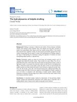
Hematocrit of β-thalassemic mice after repeated muscular electrotransfer of 1 μg and 1.5 μg of Epo-plasmidFigure 2
Hematocrit of β-thalassemic mice after repeated
muscular electrotransfer of 1 μg and 1.5 μg of Epo-
plasmid. Individual hematocrit kinetics of β-thalassemic
mice electrotransfered with NaCl 150 mM solution for con-
trol group (2-A) or with 1 μg (2-B) and 1.5 μg (2-C) of Epo-
plasmid for the other groups. Figure 2-D presents mean
hematocrit of each group with standard error of the mean
(SEM). Electrotransfer was performed at day 0, 34, 112 and
215 for the three groups. One additional electrotransfer was
performed at day 77 for the 1 μg group. Arrows indicate
electrotransfer applications.
25
30
35
40
45
50
55
60
0 30 60 90 120 150 180 210 240 270 300 330 360 390 420 450 480
Hematocrit (%)
25
30
35
40
45
50
55
60
0 30 60 90 120 150 180 210 240 270 300 330 360 390 420 450 480
Hematocrit (%)
25
30
35
40
45
50
55
60
0 30 60 90 120 150 180 210 240 270 300 330 360 390 420 450 480
Hematocrit (%)
25
30
35
40
45
50
0 30 60 90 120 150 180 210 240 270 300 330 360 390 420 450 480
Days
Mean hematocrit (%)
Control 1μg 1.5μg
D
A
Control
B
1μg
C
1.5μg
1μg
1.5μg
1μg
1.5μg
1μg
1μg
1.5μg
1μg
1.5μg
Genetic Vaccines and Therapy 2008, 6:10 />Page 5 of 6
(page number not for citation purposes)
This over one year study indicates that an appropriate
administration schedule to treat β-thalassemic anaemia in
mice could consist in a 1.5 μg Epo-plasmid dose electro-
transfer firstly repeated after 5 weeks as an initiating dose
to restore a normal hematocrit, and then repeated every 3
or 4 months to maintain this hematocrit level. The present
experiment shows that repeated electrotransfer of low
Epo-plasmid doses allows fine tuning of hematocrit
response on a more than one year period. Looking at indi-
vidual data, it appears that the hematocrit can be main-
tained at an almost constant level for each of the treated
animal. This strategy allows to avoid the deleterious initial
hematocrit peak and to maintain a normal hematocrit
with small fluctuation amplitude. Furthermore, we may
hypothesise that this administration schedule which leads
to low Epo endogenous production, may limit humoral
response which has been clearly correlated to transgene
expression level [26]. Therefore, anti-Epo antibodies pro-
duction coming along with host autoimmune reaction,
which has already been described in non-human primate
[7], might be avoided with the present repeated and light
therapeutic protocol.
Regarding possible clinical applications of the electro-
transfer technology, one may argue that repetitive use of
electric pulses might be painful. As far as we know, no sig-
nificant discomfort related to the electrotransfer technol-
ogy in humans has been reported so far. Several clinical
trials of electrochemotherapy were reported with a good
tolerance to the electric pulses delivery. Electrochemother-
apy has recently been evaluated in an European project
(ESOPE) and validated for clinical use.
As far as muscle electrotransfer is concerned, at least two
clinical trials have been approved and are being con-
ducted in the area of cancer vaccination by two different
companies, Ichor and Inovio (vaccination using tumor
antigen). The results of these first in man studies should
give us more details about the discomfort linked to this
procedure.
Conclusion
The present work indicates that plasmids can be delivered
repetitively with little or none impairment of transgene
delivery and expression, in opposite to viral vector medi-
ated gene delivery. This repeated delivery protocol allows
careful adjustments to reach the clinical endpoint and
feedback for subsequent dose delivery. This safe treatment
protocol could be applied to another anaemic context and
extend to a wide variety of gene therapy applications using
many candidate therapeutic genes such as growth factor
genes.
Competing interests
The author(s) declare that they have no competing inter-
ests.
Authors' contributions
YB, DS, PB and EP carried out the design of the study. EEF
and EP performed experimental protocols, assays and
data collection. All the authors participated in data analy-
sis. EEF drafted the manuscript with advices provided by
PB. All the authors read and approved the manuscript.
Hemoglobin and MCHC evolutions after repeated muscular electrotransfer of 1 μg and 1.5 μg of Epo-plasmidFigure 3
Hemoglobin and MCHC evolutions after repeated
muscular electrotransfer of 1 μg and 1.5 μg of Epo-
plasmid. Hemoglobin (HGB) evolution (2-A) and MCHC
evolution (2-B) in β-thalassemic mice electrotransfered with
NaCl 150 mM solution for control group (solid diamond) or
with 1 μg (solid sphere) and 1.5 μg (solid square) Epo-plas-
mid doses for the other groups. Electrotransfer was per-
formed at day 0, 34, 112 and 215 for the three groups. One
additional electrotransfer was performed at day 77 for the 1
μg group. Error bars show SEM. Arrows indicate electro-
transfer applications.
28
30
32
34
36
0 30 60 90 120 150 180 210 240 270 300
Days
MCHC (g/dl)
9
10
11
12
13
14
15
0 30 60 90 120 150 180 210 240 270 300
HGB (g/dl)
1μg
1.5μg
1μg
1.5μg
1μg
1μg
1.5μg
1μg
1.5μg
A
B
Genetic Vaccines and Therapy 2008, 6:10 />Page 6 of 6
(page number not for citation purposes)
Additional material
Acknowledgements
The authors thank Michael Bettan for preliminary study of β-thalassemic
mice treatment with Epo-plasmid muscular electrotransfer. The authors
acknowledge the Association Française contre les Myopathies (AFM) for its
financial support.
References
1. Macdougall IC: Antibody-mediated pure red cell aplasia
(PRCA): epidemiology, immunogenicity and risks. Nephrol
Dial Transplant 2005, 20 Suppl 4:iv9-15.
2. Sommer B, Rinsch C, Payen E, Dalle B, Schneider B, Deglon N, Henri
A, Beuzard Y, Aebischer P: Long-term doxycycline-regulated
secretion of erythropoietin by encapsulated myoblasts. Mol
Ther 2002, 6(2):155-161.
3. Orive G, De Castro M, Ponce S, Hernandez RM, Gascon AR, Bosch
M, Alberch J, Pedraz JL: Long-term expression of erythropoietin
from myoblasts immobilized in biocompatible and neovas-
cularized microcapsules. Mol Ther 2005, 12(2):283-289.
4. Lippin Y, Dranitzki-Elhalel M, Brill-Almon E, Mei-Zahav C, Mizrachi S,
Liberman Y, Iaina A, Kaplan E, Podjarny E, Zeira E, Harati M,
Casadevall N, Shani N, Galun E: Human erythropoietin gene
therapy for patients with chronic renal failure. Blood 2005,
106(7):2280-2286.
5. Osada S, Ebihara I, Setoguchi Y, Takahashi H, Tomino Y, Koide H:
Gene therapy for renal anemia in mice with polycystic kid-
ney using an adenovirus vector encoding the human erythro-
poietin gene. Kidney Int 1999, 55(4):1234-1240.
6. Johnston J, Tazelaar J, Rivera VM, Clackson T, Gao GP, Wilson JM:
Regulated expression of erythropoietin from an AAV vector
safely improves the anemia of beta-thalassemia in a mouse
model. Mol Ther 2003, 7(4):493-497.
7. Chenuaud P, Larcher T, Rabinowitz JE, Provost N, Cherel Y,
Casadevall N, Samulski RJ, Moullier P: Autoimmune anemia in
macaques following erythropoietin gene therapy. Blood 2004,
103(9):3303-3304.
8. Maione D, Wiznerowicz M, Delmastro P, Cortese R, Ciliberto G, La
Monica N, Savino R: Prolonged expression and effective read-
ministration of erythropoietin delivered with a fully deleted
adenoviral vector. Hum Gene Ther 2000, 11(6):859-868.
9. Payen E, Bettan M, Rouyer-Fessard P, Beuzard Y, Scherman D:
Improvement of mouse beta-thalassemia by electrotransfer
of erythropoietin cDNA. Exp Hematol 2001, 29(3):295-300.
10. Richard P, Pollard H, Lanctin C, Bello-Roufai M, Desigaux L, Escande
D, Pitard B: Inducible production of erythropoietin using
intramuscular injection of block copolymer/DNA formula-
tion. J Gene Med
2005, 7(1):80-86.
11. Rizzuto G, Cappelletti M, Maione D, Savino R, Lazzaro D, Costa P,
Mathiesen I, Cortese R, Ciliberto G, Laufer R, La Monica N, Fattori E:
Efficient and regulated erythropoietin production by naked
DNA injection and muscle electroporation. Proc Natl Acad Sci
U S A 1999, 96(11):6417-6422.
12. Maruyama H, Ataka K, Gejyo F, Higuchi N, Ito Y, Hirahara H, Imazeki
I, Hirata M, Ichikawa F, Neichi T, Kikuchi H, Sugawa M, Miyazaki J:
Long-term production of erythropoietin after electropora-
tion-mediated transfer of plasmid DNA into the muscles of
normal and uremic rats. Gene Ther 2001, 8(6):461-468.
13. Fattori E, Cappelletti M, Zampaglione I, Mennuni C, Calvaruso F,
Arcuri M, Rizzuto G, Costa P, Perretta G, Ciliberto G, La Monica N:
Gene electro-transfer of an improved erythropoietin plas-
mid in mice and non-human primates. J Gene Med 2005,
7(2):228-236.
14. Lamartina S, Roscilli G, Rinaudo CD, Sporeno E, Silvi L, Hillen W,
Bujard H, Cortese R, Ciliberto G, Toniatti C: Stringent control of
gene expression in vivo by using novel doxycycline-depend-
ent trans-activators. Hum Gene Ther 2002, 13(2):199-210.
15. Trollet C, Ibanez-Ruiz M, Bloquel C, Valin G, Scherman D, Bigey P:
Regulation of Gene Expression Using a Conditionnal RNA
Antisense Strategy. J Genome Sci Tech 2004, 3:1-13.
16. Soubrier F, Cameron B, Manse B, Somarriba S, Dubertret C, Jaslin G,
Jung G, Caer CL, Dang D, Mouvault JM, Scherman D, Mayaux JF,
Crouzet J: pCOR: a new design of plasmid vectors for nonviral
gene therapy. Gene Ther 1999, 6(8):1482-1488.
17. Kreiss P, Bettan M, Crouzet J, Scherman D: Erythropoietin secre-
tion and physiological effect in mouse after intramuscular
plasmid DNA electrotransfer. J Gene Med 1999, 1(4):245-250.
18. Sambrook J, Fritsch EF, Maniatis T: Molecular Cloning. Edited by:
Press CSHL. New York ; 1989.
19. Skow LC, Burkhart BA, Johnson FM, Popp RA, Popp DM, Goldberg
SZ, Anderson WF, Barnett LB, Lewis SE: A mouse model for beta-
thalassemia. Cell 1983, 34(3):1043-1052.
20. Mir LM, Bureau MF, Gehl J, Rangara R, Rouy D, Caillaud JM, Delaere
P, Branellec D, Schwartz B, Scherman D: High-efficiency gene
transfer into skeletal muscle mediated by electric pulses.
Proc Natl Acad Sci U S A 1999, 96(8):4262-4267.
21. Koepke JA:
Practical Laboratory Hematology. New York ,
Churchill Livingstone; 1991.
22. Popp RA, Popp DM, Johnson FM, Skow LC, Lewis SE: Hematology
of a murine beta-thalassemia: a longitudinal study. Ann N Y
Acad Sci 1985, 445:432-444.
23. de Franceschi L, Rouyer-Fessard P, Alper SL, Jouault H, Brugnara C,
Beuzard Y: Combination therapy of erythropoietin, hydroxy-
urea, and clotrimazole in a beta thalassemic mouse: a model
for human therapy. Blood 1996, 87(3):1188-1195.
24. Brugnara C: Iron deficiency and erythropoiesis: new diagnostic
approaches. Clin Chem 2003, 49(10):1573-1578.
25. Beguin Y, Loo M, R'Zik S, Sautois B, Lejeune F, Rorive G, Fillet G:
Effect of recombinant human erythropoietin on platelets in
patients with anemia of renal failure: correlation of platelet
count with erythropoietic activity and iron parameters. Eur
J Haematol 1994, 53(5):265-270.
26. Lee AH, Suh YS, Sung JH, Yang SH, Sung YC: Comparison of vari-
ous expression plasmids for the induction of immune
response by DNA immunization. Mol Cells 1997, 7(4):495-501.
Additional file 1
Changes in erythropoietin (Epo) levels after repeated muscular electro-
transfer of 1
μ
g and 1.5
μ
g of Epo-plasmid. the data provided shows the
mean EPO level reached in mice following the electrotransfer treatments,
for all three groups of mice (ie, control group, 1
μ
g treated group and 1.5
μ
g treated group). Mouse Epo changes in
β
-thalassemic mice electrotrans-
fered with NaCl 150 mM solution for control group (solid diamond) or
with 1
μ
g (solid sphere) and 1.5
μ
g (solid square) Epo-plasmid doses for
the other groups. Electrotransfer was performed at day 0, 34, 112 and
215 for the three groups. One additional electrotransfer was performed at
day 77 for the 1
μ
g group. Arrows indicate electrotransfer applications.
The EPO ELISA Medac
™
kit was used to measure mouse Epo based on
cross-reaction (detection limit of 25 mU/ml for human Epo). Data are
presented as mean Epo levels with standard error of the mean (SEM).
Click here for file
[ />0556-6-10-S1.ppt]