Báo cáo sinh học: "Genetic variability of the envelope gene of Type D simian retrovirus-2 (SRV-2) subtypes associated with SAIDS-related retroperitoneal fibromatosis in different macaque species" pot
Bạn đang xem bản rút gọn của tài liệu. Xem và tải ngay bản đầy đủ của tài liệu tại đây (464.03 KB, 15 trang )
Virology Journal
BioMed Central
Open Access
Research
Genetic variability of the envelope gene of Type D simian
retrovirus-2 (SRV-2) subtypes associated with SAIDS-related
retroperitoneal fibromatosis in different macaque species
Jeannette Philipp-Staheli1, Taya Marquardt1, Margaret E Thouless1, A
Gregory Bruce, Richard F Grant2, Che-Chung Tsai2 and Timothy M Rose*1
Address: 1Department of Pathobiology, School of Public Health and Community Medicine, University of Washington, Seattle, Washington, USA
and 2Washington National Primate Research Center, University of Washington, Seattle, Washington, USA
Email: Jeannette Philipp-Staheli - ; Taya Marquardt - ;
Margaret E Thouless - ; A Gregory Bruce - ;
Richard F Grant - ; Che-Chung Tsai - ;
Timothy M Rose* -
* Corresponding author
Published: 06 March 2006
Virology Journal2006, 3:11
doi:10.1186/1743-422X-3-11
Received: 26 October 2005
Accepted: 06 March 2006
This article is available from: />© 2006Philipp-Staheli et al; licensee BioMed Central Ltd.
This is an Open Access article distributed under the terms of the Creative Commons Attribution License ( />which permits unrestricted use, distribution, and reproduction in any medium, provided the original work is properly cited.
Abstract
Background: D-type simian retrovirus-2 (SRV-2) causes an AIDS-like immune deficiency
syndrome (SAIDS) in various macaque species. SAIDS is often accompanied by retroperitoneal
fibromatosis (RF), an aggressive fibroproliferative disorder reminiscent of Kaposi's sarcoma in
patients with HIV-induced AIDS. In order to determine the association of SRV-2 subtypes with
SAIDS-RF, and study the evolution and transmission of SRV-2 in captive macaque populations, we
have molecularly characterized the env gene of a number of SRV-2 isolates from different macaque
species with and without RF.
Results: We sequenced the env gene from eighteen SRV-2 isolates and performed sequence
comparisons and phylogenetic analyses. Our studies revealed the presence of six distinct subtypes
of SRV-2, three of which were associated with SAIDS-RF cases. We found no association between
SRV-2 subtypes and a particular macaque species. Little sequence variation was detected in SRV-2
isolates from the same individual, even after many years of infection, or from macaques housed
together or related by descent from a common infected parent. Seventy-two amino acid changes
were identified, most occurring in the larger gp70 surface protein subunit. In contrast to the
lentiviruses, none of the amino acid variations involved potential N-linked glycosylation sites.
Structural analysis of a domain within the gp22/gp20 transmembrane subunit that was 100%
conserved between SRV-2 subtypes, revealed strong similarities to a disulfide-bonded loop that is
crucial for virus-cell fusion and is found in retroviruses and filoviruses.
Conclusion: Our study suggests that separate introductions of at least six parental SRV-2
subtypes into the captive macaque populations in the U.S. have occurred with subsequent
horizontal transfer between macaque species and primate centers. No specific association of a
single SRV-2 subtype with SAIDS-RF was seen. The minimal genetic variability of the env gene within
a subtype over time suggests that a strong degree of adaptation to its primate host has occurred
during evolution of the virus.
Page 1 of 15
(page number not for citation purposes)
Virology Journal 2006, 3:11
Background
Type D simian retroviruses (SRV) are Betaretroviruses
which have been etiologically linked to a simian acquired
immune deficiency syndrome (SAIDS) of varying severity
in several Asian macaque species. SRV infections are
found in wild-caught macaques and have been endemic
in captive macaque populations in the National Primate
Research Centers (NPRC) in the United States. To date,
five macaque SRV serogroups have been identified. All of
the Type D SRVs are genetically and serologically related
to the original prototype, the Mason-Pfizer monkey virus
(MPMV), which was isolated from breast tumor tissue of
a rhesus macaque (M. mulatta) in 1970 [1]. MPMV
belongs to the SRV-3 serogroup and has been completely
sequenced [2]. The prototype SRV genomic structure consists of only four genes flanked by LTRs on the 3' and 5'
ends: the gag,prt,pol, and env genes encode the viral core
proteins, the viral protease, the reverse transcriptase/
endonuclease/integrase, and the envelope glycoproteins,
respectively.
The SRV-1 serotype was first identified in the early 1980's
in endemic infections of rhesus macaques at the California NPRC [3] and in rhesus macaques, Taiwanese rock
macaques (M. cyclopis) and cynomolgus macaques (M.
fascicularis) at the New England NPRC [4]. A California
isolate, D1/RHE/CA, was obtained from a rhesus
macaque [5] and has been completely sequenced [6]. A
New England isolate, D1/CYC/NE, was obtained from a
Taiwanese rock macaque [7]. Restriction enzyme analysis
indicated that all three macaque species infected with
SRV-1 at the New England NPRC contained the same SRV1 subtype, presumably from the introduction of the virus
into the colony from a single event [8].
The SRV-2 serotype was identified in the early 1980's in
endemic infections of pig-tailed macaques (M. nemestrina), cynomolgus macaques, and Japanese macaques (M.
fuscata) at the Washington NPRC [9-11], and in rhesus
[12] and Celebes black macaques (Macaca nigra) [13] at
the Oregon NPRC. Sequence analysis of SRV-2 isolates
from a Celebes black macaque (D2/CEL/OR) [14] and a
rhesus macaque (D2/RHE/OR) [15,16], both from the
Oregon NPRC, demonstrated the presence of distinct
SRV-2 subtypes. Partial sequence analysis of the env gene
of an additional SRV-2 isolate from a pig-tailed macaque
from the Washington NPRC (D2/MNE/WA) revealed a
close similarity to the D2/RHE/OR isolate [17].
Differences in pathogenicity have been reported for different isolates within SRV serotypes. Such differences seem
to depend on the virus subtype and the macaque species
of the infected host. The SRV-1 isolate D1/RHE/CA, for
example, was significantly more pathogenic in rhesus
macaques than the D1/CYC/NE isolate [18,19], and dif-
/>
ferences in cell tropisms as a possible cause for such varying pathogenicity have been identified [20,21]. The SRV-2
isolate, D2/CEL/OR, caused severe immunodeficiency in
Celebes black macaques but did not cause any symptoms
when transmitted to rhesus macaques [13]. The D2/RHE/
OR SRV-2 isolate was associated with mild immunodeficiency disease in rhesus macaques but caused severe fatal
immunodeficiency disease in Japanese macaques. Furthermore, a closely related variant, D2/RHE/OR/V1, isolated from another rhesus macaque in the same
endemically infected colony, caused severe illness in rhesus macaques [15]. A total of seventeen amino acid differences was detected between the two SRV-2 variants of
which ten were located in the env gene. It was speculated
that amino acid differences in the env gene could affect
virus tropism and play an important role in determining
pathogenicity.
Epidemics of SRV-2 associated SAIDS in pig-tailed
macaques at the Washington NPRC and Celebes black
macaques at the Oregon NPRC in the late 1970's and early
1980's were associated with a peculiar fibroproliferative
syndrome, histologically defined as retroperitoneal
fibromatosis (RF). RF is characterized by the aggressive
proliferation of vascular fibrous tissue subadjacent to the
peritoneum covering the ileocecal junction and the associated mesenteric lymph nodes. Two forms of RF have
been recognized: the localized form in which fibroproliferative lesions occur in multicentric isolated nodules and
the progressive form in which fibromatosis extends
throughout the abdominal cavity [9]. In some animals,
the localized form occured subcutaneously (subcutaneous fibromatosis (SF)) rather than in the usual abdominal
location [22]. Because of its multicentric nature and its
vascular and fibroproliferative features in a setting of profound immunodeficiency, RF and SF bear strong resemblance to AIDS-related Kaposi's sarcoma (KS) in humans.
In 1994, a novel gammaherpesvirus, Kaposi's sarcomaassociated herpesvirus (KSHV), was identified in both
classical KS (HIV-independent) and AIDS-KS (HIV-associated). Epidemiological studies have demonstrated that
KSHV is the etiological agent of all forms of KS, although
HIV and the associated immunodeficiency syndrome are
believed to be important co-factors in AIDS-KS. We have
previously identified the macaque homolog of KSHV,
called RF-associated herpesvirus (RFHV), in SRV-2 associated RF lesions [23] suggesting that RFHV may play an etiologic role in SAIDS-RF. However, SRV-2 is highly
associated with SAIDS-RF and SRV-2 DNA is present in RF
tumor lesions [12], suggesting that SRV-2 infection and
the resulting immunodeficiency syndrome may play an
important co-factor role in the development of RF.
Page 2 of 15
(page number not for citation purposes)
Virology Journal 2006, 3:11
/>
Table 1: Macaque sources of SRV-2 isolates
SRV
Serogroup/
Subtype
Animal/Virus Isolate
Species1
SRV-1
RM 18610/ D1/RHE/CA
Mmu
CA/cb
-
Tissue homegenate/ in vivo passage
1983
SRV-3
D3/RHE/WI
Mmu
WI
-
Breast tumor
1970
SRV-2A
D2/CEL/OR
Mni
OR
+
PBMC/Raji culture
1985
-2A
-2A
SRV-2B
Mm_Mich
NM101
D2/RHE/ORV1
Mmu
Mfa
Mmu
MI
NM/cb
OR
+
-
Spleen
Tongue
PBMC/Raji culture
1997
2004
1989
-2B
D2/RHE/OR
Mmu
OR
-
PBMC/Raji culture
1986
-2B
YN91-224
Mmu
Yerkes/cb
+
RF tumor
1991
-2B
-2B
90167
T82422
Mne
Mne
WA/tr
WA/cb
+
+
RF tumor
RF tumor
1995
1984
-2B
M78114
Mne
WA/cb
+
RF/SF tumor
1984
-2B
SRV-2C
D2/MNE/WA
442N
Mne
Mne
WA/cb
NIH/tr
+
+
RF tumor
RF tumor
1982
1996
-2C
-2C
SRV-2D
17915
91048
F90346
Mne
Mfa
Mne
NIH/tr
WA/tr
WA/cb
-
PBMC
PBMC
PBMC
1996
1997
1992
-2D
F89336
Mne
WA/cb
-
PBMC/A549 culture
1994
-2D
F91249
Mne
WA/cb
-
PBMC/A549 culture
1991
SRV -2E
A94040
Mfa
WA/tr
-
PBMC
1997
-2E
-2E
M95332
M95348
Mfa
Mfa
WA/cb
WA/cb
-
PBMC Spleen
PBMC
-2E
M96020
Mfa
WA/cb
-
Tonsils
2003
-2E
M96026
Mfa
WA/cb
-
PBMC
2003
SRV_sing31.2
Mfa
Singapore
/wc
-
PBMC
2003
SRV-2F
Origin2
RF3 Sample
Date4
1997 2003
2003
Comments
[Genbank:M11841]
[6]
Mason-Pfizer
monkey virus
(MPMV)
[Genbank:M12349]
[2]
[Genbank:M16605]
[14]
[Genbank:AF126468
] [15] → severe
SAIDS in rhesus
[Genbank:AF126467
] [16] → mild SAIDS
in rhesus
Experimentally
infected with SIV in
1989
Diagnosed with RF/
SF5
Diagnosed with RF/
SF [68]
[17]
Experimentally
infected with SHIV in
1996 [24]
Same parents as
F91249
Same father as
F91249 and F90346
Same parents as
F90346
Transferred to WA
in 1994, SRV-2
positive, mother of
M96026
Same father as
M96020 and M96026
Same father as
M95348 and M96026
Child of A94040;
same father as
M95348 and M96020
Sampled in wild
1Species
of macaque from which the sample was taken. Mne = Macaca nemestrina; Mmu = Macaca mulatta; Mfa = Macaca fascicularis; Mni = Macaca
nigra;
2Primate center origin: WA = Washington NPRC; Yerkes = Yerkes NPRC; OR = Oregon NPRC; NIH = National Institutes of Health, Bethesda
MD; MI = University of Michigan; NM = Lovelace Respiratory Research Institute, New Mexico; Singapore = sampled in the wild on the island of
Singapore; wc = wild caught; cb = colony born; tr = transferred
3RF = diagnosed with retroperitoneal fibromatosis
4Date = approximate date sample obtained
5RF/SF = diagnosed with retroperitoneal and subcutaneous fibromatosis
Page 3 of 15
(page number not for citation purposes)
Virology Journal 2006, 3:11
SRV-2 associated SAIDS-RF has been observed in a variety
of macaque species, including pig-tailed, rhesus,
cynomolgus, Japanese and Celebese black macaques at
different NPRCs in the United States. Analysis of a
number of SAIDS-RF cases at the Washington NPRC
revealed the presence of a single SRV-2 variant (D2/MNE/
WA) associated with the RF lesions in pig-tailed macaques
[17]. However, the SRV-2 variant D2/CEL/OR was also
associated with SAIDS-RF in Celebes black macaques at
the Oregon NPRC [13]. Sequence comparisons revealed
significant differences between the partial env sequence of
the D2/MNE/WA and the corresponding sequence of D2/
CEL/OR [17], suggesting that multiple SRV-2 subtypes
could be associated with SAIDS-RF. However, the molecular make-up of SRV-2 isolates associated with the various
SAIDS-RF cases in different macaque species at different
NPRCs has not been examined.
Despite the identification of KSHV as the etiological agent
of KS, much remains unknown regarding KSHV transmission, life cycle and pathogenesis, and the role of retrovirus
infection and immunodeficiency in disease progression.
This is in large part due to the lack of a relevant animal
model. Our long-term goal is to develop a macaque
model of AIDS-KS using the KSHV homolog, RFHV, as an
etiological agent to induce RF. Although it appears that
SRV-2 plays an important role in the development of RF,
it is not clear whether there is an optimal pathogenic SRV2 subtype for disease induction. In this study, we have
amplified and sequenced the complete SRV-2 env genes
from four different species of macaques, with and without
RF, from multiple NPRCs and the wild. We present here a
detailed comparative sequence analysis of the different
isolates and analyze their association with SAIDS-RF. We
further examine the possible biological impact of
sequence variation between isolates with respect to the
functional domains of the envelope glycoprotein.
Results
Amplification, cloning and sequence analysis of the
complete env genes of a wide variety of SRV-2 isolates
We have collected a number of RF tumor and non-tumor
samples from different SRV-2 infected macaque species
from a variety of sources, including captive macaques
from six different US primate research centers and wildcaught animals from Singapore (Table 1). The tissue samples ranged from freshly frozen tissue from recent necropsies to 20–30 year old formalin-fixed paraffin-embedded
tissue sections. Genomic DNA was isolated and used in
PCR amplification to obtain full-length nucleotide
sequences of the SRV-2 env genes. In some cases, sample
amount and degradation limited our ability to obtain the
full length sequence. We have analyzed the deduced
amino acid sequences from these isolates and have compared them with the complete env gene sequences previ-
/>
ously identified for an SRV-1 isolate from a rhesus
macaque from the California NPRC (D1/RHE/CA; [6]),
an SRV-3 isolate from a rhesus macaque from the Wisconsin NPRC (D3/RHE/WI; [2]), an SRV-2 isolate from a
Celebes black macaque (D2/CEL/OR; [14]), and two
closely related SRV-2 isolates from rhesus macaques (D2/
RHE/OR and D2/RHE/OR/V1; [15,16]) from the Oregon
NPRC. Additional partial env gene sequences previously
identified from SRV-2 isolates of pig-tailed macaques at
the Washington NPRC (D2/MNE/WA) [17] were also
included in the comparison.
Identification of six molecular subtypes of SRV-2 in captive
and wild-caught macaque species by phylogenetic analysis
of env gene sequences
We determined the complete sequence of the env gene
from sixteen different SRV-2 isolates and the sequence of
the C-terminal half of the env gene for an additional two
SRV-2 isolates. The resulting eighteen env gene sequences
were multiply aligned with the SRV-1, SRV-2, and SRV-3
prototype sequences obtained from the NCBI sequence
database (Genbank), as indicated above. Using the distantly related simian sarcoma virus (SSV) env gene
sequence as outgroup, we performed a phylogenetic analysis using the protein maximum-likelihood method. All
of the putative SRV-2 sequences amplified from our tissue
samples clustered closely together and were clearly distinct from the SRV-1 and SRV-3 prototype sequences (Figure 1A).
A closer phylogenetic analysis of the SRV-2 sequences
revealed the presence of six separate clusters of sequences
(Figure 1B). These clusters represent six molecular subtypes of SRV-2. The subtype SRV-2A cluster included the
original SRV-2 prototype, D2/CEL/OR, isolated from a
Celebes black macaque with SAIDS-RF at the Oregon
NPRC in 1985 [13], a closely related isolate obtained in
early 2000 from a cynomolgus macaque (NM101) with
SAIDS-RF from the Lovelace Respiratory Research Institute in New Mexico, and a more distantly related virus,
MmMich, obtained in the late1990's from a rhesus
macaque at the primate center at the University of Michigan (see Table 1 for a description of viruses and their
macaque hosts). The SRV-2B cluster included the previously characterized closely-related SRV-2 isolates
obtained in the late 1980's from the rhesus macaque colony at the Oregon NPRC, D2/RHE/OR and D2/RHE/OR/
V1 [15]. In addition, this cluster contained isolates
obtained in the early 1980's from two colony-born pigtailed macaques (M78114, T82422) at the Washington
NPRC, an isolate obtained in 1995 from a pig-tailed
macaque transferred from Indonesia to the Washington
NPRC (90167), and an isolate obtained in the mid 1990's
from a rhesus macaque (YN91-224) from the Yerkes
NPRC, which had all been diagnosed with SAIDS-RF. Also
Page 4 of 15
(page number not for citation purposes)
Virology Journal 2006, 3:11
included in this subtype were a number of isolates from
additional pig-tailed macaques with SAIDS-RF from the
WaNPRC for which only partial sequences have been
obtained (D2/MNE/WA) [17]. The SRV-2C subtype contained a previously unknown isolate from a pig-tailed
macaque (442N) housed at the NIH primate center which
had been diagnosed with SAIDS-RF in 1996 [24]. In addition, SRV-2 virus obtained in the mid-1990's from
another pig-tailed macaque (17915) from the NIH, and
from a cynomolgus macaque (91048) from the Washington NPRC, both without RF, contained similar sequences
and grouped within the SRV-2C subtype. The SRV-2D subtype consisted of three virtually identical isolates obtained
in the early 1990's from closely-related healthy pig-tailed
macaques (F89336, F90346, and F91249) at the Washington NPRC. The SRV-2E subtype included isolates obtained
from five closely related cynomolgus macaques at the
Washington NPRC. Finally, the SRV-2F subtype consisted
of an isolate obtained in 2003 from a cynomolgus
macaque which had been sampled in the wild on the
island of Singapore. Closely related isolates were identified in other cynomolgus macaques from the same geographical area (Richard Grant, unpublished data).
Genetic variation of the env gene within SRV-2 subtypes
An alignment of the complete env sequence from prototypes of each of the SRV-2 subtypes revealed identical sizes
(574 amino acids) and a high degree of conservation
throughout the entire protein (Figure 2). The genetic variation between the env genes from isolates within a specific
SRV-2 subtype was relatively small, with amino acid identities ranging from 97.3–100% (Table 2). In some cases,
few, if any, amino acid differences were detected between
the different isolates. This was true for the SRV-2F isolates
from an endemically infected group of wild macaques in
the same geographical area on the island of Singapore
(unpublished data, R. Grant) and for the SRV-2D and
SRV-2E subtypes where the different isolates came from a
single primate center during the same time period and
often consisted of macaques which were related through
the dame or sire. The SRV-2E subtype included an isolate
from A94040, a cynomolgus macaque that came to the
Washington NPRC from Texas and was SRV-2 positive at
that time, as well as isolates from descendents or siblings
of descendents sharing the same sire. The isolates from
A94040 and her child, M96026, were identical in
sequence, while only 1–3 amino differences were
observed with isolates from her child's half-siblings,
M95348 and M96020 who shared the same sire. The SRV2D subtype isolates from pig-tailed macaques F91249 and
F90346 were identical and varied at only one amino acid
position from F89336, which shared the same sire. While
the isolates from F91249 and F90346 were amplified
directly from peripheral blood leukocytes, the isolate
from F89336 was obtained from an uncloned Raji cell tis-
/>
sue co-culture which had been maintained in A549 cells
since 1996. Isolate F89336 serves as a reference strain
within the Washington NPRC.
In the other subtypes, isolates obtained from different primate centers, from different macaque species and at different time periods were remarkably similar. In subtype SRV2A, the original SRV-2 prototype, D2/CEL/OR, isolated by
Raji cell co-culture of PBMC from a Celebes black
macaque at the Oregon NPRC in the early 1980's, had
only eight amino acid differences with an isolate
(NM101) obtained 20 years later from a tissue sample of
a cynomolgus macaque from the Lovelace Respiratory
Research Institute in New Mexico. An additional SRV-2A
isolate was obtained in the mid 1990's from a tissue sample of a rhesus macaque in a primate center at the University of Michigan. Although only the C-terminal half of this
sequence was obtained, significant similarity with the
other two SRV-2A isolates was noted. In subtype SRV-2B,
two isolates (M78114, T82422) from the early 1980's,
sequenced directly from PBMCs of colony-born pig-tailed
macaques at the Washington NPRC, were identical in
sequence. A third isolate (90167) obtained from PBMCs
of a pig-tailed macaque at the same site ten years later varied by only one amino acid. This later macaque was captured in Indonesia and transferred to the Washington
NPRC, suggesting that it became infected with the SRV-2B
subtype already present in the colony. An SRV-2B isolate
obtained from RF tissue of a rhesus macaque at the Yerkes
NPRC in 1991 had only one amino acid difference compared to the M78114 and T82422 isolates from pig-tailed
macaques obtained in 1984 at the Washington NPRC. The
SRV-2B prototype, D2/RHE/OR, and the closely related
D2/RHE/OR/V1, which were obtained by Raji-cell co-culture from PBMCs, contained four and nine amino acid
differences, respectively, with the Washington isolates.
Two closely-related SRV-2D isolates were obtained from
the NIH primate center in Bethesda, MD. The SRV-2D prototype was obtained in 1996 from RF tissue of pig-tailed
macaque 442N while the 17915 isolate was obtained
from PBMCs from another NIH pig-tailed macaque. These
two sequences varied at four amino acid positions. A partial env sequence was obtained in 1997 from PBMCs from
a cynomolgus macaque (91048) which had been transferred to the Washington NPRC from Indonesia. This
sequence varied from the 442N prototype by five amino
acids within the c-terminal half.
Genetic variation of the env gene between different SRV2 subtypes
Pairwise comparisons of the different subtype env
sequences revealed amino acid conservations ranging
from 96.7% between subtypes A and E and between subtypes B and D, to 93.6% between subtypes E and F (Table
2). Seventy-two amino acid positions (13% of the entire
Page 5 of 15
(page number not for citation purposes)
Virology Journal 2006, 3:11
/>
A.
SRV-1
SRV-3
SSV
SRV-2
B.
D3/RHE/WI
F89336_MnWA
SRV-2D
F90346_MnWA
F91249_MnWA
T82422_MnWA
RF
YN91-224_MmYerkes
RF
90167_MnWA from Indonesia
M78114_MnWA
RF
RF
SRV-2B
D2/RHE/ORV1_MmOR
D2/RHE/OR_MmOR
SRV_sing31.2_MfSingapore(wild)
91048-MfWA
17915_MnNIH
442N_MnNIH
SRV-2F
SRV-2C
RF
M96020_MfWA
A94040_MfWA from TX
M96026_MfWA
SRV-2E
M95348_MfWA
M95332_MfWA
NM101_MfNM
RF
MmMich_MmMI
D2/CEL/OR_McOR
SRV-2A
RF
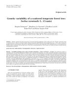
Figure
isolates 1
Phylogenetic analysis of env sequences from different SRV-2
Phylogenetic analysis of env sequences from different
SRV-2 isolates. (A) A phylogenetic tree of reference SRV-1,
SRV-2 and SRV-3 env protein sequences and sequences
obtained from the SRV-2 isolates in this study (see Table 1)
was generated from a ClustalW multiple alignment using the
protein maximum-likelihood method as implemented in the
Phylip package (v. 3.62). The sequence of the distantly related
simian sarcoma virus (SSV) env protein was used as outgroup
[Genbank:NC001514]. (B) A detailed phylogenetic tree of
the SRV-2 reference and isolate sequences was similarly generated using SRV-3 as outgroup. Emerging clusters were
labelled as subtypes SRV-2A through 2F and virus isolates
from animals diagnosed with RF are indicated.
sequence) showed differences in at least one of the
sequences analyzed, while fifty-five of these differences
occurred in more than one of the sequences. A comparison using the seventy-two variant positions visually demonstrated the basis of the sequence differences between
the six subtypes (Figure 3). Some of these sequence differences were conserved within a specific subtype. For example, at amino acid position 33 an isoleucine (I) was
conserved in all of the SRV-2B isolates, while a leucine (L)
was conserved in all of the SRV-2E isolates (Figure 3). On
the other hand, some amino acids were conserved across
subtypes, as seen in the SRV-2A, SRV-2C, SRV-2D, and
SRV-2F sequences which all contained a methionine (M)
at aa33. Frequently, there were single amino acid differ-
ences in one isolate within a subtype which were not conserved in the other isolate sequences, i.e. glycine (G) at
aa29 in the M95348 isolate of subtype SRV-2E. Due to the
fact that in most cases PCR amplification products were
sequenced directly, without cloning, these differences
would not reflect Taq polymerase errors.
Genetic variation of SRV-2 within an individual infected
macaque
In order to determine the genetic variation of SRV-2
within the same animal, multiple clones of a PCR amplification product encoding a 439 aa fragment of the env
gene were characterized from two different macaques
(F91249, T82422). Eight different clones were obtained
from each animal. Sequence analysis of each clone
revealed random nucleotide differences between each
cloned DNA within an animal (data not shown). However, no nucleotide difference occurred in more than one
clone, suggesting that the observed differences were the
result of errors induced by Taq polymerase during the PCR
amplification step. These data revealed no evidence for
the presence of multiple strains of SRV-2 within a single
individual.
We further analyzed the genetic variation within an SRV2 strain from an individual macaque over time. Two tissue
samples from macaque M95332 which contained an SRV2E subtype had been collected at two time points six years
apart, thus spanning the evolution of this virus over the
time frame from 1997–2003. The complete env gene was
amplified from each of these samples and compared. No
differences were detected between the two sequences. Furthermore, we compared the isolate in M95332 with the
isolates from two animals in the same cohort, M96020
and M96026, and the mother of M96026, A94040, who
presumably introduced this SRV-2E isolate into the Washington NPRC colony nine years before the last M95332
isolate was sequenced. This comparison revealed only one
amino acid difference between the sequence of M95332
and those of M96020, M96026 or A94040, showing very
little variation within this SRV-2E strain even between different animals.
Structural conservation of the env gene between different
SRV-2 subtypes
The SRV-2 env gene encodes a precursor polypeptide that
undergoes both glycosylation and proteolytic processing
during a maturation process that results in the expression
of the mature membrane-bound glycoprotein integrated
within the virion envelope. The envelope glycoprotein
interacts with host cellular receptors to initiate virus
adsorption and penetration, and plays an important role
in determining cell and tissue tropism. Thus, sequence
variation in the env gene can ultimately affect or determine the pathogenic potential of the virus. Interestingly,
Page 6 of 15
(page number not for citation purposes)
Virology Journal 2006, 3:11
/>
signal peptide
20
40
60
80
MTLKDIPFWRVLLIFQTARVYAGFGDPREAITMIHQQHGKPCDCAGGYVNAAPTVYLAAVSCSSHTAYQPSDSLKWRCVSNPTLANGENI
................................I................IT.......T...............................
....N.........L...Q...............................T.......T...............................
..................................................T.......T...............................
..............L...Q.............L...R.............T...A...T...............................
..P.......I......................................ST.....I.T...............................
SRV-2A
SRV-2B
SRV-2C
SRV-2D
SRV-2E
SRV-2F
:
:
:
:
:
:
SRV-1
SRV-3
: .NFNHHFT.SLVI.S.IFQ.Q.........LLE.Q.K............SSP..NS.TT....TY...SVTN....Q...T..T.SPTH.
: .NFNYHFI.SLVILS.ISQ.Q.........LAE.Q.K............SSP.INS.TT....T....SVTN....Q...T..TPSNTH.
T-cell epitope
cell receptor binding/B-and T-cell epitope
100
120
140
160
GNCPCKTFK---ESVHSSCYTAYQECFFGNKTYYTAILASNRAPTIGTSNVPTVLGNTHNLLSAGCTGN-VGQPICWNPKAPVHISDGGG
.....Q...---.........T...............................................-....................
.........---............................K......A..............T...D..-..............V.....
.....Q...---...............L.........................................-....................
......I.Q---..................................................T......-....................
.....T...---......................................................I.S-....................
SRV-2A
SRV-2B
SRV-2C
SRV-2D
SRV-2E
SRV_2F
:
:
:
:
:
:
SRV-1
SRV-3
: .S..SQCNSQSYD...AT..NH..Q.TI.....L..TMIRDKS.SS.DG....I...NQ..II...PE.KK..VV...SQPS..M.....
: .S..GECNTISYD...A...NH..Q.NI.....L..TITGD.T.A..DG.......TS...IT...PNGKK..VV...SRPS........
T-cell epitope
180
200
220
240
260
PQDKAREIAVQKRLEEIHKSLFPELRYHPLALPKARGKEKIDAQTFNLLTATYSLLNKS-NPNLANECWLCLPSGNPIPLAIPSNDSFLG
..................R........................................-.................V............
........V..................................................-..............................
..................R...........................D............-.................V.I..........
...........................................................-.....S........................
........V..................................................-..............................
SRV-2A
SRV-2B
SRV-2C
SRV-2D
SRV-2E
SRV_2F
:
:
:
:
:
:
SRV-1
SRV-3
: ....V...I.N.KF..L........S.......E.........H..D..ATVH....V.SQRQ..ED.....R..D.V...L.YDNTSCS
: ......D.I.N.KF..L.R......S.......E.........H.LD..ATVH....A.-Q.S..ED.....Q..D.V...L.Y..TLCS
SRV-2A
SRV-2B
SRV-2C
SRV-2D
SRV-2E
SRV_2F
:
:
:
:
:
:
SRV-1
SRV-3
: NSTFFFNCS.C..L.T..F....F---NFTHSV.L.ADY......I....AG.T...SYI...KPSSP---...................
: N---FACLS.H...LT..F....F---NFTDSN.L.AHY......I....AS.T...SYY.V.TASKPSN....................
280
300
320
340
S--------NLSCPIIPPLLVQPLEFMNLINASCFYSPFQNNSFDVDVGLVEFANCSTTLNIS------HSLCAPNSSVFVCGNNKAYTY
.--------.................I.......L.........G........T....I....------.....................
.--------..F..............IT.........................T....I....------Q....................
.--------.................S..T....L...S..............T....IF...------..............S......
.--------.................I..........................T....II...------.....................
.--------.................T...........S...........AG.T....I....------................R....
fusion domain
dibasic aa proteolysis site
heptad repeat
360
380
400
420
LPSNWTGTCVLATLLPDIDIVPGDAPVPVPAIDHYLHRARRAVQFIPLLVGLGITTAVSTGTAGLGYSITQYTKLSRQLISDVQAISSTI
..T......................................................................R................
..T.......................................................................................
..T.........................................................................S.............
........................V.................................................................
.......I.I................................................................................
SRV-2A
SRV-2B
SRV-2C
SRV-2D
SRV-2E
SRV_2F
:
:
:
:
:
:
SRV-1
SRV-3
: ..T....S............I..SE...I.....F.G.PK..I.....VI................V.L.......H.............
: ..T....S............I..SE...I.....F.GK.K..I.L...F............A....V.........H.............
gp70
gp22/20
immunosuppressive peptide
S-S
440
460
480
500
520
QDLQDQVDSLAEVVLQNRRGLDLLTAEQGGICLALQEKCCFYANKSGIVRDKIKRLQEDLEKRRKEIIDNPFWTGLHGLLPYLLPLLGPL
..........................................................................................
..........................................................................................
..........................................................................................
..........................................................................................
.............................................................R............................
SRV-2A
SRV-2B
SRV-2C
SRV-2D
SRV-2E
SRV_2F
:
:
:
:
:
:
SRV-1
SRV-3
: ......................................................N..D.......QL........F......VM......
: ......................................................N..D...R..RQL.......SF..F...VM......
transmembrane
gp22 processing domain
540
560
FCLLLLITFGPLIFNKIITFVKQQIDAIQAKPIQVHYHRLEQEDNGGVYLRVS
L.................A.....M............................
L.................A............................I.....
L.................A..................................
..................A............................L.....
L.................A.....ME.....................I.....
SRV-2A
SRV-2B
SRV-2C
SRV-2D
SRV-2E
SRV_2F
:
:
:
:
:
:
SRV-1
SRV-3
: L....VLS...I....LM..I.H..ES.................H..S..NLT
: L....VLS...I....LM..I.H..ES.................S..S..TLT
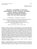
Figure 2
Multiple alignment of the complete env sequences of representative prototypes of the SRV-2 subtypes
Multiple alignment of the complete env sequences of representative prototypes of the SRV-2 subtypes. A ClustalW alignment was generated using one representative prototype member from each of the six SRV-2 subtype clusters: SRV2A (D2/CEL/OR), SRV-2B (D2/RHE/ORV1), SRV-2C (442N), SRV-2D (F90346), SRV-2E (A94040), SRV-2F (SRV_sing31.2).
The sequences of SRV-1 and SRV-3 were included for comparison. Dots represent amino acids identical to the reference
sequence of the SRV-2A prototype. Conserved cysteine residues are shaded in yellow, while putative N-linked glycosylation
sites (NXT/S) are shaded in black. The putative signal peptide, known T- and B-cell epitopes, heptad repeat, as well as the gp20
fusion and transmembrane domains are indicated and referenced in the text. A predicted disulfide linkage within the immunosuppressive peptide, and the proteolytic cleaveage sites generating the gp70 surface and gp20 transmembrane subunits are indicated. While B- and T-cell epitopes have been determined for SRV-2, the functions and locations of other domains are derived
from studies in SRV-3.
Page 7 of 15
(page number not for citation purposes)
Virology Journal 2006, 3:11
/>
Table 2: Env sequence comparison of the SRV2-subtypes
SRV-2 Subtype
Between subtypes1
Within subtype
2B
2A
2B
2C
2D
2E
2F
1The
97.3–98.6%
98.3–100%
98.5–99.4%
99.0–99.8%
99.1–99.8%
98.8–100%
2C
2D
2E
2F
96.2%
-
96.3%
94.9%
-
96.5%
96.7%
95.5%
-
96.7%
94.8%
96.3%
95.3%
-
94.8%
94.3%
94.3%
94.1%
93.6%
-
prototypes of each SRV-2 subtype, as indicated in the legend to Figure 2, were compared
our data showed that 87% of the amino acids within the
574 amino acid env sequence exhibited no variation in the
different SRV-2 isolates examined (Figures 2 and 3).
Within the variant amino acid positions, in most cases,
only conservative changes were identified. The conservative nature of the env gene variability was further highlighted by the finding that, in many of the positions, the
variant amino acid in the SRV-2 isolate was an amino acid
found in either or both of the more distantly related SRV1 or SRV-3 serogroup prototypes. For example, leucine (L)
at aa193 was found only in one SRV-2B subtype isolate
but in both SRV-1 and SRV-3 prototypes. While for the
most part the variant positions were scattered evenly
throughout the env sequence, several highly conserved
regions were identified. The N-terminal region aa60-aa95
was completely conserved between all SRV-2 isolates
examined (Figure 2).
Interestingly, this region was quite distinct from the
homologous regions in SRV-1 and SRV-3. The C-terminal
region from the putative N-linked glycosylation site at
aa345 to the C-terminus was extremely well conserved
among the SRV-2 isolates with only an occasional amino
acid variant. This conserved region within the SRV-2 isolates is homologous to regions within the SRV-3 gp22/20
protein where several domains have been studied in
detail. Such domains include the gp70/gp20 proteolytic
site (aa382) [25], the known fusion domain (aa384aa410) [26], the heptad repeat region (aa409-aa462) [27],
the immunosuppressive peptide (aa443-aa477) [25,28], a
membrane spanning domain (aa516-532) [15], and the
region spanning the gp22 processing site (aa558) [25](see
Figure 2). In contrast to the conserved N-terminal region,
the C-terminal region was highly conserved not only
between SRV-2 subtypes but also with the SRV-1 and SRV3 sequences, with one region (aa419-aa485) completely
conserved between the isolates from the different SRV
serogroups. All twenty-two cysteine (C) residues were
conserved between SRV-2 isolates and the SRV-1 and SRV-
3 prototypes (Figure 3) indicating that all SRV serogroups
maintain the same basic disulfide-linked three-dimensional env structure. SRV-1 and SRV-3 sequences contained three additional conserved cysteine residues which
were not found in the SRV-2 isolates. Eleven putative Nlinked glycosylation sites (NXS/T) were conserved in all
the SRV-2 isolates and the SRV-1 and SRV-3 prototypes.
Sequence and structural conservation of an
immunosuppressive peptide
To further explore the conservation detected in the C-terminal region of the different SRV-2 subtypes and the SRV1 and SRV-3 serotypes, we searched the existing structural
databases for similar three-dimensional structures with
3D-PSSM [29] using the D2/CEL/OR subtype 2A
sequence as probe. 3D-PSSM is a program that uses a
threading algorithm to map the input sequence onto
known 3-dimensional structures based on several parameters including amino acid sequence, multiple alignments, and secondary structure predictions. A region of
the SRV-2 env polypeptide between amino acids 420–485
that was completely conserved in all SRV-2 isolates and in
the SRV-1 and 3 prototypes was found to have strong
structural homology to the C-terminal domains in the
envelope proteins of two other retroviruses, Moloney
murine leukemia virus (MMLV) and human T-lymphotropic virus (HTLV-1), as well as to a domain in the envelope protein of Ebola virus, a ssRNA virus. The known
crystal structures of the MMLV, HTLV-1 and Ebola envelope proteins revealed that these domains form a highly
conserved hairpin loop structure stabilized by a disulfide
bond [30-32]. This loop structure is believed to be responsible for viral fusion with cellular membranes in several
virus species, some of which share little or no obvious
evolutionary relationship. Such viruses include the above
mentioned oncogenic retroviruses, the orthomyxovirus
influenza [33], the lentiviruses HIV-1 and SIV [34,35], the
paramyxovirus SV5 [36,37], and filoviruses [38]. Threedimensional structural predictions, using the Cn3D struc-
Page 8 of 15
(page number not for citation purposes)
Virology Journal 2006, 3:11
SRV-3
D3/RHE/WI
SRV-1
D1/RHE/CA
FYLSQEEQKSSNLTTNTSHIDDTAPKVVIILRDEVLLST—-SLYDASTSYYNNKTSVEAKHLNRGLKTIEQS
FHLSQEEQKSSNLTTNSSHIDDIAPKVVMILKDEVLTCT—-SLYDAGTSYISNKTSVEAKHLNKGLKTIEQS
LDVFREIHQITVLTEQTKTFNTSATNPVIAIRNNVLFSINIALFGVETTILHNKTTVAARRLRKGLKAMDQV
VDVFREIHQITVLTEQTKTFNTSATTHVIAIRNNVLFSINIALSDVETTTLHNKTTVATKRFAKRLKAMDQV
LDVFREIHQITVLTKQTKAFNTSATNPVIAIRNNVLFSINIALSDVETTILHNKTTVAAKRLRKGLKAMDQV
LDVFREIHQITVLTEQTKAFNTSATNPVIALRNNVLFSINIALSDVETTILHNKTTVAAKRLRKGLKAMDQV
LDVFREIHQITVLTEQTKAFNTSATNPVIAIRNNVLFSINIALSDVETTILHNKTTVAAKRLRKGLKAMDQV
LDVFREIHQITVLTEQTKAFNTSATNPVIAIRNNVLFSINIALSDVETTILHNKTTVAAKRLRKGLKAMDQV
SATNPGIADRNNVL
LDVFREMHQNTVLTEQTKALNTSATNPVIAIRDNVIFSSNTALSDVETTIFHSKTTVAAKSLRKGLKAIDQV
LDVFREMHQNTVLTEQTKALNTSSTNPVIAIRDNVIFSSNTALSDVETTIFHSKTTVAAKSLRKGLKAIDQV
LDVFREMHQNTVLTEQTKALNTSATNPVIAIRDNVIFSSNTALSDVETTIFHSKTTVAAKSLRKGLKAIDQV
LNVLQEMHQNTVLTEKTKAFKATADNPVVVIKNNILFFITIAFFDVETTILQNKTTVAAKRLRKGLKAIDQI
LNVLREMHQNTVLTEKTKAFKATADNPVVVIKNNILFSINIAFFDVETTILQNKTTVAAKRLRKRLKAIDQI
ILFSINIAFFDVETTILQNKATVAAKRLRKGLKAIDLV
LDVFREMHQNAVLAEKTKAFNTSATNPVIAIKNNILFSMNIAFFDVEATTLHNKSTVAAKRLRKGFKTIDQV
VDVFREMHQNAVLTEKTKAFNTSATNPIIAIKNNILVSMNIAFFDVEATTLHNKSTVAAKRLRRGLKAMDQV
RNIVFFDVEAATLHNKTSVAAKRLRKGLKSIDQV
LDVLQELHRNTALTEKIQAFNTTATNPVIAIKNSILFSINIAFFDVETTIIHNKSTVVAKRLRKGFKAIDQL
LDVLQELHRNTALTEKIQAFNTTATNPVIAIKNSILFSINIAFFDVETTIIHNKSTVVAKRLRKGFKAIDQL
LDVLQELHRNTALTEKIQAFNTTATNPVIAIKNSILFSINIAFFDVETTIIHNKSTVVAKRLRKGFRAIDQL
LDVLQGLYRNTALTEKIQAFNTTATNPVIAIKNSILFSINIAFFDVETTIIHNKSTVVAKRLRKGFRAIDQL
LDVLQELHRNTALTEKIQAFNTTATNPVIAIKNSILFSINIAFFDVETTIIHNKSTVVAKRLRKGFKAMDQL
PDIFREMHQSTVITETTKAFNTSAISPVIVIKNNILFSTNIAFSDAGTTILHNRSIIAAKRLRRGLKAMEQI
--RF
RF
RF
RF
RF
---RF
--RF
RF
-------
Mmu
Mmu
Mmu
Mne
Mne
Mne
Mne
Mne
Mne
Mne
Mne
Mne
Mfa
Mni
Mfa
Mmu
Mfa
Mfa
Mfa
Mfa
Mfa
Mfa
OR
OR
YE
WA
WA
WA
WA
WA
WA
WA
NIH
NIH
WA
OR
NM
MI
TX
WA
WA
WA
WA
wild
3
5
11
15
19
29
33
35
37
50
51
55
57
59
88
96
97
99
109
115
128
135
150
151
154
156
160
169
171
185
193
195
223
241
253
255
263
269
284
285
287
289
292
296
302
308
309
311
315
316
317
321
335
337
344
349
351
366
404
415
418
455
493
494
519
522
537
540
546
547
550
569
SRV-2
(B) D2/RHE/ORV1
D2/RHE/OR
YN91-224
90167
T81273
M78114
D2/MNE/WA
(D) F90346
F89336
F91249
(C) 442N
17915
91048
(A) D2/CEL/OR
NM101
Mm_Mich
(E) A94040
M96026
M95332
M95348
M96020
(F) SRV_sing31.2
/>
Figure 3
Alignment of variable amino acid positions within SRV-2 env sequences
Alignment of variable amino acid positions within SRV-2 env sequences. This column alignment presents only those
amino acid positions that vary in one or more of the twenty-one SRV-2 env sequences analyzed; exact position within the complete env sequence (Figure 2) is indicated at the bottom of each column. The analogous amino acid positions of the closely
related SRV-1 and SRV-3 sequences are shown for comparison. The macaque species, origin and RF status for each SRV-2 isolate are indicated on the right. Colored residues indicate the amino acid groupings upon which the phylogenetic analysis is
based. Non-conserved amino acid variants are shaded (magenta). (Mne) Macaca nemestrina, (Mni) Macaca nigra, (Mfa) Macaca
fascicularis, (Mmu) Macaca mulatta.
ture viewing program, revealed an almost perfect alignment between the structure predicted for the SRV-2
domain and the known crystal structures of the other proteins, even though only nine amino acids in a 45 aa
stretch were conserved (Figure 4). The region corresponding to the conserved domain within the SRV-2 isolates has
been previously identified as an immunosuppresive peptide in several oncogenic retroviruses capable of inducing
an immuosuppressed state in their hosts (FeLV, MuLV,
REV-A) [39]. In addition, the entire gp20 protein of SRV3 has also been found to have immunosuppressive properties [40]. Studies with SRV-3 have revealed that residues
in the immunosuppressive peptide found within the gp20
protein subunit are responsible for binding to the gp70
protein subunit and are crucial for virus-cell fusion
[25,28].
Discussion
We investigated the genetic diversity of the serogroup 2
simian retroviruses (SRV-2) in four different wild-caught
or captive macaque species from six different primate
centers within the US over a 23 year time period. We identified at least six different SRV-2 subtypes by molecular
comparison of the complete env gene from twenty-two
different isolates. Our results indicate that separate introductions of at least six parental virus subtypes have
occurred in the captive macaque populations in the U.S.
with subsequent horizontal transfer between macaque
species and primate centers.
It is most likely that divergent SRV-2 strains were introduced to the United States via importations of different
species of infected macaques from different geographical
areas. Procurement from common sources, close contact
in primate holding facilities, and traffic between primate
Page 9 of 15
(page number not for citation purposes)
Virology Journal 2006, 3:11
centers would explain the spread of virus across the captive macaque populations and between macaque species.
The introduction of the SRV-2E subtype into the Washington NPRC provides one example for such a virus transfer
that was evident from our study. In 1994, a female
cynomolgus macaque, A94040, was purchased by the
Washington NPRC for breeding purposes. At the time of
transfer, this animal was negative for SRV-2 by serology
but was later shown to be positive by virus culture. Our
analysis of DNA from PBMCs collected in 1997 revealed
that A94040 was infected with an SRV-2E subtype, that
was not present in other macaques sampled at the Washington NPRC before 1994. The offspring of A94040,
M96026, born in 1996, became infected with SRV-2 and
analysis of PBMCs collected in 2003 revealed the presence
of an SRV-2E isolate identical to that of its mother. Our
analysis demonstrated that siblings with the same father
as M96026, but a different mother, were infected with
SRV-2E isolates that were nearly identical (1–3 aa differences) to that of M96026 and its mother A94040
Our data demonstrates that the env gene of SRV-2 is very
stable suggesting a remarkable adaptation of the virus to
its host. Within the five isolates of SRV-2E obtained from
a cohort of cynomolgus macaques at the Washington
NPRC, only 0–3 amino acid differences within the 574 aa
envelope protein were detected. In addition, we found no
evidence for variation of the viral env gene within a single
individual over a 6 year period. Surprisingly, even viral
isolates from different primate centers from different
macaque species separated in time by as much as 20 years
showed a high degree of conservation. The SRV-2B isolates obtained seven years apart from the rhesus macaque,
YN91-224, at the Yerkes NPRC and the pig-tailed
macaque, T81273, at the Washington NPRC, differed by
only one amino acid. Our data confirm earlier studies
which showed a remarkable stability of the SRV-2 genome
over time by analysing partial env sequences in smaller
and more restricted samples [17,41].
The stability of the viral env gene over time within any
given subtype suggests that the different SRV-2 subtypes
evolved in the wild over long periods of time in segregrated primate hosts. Such segregation could be dictated
by constraints involving different geographical areas, different niches within the same geographical area, and/or
different natural host species. In our study, the natural
host species for only one of the SRV-2 subtypes was apparent. The SRV-2F subtype was identified in a number of
cynomolgus macaques in the wild on the island of Singapore. Interestingly, the SRV-2F subtype was clearly distinct
from all the subtypes present in captive populations of
cynomolgus, rhesus, Black celebes, or pig-tailed
macaques, suggesting that none of these five subtypes
originated from a cynomolgus macaque reservoir in Sin-
/>
gapore. To date, no SRV-2 reservoir has been identified in
wild-living rhesus macaques which are native to India
[42]. Thus, the natural host species of SRV are likely to be
found in Southeast Asia. However, further analysis of
SRV-2 isolates directly from wild-caught animals is
needed to understand the natural reservoirs for these
viruses in more detail.
Our initial impetus to study the genetic variation within
the SRV-2 serotype was to determine whether there was an
association between virus subtype and SAIDS-RF. Our
data revealed that SAIDS-RF was associated with three
SRV-2 subtypes, 2A, 2B and 2C, in multiple species of
macaque, including pig-tailed, rhesus, cynomolgus and
Black celebes. A total of eight RF cases were examined
from five primate centers including the Washington, Oregon, and Yerkes NPRCs, the NIH primate center and the
Lovelace Respiratory Research Institute in New Mexico.
While SRV-2A was associated with RF in celebes and
cynomolgus macaques, the SRV-2B subtype was associated with RF in pig-tailed and rhesus macaques. The SRV2C subtype was only associated with RF in pig-tailed
macaques. No obvious sequence similarities were
detected between the SRV-2A, -2B and -2C subtypes which
would correlate with the RF association. In two of the RF
cases, RF occurred soon after experimental infection with
SIV or SHIV. The rhesus macaque YN91-224 which was
infected with an SRV-2B subtype was diagnosed with RF
after undergoing an experimental infection with SIV at the
Yerkes NPRC (personal communication, H. McClure).
The SRV-2C infected pig-tailed macaque 442N was diagnosed with RF 24 weeks after infection with a pathogenic
strain of SHIV [24]. Thus, our studies revealed only an
association between the SRV-2A subtype and SAIDS-RF in
Black celebes macaques and between the SRV-2B subtype
and SAIDS-RF in pig-tailed macaques, in the absence of
other known immunodeficiency agents.
We have recently identified a single case of RF in a rhesus
macaque experimentally infected with a pathogenic strain
of SIV [43]. This animal was negative for all SRV serotypes
using type-specific qPCR assays. Additionally, four cases
of SAIDS-RF were reported in 1983 in a colony of Taiwanese rock macaques at the New England NPRC which were
endemically infected with SRV-1 [44]. Similarly, a single
case of SAIDS-SF, the subcutaneous form of RF, was
reported in 1983 in a colony of rhesus macaques endemically infected with the D1/RHE/CA subtype of SRV-1 at
the California NPRC [3]. Even though the vast majority of
RF cases in the different macaque colonies were associated
with SRV-2 serotypes, these findings suggest a broader role
for different SRV serotypes and possibly other retroviruses
such as lentiviruses as cofactors in the development of RF,
albeit with an apparent low efficiency.
Page 10 of 15
(page number not for citation purposes)
Virology Journal 2006, 3:11
/>
S-S
D2/CEL/OR
MMLV
HTLV-1
Ebola
sdvqAISSTIQDLQDQVDSLAEVVLQNRRGLDLLTAEQGGICLALQEKCcfyank
ddlrEVEKSISNLEKSLTSLSEVVLQNRRGLDLLFLKEGGLCAALKEECafyad~
kdisQLTQAIVKNHKNLLKIAQYAAQNRRGLDLLFWEQGGLCKALQEQCcflnit
qlanETTQALQLFLRATTELRTFSILNRKAIDFLLQRWGGTCHILGPDCriephd
Figure 4 and sequence alignment viral envelope env and
structurally similar regions of otherof thethe SRV-2proteins
putative disulfide-bonded loop region of highly conserved
Structural
Structural and sequence alignment of the highly conserved putative disulfide-bonded loop region of the
SRV-2 env and structurally similar regions of other
viral envelope proteins. Structural similarities to the SRV2 env protein were identified by querying the NCBI protein
structure database using 3D-PSSM and Cn3D. A region
within the C-terminal domain of SRV-2 env which was identical in all SRV-2 isolates and SRV-1 and SRV-3 prototypes
(aa426-471) was predicted to have structural similarities to a
disulfide-bonded loop presumed to be important for viruscell fusion in a number of RNA viruses and retroviruses,
including Ebola virus 1: 1EBO_A (Gp2); Ebola virus 2:
2EBO_A (Gp2); MMLV (Moloney murine leukemia virus):
1MOF (coat protein); HTLV-1 (human T-lymphotropic
virus): 1MG1_A (gp21) (see text). The disulfide bridge is indicated by S-S.
Factors which are likely to play a major role in the development of RF, include differences in the severity and type
of immunosuppression caused by an SRV-2 or lentivirus
infection, and co-infection with RFHV. Current data suggests that the macaque herpesvirus, RFHV, may play a
causative role in the etiology of RF [23,45,46]. We have
developed PCR assays to detect the host-specific variants
of RFHV in rhesus (RFHVMm) and pig-tailed (RFHVMn)
macaques and have identified RFHV in RF lesions from
the macaques infected with both SRV-2B (T82422, 90167,
M78114, and YN91-224) and SRV-2C (442N) in this
study [[45], and unpublished results]. It is not known, at
this point, whether the macaque cohorts infected with the
SRV-2D, -2E and -2F subtypes which were not associated
with RF, were also co-infected with RFHV.
Differences in disease pathology and severity have been
observed in SRV-2 infections which could impact RF
development. Within a cohort infected with a single SRV2 subtype, different outcomes have been reported, including a viremic state with rapid progression of SAIDS, a lowgrade viremia with a chronic milder form of the disease,
and a strong antibody response with no overt signs of disease [47]. Similarly, the same SRV-2 subtype can elicit differences in disease severities in different macaque species.
The D2/CEL/OR isolate, for example, caused severe
immunodeficiency in Celebes black macaques, but when
the same isolate was transmitted to rhesus macaques, the
animals seroconverted and remained virus- and symptom-free [13]. Conversely, the D2/RHE/OR isolate caused
mild disease in rhesus macaques but severe fatal immunodeficiency disease in Japanese macaques (Macaca fuscata). On the other hand, the closely related SRV-2B
isolates, D2/RHE/OR and D2/RHE/OR/V1, which differ
in only 17 amino acids over their entire genomes, were
found to induce vastly different disease outcomes in rhesus macaques and to also display differences in tropism in
cell culture assays [15]. The D2/RHE/OR variant was associated with only mild disease while the V1 variant caused
severe SAIDS.
In our study, the env sequence from the different SRV-2
isolates was highly conserved overall, with 93–96%
amino acid identity between isolates from different subtypes and 97–100% amino acid identity between isolates
within a subtype. A hypervariable region was detected
between aa 284–321 near the C-terminus of the gp70 protein. Even within this variable region, most amino acid
changes were conservative or conserved with the SRV-1 or
SRV-3 sequences or both, underlining the stability of the
env protein. Interestingly, all potential N-linked glycosylation sites were conserved between the SRV-2 isolates, and
even with the related SRV-1 and SRV-3 serotypes. This is
in stark contrast to HIV and SIV which exhibit extreme variation in number and precise location of N-linked glycosylation sites [48]. Such variation is believed to be a major
pathway for immune evasion for lentiviruses. Thus, the
strict conservation of glycosylation sites between the various SRV-2 isolates may help explain the high efficiency of
neutralizing antibodies against SRV-2 infections [49].
Our analysis showed that 15.5% of the amino acid positions within the receptor-binding surface-exposed (SU)
subunit gp70 were variable while only 8.7% of the amino
acid positions within the transmembrane (TM) subunit
gp22 differed between isolates. Differences were more
concentrated in the C-terminal portion of gp70 (last 100
aa) but also affected the N-terminal signal peptide
domain, as well as the known B- and T-cell epitopes
(aa96-102, aa127-153). The B- and T-cell epitope at aa96102 is of particular importance since naturally occurring
neutralizing antibodies to SRV-2 are directed against this
area [50] and it confers binding to the RD114/simian type
D retrovirus receptor, a neutral amino acid transporter
[51,52]. Three amino acid positions in this seven amino
acid domain displayed variations between isolates. We
identified three sequence variants within the T-cell
Page 11 of 15
(page number not for citation purposes)
Virology Journal 2006, 3:11
epitope at aa127-152, but only one difference in the 2nd Tcell epitope located at aa233-249 [53]. Although some of
these differences are of conservative nature, the remaining
changes could affect the ability of the virus subtypes to
elicit an immune response and lead to differences in disease outcome.
The gp22 subunit was far less variable between isolates
than the gp70 subunit which likely is the result of sterical
restrictions imposed by a string of conserved functional
domains. The TM subunits of most retroviruses, including
SRV-3, contain an N-terminal hydrophobic fusion peptide followed by a putative coiled coil-forming sequence,
a disulfide-bonded loop connected to a shorter C-terminal alpha helix, a region with one or more N-linked glycosylation sites, a hydrophobic membrane-spanning
sequence, and, as in SRVs, a cytoplasmic tail. Absolute
sequence conservation between different SRV-2 subtypes
and the SRV-1 and SRV-3 serotypes was seen within a
region of the C-terminal gp20 domain which has been
shown to be crucially important for the interaction
between the SU and TM domains and for the fusion of
viral and cellular membranes in MPMV. Using a threading
algorithm, we showed that the homologous region within
SRV-2, between aa426 and aa471, was structurally similar
to a region in other retroviruses including MMLV and
HTLV-1 [31,32,54] and the filovirus Ebola [38]. In these
and other distantly related retroviruses [34,35] as well as
in the orthomyxovirus influenza, the conserved disulfidebonded loop plays a highly pivotal role in stabilizing a
chain reversal which provides a hinge-like function that
brings the fusion peptide into proximity to the target cell
membrane during the fusion process [33]. We propose
that SRV-2, in analogy, uses a similar mechanism for host
cell membrane fusion.
Conclusion
In conclusion, our study revealed the presence of five SRV2 subtypes circulating among four macaque species held
in US primate centers. Three of these subtypes were associated with RF in some macaque species, although a correlation between sequence variation and disease could
not be established. A sixth and different subtype was
found in wild cynomolgous macaques on the island of
Singapore suggesting that the subtypes found in captive
macaques in the U.S. did not originate from this virus reservoir. Since the viral subtypes discovered in captive monkeys did not display a macaque species specificity, the
nature of the original reservoir for these viruses is unclear.
Our studies also showed that the envelope protein of SRV2 was very stable suggesting a high degree of adaptation of
SRV-2 to its host. In all likelihood, simian retroviruses
have evolved very slowly in geographically distinct
macaque species, and only the physical contact with other
/>
macaque species in captivity has allowed single virus subtypes to spread between species.
Methods
Animals
Tissue samples were obtained from a variety of macaque
species naturally infected with SRV-2 that were housed at
different primate research centers in the U.S. over the last
twenty years. Some of these macaques were asymptomatic, while others had been diagnosed with SAIDS-RF
(see Table 1). The tissue samples included RF tissue from
RF positive animals, tongue, spleen, tonsils and PBMCs or
PBMC/Raji cell co-cultures. Samples had been collected
over a wide time-range (1983–2003), starting at the
height of the SRV-2 epidemic in 1982–83. Macaque species included pig-tailed macaques (Macaca nemestrina
(Mne)), Celebes black macaques (Macaca nigra (Mni)),
cynomolgous macaques (Macaca fascicularis (Mfa)) and
rhesus macaques (Macaca mulatta (Mmu)). Tissue samples were obtained from various primate research centers
including the Washington NPRC (Seattle, WA; C.-C. Tsai
and M.E. Thouless), Yerkes NPRC (Atlanta, GA; Harold
McClure), National Institutes of Health (Bethesda, MD;
Riri Shibata), Lovelace Respiratory Research Center (Albuquerque, NM; Carole Emerson), University of Michigan
(Ann Arbor, MI; Nina Woodford); as well as one wild
cynomolgous macaque whose blood was drawn during a
catch-and-release process in the forests of Singapore (University of Washington; Lisa Jones-Engel).
DNA isolation
Frozen tissue samples or cell pellets were quickly thawed
in a standard proteinase K extraction buffer containing
0.1% SDS and vortexed. Paraffin-embedded formalin
fixed samples were first treated with xylenol to remove the
paraffin before extraction. Samples were digested over
night at 50°C and DNA was isolated by standard phenol/
chloroform extraction and ethanol precipitation.
PCR amplification, cloning and sequencing of the SRV-2
env genes
To obtain the complete sequence of the different SRV-2
env genes, template DNA was amplified by PCR using
SRV-2 specific primers. Primers srv2-env a (5'-CCTGAGATCACTCCTTTTCTTTGCTCAT-3') and srv2-env1 b (5'CCGTCATTGGCTGACCAGTTTAG-3') were used to
obtain a 1,796 bp PCR product containing the complete
SRV-2 env sequence. In some cases, the primers srv2-env a
and srv2-env b (5'-CAGTTGAGACGGCAGTGGTT-3') were
used to obtain a 1,235 bp fragment which overlapped
with a 1,041 bp fragment obtained using srv2-env1 a (5'GAGTGCTGGCTATGCTTACCATC-3') and srv2-env1 b.
PCR fragments were purified with the GeneClean III Kit
(Q-Biogen) and directly used as templates in a sequencing
reaction.
Page 12 of 15
(page number not for citation purposes)
Virology Journal 2006, 3:11
To search for SRV-2 variants in the same animal, a 439 bp
fragment of the env gene was PCR-amplified from
genomic DNA of two macaques (T82422, F91249) using
Platinum Taq polymerase and the primers: SRV2-env 1a
(5'-GAGTGCTGGCTATGCTTACCATC-3') and SRV2-env b
(5'-CAGTTGAGACGGCAGTGGTT-3'). The fragments
were subcloned into the pCR-Blunt vector (Invitrogen Life
Technologies, Carlsbad CA) using standard procedures.
Eight clones were obtained from each animal for sequence
analysis.
Sequencing was performed on an ABI model 310 automated sequencer with Prism Big Dye terminator cycle
sequencing ready reaction kit with Amplitaq DNA
polymerase FS (Applied Biosystems). DNA sequences
were analysed using Sequencher 4.1.4 (GeneCodes).
Sequence analysis
Multiple nucleotide and encoded amino acid alignments
were done with Vector NTI 9.0.0 (InforMax) based on the
ClustalW algorithm (EMBL, Heidelberg, Germany) or
with the ClustalW program [55] directly. The location of
the signal peptide was determined by SignalP 3.0 [56],
SigCleave [57], and iPSORT [58]. The location of transmembrane domains was determined with MEMSAT2 [59]
and Tmpred [60]. Putative glycosylation sites were determined with NetNGLyc 1.0 [61].
Three-dimensional structure analysis
The program 3D-PSSM [62] was used to identify proteins
with structural similarities to the SRV2 env prototype (D2/
CEL/OR). The structures for the six highest scoring proteins were obtained from the Molecular Modeling Database (MMDB)[63] which contains experimentally
determined three-dimensional biomolecular structures
obtained from the Protein Data Bank. Matching structures
were aligned with SRV2 env using NCBI's 3D-structure
viewer Cn3D v4.1 [64]. Using NCBI's vector alignment
search tool (VAST)[65], we determined further protein
structure neighbors by direct comparison of 3-dimensional protein structures stored in MMDB. This allowed us
to further evaluate possible structural and functional
domains of the SRV2 env protein.
/>
parsimony method as implemented in the PHYLIP package. All of the major branch points were strongly supported by boot-strap analysis and statistical evaluations
within ProML.
Competing interests
The author(s) declare that they have no competing interests.
Authors' contributions
JP-S carried out the sequence analysis, the three-dimensional structure analysis, the phylogenetic analysis, as well
as the cloning studies, participated in the PCR and cloning
efforts, and drafted the manuscript. TM participated in the
PCR and cloning efforts. AGB developed the PCR cloning
approach and participated in the PCR and cloning efforts.
RG provided the SRV-2 envelope sequence from the wildcaught macaque, MET supplied DNA samples and participated in the design and coordination of the study. C.C-T
provided a large number of RF and non-RF tissue samples
from his study collection. TMR conceived of the study,
and participated in its design and coordination and
helped to draft the manuscript. All authors read and
approved the final manuscript.
Acknowledgements
We would like to acknowledge N. Woodford, R. Shibata, C. Emerson, and
H. McClure for their generous gifts of macaque tissue samples. We would
also like to acknowledge the National Primate Research Centers and other
primate centers indicated in this study for their help in obtaining samples.
This work was partially supported by RR13154 and RR00166 from the
National Center for Research Resources. T. Rose is the recipient of a K02
award, AI49275, from the National Institute for Allergy and Infectious Diseases.
References
1.
2.
3.
4.
Phylogenetic analysis
Multiple sequence alignments were performed with ClustalW [55]. Phylogenetic analysis of amino acid sequences
was done with protein maximum-likelihood (ProML)
method from the PHYLIP package version 3.62 of phylogenetic analysis programs (.)[66] (University of Washington, Seattle). Bootstrap analysis was performed using the
programs Seqboot and Consense from the PHYLIP package. The treefiles were displayed with TreeView [67]. Identical tree topologies were obtained by using neighborjoining analysis of the protein distance matrices and by a
5.
6.
7.
8.
Chopra HC, Mason MM: A new virus in a spontaneous mammary tumor of a rhesus monkey.
Cancer Res 1970,
30:2081-2086.
Sonigo P, Barker C, Hunter E, Wain-Hobson S: Nucleotide
sequence of Mason-Pfizer monkey virus: an immunosuppressive D-type retrovirus. Cell 1986, 45:375-385.
Henrickson RV, Maul DH, Osborn KG, Sever JL, Madden DL, Ellingsworth LR, Anderson JH, Lowenstine LJ, Gardner MB: Epidemic of
acquired immunodeficiency in rhesus monkeys. Lancet 1983,
1:388-390.
Letvin NL, Eaton KA, Aldrich WR, Sehgal PK, Blake BJ, Schlossman SF,
King NW, Hunt RD: Acquired immunodeficiency syndrome in
a colony of macaque monkeys. Proc Natl Acad Sci USA 1983,
80:2718-2722.
Marx PA, Maul DH, Osborn KG, Lerche NW, Moody P, Lowenstine
LJ, Henrickson RV, Arthur LO, Gilden RV, Gravell M, et al.: Simian
AIDS: isolation of a type D retrovirus and transmission of the
disease. Science 1984, 223:1083-1086.
Power MD, Marx PA, Bryant ML, Gardner MB, Barr PJ, Luciw PA:
Nucleotide sequence of SRV-1, a type D simian acquired
immune deficiency syndrome retrovirus.
Science 1986,
231:1567-1572.
Daniel MD, King NW, Letvin NL, Hunt RD, Sehgal PK, Desrosiers
RC: A new type D retrovirus isolated from macaques with an
immunodeficiency syndrome. Science 1984, 223:602-605.
Desrosiers RC, Daniel MD, Butler CV, Schmidt DK, Letvin NL, Hunt
RD, King NW, Barker CS, Hunter E: Retrovirus D/New England
Page 13 of 15
(page number not for citation purposes)
Virology Journal 2006, 3:11
9.
10.
11.
12.
13.
14.
15.
16.
17.
18.
19.
20.
21.
22.
23.
24.
25.
26.
and its relation to Mason-Pfizer monkey virus. J Virol 1985,
54:552-560.
Giddens WE Jr, Tsai CC, Morton WR, Ochs HD, Knitter GH, Blakley
GA: Retroperitoneal fibromatosis and acquired immunodeficiency syndrome in macaques. Pathologic observations and
transmission studies. Am J Pathol 1985, 119:253-263.
Hefti E, Ip J, Giddens WE Jr, Panem S: Isolation of a unique retrovirus, MNV-1, from Macaca nemestrina.
Virology 1983,
127:309-319.
Stromberg K, Benveniste RE, Arthur LO, Rabin H, Giddens WE Jr,
Ochs HD, Morton WR, Tsai CC: Characterization of exogenous
type D retrovirus from a fibroma of a macaque with simian
AIDS and fibromatosis. Science 1984, 224:289-282.
Bryant ML, Marx PA, Shiigi SM, Wilson BJ, McNulty WP, Gardner MB:
Distribution of type D retrovirus sequences in tissues of
macaques with simian acquired immune deficiency and retroperitoneal fibromatosis. Virology 1986, 150:149-160.
Marx PA, Bryant ML, Osborn KG, Maul DH, Lerche NW, Lowenstine
LJ, Kluge JD, Zaiss CP, Henrickson RV, Shiigi SM, et al.: Isolation of
a new serotype of simian acquired immune deficiency syndrome type D retrovirus from Celebes black macaques
(Macaca nigra) with immune deficiency and retroperitoneal
fibromatosis. J Virol 1985, 56:571-578.
Thayer RM, Power MD, Bryant ML, Gardner MB, Barr PJ, Luciw PA:
Sequence relationships of type D retroviruses which cause
simian acquired immunodeficiency syndrome. Virology 1987,
157:317-329.
Marracci GH, Avery NA, Shiigi SM, Couch G, Palmer H, Pilcher KY,
Nichols H, Hallick LM, Axthelm MK, Machida CA: Molecular cloning and cell-specific growth characterization of polymorphic
variants of type D serogroup 2 simian retroviruses. Virology
1999, 261:43-58.
Marracci GH, Kelley RD, Pilcher KY, Crabtree L, Shiigi SM, Avery N,
Leo G, Webb MC, Hallick LM, Axthelm MK, et al.: Simian AIDS
type D serogroup 2 retrovirus: isolation of an infectious
molecular clone and sequence analyses of its envelope glycoprotein gene and 3' long terminal repeat. J Virol 1995,
69:2621-2628.
Grant RF, Malinak CJ, Wu H, Sabo A, Tsai CC: PCR amplification
and DNA sequencing of SRV-2 from archived tumor tissues.
Virus Res 1995, 36:187-200.
Heidecker G, Lerche NW, Lowenstine LJ, Lackner AA, Osborn KG,
Gardner MB, Marx PA: Induction of simian acquired immune
deficiency syndrome (SAIDS) with a molecular clone of a
type D SAIDS retrovirus. J Virol 1987, 61:3066-3071.
Letvin NL, Daniel MD, Sehgal PK, Chalifoux LV, King NW, Hunt RD,
Aldrich WR, Holley K, Schmidt DK, Desrosiers RC: Experimental
infection of rhesus monkeys with type D retrovirus. J Virol
1984, 52:683-686.
van Kuyk RW, Acevedo RA, Torres JV, Levy NB, Planelles V, Munn
RJ, Unger RE, Gardner MB, Luciw PA: Characterization of rhesus
macaque B-lymphoblastoid cell lines infected with simian
type D retrovirus. AIDS Res Hum Retroviruses 1991, 7:899-909.
Yetz JM, Letvin NL: Macaque monkey type D retrovirus replicates in vitro in a distinct subpopulation of B lymphocytes. J
Gen Virol 1987, 68(Pt 2):573-576.
Tsai CC, Warner TF, Uno H, Giddens WE Jr, Ochs HD: Subcutaneous fibromatosis associated with an acquired immune deficiency syndrome in pig-tailed macaques. Am J Pathol 1985,
120:30-37.
Rose TM, Strand KB, Schultz ER, Schaefer G, Rankin GW Jr, Thouless
ME, Tsai CC, Bosch ML: Identification of two homologs of the
Kaposi's sarcoma-associated herpesvirus (human herpesvirus 8) in retroperitoneal fibromatosis of different macaque
species. J Virol 1997, 71:4138-4144.
Shibata R, Maldarelli F, Siemon C, Matano T, Parta M, Miller G, Fredrickson T, Martin MA: Infection and pathogenicity of chimeric
simian-human immunodeficiency viruses in macaques:
determinants of high virus loads and CD4 cell killing. J Infect
Dis 1997, 176:362-373.
Brody BA, Hunter E: Mutations within the env gene of MasonPfizer monkey virus: effects on protein transport and SU-TM
association. J Virol 1992, 66:3466-3475.
Chambers P, Pringle CR, Easton AJ: Heptad repeat sequences are
located adjacent to hydrophobic regions in several types of
/>
27.
28.
29.
30.
31.
32.
33.
34.
35.
36.
37.
38.
39.
40.
41.
42.
43.
44.
45.
46.
47.
48.
virus fusion glycoproteins.
J Gen Virol 1990, 71(Pt
12):3075-3080.
Song C, Hunter E: Variable sensitivity to substitutions in the Nterminal heptad repeat of Mason-Pfizer monkey virus transmembrane protein. J Virol 2003, 77:7779-7785.
Brody BA, Kimball MG, Hunter E: Mutations within the transmembrane glycoprotein of Mason-Pfizer monkey virus: loss
of SU-TM association and effects on infectivity. Virology 1994,
202:673-683.
Kelley LA, MacCallum RM, Sternberg MJ: Enhanced genome annotation using structural profiles in the program 3D-PSSM. J
Mol Biol 2000, 299:499-520.
Center RJ, Kobe B, Wilson KA, Teh T, Howlett GJ, Kemp BE, Poumbourios P: Crystallization of a trimeric human T cell leukemia
virus type 1 gp21 ectodomain fragment as a chimera with
maltose-binding protein. Protein Sci 1998, 7:1612-1619.
Fass D, Harrison SC, Kim PS: Retrovirus envelope domain at 1.7
angstrom resolution. Nat Struct Biol 1996, 3:465-469.
Weissenhorn W, Carfi A, Lee KH, Skehel JJ, Wiley DC: Crystal
structure of the Ebola virus membrane fusion subunit, GP2,
from the envelope glycoprotein ectodomain. Mol Cell 1998,
2:605-616.
Bullough PA, Hughson FM, Skehel JJ, Wiley DC: Structure of influenza haemagglutinin at the pH of membrane fusion. Nature
1994, 371:37-43.
Chan DC, Fass D, Berger JM, Kim PS: Core structure of gp41
from the HIV envelope glycoprotein. Cell 1997, 89:263-273.
Weissenhorn W, Dessen A, Harrison SC, Skehel JJ, Wiley DC:
Atomic structure of the ectodomain from HIV-1 gp41.
Nature 1997, 387:426-430.
Baker KA, Dutch RE, Lamb RA, Jardetzky TS: Structural basis for
paramyxovirus-mediated membrane fusion. Mol Cell 1999,
3:309-319.
Joshi SB, Dutch RE, Lamb RA: A core trimer of the paramyxovirus fusion protein: parallels to influenza virus hemagglutinin
and HIV-1 gp41. Virology 1998, 248:20-34.
Volchkov VE, Blinov VM, Netesov SV: The envelope glycoprotein
of Ebola virus contains an immunosuppressive-like domain
similar to oncogenic retroviruses. FEBS Lett 1992, 305:181-184.
Cianciolo GJ, Copeland TD, Oroszlan S, Snyderman R: Inhibition of
lymphocyte proliferation by a synthetic peptide homologous
to retroviral envelope proteins. Science 1985, 230:453-455.
Blaise S, Mangeney M, Heidmann T: The envelope of MasonPfizer monkey virus has immunosuppressive properties. J
Gen Virol 2001, 82:1597-1600.
Rosenblum LL, Weiss RA, McClure MO: Virus load and sequence
variation in simian retrovirus type 2 infection. J Virol 2000,
74:3449-3454.
Nandi JS, Bhavalkar-Potdar V, Tikute S, Raut CG: A novel type D
simian retrovirus naturally infecting the Indian Hanuman
langur (Semnopithecus entellus). Virology 2000, 277:6-13.
Bielefeldt-Ohmann H, Barouch DH, Bakke AM, Bruce AG, Durning M,
Grant R, Letvin NL, Ryan JT, Schmidt A, Thouless ME, Rose TM:
Intestinal stromal tumors in a simian immunodeficiency
virus-infected, simian retrovirus-2 negative rhesus macaque
(Macaca mulatta). Vet Pathol 2005, 42:391-396.
King NW, Hunt RD, Letvin NL: Histopathologic changes in
macaques with an acquired immunodeficiency syndrome
(AIDS). Am J Pathol 1983, 113:382-388.
Schultz ER, Rankin GW Jr, Blanc MP, Raden BW, Tsai CC, Rose TM:
Characterization of two divergent lineages of macaque rhadinoviruses related to Kaposi's sarcoma-associated herpesvirus. J Virol 2000, 74:4919-4928.
Rose TM, Ryan JT, Schultz ER, Raden BW, Tsai C-C: Analysis of 4.3
Kb of the divergent locus-B of macaque retroperitoneal
fibromatosis-associated herpesvirus (RFHV) reveals close
similiarity to Kaposi's sarcoma-associated herpesvirus
(KSHV) in gene sequence and genome organization. J Virol
2003, 77:5084-5097.
Shiigi SM, Wilson BJ, Malley A, Chandler RA, Howard CF Jr, Olson
LC, Palotay JL, McNulty WP, Marx PA: Association of SAIDS/RFrelated signs with current or past SAIDS type 2 retrovirus
infection in a colony of Celebes black macaques. Lab Anim Sci
1986, 36:20-23.
Bolmstedt A, Sjolander S, Hansen JE, Akerblom L, Hemming A, Hu SL,
Morein B, Olofsson S: Influence of N-linked glycans in V4-V5
Page 14 of 15
(page number not for citation purposes)
Virology Journal 2006, 3:11
49.
50.
51.
52.
53.
54.
55.
56.
57.
58.
59.
60.
61.
62.
63.
64.
65.
66.
67.
68.
/>
region of human immunodeficiency virus type 1 glycoprotein
gp160 on induction of a virus-neutralizing humoral response.
J Acquir Immune Defic Syndr Hum Retrovirol 1996, 12:213-220.
Wilkinson RC, Murrell CK, Guy R, Davis G, Hall JM, North DC, Rose
NJ, Almond N: Persistence and dissemination of simian retrovirus type 2 DNA in relation to viremia, seroresponse, and
experimental transmissibility in Macaca fascicularis. J Virol
2003, 77:10751-10759.
Werner LL, Torres JV, Leung CY, Kwang HS, Malley A, Benjamini E:
Immunobiological properties of a recombinant simian retrovirus-1 envelope protein and a neutralizing monoclonal antibody directed against it. Mol Immunol 1991, 28:819-826.
Rasko JE, Battini JL, Gottschalk RJ, Mazo I, Miller AD: The RD114/
simian type D retrovirus receptor is a neutral amino acid
transporter. Proc Natl Acad Sci USA 1999, 96:2129-2134.
Tailor CS, Nouri A, Zhao Y, Takeuchi Y, Kabat D: A sodiumdependent neutral-amino-acid transporter mediates infections of feline and baboon endogenous retroviruses and simian type D retroviruses. J Virol 1999, 73:4470-4474.
Malley A, Torres JV, Benjamini E, Pangares N, Axthelm M: Characterization of T cell epitopes on the envelope glycoprotein of
simian retrovirus 1 and 2 (SRV-1 and SRV-2) in several
mouse strains. Mol Immunol 1992, 29:999-1004.
Kobe B, Center RJ, Kemp BE, Poumbourios P: Crystal structure of
human T cell leukemia virus type 1 gp21 ectodomain crystallized as a maltose-binding protein chimera reveals structural
evolution of retroviral transmembrane proteins. Proc Natl
Acad Sci USA 1999, 96:4319-4324.
ClustalW [ />SignalP 3.0 []
SigCleave []
iPSORT [ />MEMSAT2 []
TMPRED []
NetNGLyc 1.0 []
3D-PSSM [ />Molecular Modeling Database
[ />Structure/MMDB/mmdb.shtml]
Cn3D v4.1
[ />cn3d.shtml]
VAST [ />PHYLIP 3.62
[ />phylip.html]
TreeView [ />Tsai CC, Giddens WE Jr, Morton WR, Rosenkranz SL, Ochs HD,
Benveniste RE: Retroperitoneal fibromatosis and acquired
immunodeficiency syndrome in macaques: epidemiologic
studies. Lab Anim Sci 1985, 35:460-464.
Publish with Bio Med Central and every
scientist can read your work free of charge
"BioMed Central will be the most significant development for
disseminating the results of biomedical researc h in our lifetime."
Sir Paul Nurse, Cancer Research UK
Your research papers will be:
available free of charge to the entire biomedical community
peer reviewed and published immediately upon acceptance
cited in PubMed and archived on PubMed Central
yours — you keep the copyright
BioMedcentral
Submit your manuscript here:
/>
Page 15 of 15
(page number not for citation purposes)