Báo cáo sinh học: "Combined vascular endothelial growth factor-A and fibroblast growth factor 4 gene transfer improves wound healing in diabetic mice" pptx
Bạn đang xem bản rút gọn của tài liệu. Xem và tải ngay bản đầy đủ của tài liệu tại đây (1.43 MB, 16 trang )
RESEA R C H Open Access
Combined vascular endothelial growth factor-A
and fibroblast growth factor 4 gene transfer
improves wound healing in diabetic mice
Agnieszka Jazwa
1
, Paulina Kucharzewska
1
, Justyna Leja
1
, Anna Zagorska
1
, Aleksandra Sierpniowska
1
,
Jacek Stepniewski
1
, Magdalena Kozakowska
1
, Hevidar Taha
1
, Takahiro Ochiya
2
, Rafal Derlacz
3
, Elisa Vahakangas
4
,
Seppo Yla-Herttuala
4
, Alicja Jozkowicz
1
, Jozef Dulak
1*
Abstract
Background: Impaired wound healing in diabetes is related to decreased production of growth factors. Hence,
gene therapy is considered as promising treatment modality. So far, efforts concentrated on single gene therapy
with particular emphasis on vascular endothelial growth factor-A (VEGF-A). However, as multiple proteins are
involved in this process it is rational to test new approaches. Therefore, the aim of this study was to investigate
whether single AAV vector-mediated simultaneous transfer of VEGF-A and fibroblast growth factor 4 (FGF4) coding
sequences will improve the wound healing over the effect of VEGF-A in diabetic (db/db) mice.
Methods: Leptin receptor-deficient db/db mice were randomized to receive intradermal injections of P BS or AAVs
carrying b-galactosidase gene (AAV-LacZ), VEGF-A (AAV-VEGF-A), FGF-4 (AAV-FGF4-IRES-GFP) or both therapeutic
genes (AAV-FGF4-IRES-VEGF-A). Wound healing kinetics was analyzed until day 21 when all animals were sacrificed
for biochemical and histological examination.
Results: Complete wound closure in animals treated with AAV-VEGF-A was achieved earlier (day 19) than in
control mice or animals injected with AAV harboring FGF4 (both on day 21). However, the fastest healing was
observed in mice injected with bicistronic AAV-FGF4-IRES-VEGF-A vector (day 17). This was paralleled by
significantly increased granulation tissue formation, vascularity and dermal matrix deposition. Mechanistically, as
shown in vitro, FGF4 stimulated matrix metalloproteinase-9 (MMP-9) and VEGF receptor-1 expression in mouse
dermal fibroblasts and when delivered in combination with VEGF-A, enhan ced their migration.
Conclusion: Combined gene transfer of VEGF-A and FGF4 can improve reparative processes in the wounded skin
of diabetic mice better than single agent treatment.
Introduction
Optimum healing of a cutaneous wound requi res a well
orchestrated integration of the complex biological and
molecular events of cell m igration and proliferation,
extracellular matrix (ECM) deposition, angiogenesis and
remodeling [1,2]. One of the most common disease
states associated with impaired tissue repair is diabetes
mellitus [1]. Many factors contribute to chronic, non-
healing diabetic wounds, among which crucial is the
impairment in the production of cytokines and growth
factors, such as keratinocyte growth factor (KGF), vascu-
lar endothelial growth factor- A (VEGF-A) or platelet-
derived growth factor (PDGF) by local inflammatory
cells and fibroblasts [1,3,4].
In animal models of impaired wound healing dimin-
ished neovascularization is also associated with delayed
or diminished production of VEGF-A and other angio-
genic growth factors [5]. VEGF-A, as the most potent
angiogenic factor of the VEGF family members, exerts
its mitogenic activity via its receptors VEGF-R1 (Flt-1)
and VEGF-R2 (Flk-1), which are expressed mainly by
endothelial cells [6]. Moreover, VEGF-A may modulate
* Correspondence:
1
Department of Medical Biotechnology, Faculty of Biochemistry, Biophysics
and Biotechnology, Jagiellonian University, Krakow, Poland
Full list of author information is available at the end of the article
Jazwa et al. Genetic Vaccines and Therapy 2010, 8:6
/>GENETIC VACCINES
AND THERAPY
© 2010 Jazwa et al; licensee BioMed Central Ltd. This is an Open Access article distributed under the terms of the Creative Commons
Attribution License (http://creativecomm ons.org/licenses/by/2.0), which permits unrestricted use, distribution, and reproduction in
any medium, provided the original work is properly cited.
expression of plasminogen activator (PA) and plas mino-
gen activator inhibitor-1 (PAI-1) in microvascular
endothelial cells [7] as well as influence endothelial cell-
derived matrix metalloproteinases (MMPs) activity [8].
These actions contribute to the ability of VEGF-A to
promote endothelial cell invasion. Acc ordingly, it has
been shown that VEGF-A delivered either as a protein
[9] or as a gene [10,11] improves wound healing in dia-
betic mice through the stimulation of angiogenesis,
re-epithelialization, synthesis and maturation of extracel-
lular matrix.
Fibroblast growth factors (FGFs), a large family of more
than 20 multifunctional proteins, stimulate proliferation
in a wide range of cell types, through their binding to cell
membrane tyrosine kinase receptors [12]. These FGF
receptors (FGFRs) comprise 4 receptor tyrosine kinases
designated FGFR-1, FGFR-2, FGFR-3, and FGFR-4 [13].
Upon receptor binding, FGFs can elicit a variety of biolo-
gical responses, such as cell proliferation, differentiation
and migration. These activities are critical to a wide vari-
ety of physiological as well as pathological processes
including angiogenesis, vasculogenesis, wound healing,
tumorigenesis, and embryonic development [14].
FGF4 is a member of FGFs family and was the first
one among all FGFs to be described as an oncogene. It
is expressed during early limb development and
throughout embryogenesis [15,16]. In adults, FGF4 is
found primarily in tumors, such as stomach cancer,
Kaposi sarcoma, and breast cancer [17], but also to
some extend in the nervous system, intestines, and
testes [18]. Few years ago, also the potential therapeuti c
application of this growth factor has been highli ghted as
it has been demonstrated to play a pivotal role in the
growth of newly formed capillaries and their enlarge-
ment in the process called arteriogenesis [ 19]. The
ang iogenic effects of FGF4 are related to the up-regula-
tion of the endogenous VEGF-A expression [19,20].
Unlike FGF-1, -2, and -9, which lack a signal peptide
(but may still be released by an alternative secretion
pathway), FGF4 is efficien tly secreted [21], what is
rather advantageous ove r the other FGFs for the gene
therapy. FGF4 protein is a potent mitogen for a variety
of cell types of mesodermal and neuroectodermal origin,
including fibroblasts and melanocytes [14]. It has also
been shown to stimulate endothelial cell prolifer ation,
migration, and protea se produc tion in vitro and neovas-
cularization in vivo [22]. FGFR-2 is the preferred recep-
tor for FGF4 under restricted heparan sulfate conditions
[23]. Furthermore, FGF4 similarly to VEGF-A [6], binds
to heparan sulfate of the extracellular matrix, what leads
to its deposition near the place of synthesis [23].
So far, all efforts concentrated on single gene therapy
for the treatment of impaired wound healing. However,
as multiple proteins are involved in this process there
might be a need to efficiently deliver more than one
gene. The role of VEGF-A in the promotion of wound
closure has been well documented whereas the effect of
FGF4 has not been analyzed. Therefore, t he aim of this
study was to investigate whether FGF4 will accelerate
the wound closure and whether combined AAV-
mediated gene therapy approach with VEGF-A and
FGF4 coding sequences will improve the wound healing
over the effect of VEGF-A in genetically diabetic mice.
Materials and methods
Reagents
Cell culture reagents, Dulbecco’s Modified Eagle’s Med-
ium (DMEM) and foetal bovine serum (FBS) were from
PAA (Lodz, Poland). Recombinant human vascular
endothelial growth factor (rhVEGF-A) and recombinant
human fibroblast growth factor (rhFGF4) as well as
hVEGF-A- and hFGF4-recognizing ELISA kits were pro-
cured from R&D Systems Europe (Warszawa, Poland).
Oligo(dT) primers, dNTPs, MMLV reverse transcriptase,
b-galactosidase Enzyme Assay System and Bromodeox-
yuridine (BrdU) incorporation assay were obtained from
Promega (Gdansk, Poland). pAAV-MCS and pAAV-
LacZ plasmid vectors were obtained from Stratagene
(Piaseczno, Poland). Proliferating cell nuclear antigen
(PCNA) recognizing p rimary antibodies (clone PC10)
and Animal R esearch Kit (ARK) Peroxidase were pro-
cured from DAKO (Gdynia, Poland). Streptavidin Alexa
Fluor 546 and Alexa Fluor 488 secondary antibodies
were obtained from Invitrogen (Warszawa, Poland). All
other reagents and chemicals, unless otherwise stated,
were purchased from Sigma (Poznan, Poland).
AAV vector preparation and characterization
Four AAV serotype 2 vectors (AAV2) were used in the
present study (Figure 1a). They were carrying either
LacZ reporter (control) gene under the control of con-
stitutive CMV (cytomegalovirus) imm ediate early pro-
moter or human 165-isoform of VEGF-A under the
control of strong CMV promoter or human FGF4 under
the control of chicken b-actin promoter and CMV
enhancer. Bicistronic vector was carrying human FGF4
and human VEGF-A genes separated by internal riboso-
mal entry side ( IRES) region under the control of
chicken b-actin promoter and CMV enhancer. IRES of
the Polyoma virus 1 origin permitted simultaneous over-
expression of both genes. The cDNA for human VEGF-
A was obtained from pSG5-VEGF-A [24] cloned into
the pAAV-MCS. pTR-UF12 and pTR-UF22 were used
for cloning of bicistronic plasmid vectors carrying FGF4
and GFP or FGF4 and VEGF-A respectively, and were
kindly gifted by Dr Sergei Zolotukhin [25]. cDNA for
human FGF4 was subcloned by PCR with appropriate
primer pairs from pCAGGS-HST plasmid [26].
Jazwa et al. Genetic Vaccines and Therapy 2010, 8:6
/>Page 2 of 16
Infectious vector stocks were generated in HEK-293
cells (human embryonic kidney-293 cells), cultured in
150-mm diameter Petri dishes, by co-transfecting each
plate with 15 μg of each vector plasmid, togethe r with 45
μg of the packaging/helper plasmid pDG (kindly provided
by Dr Jurgen A. Kleinschmidt, Program of Infection and
Cancer, German Cancer Research Center; Heidelberg,
Germany) expressing AAV and adenovirus helper func-
tions. At 12 h after transfection, the medium was
replaced with fresh medium and 3 days later the cells
were harvested by scraping, centrifuged and the cell pel-
lets resuspended in 15 ml of 150 mM NaCl, 50 mM Tris-
HCl (pH 8.5). Three rounds of fast freeze-thawing were
performed on the cell lysate and 50 U ml
-1
benzonase
was added and incubated for 1 h at 37°C. The lysate was
then centrifuged at 5 000 rpm for 20 min and superna-
tant retained and transferred to an Optiseal ultracentri-
fuge tube (Beckman). An iodixanol gradient was
established with 15, 25, 40 and 57% iodixanol (Optiprep);
the 25 and 57% fractions contained phenol red so that
the 40% fraction, which contained the AAV, was easily
visualized. Ultracentrifugation of the gradient was per-
formed in a Beckman ultracentrifuge (rotor type Ti50.2)
at 40 000 rpm for 2 h 40 min at 18°C. The 40% fraction
(about 3 ml) was removed using a 21G needle and
applied to a 1 ml Heparin HP column (Amersham Bio-
sciences) connected to the high-performance liquid chro-
matography (HPLC) system. The column was washed i n
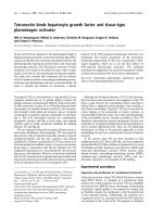
Figure 1 In vitro gene expression in AAV-transdu ced HeLa cells. (A) Schematic representation of expression cassettes in AAV vectors used
for transduction: control vector encoding b-galactosidase - AAV-LacZ; VEGF-A overexpressing vector - AAV-VEGF-A; FGF4 (cap-dependent cistron)
and GFP (IRES-dependent cistron) - AAV-FGF4-IRES-GFP; FGF4 (cap-dependent cistron) and VEGF-A (IRES-dependent cistron) - AAV-FGF4-IRES-
VEGF-A. CMV ie enhancer - cytomegalovirus immediate-early enhancer. IRES - internal ribosome entry site. (B) b-galactosidase in situ staining of
non-transduced or AAV-LacZ-transduced HeLa cells (arrows). (C) and (D) ELISA determining respectively, hVEGF-A and hFGF4 release into the cell
culture media. Production of both hVEGF-A and hFGF4 proteins was significantly up-regulated after transduction with therapeutic vectors when
compared to non-transduced (control) cells or cells transduced with AAV-LacZ vector. Representative data out of two independent experiments
performed in duplicates. Values are means ± SD; *p < 0.05 vs control and AAV-LacZ. Scale bar = 0.1 mm.
Jazwa et al. Genetic Vaccines and Therapy 2010, 8:6
/>Page 3 of 16
1×PBS-MK (1×PBS, 1 mM MgCl
2
,2.5mMKCl)and
virus was eluted in 0-1 M gradient of Na
2
SO
4
in 1×PBS-
MK. The viral preparation was desalted by dialysis
(Slyde-A-Lyser, Pierce) against 1×PBS at 4°C and stored
at -80°C. AAV titer was determined by measuring the
copy number of the viral genomes in dialyzed samples.
This was achieved by a real-time PCR procedure using
primers mapping in the target gene coding region. Pri-
mers recognizing LacZ (5′-AGA-ATCCGACGGGTTGT-
TACTCGC-3′ and 5′ -TGCGCTCAGGTCAAATTC
AGACGGC-3′ ), hVEGF-A (5′-ATGTCTATCAGCG-
CAGCTACTGCC-3′ and 5′-AGCTCATCTCTCCTAT-
GTGCTGGC-3′ )andhFGF4(5′ -TGGTGGCGCT
CTCGTTGGCG-3′ and 5′-ATCGGTGA-AGAAGGGC-
GAGCC-3′) were used. The purified viral preparations
used in the present study had particle titers of approx.
1×10
11
viral particles (vp) ml
-1
. Cells in culture and ani-
mals received the dose of AAV stated in the experimental
protocol.
Cell culture
HeLa cells (human epithelial cells from a fatal cervical
carcinoma) were maintained in low glucose (5.5 mM)
DMEM supplemented with 10% heat-inactivated FBS,
L-glutamine ( 2 mM), penicillin (100 U ml
-1
)andstrep-
tomycin (10 μgml
-1
).
Primary isolates of dermal fibroblasts were harvested
from 10-week-old diabetic (db/db) C57BLKS mice and
their wild-type (WT) littermates. The animals were sacri-
ficed and trunk skin was removed by sharp dissection
under sterile conditions. The harvested skin was then
minced and digested for 3 hours (from db/db mice) and
for 6 hours (from WT mice) in 0.2% collagenase type II
(Gibco; Warszawa, Poland) s olution in serum-free low
glucose DMEM a t 37°C. The dissociated cells were then
centrifuged and resuspended in low glucose (5.5 mM)
DMEM medium supplem ented with 20% FBS, 2 mM L-
glu tamine, 100 U ml
-1
penicillin, and 10 μgml
-1
strepto-
mycin. The cells were cultured at standard conditions:
5% CO
2
, 37°C and humidified atmosphere. After the first
or second passage cells from diabetic animals were
grown either in low (5.5 mM) or in high (25 mM) glucose
concentration for 48-72 hours. Fibroblasts from WT
mice were cultured in low glu cose DMEM. Cells at
passage 2 or 3 were used for experiments.
AAV-mediated transduction of cells in culture
HeLa cells were cultured at density 1 × 10
3
per 1 well of
the 96-well plate and exposed to 1 × 10
3
MOI (multipli-
city of infection) of AAV-LacZ, AAV-FGF4-IRES-GFP,
AAV-VEGF-A and AAV-FGF4-IRES-VEGF-A for
72 hours. After that time the transduction efficiency was
determined by b-galactosidase in situ staining and
conditioned culture media were collected for the mea-
surement of therapeutic growth factors production.
Animals
All animal procedures were in accordance with the
declaration of H elsinki and with the Guide for the Care
and Use of Laboratory Animals and were approved by
the Experimental Animal Committee at the Jagiellonian
University. Genetically diabetic C57BLKS mice homozy-
gous for a mutation in the leptin receptor (Lepr
db
)were
obtained from Jackson Laboratories (Bar Harbor, Maine
USA). Animals were 14-week-old at the start of the
experiments. Diabetic mice were obese, weighing 45 ±
5 g, hyperglycaemic with glucose concentrations in
excess of 400 mg per 100 ml. The hyperglycaemia pro-
duced classic signs of diabetes, including polydipsia, poly-
uria, and glyco suria. Animals were housed individually,
maintained under controlled environmental conditions
(12-h light/dark cycle at approx. 23°C), and provided
with standard laboratory food and water ad libitum.
Experimental protocol
After general inhalatory anesthesia with halothane, hair
on the back was shaved. Two full-thickness excisional
circular wounds (4 mm in diameter) were made using
biopsy punch on the dorsum of each mice. Animals
were randomized to receive either PBS, AAV-LacZ,
AAV-FGF4-IRES-GFP, AAV-VEGF-A or AAV-FGF4-
IRES-VEGF-A. Five animals were included into each
group (n = 5). All AAV vectors and PBS were injected
in the wound edges immediately after incision through
four (2 per each wound) intradermal injections with a
total volume of 100 μl. Animals received 3 × 10^10 vp
of an appropriate AAV vector.
Determination of wound area
Two wounds on the dorsum of each mice were photo-
graphed and measured using Image J software by an
observer blinded to t he experimental protocol at day 0
(directly after wounding), day 1 and then every second
day till the end of the observation when the last wounds
healed (day 21). Ten wounds per each group were
included into the analysis. Wound was considered
closed when it was complete ly covered with epithelium.
The wound area measured directly after wounding was
used as the reference or original area and all further
areas were recorded a s the percentage of the original
area. Once the experimental schedule was completed
(day 21) wounded skin, together with a margin of
healthy skin, was excised using 8 mm-diameter biopsy
punch. One wound was taken for histological examina-
tion (n = 5/group) and the second one for dete rmina-
tion of transgene level or activity (n = 5/group).
Jazwa et al. Genetic Vaccines and Therapy 2010, 8:6
/>Page 4 of 16
Detection of b-galactosidase activity
In situ: PBS- and AAV-LacZ-injected skin was briefly
washed in cold PBS, fixed in 2% buffered formaldehyde
and again washed in PBS. AAV-LacZ-transduced cells
growing in culture were fixed in 0.25% buffered formalin
and washed in PBS. The samples were immersed over-
night in a solution containing 1 mg ml
-1
5-bromo-4-
chloro-3-indolyl-b-D-galactopyranoside (X-gal), 2 mM
MgCl
2
,5mMK
3
Fe(Cn)
6
,5mMK
4
Fe(Cn)
6
in PBS at
37°C.
In tissue lysates: b-galactosidase activity was deter-
mined using b-galactosidase Enzyme Assay in PBS- and
AAV-LacZ-injected skin accordi ng to vendor’sprotocol.
Activity was normalized to the total protein content and
expressed in arbitrary units.
Determination of FGF4 and VEGF-A protein by ELISA
Skin samples were homogenized in 300 μl of lysis buffer
(PBS with 1% Triton and protease inhibitors - 10 mM
PMSF, 1 mg ml
-1
aprotinin and 1 mg ml
-1
leupeptin)
using an TissueLyser homogenizer (Qiagen). The homo-
genate was c entrifuged at 21 000 g for 10 min at 4°C.
The supernatant was colle cted and used for protein
determination using the Bicinchoninic Acid Protein
Assay Kit. Analysis was performed with hFGF4- and
hVEGF-A-recognizing ELISA kits. The level of hFGF4
and hVEGF-A in conditioned culture medium of AAV-
transduced HeLa cells was determined with the same
ELISA reagents. The amount of hFGF4 and hVEGF-A
was expressed in pg/mg protein (when deter mined in
tissue lysates) and in pg ml
-1
(when determined in con-
ditioned cell culture media).
Histology
Skin from the healed wo und beds surrounded by a mar-
gin of normal skin and the underlying muscle layer were
harvested and fixed in 10% neutral buffered formalin for
at least 24 h at room temperature, dehydrated in graded
ethanol, cleared in xylene and embedded in paraffin.
Perpendicular sections to the anterior posterior axis of
the wounds (3 μm t hick) were mounted on glass slides,
dewaxed, rehydrated with distilled water and stained
with haematoxylin/eosin or with Masson’ strichrome,
according to routine procedures for light microscopy.
The areas proximal to the incision were evaluated in all
skin sections in 10 random microscopic fields (1000×
magnification) by an observer blinded to the experimen-
tal protocol. The following parameters were evaluated
and scored as previously described [27,28], modified and
internally validated in our laboratory: 1) vascularity, 2)
granulation tissue formation and remodeling and 3) der-
mal matrix deposition and regeneration. We used four-
point scale to evaluate vascularity (1 - one or two vessels
per site; 2 - three vessels per site; 3 - four vessels per
site; 4 - five or more vessels per site) and three-point
scale to evaluate granulation tissue formation (1 - thin
granulation layer with up to 35 cells per site; 2 - moder-
ate granulation layer with up to 45 cells per site; 3 -
thick granulation layer with up to 55 and more cells per
site) and dermal matrix deposition and regeneration (1 -
little collagen deposition and little regeneration with up
to 10 hair follicles within the scar; 2 - moderate collagen
deposition and moderate regeneration with up to
20 hair follicles within the scar; 3 - high collagen
deposition and complete regeneration with up to 30 and
more hair follicles within the scar). The edges of the
wound in each of the sections were used as comparisons
for scoring.
Immunohistochemistry
To visualize the smallest blood vessels (<10 μmofthe
inner diameter), skin sections were deparaffinized and
subjected to antigen retrieval using 0.05 M sodium
citrate buffer (pH 6.0). Capillary endothelial cells were
detected with biotinylated Bandeiraea simplicifolia I
(BS-I) isolectin B
4
(dilution 1:100, Vector Laboratories;
Janki, Poland) . Incorporated isolectin was detected with
streptavidin- and fluoro chrome-conjugated antibodies
(Streptavidin Alexa Fluor 546). Additionally, in order to
visualize endothelial cell proliferation tissue sections
were exposed to proliferating cell nuclear antigen
(PCNA) recognizing antibodies (dilution 1:200) followed
by fluorochrome-conjugated secondary antibodies
(Alexa Fluor 488). All sections were mounted in DAPI
(4′ ,6-diamidino-2-phenylindole)-containing medium
(a fluorescent stain that strongly binds to DNA).
Proliferation assay
Mouse dermal fibroblasts were seeded in 96-well plate at
confluence 3 × 10
3
cells per well and grown in complete
DMEM medium containing low (5.5 mM) glucose (cells
from WT and db/db mice) or high (25 mM) glucose
(cells from db/db mice) for 24 hours. One hour before
stimulation complete medium was removed and cells
were overlaid with medium containing 0.5% FBS. Cells
were stimulated either with rhVEGF-A (50 ng ml
-1
)or
rhFGF4 (50 ng ml
-1
) or with both (50 ng ml
-1
each) for
24 hours. BrdU incorporation assay was performed
according to vendor’s protocol.
Migration assay
Transwell plates (8 μm po re) (Costar, Corning; Poznan,
Poland) were coated with fibronectin (20 μgml
-1
) mixed
with 0.5% gelatin in 1:1 ratio. Cells (1 × 10
4
per trans-
well) resuspended in DMEM medium containing low
(5.5 mM) glucose (WT and db/db f ibroblasts) or high
Jazwa et al. Genetic Vaccines and Therapy 2010, 8:6
/>Page 5 of 16
(25 mM) glucose (db/db fibroblasts) supplemented with
0.5% bovine serum albumin (BSA) were applied on the
coated transwell plates (upper compartment of a Boyden
chamber). Transwell plates with cells were then placed
in wells of a 24-well culture dish filled either with low
(5.5 mM) glucose (WT and db/db f ibroblasts) or high
(25 mM) glucose (db/db fibroblasts) DMEM containing
0.5% BSA supplemented either with rhVEGF-A (50 ng
ml
-1
)orrhFGF4(50ngml
-1
) or with both (50 ng ml
-1
each) (lo wer compartment of a Boyden chamber). After
20 hours of culture at 37°C each of the transwell plates
was washed with PBS, fixed in 10% formalin and stained
with haematoxylin and eosin. The non-migratory cells
from the filter surface of the upper compartment were
gently removed and only the cells that migrated to the
lower side were counted in 4 random microscopic fields
(200× magnification).
Quantitative RT-PCR
Total RNA was isolated from cells with Fenozol Total
RNA Isolation Reagent (PAA). Synthesis of cDNA was
performed using oligo-dT primers for 1 h at 42°C using
MMLV reverse transcriptase, according to vendor’ s
instruction. Quantitative RT-PCR was performed in a
Rotor Gene RG-3000 (Corbett Research) in a mixture
containing SYBR Green PCR Master Mix (SYBR Green
qPCR Kit), 50 ng of cDNA and specific primers in a
total volume of 15 μl. The primers recognizing MMP-9
(5′ -TGTGGATGTTTTTGATGCTATTG-3′ and 5′ -
CGGAGTCCAGCGTTGCA-3′ ), Flt-1 (5′ -GCACC-
TATGCSTGCAGAGC-3′ and 5′-TCTTTCAATAAA-
CAGCGTGCTG-3′ )andEF2(5′-GCGGTCAGCACA
ATGGCATA and 5′ -GACATCACCAAGGGTGTG-
CAG-3′) were used. EF2 (elongation factor 2) was used
as a housekeeping gene. After incubation for 15 min at
95°C, a three -step cycling protocol (30 s at 95°C, 45 s at
60°C and 45 s at 72°C) was used for 40 cycles. The
melting curve analysis was done using the program sup-
plied by Corbett Research. Relativ e quantification of
gene expression was calculated based on the compara-
tive C
T
(threshold cycle value) method (ΔC
T
=C
Tgene
of interest
-C
T housekeeping gene
). Comparison of gene
expression in different samples was performed basing
on the differences in ΔC
T
of individual samples (ΔΔC
T
).
Statistical analysis
Results are expressed as mean ± SEM unless otherwise
stated. One-wa y analysis of variance (ANOVA) followed
by Bonferroni’s post-hoc test or unpaired Student’ s
t-test was used to evaluate the statistical significance
between investigated groups. p < 0.05 was considered
statistically significant.
Results
VEGF-A and FGF4 are efficiently produced by
AAV-transduced HeLa cells
HeLa cells were exposed to AAV-LacZ, AAV-VEGF-A,
AAV-FGF4-IRES-GFP, and AAV-FGF4-IRES-VEGF-A
vectors (Figure 1a) each of them administered at 1 ×
10
3
MOI. This dose of vectors did not influence the cell
viability (data not shown). The analysis of gene expres-
sion was performed 72 h after transduction. As judged
from LacZ staining (Figure 1b) the in vitro transduction
efficiency with this dose of vectors was not very potent
(about 3.5%) but high enough to see the overexpression
of all introduced genes (Figure 1b, c, d). Since VEGF-A
and FGF4 are secreted proteins [6,21], their expression
was measured by ELISA in the culture supernatants col-
lected from transduced and n on-transduced HeLa cells
(Figure 1c and 1d, respectively). In adults, FGF4 is pro-
duced only under pathological conditions by certain
cancer cells, while HeLa cell line has been characterized
as non-expressing FGF4 [29]. In our hands, control
(non-transduced and AA V-LacZ-tr ansduced) HeLa cells
also did not release FGF4 into the cell culture media
(Figure 1d), while they release about 2616 ± 48 p g ml
-1
of human VEGF-A (Figure 1c). Transduction with con-
trol vector (AAV-LacZ) did not significantly affect
this production which was about 2916 ± 50 pg ml
-1
(Figure 1c). When AAV-VEGF-A or AAV-FGF4-IRES-
VEGF-A were added to th e cells the pro duction of VEGF
increased about 2-fold - up to 5667 ± 165 pg ml
-1
and
5471 ± 34 pg ml
-1
, respectively (Figure 1c). Interest-
ingly, the localization of hVEGF-A gene after CMV or
IRES sequence in the vector did not influence this pro-
tein production, as in both cases it was comparable.
Unlike hVEGF-A, hFGF4 production was much lower
and reached 56 ± 4 pg ml
-1
and 254 ± 17 pg ml
-1
after transduction with AAV-FGF4-IRES-GFP and
AAV-FGF4-IRES-VEGF-A, respectively (Figure 1d). The
experiments revealed that both VEGF-A and FGF-4
were released from the cells (data not shown) what
confirmed previously published observations [21].
Wound closure is significantly accelerated after AAV-
VEGF-A and AAV-FGF4-IRES-VEGF-A administration
Mice homozygous for a mutation in the leptin receptor
(Lepr
db
) exhibit a phenotype similar to adult-onset dia-
betes mellitus (type II), including a significant wound-
healing impairment when compared with their non-
diabetic littermates [30, 31]. In thi s study, a 4-mm
full-thickness excisional wound model was used. Ani-
mals were randomized to receive either PBS, AAV-LacZ,
AAV-FGF4-IRES-GFP, AAV-VEGF-A or AAV-FGF4-
IRES-VEGF-A (see Figure 1a). Although one of the
Jazwa et al. Genetic Vaccines and Therapy 2010, 8:6
/>Page 6 of 16
vectors (AAV-FGF4-IRES-GFP) expressed two proteins -
therapeutic (FGF4) and control (GFP) we decided to use
additional b-galactosidase (LacZ) expressing vector as
the most appr opriate control for our study. First of all,
LacZ was shown to be less immunogenic than GFP [32].
This seems to be of great importance in wound healing
studies as prolonged and dysregulated inflammatory
phase results in poor healing [33]. Moreover, IRES-
dependent gene expression in bicistronic vectors was
shown to be low er than cap-dependent gene expression
[25]. Since our therapeutic genes were mostly cap-
dependent (except VEGF-A in AAV-FGF4-IRES-VEGF-
A vector) we decided to use a control vector carrying
LacZ gene under the strong constitutive CMV promoter.
Additionally, presence of G FP sequence in AAV-FGF4-
IRES-GFP vector served as a control for VEGF-A used
in the second AAV-FGF4-IRES-VEGF-A bicistronic
vector.
The lesions were analyzed at different time-points by
measuring the wound area. Neither AAV-LacZ nor
AAV-FGF4- IRES-GFP accelerat ed wound closure at any
stage of the healing process (Figure 2a). In late stages of
the healing process wounds treated either with AAV-
VEGF-A or AAV-FGF4-IRES-VEGF-A healed signifi-
cantly faster confirming a crucial role of VEGF-A in this
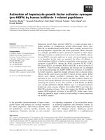
phenomenon (Figure 2a) . The reduction of the wound
area after AAV-VEGF-A injection was clearly visible
starting from day 17: 3.48 ± 1.47% of the initial wound
area vs 9.87 ± 4.95% in PBS group (p < 0.05) and vs
12.5 ± 4.05% in AAV-LacZ group (p < 0.05). At day 19
all wounds in AAV-VEGF-A group were covered with
epithelium and considered closed (Figure 2a, b). Inter-
estingly, the reduction of the wound area after AAV-
FGF4-IRES-VEGF-A injection was even more potent
starting already from day 13: 14.49 ± 3.29% of the initial
wound area vs 26.65 ± 0.1 6% in PB S group (p < 0.05)
and vs 25.7 ± 2.37% in AAV-LacZ group (p < 0.05) at
day 13; 6.86 ± 2.75% of the initial wound area vs
19.35 ± 3.07% in PB S group (p < 0.05) and vs 16.42 ±
3.2% in AAV-LacZ group (p < 0.05) at day 15. At day
17 all wounds in AAV-FGF4-IRES-VEG F-A group were
covered with epithelium and considered closed. The
healing process in some animals from PBS, AAV-LacZ
as well as AAV-FGF4-IRES-GFP groups was prolonged
until day 21 (Figure 2a).
Transgene expression in wounds of db/db mice 21 days
after AAV transduction
To study the location and the time course of AAV
expression in wounds b-galactosidase activity was de ter-
mined by histological analysis and using colorimetric
assay in skin lysates of AAV-LacZ injected mice 21 days
after treatment. Skin samples from PBS group served as
negative controls. Local b-galactosidase activity was
observed in histological skin sections close to the sites
ofwoundingandgenetransfer.Thebluestainingwas
present mostly in the dermal layer and hair follicles
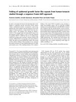
(Figure 3a). The colorimetric assay in tissue lysates indi-
cated weak statistically not significant increase in the
b-galactosidase activity when compared to the PBS
injected animals (Figure 3b).
Expression of both therapeutic genes in skin lysates
was determined at day 21 using ELISA kits recognizing
hVEGF-A and hFGF4 proteins (Figure 3c, d). Slight
increase in the level of hVEGF was detected after AAV-
VEGF-A administration (1.7 ± 1.22 pg/mg protein)
(Figure 3c). hVEGF protein was not detected in the skin
of AAV-FGF4-IRES-VEGF-A-injected mice with avail-
able EL ISA kit (Figure 3c). In case o f hFGF4 its level in
skin homogenates of diabeticmiceafterAAV-FGF4-
IRES-GFP injection was a bit higher (5.66 ± 1.27 pg/mg
protein) than after AAV-FGF4-IRES-VEGF-A (1.94 ±
0.84 pg mg
-1
protein) (Figure 3d). Of note, despite the
higher production of hFGF4 from AAV-FGF4-IRES-
GFP, the acceleration of wound healing was faster in
mice receivi ng AAV-FGF4-IRES-VEGF-A, indicating for
the significance of combined growth factors delivery.
Local AAV-FGF4-IRES-VEGF-A delivery promotes wound
healing at the histological level
Diabetic animals usually have a thicker epithelial l ayer
than normal mice [27]. In addition, the different layers
are less differentiated and adipose infil trat es are present
in the dermis, impairing the normal elasticity of the skin
and, as a consequence, it is more prone to a delayed
healing [27]. At day 21 all wounds were already covered
with epithelium therefore, by histological evaluat ion, we
were not able to observe any differences in the grade of
re-epithelialization between analyzed groups of animals.
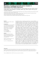
Nevertheless, the epithelial layer covering AAV-VEGF-
A- and AAV-FGF4-IRES-VEGF-A-treated wounds was
thicker and had a greater cell density when compared to
PBS or AAV-LacZ controls or AAV-FGF4-IRES-GFP-
treated wounds (Figure 4a, b, photos IV and V).
Interestingly, within the scar tissue of most of the
analyzed skin sections we found clusters of inflamma-
tory cells forming granulomas (Figure 4a, photo VI).
Granuloma represents a special type of inflammatory
reaction in which collection of immune cells is trying
to destroy a foreign substance. Apparently, this
immune response does not se em to be related to any
of the introduced transgenes or AAV capsid proteins
as granulomas were found within the healed wounds
of all investigated groups of animals including mice
injected with PBS. The real cause of such inflammatory
reaction is not known and we presume that it might be
related to wounding-induced cholesterol crystals
formation.
Jazwa et al. Genetic Vaccines and Therapy 2010, 8:6
/>Page 7 of 16
Although, plenty of inflammatory cells could still be
found in the skin sections, most of the cells within the
scar tissue of all analyzed groups were of mesenchymal
origin (fibroblasts and myofibroblasts). It indicates that
the process of tissue remodeling has already been
initiated. Granulation tissue and especially its vascularity
was enhanced after all three therapeutic vectors in com-
parison to the control PBS- and AAV-LacZ-injected ani-
mals however, statistically significant difference was
observed only after bicistronic AAV-FGF4-IRES-VEGF-
A vector administration (Figure 5a and 5b, respectively).
Thus, VEGF-A delivered in combination with FGF4 into
the wound edge reduced adipose substitution and
produced a significant improvement in the healing pro-
cess by increasing the thickness and vascularization of
granulation tissue. Additionally, single (AAV-FGF4-
IRES-GFP and AAV-VEGF-A) or combined (AAV-
FGF4-IRES-VEGF-A) gene transfer resulted in abundant
collagen deposition in comparison to the PBS- or AAV-
LacZ-treated control wounds (Figure 5c).
AAV-VEGF-A stimulates new blood vessels formation in
the skin of db/db mice
Isolectin B
4
was used to visualize the smalles t blood ves-
sels (capillaries) in the skin tissue 21 days after wounding
and gene transfer (Figure 6a). Double immunofluorescent
Figure 2 AAV-VEGF-A and AAV-FGF4-IRES-VEGF-A accelerates time to wound closure in db/db mice. (A) Quantification of the wound area
at consecutive days. Reduction of the wound area after AAV-VEGF-A injection was significantly enhanced starting from day 17. At day 19 all
wounds in AAV-VEGF-A group were covered with epithelium and considered closed (arrow, inset). The reduction of the wound area after AAV-
FGF4-IRES-VEGF-A injection was even more visible when compared to AAV-LacZ-injected controls starting already from day 13. At day 17 all
wounds in AAV-FGF4-IRES-VEGF-A group were covered with epithelium and considered closed (arrow, inset). No acceleration of the wound
closure was observed after AAV-FGF4-IRES-GFP at any time-point and the last wounds in this group were considered closed at day 21 together
with PBS- and AAV-LacZ-injected animals. (B) Representative pictures taken at day 19 showing wounds of AAV-VEGF-A and AAV-FGF4-IRES-VEGF-
A-injected animals completely covered with epithelium and prolonged healing process in PBS-, AAV-LacZ- and AAV-FGF4-IRES-GFP-treated mice.
Graph represents means ± SEM (n = 10 wounds/group); *p < 0.05 vs PBS and AAV-LacZ.
Jazwa et al. Genetic Vaccines and Therapy 2010, 8:6
/>Page 8 of 16
staining using biotinylated isolectin B
4
and PCNA-recog-
nizing antibodies revealed that administration o f AAV-
VEGF-A stimulated angi ogenesis by induction of proli f-
eration of capillary endothelial cells in the dermal area
proximal to the healed incision (10.2 ± 1.06/mm
2
vs
4.77 ± 1.77/mm
2
in PBS group; p < 0.05 and vs 5.31 ±
0.8/mm
2
in AAV-LacZ group; p < 0.05) (Figure 6b). This
was paralleled with increased total number of capillaries
(42.47 ± 1.37/mm
2
vs 33.5 ± 2.7/mm
2
in PBS group;
p < 0.05 and vs 35.41 ± 2.9/mm
2
in AAV-LacZ group;
p = 0.09) (Figure 6c). The number of skin capillaries
detected 21 days after wounding and AAV-FGF4-IRES-
GFP or AAV-FGF4-IRES-VEGF-A injection did not differ
significantly from PBS- and AAV-LacZ-treated animals
(Figure 6c).
In vitro characteristics of healthy (WT) and diabetic (db/
db) mouse dermal fibroblasts
Our observation that AAV-FGF4-IRES-VEGF-A and
AAV-VEGF-A accelerated time to wound closure in
mice prompted us to explore the underlying mechanisms.
As the efficiency of growth factors in vivo could result
from sustained production by AAV vector which
occurred during entire healing process and would require
much more animals to check in details, we decided to
investigate the migratory and proliferation capabilities of
fibroblasts using recombinant growth factors.
Proliferation of diabetic and wild-type fibroblasts was
measured using BrdU incorporation assay (Figure 7a).
Diabetic fibroblasts cultured in low glucose (5.5 mM)
DMEM proliferate d slightly but significantly slower than
wild-type cells (81.5 ± 7.4% vs 100%, respectively;
p < 0.05). Fibroblasts from diabetic mice cultured in
high glucose (25 mM) DMEM exhibited more potent
reduction in proliferation rate (61.5 ± 13% vs 100% in
WT control; p < 0.05 and vs 81.5 ± 7.4% in db/db
5.5 mM control; p < 0.05). rhFGF4 delivered alone or in
combination with rhVEGF significantly increased the
proliferatio n of wild-type fibroblasts (133.7 ± 19.2% and
121.8 ± 20.6% vs 100% WT contro l, respectively). Prolif-
eration of db/db fibroblasts cultured in low glucose in
the presence of rhFGF4 was slightly weaker than of WT
Figure 3 Weak transgene expression in the skin of db/db mice 21 days after wounding and gene transfer.(A)Representativeskin
sections demonstrating b-galactosidase activity (arrows) in AAV-LacZ injected animal and the negative control (PBS treated skin). (B) Colorimetric
assay showing slightly increased b-galactosidase activity in skin tissue homogenates from AAV-LacZ-injected animlas in comparison to the PBS-
treated mice. (C) hVEGF and (D) hFGF4 protein in skin tissue homogenates measured by ELISA. Graphs represent means ± SEM (n = 5 animals/
group); *p < 0.05 vs PBS and AAV-LacZ. Scale bar = 0.05 mm.
Jazwa et al. Genetic Vaccines and Therapy 2010, 8:6
/>Page 9 of 16
cells and, although the trend was clearly visible, did not
reach the statistical signi ficance (103.5 ± 6.3% vs 81.5 ±
7.4% db/db 5.5 mM control, p = 0.09) (Figure 7a). Inter-
estingly, combined rhFGF4 and rhVEGF-A treatment
slightly but significantly increased the proliferation rate
of db/db fibroblasts cultured in low glucose (115 ± 6.4%
vs 81.5 ± 7.4% db/db 5.5 mM control, p < 0.05). Cells
from db/db mice cultured in high glucose concentration
did respond neither to rhFGF4 or rhVEGF-A and their
proliferation did not change significantly when two
growth factors, rhFGF4 and r hVEGF-A, were used
(Figure 7a).
Differences in basal migration on fibronectin/gelatin
were observed between diabetic (db/db) and healthy
(WT) fibroblasts (Figure 7b). Migration of diabetic
fibroblasts cultured in low glucose DMEM was impaired
when compared to wild-type fibroblasts (73.5 ± 7% vs
100%, respectively; p < 0.05). When diabetic fibroblasts
were cultured in high glucose DMEM t he impairment
of migration was much more potent (49 ± 13% vs 100%
in WT control; p < 0.05 and vs 73.5 ± 7% in db/db
5.5 mM control; p < 0.05). Migration in response to sin-
gle rhFGF4 treatment increased more than 2 times in
case of wild-type fibroblasts (249.7 ± 19%; p < 0.05 vs
WT control) and about 3 times in case of diabetic fibro-
blasts cultured in low glucose DMEM (3 13.5 ± 87.4%;
p < 0.05 vs db/db 5.5 mM control). When rhFGF4 was
added in combination with rhVEGF-A the migration of
both cell types (WT and db/db cultured in low gluco se)
did not differ significantly from the one observed after
Figure 4 Skin morphology of db/db mice 21 days after wounding and gene transfer. (A) Haematoxylin/eosin staining of the skin injected
with (I) PBS; (II) AAV-LacZ; (III) AAV-FGF4-IRES-GFP; (IV) AAV-VEGF-A and (V) AAV-FGF4-IRES-VEGF-A. Analysis revealed less adipose tissue and
better organized granulation tissue with the presence of hair and restoration of normal architecture of dermis in AAV-VEGF-A and AAV-FGF4-
IRES-VEGF-A-treated mice in comparison to PBS-, AAV-LacZ- and AAV-FGF4-IRES-GFP-injected animals. Panels are representative of 5 animals per
group. Scale bar (I-V) = 0.1 mm. (VI) Higher magnification of AAV-FGF4-IRES-VEGF-A-injected skin with granulomas (arrows). Scale bar (VI) = 0.05
mm. (B) Representative Masson’s trichrome staining of the skin injected with (I) PBS; (II) AAV-LacZ; (III) AAV-FGF4-IRES-GFP; (IV) AAV-VEGF-A and
(V) AAV-FGF4-IRES-VEGF-A. Double-headed arrows indicate the thickness of the collagen layer that was significantly thicker after injection of all
three therapeutic vectors (AAV-FGF4-IRES-GFP, AAV-VEGF-A, AAV-FGF4-IRES-VEGF-A) when compared to PBS- and AAV-LacZ-treated animals.
Panels are representative of 5 animals/group. Scale bar = 0.05 mm.
Jazwa et al. Genetic Vaccines and Therapy 2010, 8:6
/>Page 10 of 16
single rhFGF4 tr eatment (264 ± 64%; p < 0.05 vs WT
control and 419.4 ± 192.7% vs db/db 5.5 mM control,
respectively) (Figure 7b). Migrati on of diabetic fibro-
blasts cultured in high glucose DMEM did change
neither upon single rhFGF4 nor rhVEGF-A treatment.
However, it was strongly increased (about 4 times) in
response to both growth factors (196.4 ± 60.4%, p <
0.05 vs db/db 25 mM control) (Figure 7b).
FGF4 stimulates MMP-9 and Flt-1 expression in primary
mouse dermal fibroblasts
Basal MMP-9 expression did not differ significantly
between analyzed groups (Figure 8a). Sole rhVEGF-A
treatment did not influence MMP-9 expression in dia-
betic dermal fibroblasts cultured in high glucose con-
centration (Figure 8b), but it was significantly up-
regulated by rhFGF4 (Figure 8b). As there was no addi-
tional up-regulation after combined rhFGF4 and
rhVEGF-A treatment, this effect most probably depends
only on FGF4. Nevertheless, this rhFGF4-mediated sti-
mulation of MMP-9 expression seems to be insufficient
to significantly increase the migration of diabetic fibro-
blasts cultured in 25 mM glucose DMEM, as its rate did
not change upon sole rhFGF4 treatment (see Figure 7b).
Thus, we have performed analysis of the expression of
one of the VEGF receptors, Flt-1, as a possible mechan-
ism responsible for this observation. Similarly to MMP-
9, there were no significant differences in the basal Flt-1
expression between WT and db/db fibroblasts kept
either in low or high glucose (Figure 8c). Sole rhVEGF
treatment did not influence Flt-1 expression in diabetic
Figure 5 Semi-quantitative evaluation for granulation tissue:
(A) granulation tissue, (B) vascularity, and (C) dermal matrix
deposition and regeneration. Graphs represent means ± SEM (n = 5
animals/group); *p < 0.05 vs PBS and AAV-LacZ.
Figure 6 AAV-VEGF-A stimulates skin neovas cularization in
db/db mice 21 days after wounding and gene transfer. (A)
Representative pictures demonstrating isolectin B4 binding and
expression of proliferating cell nuclear antigen (PCNA) (arrows). DAPI
was used to confirm the nuclear localization of PCNA (arrow). (B)
The number of proliferating capillary endothelial cells was increased
after AAV-VEGF-A injection. (C) Total number of capillaries was also
increased only after AAV-VEGF-A administration. Graphs represent
means ± SEM (n = 5 animals/group); *p < 0.05 vs PBS and AAV-
LacZ; # p < 0.05 vs PBS. Scale bar = 0.01 mm.
Jazwa et al. Genetic Vaccines and Therapy 2010, 8:6
/>Page 11 of 16
fibroblasts, but again, a s in case of MMP-9, it was
up-regulated by rhFGF4 with no further change by
combined treatment with rhVEGF-A and rhFGF4
(Figure 8d).
Discussion
The salient finding o f the present study is demonstra-
tion that combined overexpression of VEGF-A and
FGF4 results in the faster wound healing in diabetic
mice than single treatment. This can be a result of
enhanced migration of fibroblasts stimulated by two
growth factors, the process at least partially dependent
on up-regulation of MMP-9 and VEGF receptor 1
expression enhanced by FGF4.
VEGF-A has been shown to play a pivotal role in the
initiation of angiogenesis, based on its ability to induce
the expression of proteases that digest components of
the extracellular matrix that impe de angiogenesis, to
Figure 7 Combined rhFGF4 and rhVEGF-A treatment improves high glucose-impaired biological properties of diabetic mouse dermal
fibroblasts. Functional in vitro tests with age- and passage-matched fibroblasts isolated from the skin of diabetic and wild-type mice: (A)
Proliferation after 24 hours of culture. Diabetic fibroblasts cultured in 5.5 mM glucose DMEM (empty bars) show impaired basal proliferation in
comparison to wild-type fibroblasts (black bars) which is even more intense when the db/db cells are kept in 25 mM glucose (gray bars). rhFGF4
(delivered alone or in combination with rhVEGF-A) induces WT cell proliferation whereas only co-stimulation with rhVEGF-A significantly increases
proliferation of cells isolated from db/db mice and cultured in 5.5 mM glucose (similar tendency in diabetic fibroblasts kept in 25 mM glucose).
(B) Migration after 20 hours of culture. Diabetic fibroblasts cultured in 5.5 mM glucose DMEM (empty bars) show impaired basal migration on
gelatin/fibronectin in comparison to wild-type fibroblasts (black bars) which is even more intense when the cells are kept in 25 mM glucose
(gray bars). Migration towards rhFGF4 gradient (delivered alone or in combination with rhVEGF-A) is preserved in WT cells and in cells from db/
db mice cultured in 5.5 mM glucose. Fibroblasts isolated from the skin of db/db mice and cultured in 25 mM glucose show impaired migration
towards rhFGF4, that can be restored by combined rhFGF4 and rhVEGF-A treatment. C - control, V - rhVEGF-A (50 ng ml
-1
), F - rhFGF4 (50 ng ml
-
1
), V+F - rhVEGF-A (50 ng ml
-1
) and rhFGF4 (50 ng ml
-1
). Graphs represent means ± SEM from n = 5 (A) and n = 3 (B) independent experiments
performed in duplicates; *p < 0.05 vs appropriate control; # p < 0.05 vs WT control; § p < 0.05 vs WT and db/db 5.5 mM control.
Jazwa et al. Genetic Vaccines and Therapy 2010, 8:6
/>Page 12 of 16
promote endothelial cell proliferation, and to prevent
their apoptosis [6,8]. In addition, it has been demon-
strated that VEGF-A gene transfer improves diabetes-
impaired wound heali ng by stimulation of a ngiogenesis
and granulation tissue formation [9,10].
In our study, the healing process after injection of
AAV-VEGF-A was also accelerated when compared t o
PBS- or AAV-LacZ-treated animals. FGF4 delivered
alone in a form of AAV-FGF4-IRES-GFP vector did not
influence the rate of wound healing. So far, there were
no studies investigating the role of this agent in this
phenomenon. In contrary to VEG F-A, FGF4 is an onco-
gene and is not produced in adults under normal, phy-
siological conditions [17]. It is very well known that
disruption of the normal process of tissue regeneration
in diabetes mellitus is related to the decreased produc-
tion of different growth factor s and VEGF-A is one of
them [34-36]. Therefore, FGF4 delivered alone, although
possessing the ability to up-regulate the endogenous
VEGF-A expression [19,20], may be ineffective in
reparative processes in diabetes. In this sense, the com-
bined therapy us ing FGF4 and VEGF-A codi ng
sequences seemed to be a better approach. In fact,
AAV-mediated combined gene transfer of FGF4 and
VEGF-A improved diabetic wound healing over the
effect exerted by AAV-VEGF-A.
Using a vailable ELISA kits we were not able to detect
any up-regulation of hVEGF-A and only slight hFGF4
Figure 8 rhFGF4 stimulates MMP-9 and Flt-1 expression in diabetic mouse dermal fibroblasts. (A) There are no significant differences in
the basal MMP-9 expression between WT and db/db fibroblasts cultured either in low (5.5 mM) or high (25 mM) glucose. (B) MMP-9 expression
is up-regulated upon single rhFGF4 or combined with rhVEGF-A treatment in db/db fibroblasts cultured in hyperglycemic conditions. (C) There
are no significant differences in the basal Flt-1 expression between WT and db/db fibroblasts cultured either in low (5.5 mM) or high (25 mM)
glucose. (D) Flt-1 expression is up-regulated upon single rhFGF4 or combined with rhVEGF-A treatment in db/db fibroblasts cultured in
hyperglycemic conditions. C - control, V - rhVEGF-A (50 ng ml
-1
), F - rhFGF4 (50 ng ml
-1
), V+F - rhVEGF-A (50 ng ml
-1
) and rhFGF4 (50 ng ml
-1
).
Graphs represent means ± SEM from n = 3 independent experiments performed in duplicates; *p < 0.05 vs control.
Jazwa et al. Genetic Vaccines and Therapy 2010, 8:6
/>Page 13 of 16
production in the s kin of AAV-FGF4-IRES-VEGF-A-
injected mice at the end of the healing process. We sus-
pect that the turnover of transduced cells might result
in loss of introduced gene expression over time and
finally lead to very low or lack of expression especially
that at day 21 all wounds in the AAV-FGF4-IRES-
VEGF-A group were healed since couple of days already.
Also, as demonstrated by b-galactosidase in situ staining
and with the colorimetric assay in skin tissue lysates, the
expression of the reporter gene 21 days after vector
injection was local and rather low. Similar observ ation
has been made by Badillo and co-workers, who used
lentiviral vectors, capable of high and stable gene deliv-
ery, for t he transduction of wounded skin tissue. The
authors observed that 14 days post wounding and viral
treatment GFP expression was limited to isolated areas
or completely absent [37]. Importantly, we did not
detect any hVEGF-A and any hFGF-4 in the plasma of
the transduced mice (data not shown) what i ndicates
that there was no systemic exposure to these agents and
no significant side effects.
In the present study combinatory therapy with VEG F-
A and FGF4 coding sequences increased the vascularity
of granulation tissue. Concerning vascularity, only
mature vessels that contained erythrocytes were
counted. In contrast to AA V-VEGF-A combined VEGF-
A and F GF4 treatment did not significantly change the
number of isolectin-labeled capillaries and their prolif-
eration 21 days after gene transfer suggesting the lack of
stimulatory effect on the neovascularization. On the
other h and, it is very well known that upon completion
of epithelialization (what in case of AAV-FGF4-IRES-
VEGF-A-injected animals occurred a couple of days
before histological exami nation) cell proliferation and
neovascularization cease, scar tissue forms, and the
wound enters the remodeling phase [38]. One of the
typical features of transformation of the granulation tis-
sue into scar is regr ession of vascular structures. There-
fore, the lack of effect of AAV-FGF4-IRES-VEGF-A
vector on the total number of skin capillaries and their
proliferation 21 days after wounding and gene transfer
maybeexplainedbythephenomenonoftheirnatural
regression.
Very recently, Brem et al. d emonstrated that VEGF-A
was capable of stimulating the migration of activated
keratinocytes over the wound area what indicates that
this growth factor can promote epithelialization inde-
pendently of its role in recruiting and stimulating
endothelial cells in the repair process [39]. Although, in
the present study stimulation of keratinocyte migration
was not investigated, we cannot exclude that it also
contributed to the faster wound closure in AAV-VEGF-
A- and AAV-FGF4-IRES-VEGF-A-treated mice indepen-
dently of the stimulation of angiogenesis.
In our hands, VEGF-A delivered alone or in combina-
tion with FGF4 into the wound edge produced a signifi-
cant acceleration of the wound closure that was
associated with increased thickness of granulation tissue,
increased number of cells within the dermal layer and
abundant collagen deposition. Collagen is the major
connective-tissue component of granulation tissue, scar
and dermis. Its synthesis and deposition are critical for
wound closure [39].
Studies demonstrating that diabetes does have effects
on essential aspects of fibroblast biology, such as prolif-
eration and collagen synthesis, pointed at the important
and complex functions of these cells during tissue repair
[33,40,41 ]. There fore, in the present study we addressed
the question whether glucose concentration might
somehow modulate proliferation, migration and angio-
genic gene expression in diabetic dermal fibroblasts
exposed to sole o r combined rhVEGF-A and rhFGF4
treatment. Age- and pa ssage-matched fibroblasts iso-
lated from the skin of non-diabetic (WT) mice of the
same background strain served as control. Diabetic
fibroblasts were cultured either in low (5.5 mM) or high
(25 mM) glucose DMEM concentration and the later
was intended to resemble the diabetic environment. We
have observed significant differences in basal prolifera-
tion and migration on fibronectin/gelatin between dia-
betic and healthy (WT) mouse dermal fibroblasts.
Moreover, db/db fibroblasts cultured in high glucose
concentration (25 mM) exhibited even more dramatic
reduction in proliferation and migration rate than db/db
fibroblasts cultured in low glucose (5.5 mM). Addition-
ally, culture in the presence of high glucose concentra-
tion led to significantly impaired response to rhFGF4
treatment. Apparently, c o-stimulation with rhFGF4 and
rhVEGF-A significantly improved migration of these
cells.
We have examined MMP-9 (gelatinase) expression as
a possible m echanism responsible for this observation.
We did not observe any significant differences in the
basal MMP-9 expression between WT and db/db fibro-
blasts cultured either in low or in high glucose concen-
tration. This is somehow in agreement with other data
demonstrating decreased migration of db/db fibroblasts
in comparison to WT cells associated with only a selec-
tive increase in pro-MMP-9 in db/db fibroblasts but
with no difference in active MMP-9 [40].
Interestingly, we found MMP-9 gene to be signifi-
cantly up-regulated in db/db fib roblasts cultured i n high
glucose u pon rhFGF4 treatm ent. Similar acti vity of
FGF4 has been demonstrated by Anteby et al.intro-
phoblast suggesting its important role in early placental
development [42]. Therefore, we can speculate that by
increasing the invasiveness of skin fibroblasts FGF4
might also play a crucial role in the process of wound
Jazwa et al. Genetic Vaccines and Therapy 2010, 8:6
/>Page 14 of 16
healing. Sole rhVEGF-A treatment did not influence
MMP-9 expression in db/db fibroblasts cultured under
hyperglycemic conditions. Moreover, there was no addi-
tional up-regulation after combined rhFGF4 and
rhVEGF-A treatment, what indicates that this effect
depends o n FGF4. Nevertheless, FGF4-mediated stimu-
lation of MMP-9 expression seems to be insufficient to
significantly increase migration of d iabetic fibroblasts
kept in high glucose, as its rate did not change upon
sole rhFGF4 treatment. As an additional confirmation
we have performed the western blot analysis of the
levels of pro-MMP-9 in the tissue homogenates. We
observed about 3-fold up-regulation of this protein in
the skin of animals treated with FGF-4 transgene in
comparison to control animals injected with AAV-La cZ.
However, due to big variability between the samples the
difference was not statistically significant (data not
shown).
Wang and Keiser demonstrated that VEGF-A up-regu-
lates MMP-1, -3, and -9 expression in human smooth
muscle cells (SMCs) and accelerates their migration
through synthetic ECM barriers [43]. Furthermore, the
authors showed expression of the high-affinity Flt-1
receptor in human SMCs and its phosphorylation upon
VEGF-A treatment, suggesting its role in mediating
VEGF-A action. Very recentl y Brem et al. demonstrated
that VEGF-A stimulates migration of human fibroblasts
cultured in vitro, what indicates a non-angiogenic effect
of VEGF-A on wound closure [39]. In our model, we
did not observe any significant in vitro effect of
rhVEGF-A on primary diabetic or non-diabetic mouse
dermal fibroblasts migration. Moreover, rhVEGF-A did
not change Flt-1 mR NA level in thos e cells. Interest-
ingly, however, we have found up-regulation of this
gene expression in response to rhFGF4 in diabetic fibro-
blasts cultured in the presence o f high glucose concen-
tration. Moreover, there was no difference in Flt-1
expression upon single rhFGF4 or combined with
rhVEGF-A treatment, what suggests the FGF4-specific
effect.
Conclusion
On the basis of these findings, we can conclude that
VEGF-A may increase migration of diabetic fibroblasts
cultured in high glucose concentration through FGF4-
mediated up-regulation of one of the VEGF receptors,
Flt-1. In this sense, combined therapy approach with
VEGF-A and FGF4 genes may significantly improve the
delayed wound repair in diabetes over the effect exerted
by single VEGF-A treatment. Whether this finding
might have important clinical implications remains to
be established and deserves further pre-clinical and clin-
ical investigation.
Abbreviations
AAV: adeno-associated viral vector; db/db, diabetic; Flt-1: fms-like tyrosine
kinase-1 receptor (VEGF receptor 1); FGF4: fibroblast growth factor 4; MMP-9:
matrix metalloproteinase-9; VEGF-A: vascular endothelial growth factor-A.
Acknowledgements
Grzegorz Dyduch, PhD, MD (Department of Clinical and Experimental
Pathomorphology, Jagiellonian University, Poland) is kindly acknowledged
for advise and help with histological examination. The study was supported
by research grants from Ministry of Science and Higher Education, No. N302
020 31/1998 and No. PBZ-KBN-096/P05/2005 (both awarded to Jozef Dulak).
Agnieszka Jazwa is the recipient of the Foundation for Polish Science (FNP)
Fellowship. Alicja Jozkowicz is the recipient of the Wellcome Trust
International Senior Research Fellowship. The Faculty of Biochemistry,
Biophysics and Biotechnology of the Jagiellonian University is a beneficiary
of the structural funds from the European Union (grant No: POIG.02.01.00-
12-064/08 - “Molecular biotechnology for health“ and No. POIG 01.02-00-109/
99 ‘Innovative methods of stem cell applications in medicine; No:
POIG.02.02.00-014/08 - “Jagiellonian Centre of Experimental Therapeutics”)
European Union structural funds, Innovative Economy Operational
Programme.
Author details
1
Department of Medical Biotechnology, Faculty of Biochemistry, Biophysics
and Biotechnology, Jagiellonian University, Krakow, Poland.
2
Section for
Studies on Metastasis, National Cancer Center Research Institute, Tokyo,
Japan.
3
Research & Development Department, Adamed Ltd., Pienkow,
Poland.
4
A. I. Virtanen Institute, University of Kuopio and Gene Therapy Unit,
Kuopio University Hospital, Kuopio, Finland.
Authors’ contributions
AJ participated in the design of the study, carried out the practical work and
drafted the manuscript. PK, JL, AZ, AS, JS, MK, HT, TO, RD and EV participated
in the practical work and discussions. SYH and AJ participated in design of
the study and helped to draft the manuscript. JD conceived of the study,
designed it and edited the manuscript. All authors read and approved the
final manuscript.
Competing interests
The authors declare that they have no competing interests.
Received: 9 December 2009 Accepted: 30 August 2010
Published: 30 August 2010
References
1. Falanga V: Wound healing and its impairment in the diabetic foot. Lancet
2005, 366(9498):1736-1743.
2. Martin P: Wound healing–aiming for perfect skin regeneration. Science
1997, 276(5309):75-81.
3. Doxey DL, Ng MC, Dill RE, Iacopino AM: Platelet-derived growth factor
levels in wounds of diabetic rats. Life Sci 1995, 57(11):1111-1123.
4. Werner S, Breeden M, Hubner G, Greenhalgh DG, Longaker MT: Induction
of keratinocyte growth factor expression is reduced and delayed during
wound healing in the genetically diabetic mouse. J Invest Dermatol 1994,
103(4):469-473.
5. Frank S, Hubner G, Breier G, Longaker MT, Greenhalgh DG, Werner S:
Regulation of vascular endothelial growth factor expression in cultured
keratinocytes. Implications for normal and impaired wound healing. J
Biol Chem 1995, 270(21):12607-12613.
6. Ferrara N: Role of vascular endothelial growth factor in regulation of
physiological angiogenesis. Am J Physiol Cell Physiol 2001, 280(6):
C1358-1366.
7. Pepper MS, Ferrara N, Orci L, Montesano R: Vascular endothelial growth
factor (VEGF) induces plasminogen activators and plasminogen activator
inhibitor-1 in microvascular endothelial cells. Biochem Biophys Res
Commun 1991, 181(2):902-906.
8. Lamoreaux WJ, Fitzgerald ME, Reiner A, Hasty KA, Charles ST: Vascular
endothelial growth factor increases release of gelatinase A and
decreases release of tissue inhibitor of metalloproteinases by
microvascular endothelial cells in vitro. Microvasc Res 1998, 55(1):29-42.
Jazwa et al. Genetic Vaccines and Therapy 2010, 8:6
/>Page 15 of 16
9. Galiano RD, Tepper OM, Pelo CR, Bhatt KA, Callaghan M, Bastidas N,
Bunting S, Steinmetz HG, Gurtner GC: Topical vascular endothelial growth
factor accelerates diabetic wound healing through increased
angiogenesis and by mobilizing and recruiting bone marrow-derived
cells. Am J Pathol 2004, 164(6):1935-1947.
10. Galeano M, Deodato B, Altavilla D, Cucinotta D, Arsic N, Marini H, Torre V,
Giacca M, Squadrito F: Adeno-associated viral vector-mediated human
vascular endothelial growth factor gene transfer stimulates angiogenesis
and wound healing in the genetically diabetic mouse. Diabetologia 2003,
46(4):546-555.
11. Romano Di Peppe S, Mangoni A, Zambruno G, Spinetti G, Melillo G,
Napolitano M, Capogrossi MC: Adenovirus-mediated VEGF(165) gene
transfer enhances wound healing by promoting angiogenesis in CD1
diabetic mice. Gene Ther 2002, 9(19):1271-1277.
12. Galzie Z, Kinsella AR, Smith JA: Fibroblast growth factors and their
receptors. Biochem Cell Biol 1997, 75(6):669-685.
13. Zhang X, Ibrahimi OA, Olsen SK, Umemori H, Mohammadi M, Ornitz DM:
Receptor specificity of the fibroblast growth factor family. The complete
mammalian FGF family. J Biol Chem 2006, 281(23):15694-15700.
14. Ornitz DM, Itoh N: Fibroblast growth factors. Genome Biol 2001, 2(3):
REVIEWS3005.
15. Feldman B, Poueymirou W, Papaioannou VE, DeChiara TM, Goldfarb M:
Requirement of FGF-4 for postimplantation mouse development. Science
1995, 267(5195):246-249.
16. Niswander L, Tickle C, Vogel A, Booth I, Martin GR: FGF-4 replaces the
apical ectodermal ridge and directs outgrowth and patterning of the
limb. Cell 1993, 75(3):579-587.
17. Adnane J, Gaudray P, Dionne CA, Crumley G, Jaye M, Schlessinger J,
Jeanteur P, Birnbaum D, Theillet C: BEK and FLG, two receptors to
members of the FGF family, are amplified in subsets of human breast
cancers. Oncogene 1991, 6(4):659-663.
18. Yamamoto H, Ochiya T, Takahama Y, Ishii Y, Osumi N, Sakamoto H,
Terada M: Detection of spatial localization of Hst-1/Fgf-4 gene
expression in brain and testis from adult mice. Oncogene 2000,
19(33):3805-3810.
19. Rissanen TT, Markkanen JE, Arve K, Rutanen J, Kettunen MI, Vajanto I,
Jauhiainen S, Cashion L, Gruchala M, Narvanen O, et al: Fibroblast growth
factor 4 induces vascular permeability, angiogenesis and arteriogenesis
in a rabbit hindlimb ischemia model. Faseb J 2003, 17(1):100-102.
20. Deroanne CF, Hajitou A, Calberg-Bacq CM, Nusgens BV, Lapiere CM:
Angiogenesis by fibroblast growth factor 4 is mediated through an
autocrine up-regulation of vascular endothelial growth factor
expression. Cancer Res 1997, 57(24):5590-5597.
21. Delli-Bovi P, Curatola AM, Newman KM, Sato Y, Moscatelli D, Hewick RM,
Rifkin DB, Basilico C: Processing, secretion, and biological properties of a
novel growth factor of the fibroblast growth factor family with
oncogenic potential. Mol Cell Biol 1988, 8(7):2933-2941.
22. Dell
’Era P, Belleri M, Stabile H, Massardi ML, Ribatti D, Presta M: Paracrine
and autocrine effects of fibroblast growth factor-4 in endothelial cells.
Oncogene 2001, 20(21):2655-2663.
23. Aviezer D, Safran M, Yayon A: Heparin differentially regulates the
interaction of fibroblast growth factor-4 with FGF receptors 1 and 2.
Biochem Biophys Res Commun 1999, 263(3):621-626.
24. Dulak J, Jozkowicz A, Ratajska A, Szuba A, Cooke JP, Dembinska-Kiec A:
Vascular endothelial growth factor is efficiently synthesized in spite of
low transfection efficiency of pSG5VEGF plasmids in vascular smooth
muscle cells. Vasc Med 2000, 5(1):33-40.
25. Kapturczak M, Zolotukhin S, Cross J, Pileggi A, Molano RD, Jorgensen M,
Byrne B, Flotte TR, Ellis T, Inverardi L, et al: Transduction of human and
mouse pancreatic islet cells using a bicistronic recombinant adeno-
associated viral vector. Mol Ther 2002, 5(2):154-160.
26. Sakamoto H, Ochiya T, Sato Y, Tsukamoto M, Konishi H, Saito I, Sugimura T,
Terada M: Adenovirus-mediated transfer of the HST-1 (FGF4) gene
induces increased levels of platelet count in vivo. Proc Natl Acad Sci USA
1994, 91(26):12368-12372.
27. Bitto A, Minutoli L, Galeano MR, Altavilla D, Polito F, Fiumara T, Calo M, Lo
Cascio P, Zentilin L, Giacca M, et al: Angiopoietin-1 gene transfer
improves impaired wound healing in genetically diabetic mice without
increasing VEGF expression. Clin Sci (Lond) 2008, 114(12):707-718.
28. Botusan IR, Sunkari VG, Savu O, Catrina AI, Grunler J, Lindberg S, Pereira T,
Yla-Herttuala S, Poellinger L, Brismar K, et al: Stabilization of HIF-1alpha is
critical to improve wound healing in diabetic mice. Proc Natl Acad Sci
USA 2008, 105(49):19426-19431.
29. Koda T, Hasan S, Sasaki A, Arimura Y, Kakinuma M: Regulatory sequences
required for hst-1 expression in embryonal carcinoma cells. FEBS Lett
1994, 342(1):71-75.
30. Coleman DL: Diabetes-obesity syndromes in mice. Diabetes 1982,
31(Suppl 1 Pt 2):1-6.
31. Mace KA, Yu DH, Paydar KZ, Boudreau N, Young DM: Sustained expression
of Hif-1alpha in the diabetic environment promotes angiogenesis and
cutaneous wound repair. Wound Repair Regen 2007, 15(5):636-645.
32. Inoue Y, Yamazaki Y, Shimizu T: How accurately can we discriminate G-
protein-coupled receptors as 7-tms TM protein sequences from other
sequences? Biochem Biophys Res Commun 2005, 338(3):1542-1546.
33. Hehenberger K, Heilborn JD, Brismar K, Hansson A: Inhibited proliferation
of fibroblasts derived from chronic diabetic wounds and normal dermal
fibroblasts treated with high glucose is associated with increased
formation of l-lactate. Wound Repair Regen 1998, 6(2):135-141.
34. Brown LF, Yeo KT, Berse B, Yeo TK, Senger DR, Dvorak HF, van de Water L:
Expression of vascular permeability factor (vascular endothelial growth
factor) by epidermal keratinocytes during wound healing. J Exp Med
1992, 176(5):1375-1379.
35. Swift ME, Kleinman HK, DiPietro LA:
Impaired wound repair and delayed
angiogenesis in aged mice. Lab Invest 1999, 79(12):1479-1487.
36. Altavilla D, Saitta A, Cucinotta D, Galeano M, Deodato B, Colonna M,
Torre V, Russo G, Sardella A, Urna G, et al: Inhibition of lipid peroxidation
restores impaired vascular endothelial growth factor expression and
stimulates wound healing and angiogenesis in the genetically diabetic
mouse. Diabetes 2001, 50(3):667-674.
37. Badillo AT, Chung S, Zhang L, Zoltick P, Liechty KW: Lentiviral gene
transfer of SDF-1alpha to wounds improves diabetic wound healing. J
Surg Res 2007, 143(1):35-42.
38. Eming SA, Brachvogel B, Odorisio T, Koch M: Regulation of angiogenesis:
wound healing as a model. Prog Histochem Cytochem 2007, 42(3):115-170.
39. Brem H, Kodra A, Golinko MS, Entero H, Stojadinovic O, Wang VM,
Sheahan CM, Weinberg AD, Woo SL, Ehrlich HP, et al: Mechanism of
Sustained Release of Vascular Endothelial Growth Factor in Accelerating
Experimental Diabetic Healing. J Invest Dermatol 2009, 129(9):2275-87.
40. Lerman OZ, Galiano RD, Armour M, Levine JP, Gurtner GC: Cellular
dysfunction in the diabetic fibroblast: impairment in migration, vascular
endothelial growth factor production, and response to hypoxia. Am J
Pathol 2003, 162(1):303-312.
41. Goldstein S, Moerman EJ, Soeldner JS, Gleason RE, Barnett DM: Diabetes
mellitus and genetic prediabetes. Decreased replicative capacity of
cultured skin fibroblasts. J Clin Invest 1979, 63(3):358-370.
42. Anteby EY, Greenfield C, Natanson-Yaron S, Goldman-Wohl D, Hamani Y,
Khudyak V, Ariel I, Yagel S: Vascular endothelial growth factor, epidermal
growth factor and fibroblast growth factor-4 and -10 stimulate
trophoblast plasminogen activator system and metalloproteinase-9. Mol
Hum Reprod 2004, 10(4):229-235.
43. Wang H, Keiser JA: Vascular endothelial growth factor upregulates the
expression of matrix metalloproteinases in vascular smooth muscle cells:
role of flt-1. Circ Res 1998, 83(8):832-840.
doi:10.1186/1479-0556-8-6
Cite this article as: Jazwa et al.: Combined vascular endothelial growth
factor-A and fibroblast growth factor 4 gene transfer improves wound
healing in diabetic mice. Genetic Vaccines and Therapy 2010 8:6.
Jazwa et al. Genetic Vaccines and Therapy 2010, 8:6
/>Page 16 of 16