Báo cáo y học: " Identification of transcripts with enriched expression in the developing and adult'''' pptx
Bạn đang xem bản rút gọn của tài liệu. Xem và tải ngay bản đầy đủ của tài liệu tại đây (2.23 MB, 19 trang )
Genome Biology 2008, 9:R99
Open Access
2008Hoffmanet al.Volume 9, Issue 6, Article R99
Research
Identification of transcripts with enriched expression in the
developing and adult pancreas
Brad G Hoffman
*
, Bogard Zavaglia
*
, Joy Witzsche
*
, Teresa Ruiz de Algara
*
,
Mike Beach
*
, Pamela A Hoodless
†‡
, Steven JM Jones
‡§
, Marco A Marra
‡§
and Cheryl D Helgason
*¶
Addresses:
*
Department of Cancer Endocrinology, BC Cancer Research Center, West 10th Ave, Vancouver, BC, V5Z 1L3, Canada.
†
Terry Fox
Laboratory, BC Cancer Research Center, West 10th Ave, Vancouver, BC, V5Z 1L3, Canada.
‡
Department of Medical Genetics, Faculty of
Medicine, University of British Columbia, University Boulevard, Vancouver, BC, V6T 1Z3, Canada.
§
Micheal Smith Genome Sciences Centre,
BC Cancer Agency, West 7th Ave, Vancouver, BC, V5Z 4S6, Canada.
¶
Department of Surgery, Faculty of Medicine, University of British
Columbia, West 10th Avenue, Vancouver, BC, V5Z 4E3, Canada.
Correspondence: Cheryl D Helgason. Email:
© 2008 Hoffman et al.; licensee BioMed Central Ltd.
This is an open access article distributed under the terms of the Creative Commons Attribution License ( which
permits unrestricted use, distribution, and reproduction in any medium, provided the original work is properly cited.
Molecular networks in pancreas development<p>The expression profile of different developmental stages of the murine pancreas and predictions of transcription factor interactions, provides a framework for pancreas regulatory networks and development.</p>
Abstract
Background: Despite recent advances, the transcriptional hierarchy driving pancreas
organogenesis remains largely unknown, in part due to the paucity of comprehensive analyses. To
address this deficit we generated ten SAGE libraries from the developing murine pancreas spanning
Theiler stages 17-26, making use of available Pdx1 enhanced green fluorescent protein (EGFP) and
Neurog3 EGFP reporter strains, as well as tissue from adult islets and ducts.
Results: We used a specificity metric to identify 2,536 tags with pancreas-enriched expression
compared to 195 other mouse SAGE libraries. We subsequently grouped co-expressed transcripts
with differential expression during pancreas development using K-means clustering. We validated
the clusters first using quantitative real time PCR and then by analyzing the Theiler stage 22
pancreas in situ hybridization staining patterns of over 600 of the identified genes using the
GenePaint database. These were then categorized into one of the five expression domains within
the developing pancreas. Based on these results we identified a cascade of transcriptional
regulators expressed in the endocrine pancreas lineage and, from this, we developed a predictive
regulatory network describing beta-cell development.
Conclusion: Taken together, this work provides evidence that the SAGE libraries generated here
are a valuable resource for continuing to elucidate the molecular mechanisms regulating pancreas
development. Furthermore, our studies provide a comprehensive analysis of pancreas
development, and insights into the regulatory networks driving this process are revealed.
Published: 14 June 2008
Genome Biology 2008, 9:R99 (doi:10.1186/gb-2008-9-6-r99)
Received: 2 April 2008
Revised: 13 May 2008
Accepted: 14 June 2008
The electronic version of this article is the complete one and can be
found online at />Genome Biology 2008, 9:R99
Genome Biology 2008, Volume 9, Issue 6, Article R99 Hoffman et al. R99.2
Background
An understanding of the molecular and cellular regulation of
pancreas development is emerging [1-5]. Expression of the
transcription factor Pdx1 is essential for pancreas develop-
ment and is initiated at Theiler stage (TS) 13 in the region of
gut endoderm destined to become the pancreas [6-8]. At
TS14, the foregut endoderm evaginates to form the dorsal
pancreas bud [6,9,10]. The ventral bud appears somewhat
later (TS17-TS20). Expression of Ptf1a, another critical regu-
latory factor, is detected at this stage and is essential for the
generation of both exocrine and endocrine cell types [11-13].
The 'secondary transition', from TS20 to TS22, marks the dif-
ferentiation of pancreas precursors into endocrine and exo-
crine cell types. The notch signaling pathway plays a critical
role in this process through the lateral inhibition of neighbor-
ing cells [2,3,14,15]. Subsequently, endocrine progenitors
express the essential basic helix-loop-helix transcription fac-
tor Neurog3 [16-18]. In response to Neurog3 expression,
endocrine precursor cells express a number of transcriptional
regulators, including B2/NeuroD, Pax6, Isl1, Nkx2-2, Nkx6-
1, and others, that play roles in the differentiation and matu-
ration of the various endocrine cells types [8,19]. By TS24 the
majority of cell fates are established and remodeling of the
pancreas begins with initially scattered endocrine cells
formed at duct tips starting to migrate. At TS26, isletogenesis
occurs as endocrine cells fuse and form recognizable 'islets',
while acinar cells gain their mature ultrastructure. Pancreas
development continues postnatally, with β-cells gaining the
ability to sense glucose levels and respond with pulsatile insu-
lin release.
Analysis of the transcriptomes of precursor cells present at
different stages of pancreas development is expected to fur-
ther facilitate a definition of the genetic cascades essential for
endocrine and exocrine differentiation. Towards this end a
number of microarray expression profiling studies have been
carried out on the developing pancreas [20-26]. Serial analy-
sis of gene expression (SAGE), like microarrays, provides a
quantitative analysis of gene expression profiles. A major
advantage of SAGE, however, is that the data are digital, mak-
ing it easily shared amongst investigators and compared
across different experiments and tissues.
In this study we describe the construction and analyses of ten
SAGE libraries from TS17 to TS26 (embryonic days 10.5-18.5)
mouse pancreases as well as from adult islets and ducts. Pdx1
enhanced green fluorescent protein (EGFP) and Neurog3
EGFP reporter strains [22] were employed to allow fluores-
cence activated cell sorting (FACS) purification of pancreatic
and endocrine progenitor cell populations, respectively, at
early stages of mouse pancreas development. To our knowl-
edge we are the first group to generate SAGE libraries from
embryonic pancreas tissues. In sum, we sequenced over 2
million SAGE tags representing over 200,000 tag types, pro-
viding a truly comprehensive view of pancreas development.
To validate our results, we assessed the temporal expression
profiles of 44 genes by quantitative real-time PCR (qRT-PCR)
and categorized the TS22 pancreas staining patterns of 601
genes in the GenePaint database [27,28], providing insight
into the expression profiles of hundreds of transcripts previ-
ously not described in the pancreas. We then used the librar-
ies to construct a network of predicted transcription factor
interactions describing β-cell development, and validated
selected linkages in this network using chromatin immuno-
precipitation followed by qPCR (ChIP-qPCR) to detect
enrichment of binding sites. Taken together, we anticipate
these data will act as a framework for future studies on the
regulatory networks driving pancreas development and
function.
Results
Validating the biological significance of the pancreas
SAGE libraries
In order to gain further insights into pancreas development
and to provide a complementary analysis to available micro-
array data, we generated ten SAGE libraries from the mouse
pancreas tissues by sequencing a total of 2,266,558 tags
(Table 1). These libraries are publicly available at the Mouse
Atlas [29] or CGAP SAGE websites [30] and can be analyzed
using tools available through these sites. A total of 208,412
different tag types were detected in these libraries after strin-
gent quality selection.
To confirm that the libraries accurately represent the cell
types intended (Table 1), we assessed the distribution of tags
in the libraries for genes with well-characterized expression
profiles in pancreas development. Figure 1 shows that tran-
scription factors expressed in pancreas progenitor epithelial
cells, such as Pdx1 and Nkx2-2, can be found in our TS17-TS19
Pdx1 EGFP+ libraries. Tags for these genes were also found
frequently in the Neurog3 EGFP+ libraries. This is in agree-
ment with the known expression of these factors. For exam-
ple, Pdx1 is expressed in essentially all pancreas epithelial
cells prior to the secondary transition while its expression
after the secondary transition is abundant only in β-cells and
β-cell precursors [8]. Prior to the secondary transition
Neurog3 expression is quite low; however, at the start of the
secondary transition its expression increases dramatically
[31] and is subsequently lost quickly thereafter. This is pre-
cisely what we see in our data - low Neurog3 levels in the Pdx1
EGFP+ libraries, high expression in the Neurog3 EGFP+
libraries and diminishing expression in the TS22 and TS26
whole pancreas libraries, with no expression in the Neurog3
EGFP- or the adult islet or duct libraries. Neurod1, Isl1, Pax6
and Pax4 expression occurs subsequent to Neurog3, but
unlike Neurog3 their expression is maintained in endocrine
cell types [8]. In our data it is clear that the expression of all
of these genes is most abundant in the Neurog3 EGFP+
libraries, or the islet library, as would be predicted. Ptf1a and
Bhlhb8 (Mist1) are two transcription factors known to drive
exocrine cell development. Ptf1a was found only in the TS22
Genome Biology 2008, Volume 9, Issue 6, Article R99 Hoffman et al. R99.3
Genome Biology 2008, 9:R99
whole pancreas library, and while low levels of Bhlhb8 were
noted in the TS22 Neurog3 EGFP+ library, much higher lev-
els were found in the duct cell library. Markers of mature exo-
crine cells showed peak expression in the TS26 whole
pancreas or adult duct libraries, with moderate expression
also in the islet library, suggesting a low level of exocrine cell
contamination in this library. Glucagon expression peaked in
the Neurog3 EGFP+ libraries, which is not surprising as Glu-
cagon-positive cells are relatively abundant at these time
points compared to in the adult islet. Iapp, Ins1 and Ins2 were
all most abundant in the islet library, as was expected. The
expression of these genes was also noted in the duct library,
suggesting some level of islet cell contamination in this
library. In sum, the expression profiles of these selected
markers in our data match predictions based on their known
expression profiles, indicating that our libraries accurately
reflect the cell types and stages intended.
Count and specificity thresholds
In SAGE data, tags with very low counts (especially those
present as singletons) are enriched in error tags and their
counts have little statistical power. It is useful, therefore, to
use a minimum tag count threshold. To determine what count
level to threshold our data at, in order to maximize the com-
prehensiveness of the data, while at the same time ensuring a
high level of reliability, we assessed how different tag count
thresholds affected the number of tags that mapped to known
pancreas expressed transcripts or expressed sequence tags
(ESTs). This analysis revealed that a threshold of a minimum
raw count of 4 provided a good compromise between the
number of tags kept and the percentage of tags that mapped
to known pancreas expressed transcripts or ESTs (Additional
data file 1). Additionally, in comparisons using Audic and
Claverie statistics [32], tags with a count of 4 were statistically
different from 0 at p ≤ 0.05. From the 10 pancreas SAGE
libraries, 16,233 tags met this threshold. Of these, 70%
(11,656) mapped to known transcripts using the Refseq [33],
Ensembl transcript [34], and MGC [35] databases with 85%
(9,918) of these mapped unambiguously in the sense direc-
tion. These 9,918 unambiguously mapped sense tags repre-
sented 7,911 different genes, suggesting that many of the
genes have alternative transcript termination sites, although
this remains to be validated. A further 11% (1,817) of tags
mapped only to the genome and possibly represent novel
genes, leaving 17% (2,760) of tags we were unable to map.
These results suggest the comprehensive nature of our data
and suggest that our libraries are potentially a rich source of
novel pancreas expressed transcripts.
Table 1
Summary of pancreas SAGE libraries generated
Accession Stage Tissue subtype Cell types represented Library type Tags sequenced* Tag types
SM161/SM244 TS17 Pdx1 EGFP+
†
All pancreas epithelial cells with the exception of rare
Glucagon-positive cells
Long SAGElite 306,588 44,491
SM231 TS19 Pdx1 EGFP+ All pancreas epithelial cells with the exception of rare
Glucagon-positive cells
Long SAGElite 317,716 49,572
SM162/SM245 TS20 Ngn3 EGFP-
†
A mixture of pancreas cell types composed
predominantly of mesenchymal cells and pancreas
epithelial progenitors as well as those destined to
become exocrine cell types
Long SAGElite 308,745 47,695
SM243/SM160 TS20 Ngn3 EGFP+ All endocrine progenitor cells as well as endocrine cells
at various stages of maturation
Long SAGElite 320,473 51,847
SM225/SM249 TS21 Ngn3 EGFP+ All endocrine progenitor cells as well as endocrine cells
at various stages of maturation
Long SAGElite 313,503 58,864
SM232 TS22 Ngn3 EGFP+ All endocrine progenitor cells as well as endocrine cells
at various stages of maturation
Long SAGElite 301,222 37,726
SM223 TS22 Whole A mixture of pancreas cell types composed
predominantly of pancreas epithelial cells differentiating
into exocrine cell types with some endocrine cells and
mesenchymal cells
Long SAGE 98,189 13,676
SM016 TS26 Whole A mixture of pancreas cell types composed
predominantly of pancreas epithelial cells differentiating
into exocrine cell types with some endocrine cells and
mesenchymal cells
Long SAGE 81,130 17,963
SM102 DPN70 Isolated ducts Hand picked adult ducts isolated by collagenase
treatment and gradient centrifugation
Long SAGE 119,024 23,528
SM017 DPN70 Isolated islets Hand picked adult islets isolated by collagenase
treatment and gradient centrifugation composed of each
of the major endocrine cell types
Long SAGE 99,968 16,039
*After 95% quality cutoffs for all tags.
†
The Pdx1 EGFP and Ngn3 EGFP transgenic strains were obtained from Douglas Melton as described in Gu et
al. [22]. DPN, days post natal.
Genome Biology 2008, 9:R99
Genome Biology 2008, Volume 9, Issue 6, Article R99 Hoffman et al. R99.4
It was of particular interest to us to identify genes with pan-
creas specific functions, rather than genes with ubiquitous
roles in development or cellular function. We wanted, there-
fore, to institute a further threshold based on the specificity of
the tags to the pancreas libraries. For this, we obtained the
counts for the 11,735 tags that mapped unambiguously to a
specific transcript or mapped uniquely to the genome in a
total of 205 different SAGE libraries [36], including the
libraries created here. Next, we calculated the specificities (S
values) of each of these tags to each of the 205 libraries by
dividing the ratio of the tag count in the library of interest ver-
sus its mean count in all the other libraries, multiplied by the
log of its count in the library of interest, by the number of
libraries the tag was found in. Tags were then ranked on their
maximum specificity in any one of the pancreas libraries.
Table 2 lists the 25 most specific tags identified in the pan-
creas libraries. As expected, tags that map to markers of
mature pancreas cell types (that is, Ins1, Ins2, Pnlip) were
very high on the list.
To validate that these rankings accurately reflect the level of
restriction of a gene's expression pattern, we compared our
results with TS22 whole embryo in situ hybridization staining
patterns using the GenePaint database [27,28]. We did this
with sets of transcripts with high (S > 0.1, representing 5% of
the genes), medium (0.001 > S < 0.1, representing 25% of the
genes), and low (S < 0.001, representing 70% of the genes) S
values. Figure 2 indicates that the calculated S values corre-
lated extremely well with the relative restriction of the stain-
ing seen in the TS22 whole embryo sections. Genes with high
S values showed staining specifically in the pancreas, genes
with medium S values showed staining in the pancreas and a
limited number of other tissues, and genes with low S values
showed broad staining throughout the embryo. Additionally,
our metric met biological expectation and genes with known
pancreas specificity (Ins1 S = 27.9, Ins2 S = 62.7, Gcg S =
10.985, and so on) had very high S values, while housekeeping
genes (Sdha S = 0.0006, HbS1L S = 0.0002, B2m S = 0.0005)
had very low S values. Meanwhile, genes with restricted
expression to other tissues either did not meet our count
threshold (Plunc, Cldn13, Pomc, Prm2, and so on) [37] or had
very low S values (Alb S = 0.0007). Together, these observa-
tions provided confidence in our specificity metric and we set
a threshold of a minimum S of 0.002, as this value occurs
roughly at the inflection point between medium and high S
values in the plot of S value versus cumulative tag types rep-
resented (Figure 2). In sum, 2,536 (approximately 20%) tags
met this threshold.
SAGE tag clustering
We next wanted to group the tags based on their differential
expression during pancreas development so as to segregate
them based on their potential functional significance to the
different stages and cell types represented by our libraries.
First, a FOM analysis for the K-means algorithm with Eucli-
dean distance was performed on normalized data, essentially
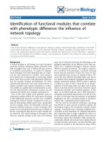
Heatmap of SAGE tag counts for genes with known expression profiles in pancreas developmentFigure 1
Heatmap of SAGE tag counts for genes with known expression profiles in
pancreas development. Tags for genes with well characterized expression
profiles in pancreas development were identified and their normalized
counts obtained in each of the ten SAGE libraries created. A heatmap,
generated using the multi-experiment viewer as described in the Materials
and methods, of these results is shown based on the counts of the tags per
hundred thousand (TPH). SAGE tags used include:
TACACGTTCTGACAACT (Nkx2-2); AAGTGGAAAAAAGAGGA
(Pdx1); TAGTTTTAACAGAAAAC (Foxa2); ACCTTCACACCAAACAT
(Hnf4a); AATGCAGAGGAGGACTC (Neurod1);
CAGGGTTTCTGAGCTTC (Neurog3); TCATTTGACTTTTTTTT (Isl1);
GATTTAAGAGTTTTATC (Pax6); CAGCAGGACGGACTCAG (Pax4);
CAGTCCATCAACGACGC (Ptf1a); AGAAACAGCAGGGCCTG
(Bhlhb8); GACCACACTGTCAAACA (Cpa1);
CCCTGGGTTCAGGAGAT (Ctrb1); TTGCGCTTCCTGGTGTT (Ela1);
ACCACCTGGTAACCGTA (Gcg); GCCGGGCCCTGGGGAAG (Ghrl);
CTAAGAATTGCTTTAAA (Iapp); GCCCTGTTGGTGCACTT (Ins1);
TCCCGCCGTGAAGTGGA (Ins2). The libraries shown include: Pdx1
EGFP+ TS17 (P+ TS17); Pdx1 EGFP+ TS19 (P+ TS19); Neurog3 EGFP-
TS20 (N- TS20); Neurog3 EGFP+ TS20 (N+ TS20); Neurog3 EGFP+ TS21
(N+ TS21); Neurog3 EGFP+ TS22 (N+ TS22); whole pancreas TS22
(WTS22); whole pancreas TS26 (WTS26); adult isolated ducts (Ducts);
adult isolated islets (Islets).
P+TS17
P+TS19
N-TS20
N+TS20
N+TS21
N+TS22
WTS22
WTS26
Ducts
Islets
Transcription factors expressed in pancreas
epithelial progenitors and endocrine cell types
Transcription factors expressed in
endocrine cell types
Transcription factors expressed in
exocrine cell types
0
TPH
20
Markers of mature exocrine cells
Markers of mature endocrine cells
0
1,000
TPH
Nkx2-2
Pdx1
Foxa2
Hnf4a
Neurod1
Neurog3
Isl1
Pax6
Pax4
Ptf1a
Bhlhb8
Cpa1
Ctrb1
Ela1
Gcg
Ghrl
Iapp
Ins1
Ins2
Genome Biology 2008, Volume 9, Issue 6, Article R99 Hoffman et al. R99.5
Genome Biology 2008, 9:R99
as described [38]. Based on these results we performed a 14-
cluster analysis using the PoissonC algorithm [39] with sub-
sequent hand curation to finalize the clusters (Figure 3 and
Additional data file 2).
A summary of the clusters (Table 3) revealed that tags for
genes with similar known pancreas function cluster together.
For example, genes essential to endocrine cell specification
were predominantly found in cluster 5, pancreatic enzyme
genes in clusters 11 and 12, and islet hormone genes in cluster
13. The clusters also showed differential enrichment for Gene
Ontology (GO) and Kyoto Encyclopedia of Genes and
Genomes (KEGG) pathway terms (Table 3). Of interest, the
clusters also had distinctively different median specificities,
Table 2
Top 25 most specific transcripts in the pancreas SAGE libraries
Tag Accession/
location
Symbol Pdx1-
GFP+
(TS17)
Pdx1-
GFP+
(TS19)
Neurog3-
GFP-
(TS20)
Neurog3-
GFP+
(TS20)
Neurog3-
GFP+
(TS 21)
Neurog3-
GFP+
(TS22)
Whole
(TS22)
Whole
(TS26)
Ducts Islets MaxS
†
TCCCGCCGT
GAAGTGGA
NM_008387 Ins2 0* 0.31 0 13.11 57.43 3,298.9 4.07 139.28 1,422.4 2,2471.19 62.72
TTCTGTCTG
GGCTTCCT
NM_023333
2210010
C04Rik
0 0 0 0 0 0 0 77.65 651.97 109.03 33.43
GCCCTGTTG
GTGCACTT
NM_008386
Ins1 0 2.83 0 45.56 19.59 839.25 6.11 9.86 207.52 3,116 27.90
TTAGGAGGC
TGCTGCTG
NM_026925
Pnlip 0 0 0 0 0 0 0 0 1,760.99 116.04 18.10
CCCTGGGTT
CAGGAGAT
NM_025583
Ctrb1 0 0.31 0 0 31.21 74.36 17.31 3,162.83 1,443.41 385.12 18.05
GCCCTGTGG
ATGCGCTT
NM_008387
Ins2 0 0 0 0 0.33 16.27 0 0 15.96 432.14 17.58
GTGTGCGCT
GGTGGCGA
NM_007919
Ela2 0 0 0 0 0 0 0 69.03 181.48 4 11.75
GCATCGTGA
GCTTCGGC
NM_007919
Ela2 0 0 0 0 0 2.32 0 1,329.96 2,680.13 1,156.37 11.24
GTGTGCGCC
GGCGGCGA
NM_026419
Ela3 0 0 0 0 0 1 1.02 636.02 369.67 23.01 11.14
ACCACCTGG
TAACCGTA
NM_008100
Gcg 7.5 63.26 0.65 2,554.97 1,952.71 550.42 34.63 25.88 124.34 326.1 10.99
AAAGTATGC
AAATAGCT
NM_026918
1810010
M01Rik
0 0 0 0 0 0 0 194.75 934.27 459.15 9.90
CAGACTAAG
TACCCATA
NM_009885
Cel 0 0 0 0 0.66 1 0 750.65 375.55 16.01 8.81
TTTTACTTCT
AAGAGTC
NM_021331
G6pc2 0 0 0 0.31 0 3.32 0 0 5.88 221.07 7.74
CCCGGGTGC
AAGAAGAA
NM_018874
Pnliprp1 0 0 0 5.93 12.62 18.26 16.3 1,135.22 250.37 8 7.40
TCCCTTCAA
CCTTAGAC
NM_011271
Rnase1 0 0 0 0 0 0.33 0 221.87 1,249.33 170.05 6.48
TTAAACCAG
AGTTCATA
NM_023333
2210010
C04Rik
0 0 0 0 0 0 0 0 10.08 0 5.66
GCCTACAAC
TAAACTGT
NM_023182
Ctrl 0 0.31 0 0 0 0 0 27.12 491.5 195.06 5.46
GCACCAAGT
ACACATAT
NM_029706
Cpb1 0 0 0 0 0 0 0 303.22 209.2 21.01 5.11
TTGCGCTTC
CTGGTGTT
NM_033612
Ela1 0 0 0 0 0 0 0 0 8.4 0 4.93
TGGGAGTGG
AGGATGCC
NM_026925
Pnlip 0 0 0 0 0 0 0 0 29.41 9 4.83
TTCCAAGTG
GAGGAGGT
NM_018874
Pnliprp1 0 0 0 0.31 0 0 10.18 163.93 36.97 1 4.78
CTAAGAATT
GCTTTAAA
NM_010491
Iapp 0 0.31 0 3.43 6.64 49.8 0 2.47 25.21 170.05 4.50
CAGTCCATC
AACGACGC
NM_018809
Ptf1a 0 0 0 0 0 0 7.13 0 0 0 4.36
CAAAGAATG
CAATCTGA
nt_039700
0 0 0 0 0 0 7.13 0 0 0 4.36
CTTGCAGTC
TGAGTTCG
nt_039413
0 0 0 0 0 0 7.13 0 0 0 4.36
*Tag counts are shown as tags per 100,000. This indicates the total number of times a given SAGE tag appears in the library per 100,000 tags and is used to normalize for
libraries of varying size.
†
S is the specificity of the tag. Specificity is calculated as described in the Materials and methods. The maximum S in any one of the libraries created
here is indicated.
Genome Biology 2008, 9:R99
Genome Biology 2008, Volume 9, Issue 6, Article R99 Hoffman et al. R99.6
with cluster 5 containing genes with the highest median S, fol-
lowed by cluster 13. These two clusters are enriched in genes
in the mature onset diabetes of the young KEGG pathway and
contain many endocrine specific factors, and this reflects the
specialized nature of these cells. Cluster 14 had the lowest
median S and the flattest expression profile of the clusters. In
sum, these data suggested that the clusters represented bio-
logically distinct gene sets.
Validation of SAGE tag clusters
To validate the identified clusters, we first compared our data
to lists of genes determined to be enriched in pancreatic pro-
genitors, endocrine cells, or islets using Affymetrix microar-
ray analysis of Pdx1 EGFP+ and Neurog3 EGFP+ cells and
islet tissues, similar to those used here [22]. There were 107
genes present in both genes sets and the representation of
each enrichment group from the array analysis in our clusters
calculated (Additional data file 3). Of the 29 genes identified
as enriched in pancreatic progenitors in the microarray anal-
ysis, we identified 13 of these in clusters 1-3 or cluster 9 that
show peak expression early in pancreas development.
Another 11 were found in clusters 10 and 11 that show peak
expression in the TS26 whole pancreas library or the duct
library, stages and tissue types that were not used in the array
analyses. Of 24 genes identified in the array study as enriched
in endocrine cells, 19 were found in cluster 5, with 2 more in
cluster 4, both of which show peak expression in the Neurog3
EGFP+ libraries here. Of the genes identified as islet enriched
in the array studies, 16 of 54 were classified as such in our
study; a further 20 were found in clusters 11 and 12 that have
Specificity threshold accurately predicts spatial expression restrictionFigure 2
Specificity threshold accurately predicts spatial expression restriction. A plot of specificity (S) versus cumulative tag types represented shows the
distribution of tags into tags with high (S > 0.1; top), medium (0.001 > S < 0.1, middle), and low (S < 0.001, bottom) S values. Representative in situ
hybridization staining patterns from TS22 whole embryo saggital sections obtained from GenePaint are shown for each specificity group. Relevant GenePaint
probe IDs can be found in Additional data file 4. Arrows indicate the location of the pancreas (p).
S=0.0002 S=0
S=0.0006
Maximum S
Cummulative tag types
represented
1,500
1,000
500
0
10
-6
10
-5
10
-4
10
-3
10
-2
10
-1
10
0
10
1
10
2
Maximum S
Cummulative tag types
represented
1,500
1,000
500
0
10
-6
10
-5
10
-4
10
-3
10
-2
10
-1
10
0
10
1
10
2
Maximum S
Cummulative tag types
represented
1,500
1,000
500
0
10
-6
10
-5
10
-4
10
-3
10
-2
10
-1
10
0
10
1
10
2
S=0
S=0.212
S=0.059
S=0.011
S=0.005
S=27.90
S=11.14
S=10.99
S=4.78
Zfp385
Jmjd3
Mfsd1
Hmgb1
Sfrp1
Rbp4
Foxa2
Onecut1
p
p
p
p
p
p
p
p
p
p
p
p
Ins1 Ela3
Pnliprp1Gcg
Genome Biology 2008, Volume 9, Issue 6, Article R99 Hoffman et al. R99.7
Genome Biology 2008, 9:R99
peak expression in the ducts, again a tissue not represented in
the array studies; and a further 10 were found in clusters 5 or
8 that show peak expression in the Neurog3 EGFP+ libraries
and islet library, respectively. Overall, the two data sets com-
pare well and the majority of genes were identified as
enriched in the same cell populations, although the differ-
ences in the tissues used in each study, specifically our inclu-
sion of developing whole pancreas and adult duct libraries,
did cause differences in some of the results.
To further confirm that our clusters accurately group genes
with similar temporal expression profiles, we analyzed the
expression of 44 genes through pancreas development using
qRT-PCR. Selected targets included Ins2, Nkx2-2, Pdx1,
Neurog3, Amy1, and Ptf1a, which all have well established
expression profiles as references. We then used a self-organ-
izing tree algorithm (SOTA) clustering analysis to group the
obtained temporal expression profiles for these genes. This
allowed us to determine if groupings similar to those found in
Median plots of identified SAGE tag K-means cluster analysis using 14 clustersFigure 3
Median plots of identified SAGE tag K-means cluster analysis using 14 clusters. We clustered 2, 536 SAGE tags with a count greater than 4 in one of the
SAGE libraries and with a minimum specificity of 0.002 and that map unambiguously to a specific transcript or genome location into 14 clusters using K-
means clustering using a PoissonC algorithm as described in the Materials and methods. The median normalized tag counts for the tags in each of the
clusters is shown plotted against the indicated SAGE libraries. The libraries shown include: Pdx1 EGFP+ TS17 (P+ TS17); Pdx1 EGFP+ TS19 (P+ TS19);
Neurog3 EGFP- TS20 (N- TS20); Neurog3 EGFP+ TS20 (N+ TS20); Neurog3 EGFP+ TS21 (N+ TS21); Neurog3 EGFP+ TS22 (N+ TS22); whole pancreas
TS22 (WTS22); whole pancreas TS26 (WTS26); adult isolated ducts (Ducts); adult isolated islets (Islets). A full list of the tags, the cluster they belong to,
and their counts in each of the libraries is shown in Additional data file 2.
Cluster 1 Cluster 2 Cluster 3 Cluster 4
Cluster 5 Cluster 6 Cluster 7 Cluster 8
Cluster 9 Cluster 10 Cluster 11 Cluster 12
Cluster 13 Cluster 14
1.0
0.8
0.6
0.4
0.2
0.0
P+TS17
P+TS19
N-TS20
N+TS20
N+TS21
N+TS22
WTS22
WTS26
Ducts
Islets
1.0
0.8
0.6
0.4
0.2
0.0
1.0
0.8
0.6
0.4
0.2
0.0
1.0
0.8
0.6
0.4
0.2
0.0
1.0
0.8
0.6
0.4
0.2
0.0
P+TS17
P+TS19
N-TS20
N+TS20
N+TS21
N+TS22
WTS22
WTS26
Ducts
Islets
P+TS17
P+TS19
N-TS20
N+TS20
N+TS21
N+TS22
WTS22
WTS26
Ducts
Islets
P+TS17
P+TS19
N-TS20
N+TS20
N+TS21
N+TS22
WTS22
WTS26
Ducts
Islets
P+TS17
P+TS19
N-TS20
N+TS20
N+TS21
N+TS22
WTS22
WTS26
Ducts
Islets
P+TS17
P+TS19
N-TS20
N+TS20
N+TS21
N+TS22
WTS22
WTS26
Ducts
Islets
P+TS17
P+TS19
N-TS20
N+TS20
N+TS21
N+TS22
WTS22
WTS26
Ducts
Islets
P+TS17
P+TS19
N-TS20
N+TS20
N+TS21
N+TS22
WTS22
WTS26
Ducts
Islets
P+TS17
P+TS19
N-TS20
N+TS20
N+TS21
N+TS22
WTS22
WTS26
Ducts
Islets
P+TS17
P+TS19
N-TS20
N+TS20
N+TS21
N+TS22
WTS22
WTS26
Ducts
Islets
P+TS17
P+TS19
N-TS20
N+TS20
N+TS21
N+TS22
WTS22
WTS26
Ducts
Islets
P+TS17
P+TS19
N-TS20
N+TS20
N+TS21
N+TS22
WTS22
WTS26
Ducts
Islets
P+TS17
P+TS19
N-TS20
N+TS20
N+TS21
N+TS22
WTS22
WTS26
Ducts
Islets
P+TS17
P+TS19
N-TS20
N+TS20
N+TS21
N+TS22
WTS22
WTS26
Ducts
Islets
1.0
0.8
0.6
0.4
0.2
0.0
1.0
0.8
0.6
0.4
0.2
0.0
1.0
0.8
0.6
0.4
0.2
0.0
1.0
0.8
0.6
0.4
0.2
0.0
1.0
0.8
0.6
0.4
0.2
0.0
1.0
0.8
0.6
0.4
0.2
0.0
1.0
0.8
0.6
0.4
0.2
0.0
1.0
0.8
0.6
0.4
0.2
0.0
1.0
0.8
0.6
0.4
0.2
0.0
Genome Biology 2008, 9:R99
Genome Biology 2008, Volume 9, Issue 6, Article R99 Hoffman et al. R99.8
the SAGE data cluster analysis were observed. In our SOTA
analysis, genes with four distinct expression profiles were
identified (Figure 4): one group with peak expression in the
islet sample, one with peak expression in the TS26 whole pan-
creas, one with peak expression from TS21-TS26, and one
with peak expression in the ducts sample. All of the genes in
the SOTA groups containing Ins2, Mafa, Pdx1, and Nkx2-2,
which are markers of the endocrine lineage, were from
clusters 1, 4, 5, and 13. Three of the six genes in the SOTA
group with peak expression at TS26 were from clusters 4 and
5, although each of these showed relatively high expression in
either the TS22 or TS26 whole pancreas libraries. Of the
Table 3
Summary of SAGE tag K-means cluster data
Cluster Number of
tags in the
cluster
Number of
genes in the
cluster
Number of
genome maps
in the cluster
Number
assessed by
GenePaint*
Number
assessed by
QPCR
Median S
†
Previously
characterized
genes in the
cluster
Selected GO categories
and KEGG pathways
enriched in the cluster
‡
1 154 85 61 37 3 0.0079 Nkx6-2 Transcriptional activator
activity p = 0.02;
development p = 0.049
24929191520.0037 Metabolism p = 0.01; cell
organization and
biogenesis p = 0.035
35840151420.0044 Receptor activity p =
0.028; development p =
0.030
4 292 115 175 45 4 0.00895 Hes6, Pdx1, Sox9 Regulation of
transcription p = 0.027;
maturity onset diabetes
of the young p = 0.002
5 1,008 427 542 175 13 0.03555 Arx, Gcg, Ghrl, Iapp,
Isl1, Nkx2-2, Myt1,
Neurog3, Neurod1,
Pax4, Pax6, Pou3f4,
Pyy
Secretory pathway p <
0.001; hormone activity
p = 0.049; maturity
onset diabetes of the
young p < 0.001
66041171610.00465
7 21 11 10 7 1 0.008
87846282950.012Pax6 Eye morphogenesis p =
0.020; type II diabetes
mellitus p = 0.001
9 23 16 6 10 2 0.0041 Cell proliferation p =
0.028
10 401 281 107 122 4 0.0158 Id2 Response to
endogenous stimulus p =
0.021
11 76 57 10 23 1 0.00555 Amy1, Cel, Clps,
Ela1, Pnliprp2
,
Reg1
Protein catabolism p =
0.002
12 154 122 13 56 3 0.0074 Ela1, Pnlip, Reg3d Growth factor binding p
= 0.005;
carboxypeptidase
activity p = 0.013;
regulation of cell growth
p = 0.027
1313684423030.01835Iapp, Ins1, Ins2 Secretion p = 0.03;
maturity onset diabetes
of the young p < 0.001;
type II diabetes mellitus
p < 0.001; type I diabetes
mellitus p = 0.003
14 56 47 4 22 0 0.00335 Protein metabolism p =
0.020
*Refers to the number of genes analyzed by in situ hybridization using GenePaint [62] on TS22 whole embryo cryo-sections that gave informative
staining.
†
S is the specificity of the tag. Specificity is calculated as described in the Materials and methods.
‡
GO term enrichments and p-values were
calculated using EASE while KEGG pathway enrichments and p-values using Webgestalt as described in the Materials and methods.
Genome Biology 2008, Volume 9, Issue 6, Article R99 Hoffman et al. R99.9
Genome Biology 2008, 9:R99
genes in the SOTA group with peak expression from TS21-
TS26, one was from cluster 3, two were from cluster 5 and one
was from cluster 9. Clusters 3 and 9 are enriched in mesen-
chymal factors (see below). Since no mesenchymal cells
should be present in the islet and duct samples, it makes
sense for these genes to have this expression profile. Two
genes from cluster 5 were in this SOTA group, including
Neurog3, which is known to be developmentally restricted in
expression, and Gast, likely reflecting the relative number of
Gastrin-producing cells in the different samples. Of the 11
genes in the SOTA group with peak expression in the ducts
sample, 4 were from clusters 7 and 12, while the rest were
found in the other clusters, although significantly excluding
clusters 13 and 8. All of the genes in this group had counts in
the duct library, despite being in clusters with peak expres-
sion in other libraries, although they all had, in general, low
overall tag counts.
GenePaint analysis
Taken together, the data suggested that the generated clusters
represent transcript sets with distinct roles in pancreas devel-
opment. To further confirm this, we assessed whether the
transcripts identified in each of the SAGE tag clusters had
spatial expression profiles consistent with these roles using
the GenePaint database [27,28]. For each of the 923 genes
present in our clusters and in the GenePaint database, we
analyzed the in situ hybridization staining pattern in the pan-
creas from TS22 whole embryo sections. In sum, 601 of the
genes showed informative staining, and these were catego-
rized based on their staining patterns into one of five expres-
SOTA clustering of temporal expression profiles from qRT-PCR analysis of 44 genes in pancreas developmentFigure 4
SOTA clustering of temporal expression profiles from qRT-PCR analysis of 44 genes in pancreas development. qRT-PCR was used to determine the
relative expression levels of the indicated genes during pancreas development at the TSs indicated. The relative level of expression of each gene was
normalized and a SOTA analysis used to group the genes. Heatmaps of the relative expression levels of the genes in the SOTA groups, including the SOTA
centroid, with peak expression in (a) the islets, (b) the TS26 developing pancreas, (c) the TS21-TS26 developing pancreas, or (d) the ducts are shown.
The data shown are averages of the results obtained from pancreases from three separate litters (pancreases from an individual litter were pooled) or
islet/duct collections with triplicate reactions from the separate RNA extractions.
Sfrp5
Crabp2
Cryab2
AI987662
Rbp4
Irx2
Abcc8
Insrr
Mlxipl
Myt3
Syt14
Rgs11
BC038479
Ins2
Mafa
Pdx1
Nkx2-2
Habp2
Cdkn1a
Tle6
Nr2f6
Ptf1a
Amy1
Onecut2
Fusip1
Rbp1
Fh1
Ambp
F11r
Tekt2
St14
E430002G05Rik
Nkx2-3
Rbpjl
P2rx1
Hhex
Clu
Dusp1
Arx
Gast
Cdkn1c
Neurog3
Sfrp1
Slc38a5
TS19
TS21 TS23 TS26
Ducts
Islets
TS19
TS21 TS23 TS26
Ducts
Islets
TS19
TS21 TS23 TS26
Ducts
Islets
TS19
TS21 TS23 TS26
Ducts
Islets
SOTA centroid
SOTA centroid
SOTA centroid
SOTA centroid
0.0
0.24
Relative expression level
(a) (b)
(c)
(d)
Genome Biology 2008, 9:R99
Genome Biology 2008, Volume 9, Issue 6, Article R99 Hoffman et al. R99.10
sion domains found in the pancreas [40] (Figure 5). For the
remaining 316 genes, either the probes did not show stain in
any sections or sections with pancreas were not present in the
database. Regardless, we identified 88 genes expressed in the
tips of epithelial branches that at E14.5 primarily contain exo-
crine progenitor cell types. A further 81 genes were identified
as expressed in the trunk of the epithelial branches that con-
tains endocrine and ductal progenitor cells; 221 genes were
identified as expressed throughout the epithelium; and a fur-
ther 51 were found only in the mesenchyme, and 42 in the vas-
culature. For a full categorization of the genes see Additional
data file 4. There were 124 (13%) genes identified in our SAGE
data that were not detected in the pancreas at the time point
assessed. The average tag count for these genes was only 6.8
while for detected genes it was 24, suggesting this is, in part,
due to the low expression levels of these genes. Moreover, the
Representative in situ staining patterns for genes expressed in each of the identified expression profilesFigure 5
Representative in situ staining patterns for genes expressed in each of the identified expression profiles. Representative genes for each of the identified
spatial expression profiles, including genes with known and previously un-described, or novel, staining profiles in pancreas development, are shown. For
this, images of in situ hybridization staining patterns for whole embryo sagittal sections were obtained from the GenePaint website and magnified to show
the pancreas (outlined in red). Relevant GenePaint probe IDs can be found in Additional data file 4.
Ins2
Gcg
Slc38a5
Cryab2
AI987662
Pam
Ela3b
Pnliprp1
P2rx1
Cckar Ctrb1
Rbpjl
Sox9
Foxa2
Tle6
Tacstd1
Serpina1a
Ambp
Sfrp1
Syt6
Cdkn1
Akap12
Ets1
Prrx1
Slc4a1
Anxa3
Hbb-y
Centd3
El
a3
b
P
n
li
prp1
P2rx1
C
cka
r
C
trb1
Rbpj
l
S
ox
9
Fox
a2
Tl
e
6
T
acst
d
1
S
er
p
ina1
a
A
mb
p
S
frp1
S
yt
6
C
dkn
1
Ak
ap1
2
E
ts1
P
rrx1
S
lc4a1
Anxa
3
H
bb-
y
C
entd
3
Trunk
Tip
Epithelial
Mesenchymal
Vasculature
Known Novel
Genome Biology 2008, Volume 9, Issue 6, Article R99 Hoffman et al. R99.11
Genome Biology 2008, 9:R99
number of genes not detected was highest in clusters 1, 2 and
13, which include genes that show low relative expression at
TS22.
Analysis of the representation of each of the five staining pro-
files in the 14 identified SAGE tag clusters (Figure 6) revealed
that some clusters are far more predictive of specific expres-
sion profiles than others. For example, clusters 9 and 12 were
not at all predictive of a given staining pattern. In contrast,
73% of the genes analyzed in cluster 4 showed pan-epithelial
staining, while 59% of those in cluster 8 showed trunk stain-
ing and a further 24% showed pan-epithelial staining. These
data suggest that several of the clusters represent genes with
distinct spatial expression profiles. Significantly, these pro-
files are consistent with the known roles of genes within the
clusters. For example, cluster 5, which is enriched in genes
involved in mature onset diabetes of the young, contains tags
with peak expression in the Neurog3 EGFP+ libraries and
genes in this cluster predominately show pan-epithelial or
trunk expression. It is apparent from these results, in
combination with the median profiles of the clusters (Figure
3), that clusters 1, 2, and 4 represent genes appropriately
expressed spatially and temporally to be functionally signifi-
cant in pancreatic progenitor cells, while genes in clusters 4,
5, and 8 are likely functionally significant in endocrine pro-
genitors, genes from clusters 6, 10, and 11 in exocrine progen-
itors, genes from cluster 3 in mesenchymal cells, and genes
from clusters 8 and 13 in adult islet cells. Taken together,
these analyses allowed us to identify lists of transcripts, many
of which have not previously been characterized in pancreas
development, that are appropriately expressed spatially and
temporally to play functionally significant roles in each of the
major phases of pancreas development.
Identification of a transcriptional cascade in endocrine
pancreas development
Transcriptional regulators, in concert with signaling factors,
provide the genetic instructions that drive endocrine
pancreas development. Based on our analyses, genes in the
endocrine lineage (that is, from pancreatic progenitor cells
through endocrine progenitors to adult islet cells) can be
identified based on their presence in clusters 1, 2, 4, 5, 8 and
13. We identified 58 tags for transcriptional regulators in
these clusters representing 43 different factors. Eliminating
factors only expressed in one library, and for which we could
not find additional support for their expression, left 38 differ-
ent factors for which there is good evidence of their expres-
sion in the endocrine pancreas lineage. Eight of these genes
were represented by multiple tag types, with four separate
tags mapping to Neurod1 and Isl1. Figure 7a shows a heatmap
of the expression of these factors in endocrine cell develop-
ment as detected in our SAGE data. Many of these factors
have well-established roles in pancreas development, includ-
ing Ngn3, Pax4, Nkx2-2, Pdx1, Isl1, and Neurod1. However,
several factors with uncharacterized roles were also identi-
fied, including: Tcf12, Zfp326 and Meox1, which were most
abundant in Pdx1 EGFP+ cells; Zfp446, Rnf6, and Son, which
were most abundant in Neurog3 EGFP+ cells; and Nr1d1,
Myt3, and Bcl6b, which were most abundant in islet cells.
The transcriptional circuits essential to β-cell development
are only beginning to be elucidated [8,41] and the majority of
regulatory interactions driving pancreas development remain
unknown. Expression profiling has been used extensively to
generate networks with co-expressed genes 'linked' using the
assumption that co-expression implies co-regulation [42-45].
The promoters of co-expressed genes are often then analyzed
The percent association of genes in each K-means cluster with the five expression domains in the pancreasFigure 6
The percent association of genes in each K-means cluster with the five expression domains in the pancreas. The in situ hybridization staining profiles of 605
genes with informative stain in TS22 pancreas tissue were classified into the groups shown using the GenePaint database. Additional data file 4 lists the full
categorization of each of these genes. The percentage of genes with each staining profile in each of the SAGE tag K-means cluster is shown.
Trunk
Tip
Epithelial
Vasculature
Mesenchymal
Not detected
80
60
40
20
0
1234567891011121314
Cluster
Percentage association
Genome Biology 2008, 9:R99
Genome Biology 2008, Volume 9, Issue 6, Article R99 Hoffman et al. R99.12
Figure 7 (see legend on following page)
Fold enrichment (/IgG)
Fold enrichment (/IgG)
250
200
150
100
50
0
Foxa2
Pdx1
Myt1
Myt3
Neurod1
Nkx2-2
Nkx6-1
NegF1
NegF2
Foxa2
Ins1
15
10
5
0
Pdx1
Myt1
Neurod1
Nkx2-2
Nkx6-1
NegP1
NegP2
(d)
Hoxb6
Tcf12
Nkx2-2 Pdx1
Foxa2
Foxa1
Nkx6-1
Nkx6-2
Meox1
Hes6
Hoxb5
Hnf4a
Onecut1
Hes1
Arid3a
Neurog3
Nkx2-2
Pdx1
Myt1
Myt3
Foxa2
Isl1
Nkx6-1
Sox9
Hnf4a
Neurod1
Arx
Pou3f4
Meox1
Hes6
Pax4
Pax6
Fos
Mlxipl
Foxa3
Mafb
Mlxipl
Nkx2-2
Pax4
Pax6
Myt1
Mafa
Pdx1
Myt3
Foxa2
Nkx6-1
Neurod1
Ins1
Ins2
(a)
(c)
(b)
0
0.5
Relative
expression level
P+TS17
P+TS19
N+TS20
N+TS21
N+TS22
Islets
Hoxb5
Hoxb6
Tcf12
Zfp326
Meox1
Onecut2
Nkx6-2
Onecut1
Onecut2
Onecut2
Foxa1
Nkx6-2
Sox9
Isl1
Son
Isl1
Zfp446
Elf3
Arid3a
Rnf6
Preb
Sox9
Neurod1
Asb4
Pou3f4
Hes6
Isl1
Pax4
Hes6
Isl1
Mafb
Foxa3
Nkx2-2
Myt1
Foxa2
Fev
Neurog3
Neurod1
Fos
Hnf4a
Arx
Rorc
Rorc
Neurod1
Neurod1
Myt3
Pax6
Mlxipl
Pax6
Bcl6b
Nr1d1
PP
EP
β-cell
Genome Biology 2008, Volume 9, Issue 6, Article R99 Hoffman et al. R99.13
Genome Biology 2008, 9:R99
for common sequence elements and the presence of known
binding sites. One advancement on this technique is to
include analysis of sequence conservation under the elements
to better predict 'real' sites [46]. Such methods have been suc-
cessfully applied to mammalian cells to identify networks
active in B-cells and during macrophage activation
[44,45,47]. Therefore, to similarly gain further insight into β-
cell development, we utilized our transcription factor cascade
as it represents sets of co-expressed transcription factors.
For this, we first eliminated factors for which we could not
find direct evidence of expression in the cell types in the β-cell
lineage, that is, pancreas progenitors, endocrine progenitors,
and mature β-cells, using the GenePaint in situ data, litera-
ture, or the EPConDB/T1Dbase [48], or as mentioned above
were found in only one library. We then searched the proxi-
mal promoters of each of the remaining transcription factors,
using the CisRED database of highly conserved sequence ele-
ments in the proximal promoters of genes from multiple spe-
cies [49], for binding sites of co-expressed transcription
factors or of factors expressed in the parent cell type. From
this we generated predicted transcription factor interaction
lists. Overall, our predictions identified 28% of transcription
factor interactions reported in the literature [8], with some
binding sites less predictive than others. Randomly generated
interaction lists using the same transcription factors identi-
fied only 3% of literature reported sites, indicating that our
interaction predictions are significantly enriched for true
sites (p = 5.7 × 10
-6
using Fisher's exact test). These predicted
interactions, as well as literature reported interactions, were
then used to build a transcriptional network describing endo-
crine pancreas development (Figure 7b). In sum, a total of 217
novel predicted interactions are shown in this network, sug-
gesting that it is a rich source of hypotheses for future valida-
tion. To validate this and to gain an understanding of the false
positive rate associated with our interaction predictions, we
utilized ChIP-qPCR to validate predicted targets of Foxa2 and
Pdx1 in the β-cell portion of the network using Min6 cells. For
Foxa2 we found high levels of enrichment for predicted sites
in the promoters of Pdx1, Myt1, Myt3, and Nkx2-2, but not for
predicted sites in the promoters of Foxa2, Neurod1, or Nkx6-
1 (Figure 7c). For Pdx1 modest levels of enrichment were
achieved for sites in the promoters of Ins1, Pdx1, Myt1,
Neurod1, Nkx2-2, and Nkx6-1, although no enrichment was
seen for a predicted site in the Foxa2 promoter (Figure 7d).
No enrichment was seen at either of two sites that were not
predicted to contain a binding site for either Foxa2 or Pdx1. In
total, 10 out of 14 (71%) predicted interactions proved valid,
with 4 of 7 (57%) validating for Foxa2 and 6 of 7 (86%) for
Pdx1.
Discussion
Taken together, our data suggest that the pancreas libraries
generated and analyzed in this study accurately reflect the cell
types intended and are highly comprehensive. Our analysis of
the tag distribution of genes with well characterized expres-
sion profiles in the libraries confirmed their known expres-
sion. As well, 79% of the genes we identified, after instituting
a count threshold, have independent validation of pancreas
expression via EST information. Of the genes we analyzed
using the GenePaint database, 20% were not detected, com-
parable with the expected detection rate of this technique,
with this number as low as 13% for clusters with expression at
TS22, the time point analyzed. It has been suggested that a
SAGE library of 120,000-160,000 tags is roughly equivalent
in sensitivity to a microarray [50]. Six of the ten libraries gen-
erated here were sequenced to greater than 300,000 tags per
library with the remaining four sequenced to approximately
100,000 tags per library. It is clear that these libraries are a
rich source of information on the expression profiles of a large
diversity of known and unknown transcripts throughout
pancreas development. Interestingly, multiple tags were
identified for many genes with critical roles in pancreas
development and function, including Ins1, Ins2, Isl1,
Neurod1, and Pax6; the biological relevance of this remains to
be elucidated. Additionally, we identified 1,049 tags that meet
both our count and specificity thresholds and mapped only to
the genome. Although some of these tags are likely to be error
tags, they represent a rich source of information on com-
pletely novel transcripts.
A cascade of transcription factors expressed in endocrine cell developmentFigure 7 (see previous page)
A cascade of transcription factors expressed in endocrine cell development. Tags that mapped unambiguously to transcription factors and met our count
and specificity thresholds were identified and their normalized counts obtained in each of the SAGE libraries that represent the development of the
endocrine lineage. (a) A heatmap, generated using the multi-experiment viewer as described in the Materials and methods, of these results is shown. Tags
are organized by the order of their expression in the libraries. The libraries shown include: Pdx1 EGFP+ TS17 (P+ TS17); Pdx1 EGFP+ TS19 (P+ TS19);
Neurog3 EGFP+ TS20 (N+ TS20); Neurog3 EGFP+ TS21 (N+ TS21); Neurog3 EGFP+ TS22 (N+ TS22); adult isolated islets (Islets). (b) A transcription
factor network in β-cell development. The network was generated as described in the materials and methods using Biotapestry to visualize the network
[65]. Ins1 and Ins2 are shown as the final products expressed in mature β-cells. Literature reported interactions [8,41] are included with arrow heads
indicating positive regulation and perpendicular lines repression. Interactions in pancreatic progenitors (PP), endocrine progenitors (EP) and mature β-cells
are shown. (c) The fold enrichment of predicted Foxa2 binding sites, as detected by qPCR, in the promoters of Pdx1, Myt1, Myt3, Neurod1, Nkx2-2, Nkx6-
1, and of two sites not predicted to contain a Foxa2 site (NegF1 and NegF2) in ChIP experiments using an anti-Foxa2 antibody compared to IgG. (d) The
fold enrichment of predicted Pdx1 binding sites, as detected by qPCR, in the promoters of Foxa2, Ins1, Myt1, Neurod1, Nkx2-2, Nkx6-1, and of two sites not
predicted to contain a Pdx1 site (NegP1 and NegP2) in ChIP experiments using an anti-Pdx1 antibody compared to IgG. The data shown are averages of the
results obtained from three separate ChIP experiments each with duplicate reactions. Error bars indicate the standard deviation of the averaged replicates.
Genome Biology 2008, 9:R99
Genome Biology 2008, Volume 9, Issue 6, Article R99 Hoffman et al. R99.14
To fully appreciate the data generated in this study it is
important to consider the cell types the libraries represent.
Two of the libraries generated use a transgenic mouse strain
that expresses EGFP under control of the Pdx1 promoter,
while four libraries were generated using a transgenic strain
expressing EGFP under control of the Neurog3 promoter
[22]. Prior to the secondary transition, Pdx1 marks all pan-
creas epithelial cells, with the exception of rare glucagon-
expressing cells. At the stages used here, TS17 and TS19, the
collected cells therefore represent a relatively pure popula-
tion of pancreas epithelial progenitor cells, which will eventu-
ally give rise to all subsequent endocrine and exocrine cell
types in the pancreas. Pdx1 is also expressed in the duodenum
at these time points and some level of duodenum specific
transcripts may also be present, although care was taken in
the dissections to minimize this. Neurog3 is extremely spe-
cific in its expression and in the pancreas is exclusively
expressed in endocrine precursors, the cells that give rise to
all of the hormone-producing cell types in the pancreas.
Neurog3 expression itself is transient and decreases
substantially after TS23 [17]. However, the EGFP protein is
relatively stable and continues to mark endocrine cells as late
as TS26 [26]. Thus, the Neurog3 EGFP+ libraries collected
here, from TS20 to TS22, represent a mixture of endocrine
progenitor cell-types, predominantly α and β cell precursors,
in various stages of differentiation and maturation. The
Neurog3 EGFP- TS20 library contains a variety of pancreas
cell types, including mesenchymal cells, as well as epithelial
and exocrine progenitors. Similarly, the TS22 and TS26
whole pancreas libraries are composed of various cell types, at
TS22 predominantly mesenchymal cells and various epithe-
lial cell types, including endocrine and exocrine progenitors,
while at TS26 a greater relative abundance of exocrine cells is
found. The Islet and Duct libraries represent fully mature cell
types. These libraries were constructed from hand-picked
islets and ducts isolated by collagenase digestion and gradient
centrifugation and, as such, contain a minimum of contami-
nating exocrine cells. The Islet library is predominantly com-
posed of β-cells that compose approximately 80% of a rodent
islet [51], with α-cells composing much of the remaining cells,
with smaller numbers of δ, ε, and PP cells also present. For
the Duct library, epithelial cells from duct tips were collected,
which have been proposed to harbor pancreas stem cells [52],
although transcripts from contaminating exocrine and islet
cells were inevitably identified as well.
One of the major strengths of SAGE data is the ability to easily
compare data amongst different libraries. We took advantage
of this strength and were able to institute a specificity-based
threshold for our data by obtaining the counts for tags that
met our count threshold in 205 different SAGE libraries. In
general, metrics for specificity assess the level of expression
in the tissue of interest versus the total expression. One study
used a metric based on Shannon entropy to account for bias
from differing levels of expression in different tissues [53].
This metric is not, however, directly applicable to SAGE data
due to the large number of tags with no counts in many sam-
ples. Here we use a metric that accounts for the relative
expression of a tag, the number of libraries it is found in, as
well as its absolute expression level. Thus, tags expressed at
high-count levels in the library of interest and with low levels
in very few other libraries have the highest specificity, while
tags with low counts in numerous samples have the lowest
specificity. We show that this metric performs well and that it
accurately identifies genes with relative levels of restriction
compared with available GenePaint data, and meets biologi-
cal expectation.
The use of a specificity threshold to assess data from tran-
scriptome-based approaches to pancreas development is
novel and previous studies have relied on the identification of
genes of interest based on their differential expression in pan-
creas development. However, differential expression can
occur for many reasons and many of the tags that did not
meet our specificity threshold showed large changes in count
levels between the libraries (data not shown), suggesting that
assessing differential expression alone does not screen out
many widely expressed genes and that the addition of a spe-
cificity threshold is helpful in producing gene lists with the
highest functional relevance. For example, of 30 genes iden-
tified as being the most significantly differentially expressed
from TS21-TS25 in Neurog3 EGFP+ cells using the PancChip
6 [26], 10 did not make our count cut-off and 15 did not make
our specificity cut-off. Additionally, with a few exceptions -
tags for Mafa were found at a count of only one in the islet and
TS22 Neurog3 EGFP+ library and, thus, missed our count
threshold - almost all of the transcription factors known to
play significant roles in pancreas development were present
on our lists. Thus, by instituting our count and specificity
thresholds we feel we have generated a tag list more enriched
for genes of biological significance to pancreas development
and function than previously accomplished.
Our data in general confirm the accuracy of our SAGE tag
clustering. In our qRT-PCR validations, 10 of the 44 genes
assessed had a maximum tag count of only 10 and our qRT-
PCR data did not match the SAGE data as well, for these
genes, as it did using genes with higher tag counts. Thus,
some caution in interpreting the expression profiles of tran-
scripts with low tag counts is necessary. Regardless, it was
clear that tags with even very low counts were still indicative
of actual expression. Clearly, deeper sequencing, particularly
of the libraries at 100,000 tag depth, would improve the sta-
tistical power of the observed expression differences and pro-
vide a more robust detection of rare transcripts.
Five distinct domains of expression have been identified in
the developing pancreas [40]. Although, several factors
expressed in endocrine cells, which are found in the epithelial
trunk, have been identified, relatively few factors are known
to be expressed in the remaining expression domains [8,40].
Here we identify and categorize into the 5 expression
Genome Biology 2008, Volume 9, Issue 6, Article R99 Hoffman et al. R99.15
Genome Biology 2008, 9:R99
domains the expression profiles of over 400 genes in the
developing pancreas. In sum, we provide spatial and tempo-
ral expression data on hundreds of transcripts not previously
characterized in pancreas development. Combining our
SAGE tag clustering and GenePaint data provides a clear
indication as to the stages of pancreas development at which
many of these genes are likely functionally significant. This,
and studies on retinal development [54], demonstrate the
power of combining SAGE tag clustering with large scale in
situ hybridization data.
Further, we utilized predicted and literature reported binding
data to gain insight into the cascade of transcription factors
that drives endocrine pancreas development. As already men-
tioned, our predicted interactions identified 28% of literature
reported sites. The CisRED database used to generate our
interaction lists identifies conserved sequence elements only
in proximal promoters (approximately 1 kb upstream). Of
course, not all regulatory elements are in proximal promoters
and this is likely a major reason a higher percentage of known
interactions was not identified. Future experimentation to
locate and analyze regulatory elements across the genome
will better predict all possible interactions. Optimally false
positive error rates in such networks are calculated using
comparisons with comprehensive binding data for multiple
factors. However, this type of data is not available for the
relevant transcription factors in pancreas tissues. We there-
fore attempted to gain an understanding of our false positive
rate by utilizing ChIP-qPCR to validate predicted binding
sites for Foxa2 and Pdx1. Although limited in scale, these val-
idation experiments give us a reasonable estimation of our
false positive rate as approximately 29%. However, this error
rate varied significantly from 14% for Pdx1 to 46% for Foxa2.
In part, this is due to our reliance on available binding data
for the different transcription factors in the network, and thus
the reliability of the predicted interactions is highly depend-
ent on the quality of the available binding data, which varies
significantly. It is also possible that some of the interactions
we tested do not occur in the cell line used, but do in fact occur
in vivo, although this remains to be tested. Regardless, it is a
given that better binding data for a greater number of tran-
scription factors are essential to the construction of more
accurate networks in this fashion. Despite this, it is clearly of
great interest to begin to elucidate the relevance of many of
these predicted interactions in pancreas development and
function.
Conclusion
Previous studies have used microarrays to assess expression
profiles in the developing pancreas. Here we utilized SAGE to
provide a more comprehensive and complementary analysis,
and to yield further insights into signals critical in regulating
pancreas development. By instituting a specificity threshold
for our data we have isolated a list of tags highly enriched for
genes with significant roles in pancreas development and
function. Equivalent analyses have not been performed in
previous array studies, and highlight one of the strengths of
SAGE as such comparisons are relatively straightforward
with this technique. Our data represent a highly comprehen-
sive analysis of pancreas development. Our classification of
the in situ staining patterns of over 400 genes, in combina-
tion with our cluster analysis, allowed us to identify gene sets
describing each of the major stages of pancreas development
represented by our libraries. These data provide evidence for
the pancreas enriched expression of hundreds of factors that
have not previously been implicated in pancreatic develop-
ment, suggesting that our data are a rich source of genes with
novel roles in pancreas development. Further, from our data
we constructed a predictive transcriptional network driving
endocrine pancreas development that predicts many novel
interactions that are of great interest for future analysis. In
sum, these data provide insight into the regulatory networks
driving pancreas development and function and provide a
framework for future studies.
Materials and methods
Mouse maintenance and islet isolation
All mice were bred and maintained at the British Columbia
Cancer Research Centre animal facility according to the
guidelines of the Canadian Council on Animal Care. All proto-
cols were approved by the University of British Columbia Ani-
mal Care Committee. Mice were housed in micro-isolator
units, provided with Purina mouse food and autoclaved water
ad libidum, and were maintained at 20°C ± 2°C under a light/
dark cycle (light, 05:00-19:00; dark, 19:00-05:00). Males
were mated overnight with up to three females and females
were checked for plugs before 9:30 the following morning.
Plugged mice were considered to be 0.5 days post coitum.
Generation of SAGE libraries
Embryos, obtained from timed pregnant female Pdx1 EGFP
and Ngn3 EGFP mice mated with C57Bl/6J mice, were staged
using Theiler criteria. Embryos were collected in ice cold
phosphate buffered saline (PBS) solution. The developing
pancreas was isolated by micro-dissection while the embryo
was submerged in PBS. For TS17-TS22 Pdx1 EGFP and Ngn3
EGFP FACS sorted libraries, a single cell suspension was gen-
erated by Trypsin/EDTA digestion. The sample was prepared
for sorting by staining with propidium iodide and passing
through a 40 μm filter with a final resuspension in FACS
buffer (PBS containing 2% fetal bovine serum and 2 mM
EDTA). EGFP positive or negative cells were sorted and col-
lected as appropriate into a microcentrifuge tube containing
Trizol
®
using BD FACSVantage instruments (operated by the
Terry Fox Laboratory at the BC Cancer Research Centre).
Between three and eight litters of mice were used to generate
each library, depending on the stage. Islets and ducts from at
least 10 separate mice were purified from 8-10-week-old ICR
males by collagenase digestion and gradient centrifugation as
previously described [55] and placed in Trizol
®
. Total RNA
Genome Biology 2008, 9:R99
Genome Biology 2008, Volume 9, Issue 6, Article R99 Hoffman et al. R99.16
was extracted using Trizol
®
and RNA quality was assessed
using a Bioanalyser (Agilent, Santa Clara, CA, USA). For the
TS22 and TS26 whole pancreas as well as the islet and duct
libraries roughly 10 μg of total RNA was used to construct
each SAGE library. For the FACS sorted libraries a SAGElite
approach [56] was used with a minimum of 20 ng of total
RNA. All libraries were constructed as previously described
[36].
SAGE data analysis
SAGE data were analyzed using DiscoverySpace4 [57]. All
SAGE libraries were generated as part of the Mouse Atlas of
Gene Expression project [36] and filtered for sequence qual-
ity so that each tag had a 95% or greater probability of being
correct, using the PHRED score quality assessment software
[58]. Tag to gene mapping was performed using the mouse
Refseq, MGC, and Ensembl databases using the Discov-
erySpace program. Tags were considered sense position
matches if they mapped in the sense orientation to the gene
and antisense matches if they mapped in the opposite orien-
tation. A tag was considered unambiguous if it matched a
single sense position gene in all of the databases, and ambig-
uous if it mapped to multiple genes in a sense position regard-
less of the mapping position.
The specificity of tags was determined by first obtaining the
counts for the tags in 205 different Mouse Atlas libraries.
From this the mean of the tag counts in all the libraries (M
a
)
was determined and compared to tag count in the library of
interest (C
i
) to obtain the mean ratio (M
r
). The total number
of libraries the tag was found in, or library count, was next
determined (L
a
) as was the total counts of the tag in the
library under analysis (C
i
). The specificity (S) was then calcu-
lated as:
S = M
r
log
1.3
(C
i
)/L
a
Thus, tags with a high mean ratio that appear in relatively few
libraries and are expressed more abundantly will have the
highest specificities.
In short, the data were first normalized and FOM analysis for
the K-means algorithm with Euclidean distance was per-
formed on normalized data using 5 iterations and a maximum
of 20 clusters. For this we used the multiexperiment viewer
from the Institute for Genomic Research [59]. From this a
cluster number of 14 was chosen and used in a PoissonC-
based clustering strategy specifically designed for SAGE data
[39]. GO term comparisons using EASE [60] and KEGG path-
way enrichments were calculated using Webgestalt [61], com-
paring against the Webgestalt mouse database using a
hypergeometric test to calculate p-values and a minimum of
two genes required in a gene set. Heatmaps were generated
using relative tag abundances using the multiexperiment
viewer from the Institute for Genomic Research [59].
GenePaint analyses
Images of in situ hybridization staining patterns for whole
embryo sagittal sections were obtained from the GenePaint
website [28,62] for 923 genes identified in our clusters. Addi-
tional data file 4 lists the gene names and GenePaint probe
IDs for all of these genes. When appropriate, higher magnifi-
cation images of the area of the embryo containing the pan-
creas were obtained and the pancreas outlined. The
brightness and contrast of some of the images were altered
using Photoshop to better assess the staining pattern. Genes
were classified as showing trunk, tip, epithelial, mesenchmy-
mal or vasculature staining, based in part on the similarity of
their staining pattern to staining patterns for Ins2, Gcg,
Neurog3, Ela3, Pnliprp1, Foxa2, Sox9, Prrx1, or Ets1.
Quantitative real-time PCR
Probes for Ins1, Ins2, Pdx1, Neurog3, Ptf1a, Nkx2-2, Amy,
and
β
-actin were purchased from Applied Biosystems (Foster
City, CA, USA). All other primers were designed using
Primer3 [63] and spanned introns where possible (Additional
data file 5). Amplicons were between 80 and 120 bp for effi-
cient amplification. Primer efficiencies were determined
using a dilution series of TS24 cDNA. Only primer pairs with
an efficiency greater than 0.8 were used in subsequent analy-
ses. An ABI 7500 real-time PCR system (Applied Biosystems)
and SYBR
®
Green supermix (Applied Biosystems) or Univer-
sal PCR Master Mix (Applied Biosystems) was used for all
reactions. Triplicate cDNAs were obtained by reverse tran-
scription of 1 μg of total RNA from newly isolated pancreas
tissue for each reverse transcription. Generated cDNA (10 ng)
was used in each reaction with all reactions done in duplicate.
Samples were normalized to GAPDH, and the fold increase
compared to TS23 as calculated using 2
-ΔΔCt
[64]. To deter-
mine significance, the non-parametric Mann Whitney test
was used to compare the ΔCt values of the reference and the
sample using Prism4 (GraphPad Software, La Jolla, CA,
USA). SOTA analysis was performed using the multiexperi-
ment viewer from the Institute for Genomic Research [59]
using Euclidean distance, 4 max. cycles and default
parameters.
Network generation
To generate networks, transcription factors with evidence of
expression in the relevant cell types in addition to being in our
SAGE data (in at least two libraries to eliminate possible arti-
facts), from the literature or in the EPConDB/T1Dbase [48]
were identified. The binding site preferences of the identified
transcription factors were then obtained from Transfac or the
literature (Additional data file 6). These sites were then used
to search the CisRED database [49] to identify conserved
motifs in the proximal promoters of factors that were co-
expressed or expressed in daughter cells. This allowed us to
create predicted transcription factor interaction lists, with lit-
erature reported interactions layered on these predictions
[8,41]. In cases where two factors with the same binding site
were co-expressed - for example, Neurog3 and Neurod1 - both
Genome Biology 2008, Volume 9, Issue 6, Article R99 Hoffman et al. R99.17
Genome Biology 2008, 9:R99
are shown to be linked to the target site. The resulting net-
work was then visualized using Biotapestry [65].
Chromatin immunoprecipitation
Min6 cells were homogenized in 1% formaldehyde and incu-
bated at room temperature for 10 minutes, prior to the addi-
tion of 0.125 M glycine followed by incubation at room
temperature for 5 minutes. Cells were pelleted and resus-
pended in 5 volumes ChIP cell lysis buffer (10 mM Tris-Cl, pH
8.0, 10 mM NaCl, 3 mM MgCl
2
, 0.5% NP-40) containing pro-
tease inhibitor cocktail (Roche, Laval, QC, Canada). Cells
were then rehomogenized, incubated on ice for 5 minutes and
repelleted. The pellet was resuspended in 1.5 volumes ChIP
nuclear lysis buffer (1% SDS, 5 mM EDTA, 50 mM Tris-Cl, pH
8.1) supplemented with protease inhibitor cocktail tablets
(Roche). Cells were passed through a 26.5 gauge needle prior
to sonication in a water-ice bath (Sonicator 3000, Misonix,
Farmingdale, NY, USA) for 6 cycles of 30 s on, 40 s off, and 60
μg of chromatin was pre-cleared with 250 μl Protein G beads
(100 μl, Active Motif, Carlsbad, CA, USA), Protein Inhibitor
Cocktail (0.5 μl, Active Motif) and supplemented with 7.5 μl
ChIP dilution buffer (0.1% SDS, 10% Triton X-100, 1.67 M
NaCl, 167 M Tris-Cl, pH 8.1). Beads were precipitated and 3
μg of rabbit α-Foxa2 (M-20; Santa Cruz, Santa Cruz, CA,
USA), rabbit α-Pdx1 (Millipore, Billerica, MA, USA) antibody
or normal rabbit IgG (Santa Cruz) was added to supernatants.
Fresh Protein G beads were also blocked with 1 mg/mg bovine
serum albumin and 0.1 mg/mg salmon sperm DNA in ChIP
dilution buffer. Following overnight incubation, rocking at
4°C, the samples were incubated with the blocked beads for 4
h, rocking at 4°C. The beads were then precipitated and
washed in low salt buffer (0.1% SDS, 1% Triton X-100, 2 mM
EDTA, 20 mM Tris-Cl, pH 8.1, 150 mM NaCl), high salt buffer
(low salt buffer with 500 mM NaCl), lithium chloride buffer
(0.25 M LiCl, 1% NP-40, 1% deoxycholate, 1 mM EDTA, 10
mM Tris-Cl, pH 8.1) and 2× TE buffer (10 mM Trix-HCl, ph
8.0, 1 mM EDTA). Beads were resuspended in 125 μl elution
buffer (1% SDS, 0.1 M NHCO
3
) and rotated for 1 h at room
temperature. NaCl (0.192 M) was added to reverse crosslinks
and samples were incubated overnight at 65°C. Samples were
then incubated with Proteinase K (Invitrogen, Carlsbad, CA,
USA) and RNaseA (Sigma, Oakville, ON, Canada) for 1 h at
50°C. DNA was purified by two rounds of phenol-chloroform
extraction and ethanol precipitation and resuspended in 50 μl
dH
2
O. To detect enrichment, we used qRT-PCR, as described
above, using the primers listed in Additional data file 5.
Accession numbers
The GenBank [33] accession numbers for the genes discussed
are: Pdx1 (NM_008814), Ptf1a (NM_018809), Neurog3
(NM_009719
), Neurod1 (NM_010894), Pax6 (NM_013627),
Isl1 (NM_021459
), Nkx2-2 (NM_010919), Nkx6-1
(NM_144955
), Myt3 (NM_173868), Foxa2 (NM_010446),
Mafa (NM_194350
), Pax4 (NM_011038), Bhlhb8
(NM_010800
), Gcg (NM_008100), Iapp (NM_010491),
Ins1 (NM_008386
), Ins2 (NM_008387), Pnlip
(NM_026925
), Sdha (NM_023281), HbS1L
(NM_001042593
), B2m (NM_009735), Plunc
(NM_011126
), Cldn13 (NM_020504), Pomc (NM_008895),
Prm2 (NM_008933
), Alb (NM_009654), Amy1
(NM_007446
), Gast (NM_010257), Tcf12 (NM_011544),
Zfp326 (NM_018759), Meox1 (NM_010791), Zfp446
(NM_175558
), Rnf6 (NM_028774), Son
(ENSMUSG00000022961
), Nr1d1 (NM_145434), Bcl6b
(NM_007528
), Arx (ENSMUSG00000035277), Mfng
(NM_008595
), Hes1 (NM_008235), Notch1 (NM_008714),
β
-actin (NM_007393), Foxa2 (NM_010446), Myt1
(NM_008665), Myt1L (NM_001093775), Ela3
(NM_026419
), Pnliprp1 (NM_018874), Sox9 (NM_011448),
Prrx1 (BC092372
), Ets1 (BC114351).
Abbreviations
ChIP, chromatin immunoprecipitation; EGFP, enhanced
green fluorescent protein; EST, expressed sequence tag;
FACS, fluorescence activated cell sorting; GO, Gene Ontol-
ogy; KEGG, Kyoto Encyclopedia of Genes and Genomes; PBS,
phosphate buffered saline; qRT-PCR, quantitative real time
PCR; S, specificity; SAGE, Serial analysis of gene expression;
SOTA, self-organizing tree algorithm; TS, Theiler stage.
Authors' contributions
BH was involved in all aspects of the project, including
design, tissue collection, data analysis, qRT-PCR validations,
GenePaint analysis, and so on. BZ assisted with qRT-PCR and
in situ studies, and performed the Foxa2 ChIP experiment.
JW performed tissue collection. TA performed tissue collec-
tion. MB performed the Pdx1 ChIP experiment. PH is a senior
author. SJ is a senior author. MM is a senior author. CH is a
senior author and was involved in experimental design and
planning.
Additional data files
The following additional data files are available. Additional
data file 1 is a figure showing a comparison of the number and
percentage of tags that map to known pancreas ESTs or tran-
scripts as a function of minimum tag count. Additional data
file 2 is an Excel spreadsheet showing the tag counts for the
2,536 tags used in our SAGE tag K-means cluster analysis.
Additional data file 3 is a figure showing the percentage of
genes identified as Pdx1 EGFP+, Neurog EGFP+, or Islet
enriched by Gu et al. [22] in each of the identified K-means
cluster groups. Additional data file 4 is a table showing the
GenePaint-based staining classification results. Additional
data file 5 is a table listing the genes and primer sequences
used in qRT-PCR validation studies. Additional data file 6 is a
table listing the obtained binding information for identified
transcription factors.
Additional data file 1Number and percentage of tags that map to known pancreas ESTs or transcripts as a function of minimum tag countNumber and percentage of tags that map to known pancreas ESTs or transcripts as a function of minimum tag count.Click here for fileAdditional data file 2Tag counts for the 2,536 tags used in our SAGE tag K-means cluster analysisTag counts for the 2,536 tags used in our SAGE tag K-means cluster analysis.Click here for fileAdditional data file 3Percentage of genes identified as Pdx1 EGFP+, Neurog EGFP+, or Islet enriched by Gu et al. [22] in each of the identified K-means cluster groupsPercentage of genes identified as Pdx1 EGFP+, Neurog EGFP+, or Islet enriched by Gu et al. [22] in each of the identified K-means cluster groups.Click here for fileAdditional data file 4GenePaint-based staining classification resultsGenePaint-based staining classification results.Click here for fileAdditional data file 5Genes and primer sequences used in qRT-PCR validation studiesGenes and primer sequences used in qRT-PCR validation studies.Click here for fileAdditional data file 6Binding information for identified transcription factorsBinding information for identified transcription factors.Click here for file
Genome Biology 2008, 9:R99
Genome Biology 2008, Volume 9, Issue 6, Article R99 Hoffman et al. R99.18
Acknowledgements
The authors would like to acknowledge members of the BC Cancer Agency
Genome Sciences Centre sequencing, bioinformatics, and SAGE library
construction teams. In addition, we are particularly grateful to Douglas
Melton and Guoqiang Gu for providing the Pdx1 EGFP and Ngn3 EGFP
mouse strains, as well as to Bruce Verchere and Galina Smirnova for their
assistance in islet and duct collection. We would also like to thank Genome
BC for their assistance in project management and the staff of the Animal
Resource Center at the BBCRC. MAM, PAH, SJ, and CDH are scholars of
the Michael Smith Foundation for Health Research. PAH is also a CIHR
New Investigator. Funding was provided by the Genome Canada contracts
'Mouse Atlas of Development' and 'MORGEN' and the National Institute of
Health (USA), with infrastructure support provided by the BC Cancer
Foundation.
References
1. Edlund H: Pancreatic organogenesis developmental mecha-
nisms and implication for therapy. Nat Rev Genet 2002,
3:524-532.
2. Lammert E, Brown J, Melton DA: Notch gene expression during
pancreatic organogenesis. Mech Dev 2000, 94:199-203.
3. Jensen J, Pedersen EE, Galante P, Hald J, Heller R, Ishibashi M,
Kegeyama R, Guillemot F, Serup P, Madsen OD: Control of endo-
dermal endocrine development by Hes-1. Nat Genet 2000,
24:36-44.
4. Edlund H: Transcribing pancreas. Diabetes 1998, 47:1817-1823.
5. Edlund H: Pancreas: how to get there from the gut? Curr Opin
Cell Biol 1999, 11:663-668.
6. Kim SK, Hebrok M, Melton DA: Notochord to endoderm signal-
ing is required for pancreas development. Development 1997,
124:4243-4252.
7. Lammert E, Cleaver O, Melton D: Induction of pancreatic differ-
entiation by signals from blood vessels. Science 2001,
294:564-567.
8. Jensen J: Gene regulatory factors in pancreatic development.
Dev Dyn 2004, 229:176-200.
9. Hebrok M, Kim SK, Melton DA: Notochord repression of endo-
dermal Sonic hedgehog permits pancreas development.
Genes Dev 1998, 12:1705-1713.
10. Wells JM, Melton DA: Early mouse endoderm is patterned by
soluble factors from adjacent germ layers. Development 2000,
127:1563-1572.
11. Yoshitomi H, Zaret KS: Endothelial cell interactions initiate
dorsal pancreas development by selectively inducing the
transcription factor Ptf1a. Development 2004, 131:807-817.
12. Zecchin E, Mavropoulos A, Devos N, Filippi A, Tiso N, Meyer D,
Peers B, Bortolussi M, Argenton F: Evolutionary conserved role
of ptf1a in the specification of exocrine pancreatic fates. Dev
Biol 2004,
268:174-184.
13. Kawaguchi Y, Cooper B, Gannon M, Ray M, MacDonald RJ, Wright
CV: The role of the transcriptional regulator Ptf1a in con-
verting intestinal to pancreatic progenitors. Nat Genet 2002,
32:128-134.
14. Apelqvist A, Li H, Sommer L, Beatus P, Anderson D, Honjo T, Hrabe
de Angelis M, Lendahl U, Edlund H: Notch signalling controls pan-
creatic cell differentiation. Nature 1999, 400:877-881.
15. Jensen J, Heller RS, Funder-Nielsen T, Pedersen EE, Lindsell C, Wein-
master G, Madsen OD, Serup P: Independent development of
pancreatic alpha- and beta-cells from neurogenin3-express-
ing precursors: a role for the notch pathway in repression of
premature differentiation. Diabetes 2000, 49:163-176.
16. Gu G, Dubauskaite J, Melton DA: Direct evidence for the pancre-
atic lineage: NGN3+ cells are islet progenitors and are
distinct from duct progenitors. Development 2002,
129:2447-2457.
17. Gradwohl G, Dierich A, LeMeur M, Guillemot F: neurogenin3 is
required for the development of the four endocrine cell lin-
eages of the pancreas. Proc Natl Acad Sci USA 2000, 97:1607-1611.
18. Jacquemin P, Durviaux SM, Jensen J, Godfraind C, Gradwohl G, Guil-
lemot F, Madsen OD, Carmeliet P, Dewerchin M, Collen D, Rousseau
GG, Lemaigre FP: Tranctiption factor hepatocyte nuclear fac-
tor 6 regulates pancreatic endocrine cell differentiation and
controls expression of the proendocrine gene ngn3. Mol Cell
Biol 2000, 20:4445-4454.
19. Soria B: In-vitro differentiation of pancreatic beta-cells. Differ-
entiation 2001, 68:205-219.
20. Wells JM: Genes expressed in the developing endocrine pan-
creas and their importance for stem cell and diabetes
research. Diabetes Metab Res Rev 2003, 19:191-201.
21. Bernal-Mizrachi E, Cras-Meneur C, Ohsugi M, Permutt MA: Gene
expression profiling in islet biology and diabetes research.
Diabetes Metab Res Rev 2003, 19:32-42.
22. Gu G, Wells JM, Dombkowski D, Preffer F, Aronow B, Melton DA:
Global expression analysis of gene regulatory pathways dur-
ing endocrine pancreatic development.
Development 2004,
131:165-179.
23. Chiang MK, Melton DA: Single-cell transcript analysis of pan-
creas development. Dev Cell 2003, 4:383-393.
24. Kaestner KH, Lee CS, Scearce LM, Brestelli JE, Arsenlis A, Le PP,
Lantz KA, Crabtree J, Pizarro A, Mazzarelli J, Pinney D, Fischer S, Man-
duchi E, Stoeckert CJ Jr, Gradwohl G, Clifton SW, Brown JR, Inoue
H, Cras-Meneur C, Permutt MA: Transcriptional program of the
endocrine pancreas in mice and humans. Diabetes 2003,
52:1604-1610.
25. Scearce LM, Brestelli JE, McWeeney SK, Lee CS, Mazzarelli J, Pinney
DF, Pizarro A, Stoeckert CJ Jr, Clifton SW, Permutt MA, Brown J,
Melton DA, Kaestner KH: Functional genomics of the endocrine
pancreas: the pancreas clone set and PancChip, new
resources for diabetes research. Diabetes 2002, 51:1997-2004.
26. White P, May CL, Lamounier RN, Brestelli JE, Kaestner KH: Defining
pancreatic endocrine precursors and their descendants. Dia-
betes 2008, 57:654-668.
27. Visel A, Carson J, Oldekamp J, Warnecke M, Jakubcakova V, Zhou X,
Shaw CA, Alvarez-Bolado G, Eichele G: Regulatory pathway anal-
ysis by high-throughput in situ hybridization. PLoS Genet 2007,
3:1867-1883.
28. Visel A, Thaller C, Eichele G: GenePaint.org: an atlas of gene
expression patterns in the mouse embryo. Nucleic Acids Res
2004, 32(Database issue):D552-D556.
29. The Mouse Atlas Website []
30. CGAP SAGE Website [ />31. Habener JF, Kemp DM, Thomas MK: Minireview: transcriptional
regulation in pancreatic development. Endocrinology 2005,
146:1025-1034.
32. Audic S, Claverie JM: The significance of digital gene expression
profiles. Genome Res 1997, 7:986-995.
33. The Refseq Database [ />34. The Ensembl Database [ />35. The Mammalian Gene Collection [ />36. Siddiqui AS, Khattra J, Delaney AD, Zhao Y, Astell C, Asano J,
Babakaiff R, Barber S, Beland J, Bohacec S, Brown-John M, Chand S,
Charest D, Charters AM, Cullum R, Dhalla N, Featherstone R, Ger-
hard DS, Hoffman B, Holt RA, Hou J, Kuo BY, Lee LL, Lee S, Leung D,
Ma K, Matsuo C, Mayo M, McDonald H, Prabhu AL, et al.: A mouse
atlas of gene expression: large-scale digital gene-expression
profiles from precisely defined developing C57BL/6J mouse
tissues and cells. Proc Natl Acad Sci USA 2005, 102:18485-18490.
37. Kouadjo KE, Nishida Y, Cadrin-Girard JF, Yoshioka M, St-Amand J:
Housekeeping and tissue-specific genes in mouse tissues.
BMC Genomics 2007, 8:127.
38. Wang H, Zheng H, Simpson D, Azuaje F: Machine learning
approaches to supporting the identification of photorecep-
tor-enriched genes based on expression data. BMC
Bioinformatics 2006, 7:116.
39. Cai L, Huang H, Blackshaw S, Liu JS, Cepko C, Wong WH: Cluster-
ing analysis of SAGE data using a Poisson approach. Genome
Biol 2004, 5:R51.
40. Zhou Q, Law AC, Rajagopal J, Anderson WJ, Gray PA, Melton DA: A
multipotent progenitor domain guides pancreatic
organogenesis. Dev Cell 2007, 13:103-114.
41. Servitja JM, Ferrer J: Transcriptional networks controlling pan-
creatic development and beta cell function. Diabetologia 2004,
47:597-613.
42. Sinner D, Kirilenko P, Rankin S, Wei E, Howard L, Kofron M, Heas-
man J, Woodland HR, Zorn AM: Global analysis of the transcrip-
tional network controlling Xenopus endoderm formation.
Development 2006, 133:1955-1966.
43. Inoue T, Wang M, Ririe TO, Fernandes JS, Sternberg PW: Transcrip-
tional network underlying Caenorhabditis elegans vulval
development. Proc Natl Acad Sci USA 2005, 102:4972-4977.
44. Nilsson R, Bajic VB, Suzuki H, di Bernardo D, Bjorkegren J, Katayama
S, Reid JF, Sweet MJ, Gariboldi M, Carninci P, Hayashizaki Y, Hume
DA, Tegner J, Ravasi T: Transcriptional network dynamics in
Genome Biology 2008, Volume 9, Issue 6, Article R99 Hoffman et al. R99.19
Genome Biology 2008, 9:R99
macrophage activation. Genomics 2006, 88:133-142.
45. Basso K, Margolin AA, Stolovitzky G, Klein U, Dalla-Favera R, Califano
A: Reverse engineering of regulatory networks in human B
cells. Nat Genet 2005, 37:382-390.
46. Beer MA, Tavazoie S: Predicting gene expression from
sequence. Cell 2004, 117:185-198.
47. Hume DA, Wells CA, Ravasi T: Transcriptional regulatory net-
works in macrophages. Novartis Found Symp 2007, 281:2-18.
48. EPConDB/T1DBase [ />49. Robertson G, Bilenky M, Lin K, He A, Yuen W, Dagpinar M, Varhol
R, Teague K, Griffith OL, Zhang X, Pan Y, Hassel M, Sleumer MC, Pan
W, Pleasance ED, Chuang M, Hao H, Li YY, Robertson N, Fjell C, Li
B, Montgomery SB, Astakhova T, Zhou J, Sander J, Siddiqui AS, Jones
SJ: cisRED: a database system for genome-scale computa-
tional discovery of regulatory elements. Nucleic Acids Res 2006,
34(Database issue):D68-D73.
50. Lu J, Lal A, Merriman B, Nelson S, Riggins G: A comparison of gene
expression profiles produced by SAGE, long SAGE, and oli-
gonucleotide chips. Genomics 2004, 84:631-636.
51. Kulkarni RN: The islet beta-cell. Int J Biochem Cell Biol 2004,
36:365-371.
52. Bonner-Weir S: Perspective: postnatal pancreatic β cell
growth. Endocrinology 2000, 141:1926-1929.
53. Schug J, Schuller WP, Kappen C, Salbaum JM, Bucan M, Stoeckert CJ
Jr: Promoter features related to tissue specificity as meas-
ured by Shannon entropy. Genome Biol 2005, 6:R33.
54. Blackshaw S, Harpavat S, Trimarchi J, Cai L, Huang H, Kuo WP,
Weber G, Lee K, Fraioli RE, Cho SH, Yung R, Asch E, Ohno-Machado
L, Wong WH, Cepko CL: Genomic analysis of mouse retinal
development. PLoS Biol 2004, 2:E247.
55. Marzban L, Soukhatcheva G, Verchere CB: Role of
carboxypeptidase E in processing of pro-islet amyloid
polypeptide in β-cells. Endocrinology 2005, 146:1808-1817.
56. Khattra J, Delaney AD, Zhao Y, Siddiqui A, Asano J, McDonald H, Pan-
doh P, Dhalla N, Prabhu AL, Ma K, Lee S, Ally A, Tam A, Sa D, Rogers
S, Charest D, Stott J, Zuyderduyn S, Varhol R, Eaves C, Jones S, Holt
R, Hirst M, Hoodless PA, Marra MA: Large-scale production of
SAGE libraries from microdissected tissues, flow-sorted
cells, and cell lines. Genome Res 2007, 17:108-116.
57. Robertson N, Oveisi-Fordorei M, Zuyderduyn SD, Varhol RJ, Fjell C,
Marra M, Jones S, Siddiqui A: DiscoverySpace: an interactive
data analysis application. Genome Biol 2007, 8:R6.
58. Ewing B, Green P: Base-calling of automated sequencer traces
using phred. II. Error probabilities. Genome Res 1998,
8:186-194.
59. Saeed AI, Sharov V, White J, Li J, Liang W, Bhagabati N, Braisted J,
Klapa M, Currier T, Thiagarajan M, Sturn A, Snuffin M, Rezantsev A,
Popov D, Ryltsov A, Kostukovich E, Borisovsky I, Liu Z, Vinsavich A,
Trush V, Quackenbush J: TM4: a free, open-source system for
microarray data management and analysis. Biotechniques 2003,
34:374-378.
60. Hosack DA, Dennis G Jr, Sherman BT, Lane HC, Lempicki RA: Iden-
tifying biological themes within lists of genes with EASE.
Genome Biol 2003, 4:R70.
61. Zhang B, Kirov S, Snoddy J: WebGestalt: an integrated system
for exploring gene sets in various biological contexts. Nucleic
Acids Res 2005, 33(Web Server issue):W741-W748.
62. GenePaint Database []
63. Primer3 [ />primer3_www.cgi]
64. Pfaffl MW: A new mathematical model for relative quantifica-
tion in real-time RT-PCR. Nucleic Acids Res 2001, 29:e45.
65. Longabaugh WJ, Davidson EH, Bolouri H: Computational repre-
sentation of developmental genetic regulatory networks.
Dev Biol 2005, 283:1-16.
66. Patsialou A, Wilsker D, Moran E: DNA-binding properties of
ARID family proteins. Nucleic Acids Res 2005, 33:66-80.
67. Kopp JL, Wilder PJ, Desler M, Kim JH, Hou J, Nowling T, Rizzino A:
Unique and selective effects of five Ets family members, Elf3,
Ets1, Ets2, PEA3, and PU.1, on the promoter of the type II
transforming growth factor-beta receptor gene. J Biol Chem
2004, 279:19407-19420.
68. Hendricks T, Francis N, Fyodorov D, Deneris ES: The ETS domain
factor Pet-1 is an early and precise marker of central serot-
onin neurons and interacts with a conserved element in ser-
otonergic genes. J Neurosci 1999, 19:10348-10356.
69. Pearson JC, Lemons D, McGinnis W: Modulating Hox gene func-
tions during animal body patterning. Nat Rev Genet 2005,
6:893-904.
70. Dodou E, Verzi MP, Anderson JP, Xu SM, Black BL: Mef2c is a direct
transcriptional target of ISL1 and GATA factors in the ante-
rior heart field during mouse embryonic development. Devel-
opment 2004, 131:3931-3942.
71. Chang CP, Jacobs Y, Nakamura T, Jenkins NA, Copeland NG, Cleary
ML: Meis proteins are major in vivo DNA binding partners for
wild-type but not chimeric Pbx proteins. Mol Cell Biol 1997,
17:5679-5687.
72. Rodrigo I, Bovolenta P, Mankoo BS, Imai K: Meox homeodomain
proteins are required for Bapx1 expression in the scle-
rotome and activate its transcription by direct binding to its
promoter. Mol Cell Biol 2004, 24:2757-2766.
73. Cairo S, Merla G, Urbinati F, Ballabio A, Reymond A: WBSCR14, a
gene mapping to the Williams Beuren syndrome deleted
region, is a new member of the Mlx transcription factor
network. Hum Mol Genet 2001, 10:617-627.
74. Bellefroid EJ, Bourguignon C, Hollemann T, Ma Q, Anderson DJ, Kint-
ner C, Pieler T: X-MyT1, a Xenopus C2HC-type zinc finger
protein with a regulatory function in neuronal
differentiation. Cell 1996, 87:1191-1202.
75. Yee KS, Yu VC: Isolation and characterization of a novel mem-
ber of the neural zinc finger factor/myelin transcription fac-
tor family with transcriptional repression activity. J Biol Chem
1998, 273:5366-5374.
76. Watada H, Scheel DW, Leung J, German MS: Distinct gene expres-
sion programs function in progenitor and mature islet cells.
J Biol Chem 2003, 278:17130-17140.
77. Preitner N, Damiola F, Lopez-Molina L, Zakany J, Duboule D, Albre-
cht U, Schibler U: The orphan nuclear receptor REV-ERBalpha
controls circadian transcription within the positive limb of
the mammalian circadian oscillator. Cell 2002, 110:251-260.
78. Jacquemin P, Lannoy VJ, Rousseau GG, Lemaigre FP: OC-2, a novel
mammalian member of the ONECUT class of homeodo-
main transcription factors whose function in liver partially
overlaps with that of hepatocyte nuclear factor-6. J Biol Chem
1999, 274:2665-2671.
79. Wang P, Liu T, Li Z, Ma X, Jin T: Redundant and synergistic effect
of Cdx-2 and Brn-4 on regulating proglucagon gene
expression. Endocrinology 2006, 147:1950-1958.
80. Lopez P, Vidal F, Martin L, Lopez-Fernandez LA, Rual JF, Rosen BS,
Cuzin F, Rassoulzadegan M: Gene control in germinal differenti-
ation: RNF6, a transcription regulatory protein in the mouse
sertoli cell. Mol Cell Biol 2002, 22:3488-3496.
81. Sun CT, Lo WY, Wang IH, Lo YH, Shiou SR, Lai CK, Ting LP: Tran-
scription repression of human hepatitis B virus genes by neg-
ative regulatory element-binding protein/SON. J Biol Chem
2001, 276:24059-24067.