Báo cáo y học: "Exon creation and establishment in human genes" doc
Bạn đang xem bản rút gọn của tài liệu. Xem và tải ngay bản đầy đủ của tài liệu tại đây (543.98 KB, 17 trang )
Genome Biology 2008, 9:R141
Open Access
2008Corvelo and EyrasVolume 9, Issue 9, Article R141
Research
Exon creation and establishment in human genes
André Corvelo
*†
and Eduardo Eyras
*‡
Addresses:
*
Computational Genomics, Universitat Pompeu Fabra, Dr. Aiguader 88, Barcelona, 08003, Spain.
†
Graduate Program in Areas of
Basic and Applied Biology, Universidade do Porto, Praça Gomes Teixeira, Porto, 4099-002, Portugal.
‡
Catalan Institution for Research and
Advanced Studies, Passeig Lluís Companys 23, Barcelona, 08010, Spain.
Correspondence: Eduardo Eyras. Email:
© 2008 Corvelo and Eyras; licensee BioMed Central Ltd.
This is an open access article distributed under the terms of the Creative Commons Attribution License ( which
permits unrestricted use, distribution, and reproduction in any medium, provided the original work is properly cited.
Regulation of exon creation<p>A comparative genomics study of alternatively spliced exons showing that the relative local abundance of splicing regulatory motifs influences splicing decisions in humans.</p>
Abstract
Background: A large proportion of species-specific exons are alternatively spliced. In primates,
Alu elements play a crucial role in the process of exon creation but many new exons have appeared
through other mechanisms. Despite many recent studies, it is still unclear which are the splicing
regulatory requirements for de novo exonization and how splicing regulation changes throughout
an exon's lifespan.
Results: Using comparative genomics, we have defined sets of exons with different evolutionary
ages. Younger exons have weaker splice-sites and lower absolute values for the relative abundance
of putative splicing regulators between exonic and adjacent intronic regions, indicating a less
consolidated splicing regulation. This relative abundance is shown to increase with exon age, leading
to higher exon inclusion. We show that this local difference in the density of regulators might be
of biological significance, as it outperforms other measures in real exon versus pseudo-exon
classification. We apply this new measure to the specific case of the exonization of anti-sense Alu
elements and show that they are characterized by a general lack of exonic splicing silencers.
Conclusions: Our results suggest that specific sequence environments are required for
exonization and that these can change with time. We propose a model of exon creation and
establishment in human genes, in which splicing decisions depend on the relative local abundance
of regulatory motifs. Using this model, we provide further explanation as to why Alu elements serve
as a major substrate for exon creation in primates. Finally, we discuss the benefits of integrating
such information in gene prediction.
Background
It is well established that alternative splicing (AS) is a wide-
spread mechanism responsible for increased protein diversity
and complexity among eukaryotes. The importance of this
mechanism in the regulation of gene function has raised the
question of its role in the context of evolution. Recent studies
separating exons by evolutionary ages have shown that spe-
cies-specific exons are mostly alternatively spliced [1,2] and
previous analyses have shown that the converse seems to be
the case, that is, many alternative exons are species-specific
[3,4]. Moreover, evolutionary rate measurements show dif-
ferences between alternatively and constitutively spliced
regions [5,6]. These have been linked to positive selection on
alternatively spliced regions that accelerates the evolution of
Published: 23 September 2008
Genome Biology 2008, 9:R141 (doi:10.1186/gb-2008-9-9-r141)
Received: 8 August 2008
Revised: 16 August 2008
Accepted: 23 September 2008
The electronic version of this article is the complete one and can be
found online at /> Genome Biology 2008, Volume 9, Issue 9, Article R141 Corvelo and Eyras R141.2
Genome Biology 2008, 9:R141
protein sequences [7,8] and to a selective constraint due to
splicing regulation [9-11]. Thus, changes in the content of
splicing regulatory motifs play an important role in shaping
the exon-intron structures of genes. In particular, these
changes give rise to species-specific exons, which can account
for phenotypic variations between organisms [12]. These
exons may occur as fortuitous additions to existing tran-
scripts, but they confer an opportunity to explore new func-
tions with negligible disruption of the usual protein function
[3]. The study of the mechanisms by which these species-spe-
cific exons can appear and become established is therefore
key for the understanding of splicing regulation.
Three main mechanisms have been identified as being
responsible for the appearance of new exons: gene duplica-
tion events, tandem exon duplication events [13], and exapta-
tion, whereby a genomic sequence that did not function as an
exon becomes exonized. This last mechanism is mostly driven
by transposable elements (TEs) in mammals [14-18]. In par-
ticular, Alu elements play a prominent role in exon creation in
primates [19-21]. These elements have motifs that resemble
splice sites as part of their consensus sequence, especially in
the opposite orientation, which can become functional
through specific mutations [22-24], allowing exonization of
part of the element. RNA editing has also been identified as a
mechanism triggering exon creation from Alu elements [25].
In this case, however, the splice site is not in the genomic
sequence, but it is instead created during the RNA editing
process.
The fact that species-specific exons are, in general, poorly
included suggests that they mainly appear with weakly recog-
nized splicing signals. In particular, this is the case for some
examples of exonized Alu elements [20], for which the
strength of the base pairing between the U1 snRNA and the
functional 5' splice site of the Alu determines the level of
inclusion [23]. Although alternative exons are generally asso-
ciated with weaker splice sites compared to constitutive exons
[26], the distributions of splice site scores for both types of
exon greatly overlap, suggesting that the strength of the splice
site alone cannot explain the observed differences in inclu-
sion levels between species-specific and evolutionarily con-
served exons. Indeed, splice sites are not the only signals
governing the recognition of an exon. There are also splicing
enhancers and silencers, which function as activators and
repressors of the splicing mechanism, respectively. These can
occur in exons as exonic splicing enhancers (ESEs) or silenc-
ers (ESSs), and in introns as intronic splicing enhancers or
silencers. Many of these regulators have been identified using
experimental [27] and computational [28,29] methods, and
recent analyses have recognized their changing role depend-
ing on their position along the exon or the intron [30,31].
These results highlight the variety of sequences that can func-
tion as splicing cis-regulatory elements, and their position-
specific effects. This raises the question of whether the low
inclusion observed for species-specific exons is related to a
form of splicing regulation that is essentially different from
that of evolutionarily conserved exons. Moreover, it is known
that for alternative exons the density of ESEs is significantly
lower compared with constitutive ones [29,32]. However, the
minimal splicing regulatory requirements for de novo exoni-
zation are poorly understood and it is not yet known how this
regulation changes with exon age.
In this article we investigate the regulatory content governing
the definition of the new exon and how the splicing regulatory
properties of exons change with time. Additionally, we show
how the local differences in the density of splicing regulatory
motifs characterize real exons with respect to pseudo-exons
better than taking into account the exonic or intronic content
alone. Finally, we study the case of Alu exonization, comple-
menting prior analyses [33-36], and provide further explana-
tions as to why this element is the most commonly exonized.
Results
Three age sets
We separated a set of internal and fully protein-coding
human exons into three age groups according to their pres-
ence or absence in other species. We classified exons as pri-
mate specific (PS) if they were found in human but not in
mouse and cow; mammalian specific (MS) if they were found
in human, mouse and cow, but not in chicken or Tetraodon;
and vertebrate and older (VO) if they were found in all these
five species. Using this approach (see Materials and methods
for details) we collected three mutually exclusive sets of 359
PS exons, 323 MS exons and 13,249 VO exons. Additionally,
we did not include any exons for which the expressed
sequence tag (EST) or cDNA evidence indicated variable
splice sites. These sets represent human protein-coding exons
of three different ages and constitute the basis of our analysis.
For the three categories, we calculated inclusion levels using
ESTs. In accordance with previously published results for PS
cassette exons [1,2], our PS exons have lower EST inclusion
levels than MS and VO exons (Mann-Whitney, p = 7 × 10
-65
),
whereas these two other sets show no significant differences
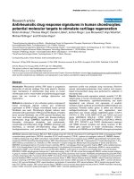
(Figure 1a). PS exons are included, on average, in less than
10% of the transcripts, with only about 5% of them being con-
stitutive. Even though PS exons are included at very low fre-
quencies, the pressure for reading frame maintenance is
higher than in MS and VO exons (Chi-square, p = 0.006 and
p = 1.6 × 10
-10
, respectively; Figure 1b). More than half of PS
exons (56.27%) have a length multiple of three, also called
symmetric. On the other hand, the percentages of MS
(45.51%) and VO (39.39%) symmetric exons are smaller. It
has been previously reported that conserved alternative
exons present a bias towards symmetry [6,37,38]. As most of
the PS exons are alternative, these numbers could just reflect
a relationship between reading frame preservation and inclu-
sion levels, regardless of exon age. We thus investigated the
relationship between exon symmetry and EST inclusion
Genome Biology 2008, Volume 9, Issue 9, Article R141 Corvelo and Eyras R141.3
Genome Biology 2008, 9:R141
levels for alternative exons belonging to the three age groups.
MS and VO exons tend to be more frequently symmetric at
lower inclusion levels (Chi-square, p = 1.7 × 10
-4
and p = 3.4 ×
10
-3
, respectively; Figure 1c). This agrees with previous
reports of a bias towards symmetry in evolutionarily con-
served alternative exons [37,38]. However, we observed the
opposite behavior for PS exons, although the observed differ-
ences are not significant, probably due to the small number of
cases in the high inclusion level categories. This suggests that
the pressure for reading frame maintenance may be related to
exon age. A study of the dependency on the inclusion level
would require further analysis with larger sets of exons.
Exon creation from repetitive sequences
Along with tandem duplication events [13], exonization of
TEs is one of the most important mechanisms of exon crea-
tion [17,35,39,40]. Therefore, we assessed the overlap
between exons from the three age sets and TEs, considering
as overlap the cases in which the TE covers at least one of the
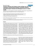
splice sites. We found that PS exons have a high density of
TEs in their flanking intronic regions (Figure 2a) and about
43% of the cases overlap TEs (Table 1). On the other hand, MS
and VO exons have a very low density of TEs in the proximal
adjacent intronic regions (Figure 2b, c) and show negligible
overlap of TEs with their splice sites. Additionally, excluding
the eight cases in which the exon overlaps more than one TE,
we found that for 116 (79.5%) of the PS exons overlapping
TEs, the TE is in the opposite strand of the exon. Although Alu
elements, unlike other TEs such as L1 and Long Terminal
Repeat (LTR) retrotransposons [41], were not found to have a
bias in the strand of insertion in human introns [40], we find
that most of the Alu elements (88.3%) overlapping a PS exon
occur in the strand opposite to the gene (anti-sense). In only
9 out of the 77 cases (11.7%) we found sense Alu elements, and
in only 4 of these is the overlap complete. Moreover, the per-
centage of anti-sense cases for non-Alu TEs is 69.6%. This
suggests that for TEs and, especially, for Alu elements,
although insertion can potentially occur in either strand,
exonization occurs mainly in the opposite strand. Interest-
ingly, although we found no overlap in the MS set, we found
19 cases (less than 0.15%) in the VO set; many of these were
simple-repeats (Table 1). More details about the type of TE
overlap are given in Table A1 in Additional data file 1.
Remarkably, more than 50% of PS exons do not overlap a TE
and cannot be explained by tandem duplication, as those
cases were discarded during the exon classification.
Analysis of the splicing regulatory content of exons
In order to understand the properties of the splicing regula-
tory content that determine the observed differences in inclu-
sion between exon sets, we conducted an analysis of splicing
cis-regulatory elements in exons and their flanking introns.
For this analysis we used three sets of splicing regulatory ele-
ments (SREs): 666 ESE hexamers [42], which we call ESE-
comb; all possible words obtained from the four position-
specific weight matrices for SR-protein binding sites from
ESE-finder (SF2/ASF, SC35, Srp40 and Srp55) using the pro-
posed thresholds [43], which we call SRall; and 386 ESS hex-
amers [42], which we call ESScomb (see Materials and
methods for a detailed description). Previous research has
pointed out that ESEs are generally more abundant in exons
than in introns [29,32,44], whereas ESSs are generally more
frequent in introns than in exons [29,31]. In fact, some of the
sets used here were partially defined based on exon/intron
and on exon/pseudo-exon enrichment [28,29]. In order to
better understand how these motifs distribute on both real/
pseudo-exons and introns, we defined a set of real exons mak-
ing use of the total set of exons from the three age groups.
Additionally, we built a set of pseudo-exons from intronic
regions that fall between protein-coding exons and are devoid
of TEs (pseudo-INT). For both real and pseudo-exons, den-
sity profiles for each SRE set are plotted in Figure A1 in Addi-
tional data file 1. Real exons, as expected, show higher
ESEcomb exonic densities when compared to pseudo-exons.
Interestingly, the densities are lower in adjacent intronic
regions. The inverse seems to be true for ESScomb. Relative
to SRall, only intronic differences were observed between real
and pseudo-exons.
This pattern suggests that the previously reported differences
between exonic and intronic content in real exons, something
not observed in pseudo-exons, are not merely due to an
increase of ESEs and a decrease of ESSs in the exonic regions,
but also to opposite changes in the adjacent intronic regions.
Taking this into account, it is plausible to hypothesize that the
effect exerted by SREs is context dependent. Splicing deci-
sions depend on the correct discrimination between exonic
and intronic regions and this is ultimately determined by
sequence features and their positioning relative to the splice
sites. Therefore, we define a measure, the exonic relative
abundance (ERA), which encapsulates both exonic and
intronic information. This measure is defined for each exon
as the relative difference between exonic and intronic densi-
ties for a given set of regulators (see Materials and methods
for details). This measure is such that, for signals that are
more abundant in the exon than in the flanking intronic
region, it takes on positive values. On the other hand, for sig-
nals that are more abundant in the flanking introns, the ERA
values distribute around a negative mean. In addition, and
contrary to the overall exonic or intronic density, this meas-
ure does not depend on SRE set size, which makes it useful for
comparing the contribution from different SRE sets to the
splicing phenotype.
Relative abundance of splicing regulators improves the
discrimination between real and pseudo-exons
We find that the ERA can discriminate better between real
and pseudo-exons than the overall density measures. For this
analysis, we considered 10,000 real exons sampled from our
three age groups and 10,000 pseudo-exons sampled from the
pseudo-INT set. Each set was randomly split into 10 non-
redundant groups. For each SRE set (ESEcomb, SRall and
Genome Biology 2008, Volume 9, Issue 9, Article R141 Corvelo and Eyras R141.4
Genome Biology 2008, 9:R141
EST inclusietryFigure 1
EST inclusion level and symmetry. (a) EST inclusion levels for the three age groups. The x-axis shows the inclusion levels in ranges of 10, and the y-axis
shows the proportion of exons from each subset falling within each range. For each exon, the EST inclusion level is defined as N
i
/(N
i
+ N
s
) × 100%, where
N
i
is the number of ESTs including the exon and N
s
the number of ESTs skipping the exon. Only exons with N
i
+ N
s
10 were considered. On the left of
the dashed line we plot the frequencies for exons with zero EST inclusion level. (b) Percentage of symmetric exons (length multiple of three) for each age
group. (c) Percentage of symmetric exons by EST inclusion level category for each age group. Only alternative spliced exons with N
i
+ N
s
10 were
considered.
0−10 10−20 20−30 30−40 40−50 50−60 60−70 70−80 80−90 90−100
EST inclusion level (%)
Frequency (% )
0
20
40
60
Primate specific
Mammalian specific
Vertebrate and older
(a)
56.27
45.51
39.39
0−30 30−60 60−90 0−30 30−60 60−90
EST inclusion level ( % )
Symmetric exons ( % )
0−30 30−60 60−90
(b)
(c)
EST inclusion level ( % )EST inclusion level ( % )
Symmetric exons ( % )
Symmetric exons ( % )
PS
PS
VO
VO
MS
MS
0
20
40
60
80
100
0
20
40
60
80
100
0
20
40
60
80
100
0
10
30
50
Genome Biology 2008, Volume 9, Issue 9, Article R141 Corvelo and Eyras R141.5
Genome Biology 2008, 9:R141
ESScomb), we scored the exons on three measures: exonic
density; intronic density; and ERA. Figure 3 shows the
receiver operating characteristic (ROC) curves for each of the
SRE sets (Figure 3a–c), vertically averaged on each false pos-
itive rate (FPR) for the 10 subsets, and the corresponding
areas under the curve (AUCs) (Figure 3d). These ROC curves
allow comparison between classifiers for all possible thresh-
olds and AUCs summarize global performance. We also used
the 10 splits for a 10-fold cross-validation test; for each group
used as a test set we used the other 9 as training sets. Accuracy
results and corresponding thresholds of the tests can be
found in Table 2 (see Table A2 in Additional data file 1 for the
complete list of accuracy values using combined and individ-
ual SRE sets). The precision-recall curves for each classifier
can be found in Figure A2 in Additional data file 1.
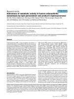
We observe that ESEcomb exonic density performs, in gen-
eral, better than intronic density (AUC, 0.727 and 0.619,
respectively; Figure 3a). Surprisingly, we found that the
opposite occurs for SRall at almost all FPR values (Figure 3b).
That is, the intronic density of SRall is more informative than
the exonic densities. Regarding ESScomb, even though
exonic and intronic densities show different behaviors (Fig-
ure 3c), no differences in AUCs were observed. Interestingly,
we found that ESEcomb and ESScomb perform better than
each individual set from which they were built and consist-
ently better than SRall (see Table A2 in Additional data file 1
for the performances of the individual sets).
Moreover, we found that ERA performs superiorly in discrim-
inating real from pseudo-exons than intronic and exonic den-
sities independently, on both ESEcomb and ESScomb sets at
all FPR values (AUC, 0.773 and 0.755). Additionally, ERA
(AUC, 0.619) provides a marginal improvement with respect
to the information provided by the intronic density of SRall
(AUC, 0.600).
Differences in the relative abundance of regulators
with age and exon establishment
In order to investigate the regulatory features that determine
the observed differences in EST inclusion levels between
recently created and older exons, we studied the splice site
strengths for each exon group. The distributions of the splice
site score for the three age groups, calculated as the sum of the
acceptor and donor scores for each exon, can be found in Fig-
ure A3A in Additional data file 1. PS exons show significantly
weaker splice sites (mean = 5.061; Mann-Whitney, p = 1.18 ×
10
-8
) than MS (6.907) and VO (7.394) exons. Moreover, the
difference between the MS and VO groups was also found to
be significant (Mann-Whitney, p = 3.63 × 10
-3
). These differ-
ences are mainly supported by lower frequencies of pyrimi-
dines upstream of the acceptor site and also by more
degenerated donor signals in PS exons (Figure A3B in Addi-
tional data file 1). This suggests that the observed differences
in exon inclusion may be related to the differences in splice
site strength. However, these distributions largely overlap.
We also observe that EST inclusion levels for PS exons seem
to be more dependent on the splice site score than for MS or
VO exons. Still, no clear, strong correlation between these two
variables could be observed (Spearman's rank correlation, PS
rho = 0.22, p = 3.81 × 10
-5
; MS rho = 0.12, p = 0.026; and VO
rho = 0.09, p = 2.23 × 10
-27
). Thus, the change from low to
high inclusion cannot be fully attributed to an increase in
splice site strength.
Accordingly, we considered SREs as additional contributors
to the splicing phenotype. We calculated ERA values for each
age group of exons (Figure 4), for the same SRE sets as before.
As a control, we used the set of pseudo-exons not overlapping
TEs, which we determined before (pseudo-INT). Figure 4
shows that pseudo-exons have ERA values distributed around
zero for all SREs tested (ESEcomb, -0.029; SRall, -0.006; and
ESScomb, -0.055). On the other hand, all real exons show
positive values for ESEs and negative for ESSs. In particular,
PS exons show the closest ERA values to pseudo-INT, but
they are still significantly different (Mann-Whitney, ESE-
comb p = 1.45 × 10
-20
, SRall p = 2.88 × 10
-8
, and ESScomb p
0). Interestingly, we also observe differences between PS
exons and MS/VO for two out the three SRE sets used. For
ESEcomb and ESScomb, PS exons show lower absolute ERA
values (0.164 and -0.302, respectively) than MS (0.284 and -
0.499) and VO (0.258 and -0.387) (see Table A3 in Additional
Table 1
Overlap with repetitive elements
SINE
Exon set N Alu Other LINE DNA LTR Other Mixed Total
PS 359 77 17271015 - 8 154
21.45% 4.74% 7.52% 2.79% 4.18% 2.23% 42.90%
MS 323- -
VO 13,249 - 5 14 - - 10 - 29
0.04% 0.11% 0.08% 0.22%
For each age group, we give the number and corresponding percentage of exons that overlap with different repetitive elements: SINEs (Alu and
other), LINE, DNA, LTR and Other. Eight PS exons overlap more than one element (Mixed). We count as overlap when the element covers at least
one of the splice sites of the exon.
Genome Biology 2008, Volume 9, Issue 9, Article R141 Corvelo and Eyras R141.6
Genome Biology 2008, 9:R141
data file 1). Relative to SRall, no significant differences
between age groups were observed. ERA was also calculated
for the individual SRE sets (see Materials and methods for
details). These results can be found in Figure A4 in Additional
data file 1.
Intronic densities for the main classes of repetitive elementsFigure 2
Intronic densities for the main classes of repetitive elements. (a) Primate specific, (b) mammalian specific and (c) vertebrate and older. At each intronic
position, the density was calculated as the proportion of cases in which the base was covered by a given type of repetitive element. We give on the x-axis
the relative position from the splice junctions as negative if upstream of the acceptor site or positive if downstream of the donor site.
−400 −200 0 200 400
Rel. position from splice junctions (bp)
Density
−400 −200 0 200 400
Rel position from splice junctions (bp)
Density
SINE LTR LINE
−400 −200 0 200 400
0.0
0.1
0.2
0.3
0.4
0.5
Rel. position from splice junctions (bp)
Density
0.0
0.1
0.2
0.3
0.4
0.5
0.0
0.1
0.2
0.3
0.4
0.5
0.0
0.1
0.2
0.3
0.4
0.5
0.0
0.1
0.2
0.3
0.4
0.5
0.0
0.1
0.2
0.3
0.4
0.5
DNA
(a)
(b)
(c)
Primate specific
Mammalian specific
Vertebrate and older
Genome Biology 2008, Volume 9, Issue 9, Article R141 Corvelo and Eyras R141.7
Genome Biology 2008, 9:R141
Focusing on MS and VO exons, we observe a surprising differ-
ence in the content of ESScomb motifs. VO exons present
lower absolute ERA values than MS (Mann-Whitney, p = 3.06
× 10
-10
). This result derives from the fact that VO exons show
relatively higher exonic densities of ESSs (0.272) compared to
MS (0.213), while for intronic content no significant differ-
ences were found (Table A3 in Additional data file 1). Also, VO
exons show slightly lower exonic densities for ESEcomb with
respect to MS (MS 0.665, and VO 0.633; Mann-Whitney, p =
4.56 × 10
-6
). These results can be partially explained by the
fact that VO exons have stronger splice sites. On the other
hand, it also suggests that AS of VO exons may be more
dependent on ESS content.
In order to understand if these regulatory elements were
under different, possibly functional, constraints depending
on the exon age, we investigated their conservation in the
mouse orthologous exons (Figure 5). For this purpose, we
have calculated the functional conservation score (FCS; see
Materials and methods for detailed description) for all three
SRE sets on both MS and VO exon sets. This measure reflects
the fraction of nucleotides that are covered by motifs from the
same SRE set in both human and mouse. This measure corre-
lates with the percentage of sequence conservation but also
takes into account cases where a substitution does not change
the regulatory character of a region. In general, VO exons
have higher FCS values compared to MS exons for ESEcomb
(Mann-Whitney p = 8.42 × 10
-13
), SRall (p = 4.64 × 10
-14
) and
ESScomb (p = 2.99 × 10
-16
). Additionally, FCS is higher for
ESEcomb than for ESScomb for both MS and VO exons
(Mann-Whitney, p 0), which might reflect the importance of
the conservation of the amount and position of ESEs in exons.
In summary, although VO exons have lower density of ESEs,
these are more conserved than in MS exons, indicating that
ESE turnover is more frequent in MS compared to VO exons,
in agreement with recent analyses [45]. Moreover, VO exons
present a larger fraction of ESSs that are highly conserved,
suggesting possible constraints due to AS regulation.
Interestingly, considering all exons from the three age
groups, ERA values tend to increase for ESEs (ESEcomb and
SRall) and decrease for ESSs (ESScomb). Figure 6 shows the
mean ERA values plotted for bins of increasing EST inclusion
levels. For ESEcomb (Figure 6a) and SRall (Figure 6b) we
observe a consistent increase except at high EST inclusion
levels, where SRall values slightly decrease. On the other
hand, there is a consistent decrease for ESScomb at all EST
inclusion levels (Figure 6c). Exonic and intronic densities do
not show such gradients with EST inclusion levels (data not
shown). Thus, inclusion levels seem to be determined by the
local differences in the densities of motifs.
Study case: why Alu elements are a good substrate for
exonization
It has been recently reported that all TEs have approximately
the same exonization levels with the exception of Alu ele-
ments, which are almost three times higher than other TE
families [40]. Additionally, the high number of Alu copies in
the human genome and their propensity to accumulate in
intronic regions[40] make this element the main source of
new exons originating from TEs. It has been shown that in
some cases, cryptic splice sites are enough to incorporate part
of an Alu element in the mature transcript [22,23] and that in
other cases, specific splicing enhancers are needed for their
inclusion [34]. We thus applied the ERA measure in order to
understand which regulatory features, besides the presence
of splice sites, may be responsible for the increased Alu exoni-
zation rate.
We compared the SRE densities between the subset of PS
overlapped by Alu elements (PS-Alu) and a set of Alu pseudo-
exons bigger than 80 bp (pseudo-Alu) (see Materials and
Table 2
Mean thresholds and accuracy for pseudo/real exon classification (10-fold cross-validation)
Threshold Accuracy
SRE set Measure Mean SD Mean SD
ESEcomb Exonic density 0.564* 0.000 0.672 0.008
Intronic density 0.450
†
0.010 0.588 0.010
Exonic relative abundance 0.136* 0.014 0.699 0.009
SRall Exonic density 0.486* 0.003 0.535 0.006
Intronic density 0.481
†
0.002 0.585 0.010
Exonic relative abundance 0.163* 0.019 0.580 0.012
ESScomb Exonic density 0.358
†
0.017 0.613 0.013
Intronic density 0.492* 0.005 0.614 0.009
Exonic relative abundance -0.216
†
0.007 0.707 0.010
*Minimum score cut-off for predicted real exons.
†
Maximum score cut-off for predicted real exons.
Genome Biology 2008, Volume 9, Issue 9, Article R141 Corvelo and Eyras R141.8
Genome Biology 2008, 9:R141
methods for details). Figure 7a, b show the mean exonic and
intronic densities of the two ESE sets considered (ESEcomb
and SRall) for PS-Alu and pseudo-Alu. The mean exonic den-
sities of ESEcomb and SRall for PS-Alu (0.597 and 0.649,
respectively) were significantly higher (Mann-Whitney, p =
4.89 × 10
-12
and p = 9.78 × 10
-6
) than the mean exonic densi-
ties for pseudo-Alu (0.514 and 0.593). Relative to ESScomb
(Figure 7c), PS-Alu shows a mean value of exonic density of
0.150 while pseudo-Alu shows a mean value of 0.190 (Mann-
Whitney, p = 1.09 × 10
-4
).
Surprisingly, we observe the opposite behavior when consid-
ering adjacent intronic regions. The mean values of the
intronic density of ESEs are significantly lower for PS-Alu
when compared to pseudo-Alu (Mann-Whitney, ESEcomb p
= 3.64 × 10
-4
and SRall p = 2.02 × 10
-5
), while for ESScomb
Performance comparison in real/pseudo-exon discrimination between different measuresFigure 3
Performance comparison in real/pseudo-exon discrimination between different measures. ROC curves (vertically averaged) for exonic density, intronic
density and ERA, using (a) ESEcomb, (b) SRall and (c) ESScomb as informative features. The average was calculated from 10 different subsets of the data
(see text for details). (d) The corresponding AUCs. The error bars represent the standard error. FPR, false positive rate; TPR, true positive rate.
0.0 0.2 0.4 0.6 0.8 1.0
0.0
0.2
0.4
0.6
0.8
1.0
FPR
TPR
ERA
Exon density
Intron density
FPR
TPR
FPR
TPR
ESEcomb SRall ESScomb
AUC
0.5
0.6
0.7
0.8
0.9
0.0
0.2
0.4
0.6
0.8
1.0
0.0
0.2
0.4
0.6
0.8
1.0
0.0 0.2 0.4 0.6 0.8 1.0
0.0 0.2 0.4 0.6 0.8 1.0
ERA
Exon density
Intron density
ERA
Exon density
Intron density
ERA
Exon density
Intron density
(a) (b)
(c) (d)
ESEcomb SRall
ESScomb
Genome Biology 2008, Volume 9, Issue 9, Article R141 Corvelo and Eyras R141.9
Genome Biology 2008, 9:R141
the mean density values are higher (Mann-Whitney, p = 1.12
× 10
-11
). All these results suggest that ESEs and ESSs play a
role in Alu exonization. In Figure 7d we can observe that for
PS-Alu, the mean ERA values for ESEcomb and SRall distrib-
ute around positive values (0.276 and 0.177) while the ESS-
comb values tend to distribute around a negative mean (-
0.625). The absolute values are significantly greater than
those obtained for pseudo-Alu (Mann-Whitney, p = 8.26 × 10
-
10
, p = 1.31 × 10
-7
and p = 3.75 × 10
-10
). Furthermore, the fact
that ESScomb produces the greatest difference of means sug-
gests that this sequence feature might be the main determi-
nant in the exonization of Alu elements. Comparing PS exons
overlapped and non-overlapped by Alus, we observe that the
latter have higher exonic (0.247) and lower intronic (0.383)
densities for ESScomb (Mann-Whitney, p = 6.29 × 10
-8
and p
= 1.83 × 10
-4
, respectively). Consequently, their absolute ERA
mean values (-0.302) are lower than those observed for Alu
overlapped exons and, surprisingly, lower than those
observed for pseudo-Alu (-0.407) (Mann-Whitney, p = 3.94 ×
10
-10
and p = 6.03 × 10
-5
).
Finally, in order to test whether the found properties are Alu
specific, we analyzed sets of pseudo-exons overlapping the
other major families of mobile elements in the human
genome: Long Interspersed Nuclear Elements (LINEs),
LTRs, DNA transposons and non-Alu Short Interspersed
Nuclear Elements (SINEs) (see Materials and methods for
details). For each of these sets, we calculated the ERA distri-
butions for the same SRE sets as before. As can be seen in Fig-
ure 7e, all the pseudo-exon sets show absolute ERA values
close to zero. Moreover, they do not present the ERA pattern
expected to favor exonization. Indeed, pseudo-exons overlap-
ping DNA transposons and LINEs have negative ERA mean
values for ESEcomb. The exception seems to be for LTR
pseudo-exons, which have positive ERA values for ESEcomb
and negative for ESScomb, but with very low absolute values.
This suggests that the high rate of Alu exonization may simply
be due to their lack of silencers.
Although Alu elements do not seem to have a strand bias
inserting within introns in human genes, protein-coding
exons are mostly created from anti-sense Alu elements [40].
In fact, we could only find 64 cases of sense Alu pseudo-
exons. In comparison, we could find more than 30,000 Alu
pseudo-exons with the Alu in anti-sense. This difference can
be explained by the efficiency of the splice sites [22,23], as
sense Alu exons do not contain the strong poly-pyrimidine
tract typical of anti-sense ones. Furthermore, most PS exons
overlapping anti-sense Alu elements are normally 80 bp long
or greater. These lengths correspond, in most cases, with the
most commonly used splice sites created by the anti-sense
Alu [46] (data not shown). In order to understand the differ-
ences in exonization levels, we compared the properties of
these two under-represented cases, sense Alu exons and anti-
sense Alu exons shorter than 80 bp, making use of pseudo-
exons overlapping these elements: pseudo-exons overlapping
SRE ERA changes with ageFigure 4
SRE ERA changes with age. Mean exonic relative abundance values for the three age groups (PS, MS and VO) and a set of pseudo-exons not overlapping
any repeats (pseudo-INT) calculated for the three motif sets (ESEcomb, ESScomb and SRall). Exons overlapping Alu elements were excluded from the PS
set. The standard error is also shown.
ESEcomb SRall ESScomb
Ex. rel. abundance
−0.6
−0.4
−0.2
0.0
0.2
0.4
pseudo-INT
PS
MS
VO
Genome Biology 2008, Volume 9, Issue 9, Article R141 Corvelo and Eyras R141.10
Genome Biology 2008, 9:R141
and Alu in the same orientation (pseudoSS-Alu) and pseudo-
exons smaller than 80 bp that overlap an Alu in the opposite
strand (pseudoSH-Alu) (see Materials and methods for
details). Interestingly, both sets have a different content of
splicing regulatory motifs with respect to anti-sense Alu
pseudo-exons (pseudo-Alu) bigger than 80 bp (Figure A5 in
Additional data file 1). Even though pseudoSS-Alu shows for
both sets of ESEs higher exonic densities with respect to the
adjacent intronic regions (Figure A5A and A5B in Additional
data file 1), no differences are observed for ESSs (Figure A5C
in Additional data file 1). This leads to positive ERA values for
ESEs (0.091 and 0.086) but close to zero values for ESSs (-
0.023). On the other hand, pseudoSH-Alu shows negative
ERA values for ESEs (-0.167 and -0.168) and close to zero
mean ERA values (-0.040) for ESSs (Figure A5D in Addi-
tional data file 1). Thus, both pseudoSS-Alu and pseudoSH-
Alu exons have ERA values for ESSs close to zero, as opposed
to anti-sense Alu pseudo-exons and PS exons overlapping
Alus, which have very large negative ERA values for ESSs.
This suggests that the higher content ESSs make sense Alus
and regions smaller than 80 bp within anti-sense Alus less
prone to exonization.
Discussion
We have analyzed the regulatory requirements for exoniza-
tion and how splicing regulation changes throughout the exon
lifespan by comparing the splicing regulatory properties of
human internal protein-coding exons classified into three age
groups: primate specific (PS), mammalian specific (MS) and
vertebrate and older exons (VO). Most of the PS exons are
alternatively spliced and show low inclusion levels. We find
only about 5% of PS exons to be constitutive, whereas previ-
ous analyses [1] report about 60% of exons to be constitutive
in a PS set. This difference can be explained by the fact that
our method is more stringent; hence it is less likely that older
exons are misclassified as PS ones; and could also be due to
the fact that we discarded exons that may have originated
from tandem duplication events, which are copies of pre-
existing exons and would be similar to older ones. Further-
more, we find that PS exons are more likely to maintain the
reading frame, indicating an additional pressure to reduce
their impact in protein-coding regions. This increased fre-
quency of symmetric exons observed in the PS set, especially
in highly included exons, is likely to be related to the fact that
the isoform including the exon is a novel one. On the contrary,
for MS and VO, lowly included exons are more frequently
symmetric. This suggests that in these cases, or in a signifi-
cant fraction of them, the ancestral form might have been
constitutively spliced, having more recently become alterna-
tive. This provides extra evidence supporting the hypothesis
that the appearance of novel isoforms is favored when their
impact is reduced. In this scenario AS acts as a key player
allowing the incorporation of novel regions in mature tran-
scripts and resulting products, establishing a close relation-
ship with the process of exon creation [3].
We have also investigated the splicing regulatory require-
ments for de novo exonization. We observed that real exons
have significantly different content of regulatory elements
compared with pseudo-exons. However, there are also signif-
icant differences in the flanking introns. Indeed, we observe
significant differences in the adjacent intronic content of
SREs that were originally classified as exonic. Intronic
regions adjacent to real splice junctions present lower densi-
ties of ESEs and higher densities of ESSs when compared to
regions adjacent to pseudo-exons. This does not necessarily
imply that such motifs are active in these regions. However,
these differences could be the result of a balance with other
nearby regulatory elements.
As exonization is related to changes in the exonic and in the
adjacent intronic regions, they should both be taken into
account. Accordingly, we defined a single measure, ERA,
which encapsulates the regulatory content of each exon and
its flanking introns. We have shown that this measure can dif-
ferentiate better real exons from pseudo-exons than the
exonic or intronic densities alone. For the three motif sets
used, ERA provides the best discriminatory power. We also
found that ESEcomb and ESScomb, which are combined sets
of ESEs and ESSs, respectively, performed better than the
individual sets alone. Another result worth mentioning is the
fact that these two computational defined sets, performed
better than the experimentally determined SRall set. The fact
that these two sets have been partly defined based on exon
versus intron and exon versus pseudo-exon comparisons
might favor their discriminative power when using exonic
density as a factor. Interestingly, the same holds true for
intronic density at a lower extent. Relative to a third set of SR
protein binding sites (SRall), we observed that SF2/ASF
SRE functional conservation between human and mouseFigure 5
SRE functional conservation between human and mouse. SRE FCS
between human and mouse of exonic regions covered by ESEcomb, SRall
and ESScomb motifs for mammalian specific and vertebrate or older
exons. See Materials and methods section for formula.
10 0.6 0.80.2 0.4
Functional conservation score
Mammalian
specific
Vertebrate
and older
ESEcomb
ESScomb
SRall
Genome Biology 2008, Volume 9, Issue 9, Article R141 Corvelo and Eyras R141.11
Genome Biology 2008, 9:R141
binding motifs perform consistently better than SC35, SRp40
and SRp55 binding sites. We thus expect that ERA or any
other measure that takes into account local differences in
motif content will contribute to the improvement of current
methods of splice site and exon prediction.
We observed that the difference in inclusion levels between
the different exon age groups cannot be fully attributed to the
splice site strength. Further studies on regulatory content
have shown that PS exons have smaller differences in ESE
motifs between exons and flanking introns than conserved
exons, that is, they are more similar to pseudo-exons than to
older exons. This indicates that a minimal amount of regula-
tory motifs is needed for exonization. Moreover, the greater
difference in the local density of regulators for older exons
means that they have acquired a consolidated set of regula-
tors. In fact, our results indicate that the relative density of
regulatory motifs increases with time, and at a higher rate in
MS exons compared to VO exons. Additionally, we found that
exons become more established, that is, exhibit higher inclu-
SRE exonic relative abundance and EST inclusion levelsFigure 6
SRE exonic relative abundance and EST inclusion levels. Cumulative plot of ERA variation (y-axis) for bins of increasing maximum EST inclusion levels (x-
axis) for (a) ESEcomb, (b) SRall and (c) ESScomb. The standard errors are also shown.
0.18
0.20
0.22
0.24
0.26
0.06
0.08
0.10
0.12
q
−0.38
−0.36
−0.34
−0.32
−0.30
ESEcomb
SRall
ESScomb
(a)
(b)
(c)
Ex. rel. abundanceEx. rel. abundanceEx. rel. abundance
20 40 80 10060
Maximum EST inclusion level (%)
20 40 80 100
60
Maximum EST inclusion level (%)
20 40 80 10060
Maximum EST inclusion level (%)
Genome Biology 2008, Volume 9, Issue 9, Article R141 Corvelo and Eyras R141.12
Genome Biology 2008, 9:R141
Alu's unique cis-reguFigure 7
Alu's unique cis-regulatory context. Exonic and intronic densities of (a) ESEcomb, (b) SRall and (c) ESScomb motifs on primate specific exons overlapping
Alu elements (PS-Alu) and on Alu pseudo-exons (pseudo-Alu). (d) Exonic relative abundance of ESEcomb, SRall and ESScomb motifs for primate specific
exons overlapping Alu elements (PS-Alu) and for Alu pseudo-exons (pseudo-Alu). (e) Exonic relative abundance for the same sets of motifs in pseudo-exons
overlapping other classes of repeats, namely DNA, LTR, LINE and SINE non-Alu (MIR) repeats. The error bars represent the standard error.
exon intron
Density
0.2
0.3
0.4
0.5
0.6
0.7
0.8
PS-Alu pseudo-Alu
exon intron
Density
0.2
0.3
0.4
0.5
0.6
0.7
0.8
exon intron
Density
0.0
0.1
0.2
0.3
0.4
0.5
0.6
PS-Alu
pseudo-Alu
Ex. rel. abundance
−0.8
−0.6
−0.4
−0.2
0.0
0.2
0.4
Ex. rel. abundance
−0.4
−0.2
0.0
0.2
0.4
(a)
(b)
(c) (d)
(e)
ESEcomb
SRall
ESScomb
ESEcomb
SRall
ESScomb
PS-Alu pseudo-Alu
PS-Alu pseudo-Alu
pseudo-DNA pseudo-LTR pseudo-LINE pseudo-MIR
ESEcomb
SRall
ESScomb
Genome Biology 2008, Volume 9, Issue 9, Article R141 Corvelo and Eyras R141.13
Genome Biology 2008, 9:R141
sion, by acquiring more enhancers relative to the flanking
introns and by increasing the density of silencers in introns
relative to the exons they flank. This is ultimately reflected in
the higher ERA absolute values obtained.
Our analyses suggest that the local sequence context in which
the exon is located plays a role in how splicing is regulated.
Although there is no direct experimental evidence of a mech-
anism in which the spliceosome senses the local densities of
splicing motifs, there is plenty of evidence of how the relative
abundance of motifs can determine the splicing phenotype. It
has been shown previously that the density of motifs close to
a splice site affects the splicing outcome [31]. In particular,
exonic regions that were intronized due to mutations to
splice-sites have less ESEs and more ESSs than average
exons, and that intronic regions that were exonized upon cre-
ation of cryptic splice-sites in introns had more ESEs and less
ESSs than normal introns [47]. This establishes a gradient of
densities between the different regions classified according to
splicing phenotype, similar to the one we find here. There is
also evidence that some splicing regulatory motifs in exons
and introns function in clusters [48-50], and that multiple
ESEs increase additively the efficiency of splicing [51,52].
Since we observe that ESEs and ESSs can occur by chance
almost anywhere in exons and introns [29,31], a local com-
pensation in the density of motifs seems to be necessary to
maintain a specific regulation [53], and this is reflected in the
local differences between exons and introns, which we can
measure using ERA.
Finally, we have also investigated the role of splicing regula-
tory elements in the exonization of TEs, which may account
for 42.9% of PS exons. When untranslated regions (UTRs) are
considered, the proportion of PS exons overlapping with TEs
is higher [1]. In fact, it has been recently reported that exoni-
zation of TEs occurs more abundantly in UTRs [40]. Thus,
new exons originating from TEs are accepted in protein-cod-
ing regions at a much lower rate than in UTRs. On the other
hand, most of the new exons overlapping TEs have been
found to introduce in-frame stop codons [40]. Many exoniza-
tions of TEs may occur as errors of the splicing mechanism,
and are, therefore, less frequently included in the protein and,
subsequently, are more often tolerated in UTRs. Since we
started from a set of protein-coding exons, our PS exons are
already part of an open reading frame, and can be considered
as recently established, that is, have become accepted into the
protein-coding region at low inclusion rates.
We observed that in most of the cases the Alu element over-
laps the PS exon on the anti-sense strand, and that these are
characterized by having a striking lack of silencers compared
to the surrounding introns. As introns can be considered as
regions with a basal density of splicing silencers [27,29], the
insertion of an anti-sense Alu therefore creates a local desert
of splicing silencers in the intronic region into which they are
inserted. Thus, the frequently observed Alu exonization
might not only stem from the presence of optimal splice sites,
but also from the creation of an environment favorable for
exonization. Interestingly, Alu pseudo-exons with overlap on
the sense strand and those in anti-sense shorter than 80 bp
have over-representation of ESSs in the exonic region, pro-
viding a possible explanation as to why they are not so fre-
quently exonized.
In the human genome there are around one million Alu cop-
ies, 66% of which accumulate in intronic regions[40]. We
found approximately 256,000 Alu pseudo-exons with splice
sites scoring above the first quartile of the distribution of
scores for real splice sites, which fall within an intron flanked
by protein-coding exons, and for which there is no evidence of
exonization from ESTs, cDNAs or proteins. From these
pseudo-exons, 15,048 (5.9%) are bigger than 80 bp, have a
length multiple of three and have no stop-codons in frame.
Moreover, 6795 (45,1%) of these are conserved in chimp and
macaque with conserved flanking AG and GT dinucleotides.
One possible reason why these conserved Alu pseudo-exons
do not appear to be included in the mature transcript is
because they have not been detected yet in EST/cDNA
sequencing experiments. However, considering the extensive
EST evidence that is available for human, one can assume
that most of these pseudo-exons are, in fact, silenced or are
not recognized by the spliceosome. After analyzing the regu-
latory content of these candidates, we observed that the ERA
values differ strikingly from the Alu exons in all sets of SREs,
suggesting that insufficient difference in density of SREs
between the potential exon and corresponding flanking
introns prevent their exonization (Table A4 in Additional
data file 1). This provides further support to the idea that a
minimum regulatory content is required for de novo exoniza-
tion.
Conclusions
Our results suggest that specific sequence environments
might be required for exonization. Namely, regions with
lower ESS content contrasting with the surroundings may be
more prone to exonization. Also, exon creation may require
the acquisition of a sufficient number of ESEs. All this sup-
ports the notion that de novo exonization is more likely to
occur when there is a sufficient difference in the density of
splicing regulatory elements on either side of optimal splice
sites. This, in fact, suggests a mechanism of exon creation and
establishment in human. New exons appear with low inclu-
sion level, as they do not have a sufficient amount of ESEs. In
this context, Alu elements play a crucial role in de novo exon
creation in primates. With time, the establishment of an exon
is determined by the accumulation of ESEs. In parallel, the
lack of ESSs plays an important role in distinguishing an exon
from the adjacent introns. This acquisition of regulatory ele-
ments along with the differentiation with respect to the
intronic context determine the establishment of an exon in
the mature transcript.
Genome Biology 2008, Volume 9, Issue 9, Article R141 Corvelo and Eyras R141.14
Genome Biology 2008, 9:R141
In summary, exon establishment is determined by the acqui-
sition of splicing regulation at a local level and, as shown, this
can be measured using a specifically devised measure, the
ERA. This measure can, in fact, distinguish better real exons
from pseudo-exons than exonic or intronic densities of splic-
ing motifs alone. We therefore conclude that local differences
in motif densities affect splicing decisions and, subsequently,
the recognition of exons. We expect that measures that take
these differences into account will provide an improvement
on standard exon and gene prediction methods.
Materials and methods
Datasets
Gene annotations for Homo sapiens (NCBI36, Apr 2006),
Mus musculus (NCBI m36, Apr 2006), Bos taurus (Btau 2.0,
Dec 2005), Gallus gallus (WASHUC 1, Dec 2005) and Tetrao-
don nigroviridis (TETRAODON 7, Sep 2004), and ortholo-
gous gene pairs between these species were downloaded from
Ensembl [54]. From the set of orthologs, only unique best
reciprocal hits were kept. Genes that had ambiguous ortholo-
gous assignations, that is, linked to more than one potential
orthologous sequence in the other genome, were eliminated.
EST, mRNA and RepeatMasker mappings were retrieved
from UCSC Genome Browser Database [55].
Alignment of exon-intron structures
Transcripts and coding sequences for each gene were pro-
jected onto the genomic sequence producing an array-like
structure of genomic regions. These structures were then
aligned between pairs of orthologous genes using information
about the splice sites and exon phases. Orthologous genes
from closely related species generally have high conservation
of their exonic structure. Taking this into account, we per-
formed comparisons between all splice sites from one gene
against all those from its orthologue. A score was defined
using the sequence identity between 40 nucleotides around
the splice junctions (20 nucleotides upstream and 20 nucle-
otides downstream of each splice site) and the exon phase. All
these scores were placed in a matrix, where every entry repre-
sents the score from the comparison of two splice sites from
the orthologous gene pair. Subsequently, using a dynamic
programming algorithm with this matrix, we identified the
putative orthologous splice sites. This was done pair-wise
between all five species. From this calculation we could detect
orthologous exons and exons with potentially no orthologue
in another genome.
Classification of exons according to evolutionary ages
We considered those exons with the following properties:
internal, protein-coding, longer than 30 nucleotides and
without 3' or 5' AS. This last condition was required to guar-
antee that both regions upstream and downstream of the
exon are fully intronic. The flanking introns were also
required to be longer than 30 nucleotides each. Additionally,
only exons with canonical splice sites (AG/GT) were consid-
ered. These requirements were necessary for the correct anal-
ysis of the densities of regulatory sequences (see below). In
order to obtain the exons belonging to the three different age
classes, comparisons using three species were performed. If a
particular exon was present in one species (reference species)
and absent in the most closely related one (target), this could
mean that either that exon was created in the reference spe-
cies or that it was lost in the target one. To resolve this ques-
tion a third species (more distantly related to the reference)
was used as out-group, to infer if the exon was present in the
common ancestor of the first two species. Three different age
classes were defined: primate specific (PS), mammalian spe-
cific (MS) and vertebrate and older (VO). PS exons were
defined as human exons that were not present in mouse or
cow (strictly speaking, PS exons are human exons that are
possibly also present in other primates). MS exons were
defined as human exons conserved in mouse and cow, but not
present in chicken or Tetraodon. Finally, VO exons were
defined as human exons that are conserved in all the other
four species. Exons that were aligned to orthologous exons
were considered as conserved. Exons that did not have an
alignment and were located between, but not necessarily
adjacent to, conserved exons were considered to be candi-
dates for PS or MS exons. These candidates were then com-
pared with TBLASTN against the region in the orthologous
genes spanned between the nearest alignable splice sites. If
any significant result was produced (e-value < 0.0005), that
exon was discarded. In this way we do not consider as non-
conserved exons that are evolving at a faster rate. In order to
reduce the possibility that the remaining exons could have
been originated by segmental duplication, exons that showed
more than 80% similarity over 40% of coverage with respect
to other exons from the same gene were discarded. As a final
filter, we only kept exons that were supported by EST or
mRNA evidence. As the search uses very stringent criteria of
sequence conservation, we do not expect the sizes of the
obtained age groups to necessarily reflect the real number of
exons belonging to these age categories in the human
genome.
Pseudo-exon sets
In addition to the three age groups, we built sets of pseudo-
exons overlapping and not overlapping TEs. Pseudo-exons
are defined as intronic sequences of length comparable to
exons, flanked by canonical splice sites and not present in any
ESTs or cDNAs. Moreover, these have have a length multiple
of three and with no stop codons in frame. Using the Repeat-
Masker annotations retrieved from UCSC Genome Browser
Database [55], repetitive and repetitive-free regions were
determined from intronic regions located between protein-
coding exons. As we needed to score splice sites and obtain
pseudo-exons of size 30 nucleotides or longer, we considered
regions bigger than 56 nucleotides (20 on the acceptor side +
30 exonic + 6 on the donor side). Then, all the candidate
splice sites in the sense strand that score above the first quar-
tile of all human protein-coding exons were taken and all the
Genome Biology 2008, Volume 9, Issue 9, Article R141 Corvelo and Eyras R141.15
Genome Biology 2008, 9:R141
pairs of acceptor and donor producing an exon bigger than 30
nucleotides were determined. Finally, we also applied filters
to extract exons with a length multiple of three and that did
not produce a stop codon in frame. We obtained a set of
pseudo-exons not overlapping any TE (pseudo-INT) and five
sets of pseudo-exons overlapping the four main classes of
repeats (SINEs: pseudo-MIR and pseudo-Alu; LINEs:
pseudo-LINE; DNA repeats: pseudo-DNA; and LTRs:
pseudo-LTR).
Alu elements contain several possible 5' and 3' splice sites
[22,23]. However, not all are commonly used. The splice sites
most generally used in exonized anti-sense Alus make up for
exons of a size of around 80 bp and bigger [46]. From our PS
set, 95% of exons overlapping Alus are of length 80 bp or
longer. Accordingly, all pseudo-exons analyzed were taken to
be 80 bp or longer. We also created two additional sets of
pseudo-exons following the above defined criteria: pseudo-
exons overlapping sense Alu elements (pseudoSS-Alu) and a
set of short (smaller than 80 nucleotides) pseudo-exons over-
lapping anti-sense Alu elements (pseudoSH-Alu).
EST inclusion level
EST alignments were retrieved from UCSC Genome Browser
Database [55] and compared with the Ensembl [54] annota-
tions. For each exon, the percentage of EST inclusion level is
defined as:
where N
i
is the number of ESTs including the exon and N
s
the
number of ESTs that cover the genomic region of the exon but
skip it. Only exons with N
i
+ N
s
10 were considered. Some
exons have zero EST inclusion, as all the corresponding ESTs
show exon skipping, but their existence is supported by
mRNA and/or protein evidence.
Density of repetitive elements
RepeatMasker mappings overlapping exons and both
upstream and downstream introns were retrieved from UCSC
Genome Browser Database [55]. For the main four categories
of elements (SINEs, LINEs, LTRs, DNA) we calculated the
intronic densities as the fraction of cases where a particular
type of element overlaps each base. Also, we tested whether
exons belonging to different age groups overlapped any of
these elements.
Splice site strength
We scored all splice sites using position weight matrices for
the human donors and acceptors. We considered positions (-
20 nucleotides to +3 nucleotides) relative to the acceptor site
and (-3 nucleotides to +6 nucleotides) to the donor site.
Relative abundance of regulatory motifs
We used three sets of regulatory motifs: 666 ESE hexamers
[42], which we call ESEcomb, built from the combination of
238 RESCUE-ESE hexamers [28] and 2,069 PESE octamers
[29]; all possible words obtained from the four position-spe-
cific weight matrices for SR-protein binding sites from ESE-
finder (SF2/ASF, SC35, Srp40 and Srp55) using the proposed
thresholds [43], which we called SRall; and 386 ESS hexam-
ers [42], which we called ESScomb and which were built from
a combination of 176 FAS-ESS hexamers [27] and 974 PESS
octamers [29]. Some of these sets were partially defined
based on exon/intron and on exon/pseudo-exon enrichment
[28,29]. Further, we introduced a new measure called the
ERA. For each exon, and for a given set of motifs, we define
the value r, calculated from the density of motifs in the exon
(density
exon
) and surrounding intronic sequences (density
in-
tron
) as follows:
where density
exon
and density
intron
are calculated as the frac-
tion of positions covered by the motifs in an exonic and
intronic sequence, respectively. To calculate exonic densities
we considered the whole exon length and for the intronic den-
sities we took 200 bp from adjacent intronic regions (100 on
each side). The results did not differ when considering only
the regions from both exon ends (Figure A6 in Additional
data file 1). We did not take into account positions that are
part of the splice site signals - namely, 3 exonic and 6 intronic
for the donor site, and 3 exonic and 20 intronic for the accep-
tor site - as these are biased in sequence content. We consid-
ered only exons of at least 46 bp and with flanking introns of
at least 126 bp. The analyses performed on the SRE sets were
also performed on the individual sets from which they were
built (see Additional data file 1 for further details).
Classification of real versus pseudo-exons
We considered two initial groups consisting of 10,000 real
exons and 10,000 pseudo-exons not overlapping any TE.
These were merged into a single group for assessment of clas-
sification accuracy based on SRE content. Three sets of SREs
were taken (ESEcomb, SRall and ESScomb) and three differ-
ent measures (exonic density, intronic density and ERA) were
tested as real/pseudo-exon classifiers. A 10-fold cross-valida-
tion was performed by randomly splitting the initial set into
10 parts of equal size. Each of these parts was scored using the
remaining nine as training data for determining the cut-off
leading to the highest accuracy. The performance was deter-
mined by calculating the accuracy value obtained in the test
set. Additionally, in order to estimate the performance of each
classifier, for all possible cut-off values, false positive rates
and true positive rates were determined for each subset and
ROC curves and AUCs were calculated.
% inclusion
N
i
N
i
+ N
s
= 100
r =
−density
exon
density
intron
density
exon
,density
intron
max ( )
Genome Biology 2008, Volume 9, Issue 9, Article R141 Corvelo and Eyras R141.16
Genome Biology 2008, 9:R141
SRE functional conservation score
For a given alignment of a human/mouse orthologous exon
pair and a given SRE set, we calculate the FCS as defined in
[11], that is, FCS = N/M, where N is the number of positions
in the alignment that are covered by motifs in both species
and M is the number of positions in the alignment that are
covered in either human, mouse or both. FCS varies between
0 and 1, where 1 means that all bases covered by motifs in
human are also covered by motifs in mouse; and 0 that none
of the bases covered by motifs in one species is covered in the
other.
Abbreviations
AS: alternative splicing; AUC: area under the curve; ERA:
exonic relative abundance; ESE: exonic splicing enhancer;
ESS: exonic splicing silencer; EST: expressed sequence tag;
FCS: functional conservation score; FPR: false positive rate;
LINE: long interspersed nuclear element; LTR: long terminal
repeat; MS: mammalian specific; PS: primate specific; ROC:
receiver operating characteristic; SINE: short interspersed
nuclear element; SRE: splicing regulatory element; TE: trans-
posable element; UTR: untranslated region; VO: vertebrate
and older.
Authors' contributions
AC and EE conceived the project and wrote the manuscript.
AC carried out the analyses. All authors read and approved
the final manuscript.
Additional data files
The following additional data are available with the online
version of this paper. Additional data file 1 contains all addi-
tional figures (Figures A1-6), additional tables (Tables A1-4)
and corresponding captions. Additional data file 2 contains
two tab separated files with table listings of the exons and Alu
pseudo-exons used.
Additional data file 1Additional figures (Figures A1-6), and additional tables (Tables A1-4)Additional figures (Figures A1-6), and additional tables (Tables A1-4)Click here for fileAdditional data file 2Exons and Alu pseudo-exons usedExons and Alu pseudo-exons used.Click here for file
Acknowledgements
The authors would like to thank J Brosius for useful comments on the man-
uscript, M Plass (funded by the Spanish Health Institute Carlos III) for EST
data handling and R Castelo (funded by the Spanish Ministry of Science) for
the splice site position weight matrices. AC received support from the
Graduate Program in Areas of Basic and Applied Biology (GABBA) and the
Portuguese Foundation for Science and Technology. EE is supported by the
Catalan Institution of Research and Advanced Studies (ICREA). This work
is partly supported by the grant BIO2005-01287 from the Spanish Ministry
of Science and by the project EURASNET from the European Commission.
References
1. Zhang XH, Chasin LA: Comparison of multiple vertebrate
genomes reveals the birth and evolution of human exons.
Proc Natl Acad Sci USA 2006, 103:13427-13432.
2. Alekseyenko AV, Kim N, Lee CJ: Global analysis of exon creation
versus loss and the role of alternative splicing in 17 verte-
brate genomes. RNA 2007, 13:661-670.
3. Modrek B, Lee CJ: Alternative splicing in the human, mouse
and rat genomes is associated with an increased frequency of
exon creation and/or loss. Nat Genet 2003, 34:177-180.
4. Nurtdinov RN, Artamonova II, Mironov AA, Gelfand MS: Low con-
servation of alternative splicing patterns in the human and
mouse genomes. Hum Mol Genet 2003, 12:1313-1320.
5. Iida K, Akashi H: A test of translational selection at 'silent' sites
in the human genome: base composition comparisons in
alternatively spliced genes. Gene 2000, 261:93-105.
6. Xing Y, Lee C: Evidence of functional selection pressure for
alternative splicing events that accelerate evolution of pro-
tein subsequences. Proc Natl Acad Sci USA 2005, 102:13526-13531.
7. Chen FC, Wang SS, Chen CJ, Li WH, Chuang TJ: Alternatively and
constitutively spliced exons are subject to different evolu-
tionary forces. Mol Biol Evol 2006, 23:675-682.
8. Xing Y, Lee C: Alternative splicing and RNA selection pressure
- evolutionary consequences for eukaryotic genomes. Nat Rev
Genet 2006, 7:499-509.
9. Chen H, Blanchette M: Detecting non-coding selective pressure
in coding regions. BMC Evol Biol 2007, 7(Suppl 1):S9.
10. Cusack BP, Wolfe KH: Changes in alternative splicing of human
and mouse genes are accompanied by faster evolution of
constitutive exons. Mol Biol Evol 2005, 22:2198-2208.
11. Plass M, Eyras E: Differentiated evolutionary rates in alterna-
tive exons and the implications for splicing regulation. BMC
Evol Biol 2006, 6:50.
12. Artamonova II, Gelfand MS: Evolution of the exon-intron struc-
ture and alternative splicing of the MAGE-A family of cancer/
testis antigens. J Mol Evol 2004, 59:620-631.
13. Kondrashov FA, Koonin EV: Origin of alternative splicing by tan-
dem exon duplication. Hum Mol Genet 2001, 10:2661-2669.
14. Almeida LM, Silva IT, Silva WA Jr, Castro JP, Riggs PK, Carareto CM,
Amaral ME: The contribution of transposable elements to Bos
taurus gene structure. Gene 2007, 390:180-189.
15. DeBarry JD, Ganko EW, McCarthy EM, McDonald JF: The contribu-
tion of LTR retrotransposon sequences to gene evolution in
Mus musculus. Mol Biol Evol 2006, 23:479-481.
16. Makalowski W, Mitchell GA, Labuda D: Alu sequences in the cod-
ing regions of mRNA: a source of protein variability. Trends
Genet 1994, 10:188-193.
17. Nekrutenko A, Li WH: Transposable elements are found in a
large number of human protein-coding genes. Trends Genet
2001, 17:619-621.
18. Piriyapongsa J, Polavarapu N, Borodovsky M, McDonald J: Exoniza-
tion of the LTR transposable elements in human genome.
BMC Genomics 2007, 8:291.
19. Makalowski W: Genomic scrap yard: how genomes utilize all
that junk. Gene 2000, 259:61-67.
20. Sorek R, Ast G, Graur D: Alu-containing exons are alternatively
spliced. Genome Res 2002, 12:1060-1067.
21. Krull M, Brosius J, Schmitz J: Alu-SINE exonization: en route to
protein-coding function.
Mol Biol Evol 2005, 22:1702-1711.
22. Lev-Maor G, Sorek R, Shomron N, Ast G: The birth of an alterna-
tively spliced exon: 3' splice-site selection in Alu exons. Sci-
ence 2003, 300:1288-1291.
23. Sorek R, Lev-Maor G, Reznik M, Dagan T, Belinky F, Graur D, Ast G:
Minimal conditions for exonization of intronic sequences: 5'
splice site formation in alu exons. Mol Cell 2004, 14:221-231.
24. Singer SS, Mannel DN, Hehlgans T, Brosius J, Schmitz J: From "junk"
to gene: curriculum vitae of a primate receptor isoform
gene. J Mol Biol 2004, 341:883-886.
25. Lev-Maor G, Sorek R, Levanon EY, Paz N, Eisenberg E, Ast G: RNA-
editing-mediated exon evolution. Genome Biol 2007, 8:R29.
26. Itoh H, Washio T, Tomita M: Computational comparative anal-
yses of alternative splicing regulation using full-length cDNA
of various eukaryotes. RNA 2004, 10:1005-1018.
27. Wang Z, Rolish ME, Yeo G, Tung V, Mawson M, Burge CB: System-
atic identification and analysis of exonic splicing silencers.
Cell 2004, 119:831-845.
28. Fairbrother WG, Yeh RF, Sharp PA, Burge CB: Predictive
identification of exonic splicing enhancers in human genes.
Science 2002, 297:1007-1013.
29. Zhang XH, Chasin LA: Computational definition of sequence
motifs governing constitutive exon splicing. Genes Dev 2004,
18:1241-1250.
30. Goren A, Ram O, Amit M, Keren H, Lev-Maor G, Vig I, Pupko T, Ast
G: Comparative analysis identifies exonic splicing regulatory
sequences - the complex definition of enhancers and
Genome Biology 2008, Volume 9, Issue 9, Article R141 Corvelo and Eyras R141.17
Genome Biology 2008, 9:R141
silencers. Mol Cell 2006, 22:769-781.
31. Wang Z, Xiao X, Van NE, Burge CB: General and specific func-
tions of exonic splicing silencers in splicing control. Mol Cell
2006, 23:61-70.
32. Wang J, Smith PJ, Krainer AR, Zhang MQ: Distribution of SR pro-
tein exonic splicing enhancer motifs in human protein-cod-
ing genes. Nucleic Acids Res 2005, 33:5053-5062.
33. Gal-Mark N, Schwartz S, Ast G: Alternative splicing of Alu exons
- two arms are better than one. Nucleic Acids Res 2008,
36:2012-2023.
34. Lei H, Day IN, Vorechovsky I: Exonization of AluYa5 in the
human ACE gene requires mutations in both 3' and 5' splice
sites and is facilitated by a conserved splicing enhancer.
Nucleic Acids Res 2005, 33:3897-3906.
35. Murnane JP, Morales JF: Use of a mammalian interspersed
repetitive (MIR) element in the coding and processing
sequences of mammalian genes. Nucleic Acids Res 1995,
23:2837-2839.
36. Ram O, Schwartz S, Ast G: Multifactorial interplay controls the
splicing profile of Alu derived exons. Mol Cell Biol 2008,
28:3513-3525.
37. Magen A, Ast G: The importance of being divisible by three in
alternative splicing. Nucleic Acids Res 2005, 33:5574-5582.
38. Resch A, Xing Y, Alekseyenko A, Modrek B, Lee C: Evidence for a
subpopulation of conserved alternative splicing events under
selection pressure for protein reading frame preservation.
Nucleic Acids Res 2004, 32:1261-1269.
39. Michel D, Chatelain G, Mauduit C, Benahmed M, Brun G: Recent
evolutionary acquisition of alternative pre-mRNA splicing
and 3' processing regulations induced by intronic B2 SINE
insertion. Nucleic Acids Res 1997, 25:3228-3234.
40. Sela N, Mersch B, Gal-Mark N, Lev-Maor G, Hotz-Wagenblatt A, Ast
G: Comparative analysis of transposed element insertion
within human and mouse genomes reveals Alu's unique role
in shaping the human transcriptome. Genome Biol 2007, 8:R127.
41. Smit AF: Interspersed repeats and other mementos of
transposable elements in mammalian genomes. Curr Opin
Genet Dev 1999, 9:657-663.
42. Stadler MB, Shomron N, Yeo GW, Schneider A, Xiao X, Burge CB:
Inference of splicing regulatory activities by sequence neigh-
borhood analysis. PLoS Genet 2006, 2:e191.
43. Liu HX, Zhang M, Krainer AR: Identification of functional exonic
splicing enhancer motifs recognized by individual SR
proteins. Genes Dev 1998, 12:1998-2012.
44. Wu Y, Zhang Y, Zhang J: Distribution of exonic splicing
enhancer elements in human genes. Genomics 2005,
86:329-336.
45. Ke S, Zhang XH, Chasin LA: Positive selection acting on splicing
motifs reflects compensatory evolution. Genome Res 2008,
18:533-543.
46. Kreahling J, Graveley BR: The origins and implications of Aluter-
native splicing. Trends Genet 2004, 20:1-4.
47. Kralovicova J, Vorechovsky I: Global control of aberrant splice-
site activation by auxiliary splicing sequences: evidence for a
gradient in exon and intron definition. Nucleic Acids Res 2007,
35:6399-6413.
48. Ule J, Stefani G, Mele A, Ruggiu M, Wang X, Taneri B, Gaasterland T,
Blencowe BJ, Darnell RB: An RNA map predicting Nova-
dependent splicing regulation. Nature 2006, 444:580-586.
49. Wagner EJ, Garcia-Blanco MA: Polypyrimidine tract binding pro-
tein antagonizes exon definition. Mol Cell Biol 2001,
21:3281-3288.
50. Zhu J, Mayeda A, Krainer AR: Exon identity established through
differential antagonism between exonic splicing silencer-
bound hnRNP A1 and enhancer-bound SR proteins. Mol Cell
2001,
8:1351-1361.
51. Graveley BR, Hertel KJ, Maniatis T: A systematic analysis of the
factors that determine the strength of pre-mRNA splicing
enhancers. EMBO J 1998, 17:6747-6756.
52. Hertel KJ, Maniatis T: The function of multisite splicing
enhancers. Mol Cell 1998, 1:449-455.
53. Sironi M, Menozzi G, Riva L, Cagliani R, Comi GP, Bresolin N, Giorda
R, Pozzoli U: Silencer elements as possible inhibitors of pseu-
doexon splicing. Nucleic Acids Res 2004, 32:1783-1791.
54. Ensembl []
55. UCSC Genome Browser Database []