Báo cáo y học: " Profiling RE1/REST-mediated histone modifications in the human genome" pps
Bạn đang xem bản rút gọn của tài liệu. Xem và tải ngay bản đầy đủ của tài liệu tại đây (601.45 KB, 20 trang )
Genome Biology 2009, 10:R9
Open Access
2009Zhenget al.Volume 10, Issue 1, Article R9
Research
Profiling RE1/REST-mediated histone modifications in the human
genome
Deyou Zheng
*†
, Keji Zhao
‡
and Mark F Mehler
*§
Addresses:
*
Institute for Brain Disorders and Neural Regeneration, Department of Neurology, Rose F Kennedy Center for the Study of
Intellectual and Developmental Disabilities, Albert Einstein College of Medicine, Morris Park Avenue, Bronx, NY 10461, USA.
†
Department of
Genetics and Neuroscience, Albert Einstein College of Medicine, Morris Park Avenue, Bronx, NY 10461, USA.
‡
Laboratory of Molecular
Immunology, National Heart, Lung and Blood Institute, National Institute of Health, Rockville Pike, Bethesda, MD 20892, USA.
§
Departments
of Neuroscience, and Psychiatry and Behavioral Sciences, Einstein Cancer Center, Albert Einstein College of Medicine, Morris Park Avenue,
Bronx, NY 10461, USA.
Correspondence: Deyou Zheng. Email:
© 2009 Zheng et al.; licensee BioMed Central Ltd.
This is an open access article distributed under the terms of the Creative Commons Attribution License ( which
permits unrestricted use, distribution, and reproduction in any medium, provided the original work is properly cited.
Abstract
Background: The transcriptional repressor REST (RE1 silencing transcription factor, also called
NRSF for neuron-restrictive silencing factor) binds to a conserved RE1 motif and represses many
neuronal genes in non-neuronal cells. This transcriptional regulation is transacted by several
nucleosome-modifying enzymes recruited by REST to RE1 sites, including histone deacetylases (for
example, HDAC1/2), demethylases (for example, LSD1), and methyltransferases (for example,
G9a).
Results: We have investigated a panel of 38 histone modifications by ChIP-Seq analysis for REST-
mediated changes. Our study reveals a systematic decline of histone acetylations modulated by the
association of RE1 with REST (RE1/REST). By contrast, alteration of histone methylations is more
heterogeneous, with some methylations increased (for example, H3K27me3, and H3K9me2/3) and
others decreased (for example, H3K4me, and H3K9me1). Furthermore, the observation of such
trends of histone modifications in upregulated genes demonstrates convincingly that these changes
are not determined by gene expression but are RE1/REST dependent. The outcomes of REST
binding to canonical and non-canonical RE1 sites were nearly identical. Our analyses have also
provided the first direct evidence that REST induces context-specific nucleosome repositioning,
and furthermore demonstrate that REST-mediated histone modifications correlate with the affinity
of RE1 motifs and the abundance of RE1-bound REST molecules.
Conclusions: Our findings indicate that the landscape of REST-mediated chromatin remodeling is
dynamic and complex, with novel histone modifying enzymes and mechanisms yet to be elucidated.
Our results should provide valuable insights for selecting the most informative histone marks for
investigating the mechanisms and the consequences of REST modulated nucleosome remodeling in
both neural and non-neural systems.
Published: 27 January 2009
Genome Biology 2009, 10:R9 (doi:10.1186/gb-2009-10-1-r9)
Received: 24 November 2008
Accepted: 27 January 2009
The electronic version of this article is the complete one and can be
found online at /> Genome Biology 2009, Volume 10, Issue 1, Article R9 Zheng et al. R9.2
Genome Biology 2009, 10:R9
Background
The repressor element 1 (RE1) silencing transcription factor
(REST; also known as neuron-restrictive silencing factor
(NRSF) or X box repressor (XBR)) is the first system-wide
transcription repressor implicated in vertebrate neuronal
development [1-5]. Since its initial discovery as a repressor
binding to RE1 sites in the SCG10 [2], type II sodium channel
[5], and synapsin I [6] genes, REST has been shown to repress
expression of more than 30 neuronal genes in non-neuronal
cells [7]. Its roles have also expanded from the original pro-
posed master regulator of neuronal gene expression [7] to
include diverse biological processes and various disease
states, including neurodevelopmental and neurodegenerative
diseases, stroke, epilepsy, cardiomyopathies, and cancer [8-
12]. The profound context-specificity of the functional reper-
toire of REST and its intricate and evolving regulatory net-
work are further underscored by its dual role as a tumor
suppressor and concurrently as an oncogene [11,13,14].
The Kruppel-type zinc finger domain of REST recognizes the
RE1 (also known as neuron-restrictive silencer element
(NRSE)), a 21 bp DNA element. RE1 nucleotide composition
has been characterized extensively and several probabilistic
models (that is, position specific frequency matrices
(PSFMs)) for the RE1 motifs have been independently devel-
oped by several research groups [7,15-18]. An extensive com-
parison of these models and their relative successes in
detecting functional RE1 motifs has so far not been
addressed, but the high information content in the 21 bp RE1
motif, due in large part to its long length and high sequence
conservation, suggests that high-affinity RE1s can be identi-
fied by any of the proposed models. Nevertheless, these mod-
els will certainly show differences in recognizing functional
but low-affinity RE1s because of the prevalence of non-func-
tional sequences that contain only one or two mismatches to
genuine RE1 motifs. Such RE1 mimic sites are especially
enriched in repetitive sequences of the human and mouse
genomes [16,19,20]; moreover, they have been proposed as a
genomic reservoir for the evolution of novel RE1 functional
sites [16,19]. For instance, a significant number of human
endogenous retroviruses and long interspersed nuclear ele-
ments (particularly type 2 (L2)) contain sequences matching
RE1 motifs [16]. The presence of RE1 motifs in L2 is very
interesting because L2 is an ancient transposon present
before the divergence of the human and rodent lineages.
Some of these L2 RE1s have been shown to interact with
REST in vitro [16], although their in vivo activities and func-
tional repertoires remain to be defined.
Recently, the association of REST with RE1s in vivo has been
characterized genome-wide using chromatin immunoprecip-
itation (ChIP) assays coupled with high-throughput sequenc-
ing - ChIP-Seq [19], ChIP-PET [21], or SACO (serial analysis
of chromatin occupancy) [20]. In addition to the identifica-
tion of several thousands of REST bound regions in the
human and mouse genomes, these studies have also uncov-
ered a new type of REST binding motif. Unlike many tran-
scription factor binding sites with palindromic sequences, the
RE1 motif is not symmetrical and can be divided into two dis-
tinct halves, each consisting of a 10 bp sequence. The canoni-
cal RE1s (cRE1s) contain a single non-conserved residue
between the two halves; the new motifs from genome ChIP
assays, however, are not 21 bp long, as the middle insertion
varies from 0, or 3-9 bp [16,20]. Not only are these non-
canonical RE1s (ncRE1s) able to interact with REST, but they
can also mediate gene regulation just like their canonical
counterparts [16,20]. Furthermore, some REST bound
regions contained only half of the cRE1 motif [19,21], suggest-
ing that local chromatin environment might affect the inter-
action between RE1 and REST. Nevertheless, the nucleotide
composition of the ncRE1s appears highly similar to that of
the cRE1s, indicating that the binding of REST is very
sequence-specific. No significant differences have as yet been
identified in comparing the functional categories of genes
with canonical or ncRE1s [19,20].
With recent advances in characterizing the interaction
between REST and its cognate DNA (that is, RE1s), our
understanding of REST functions has also evolved from the
original view of its seminal role in repressing neuronal genes
in non-neuronal cells to a more elaborate comprehension of
the overall REST regulatory network. The fact that the major-
ity of RE1s are not located in promoters but rather in regions
distant (>50 kb) from promoters [16,19,20] suggests that
REST functions can be complex, multi-layered, and genome-
wide. First of all, REST expression itself is tightly regulated at
multiple steps, ranging from transcriptional and post-tran-
scriptional to translational and post-translational processes
[11,12,22]. For example, the REST gene is highly expressed in
most embryonic and adult non-neuronal cells but at much
lower levels in differentiated neurons [22]. This regulation is
achieved, in part, through the use of three alternative 5'
exons, the production of four protein isoforms, and the pres-
ence of multiple regulatory elements in the promoter regions
[10], including a retinoic acid receptor element [23]. REST
isoforms can interact differently with RE1s and at least one
isoform (REST4) has even been implicated in differential
nuclear localization, modular function, and gene activation in
neurons [24-26]. Interestingly, the inductive role of REST4 is
mediated, in part, by the nucleosome remodeling factor BRG1
(see below), which is recruited to the REST complex in the
presence of glucocorticoid ligand-dependent transcription
[25]. Also, the REST-interacting LIM domain protein (RILP)
has been implicated in the traffic of REST isoforms between
nucleus and cytoplasm [27]. Moreover, the existence of a
ncRE1 in the REST gene suggests a possible autoregulation of
REST via a negative feedback loop [19], and the presence of a
retinoic acid receptor element in the REST promoter indi-
cates the role of retinoic acid receptor in the repression of the
REST gene during neuronal differentiation [23]. Adding yet
another layer of complexity to the REST regulatory network is
its involvement in regulating many non-coding RNAs [17-
Genome Biology 2009, Volume 10, Issue 1, Article R9 Zheng et al. R9.3
Genome Biology 2009, 10:R9
20,28]. For example, REST has been shown to regulate the
expression of several mouse microRNAs (mir-9, mir-124 and
mir-132), all of which promote neuronal differentiation [28].
More intriguingly, a small double-stranded RNA containing
RE1 (dsNRSE or RE1 dsRNA) has been identified and shown
to interact with REST and modify its function from silencing
to activating neuronal genes in adult rat neuronal stem cells
[29].
Nevertheless, central to the REST regulatory network is chro-
matin remodeling mediated by a variety of proteins that inter-
act with REST either directly or indirectly. It is now clear that
REST does not act alone; the dynamic and multi-faceted roles
of REST are achieved through distinct modular macromo-
lecular complexes recruited by REST. Thus, REST serves as a
hub for recruiting multiple chromatin modifying proteins,
including multiple histone deacetylases (HDACs) and lysine
specific demethylases (LSDs; for example, LSD1) [8,10,30].
These histone modifiers interact either directly with REST or
its corepressors, CoREST [31] and mSin3 [32-35]. The his-
tone methyltransferase G9a, the NADH-binding factor CtBP,
the methyl-CpG binding protein MeCP2, and the SWI/SNF
ATP-dependent nucleosome remodeling factor BRG1 are
other currently known factors recruited to the REST com-
plexes for chromatin remodeling [10]. Several histone resi-
dues and their modifications have been identified as targets of
these REST recruits: H3 and H4 lysine acetylations for
HDAC1/2 [32-35], H3K4 methylations for LSD1 [36], H3K9
and H3K27 methylations for G9a [37], and H4K8 acetylations
for BRG1 [38,39]. A second lysine demethylase, SMCX, has
also been found to interact with REST to facilitate the
removal of tri-methyl modifications on H3K4 (H3K4me3)
and has specifically been implicated in autism as well as men-
tal retardation [40]. Heterochromatin protein 1 via its associ-
ation with G9a and methylated H3K9 is also functionally
linked to RE1/REST regions [41]. As a result of the recruit-
ment of these diverse chromatin-modifying factors, several
histone post-translational modifications implicated in gene
activation are removed from the nucleosomes in RE1 regions
upon REST binding whereas other modifications associated
with gene repression are added. These modifications in turn
create a platform for readers (or effectors) of histone code
[42] to orchestrate key biological processes for the establish-
ment and maintenance of short- and long-term silencing of
genes harboring RE1 motifs. The considerable degrees of
interdependence and cooperation between multiple DNA,
histone and nucleosome modifying enzymes recruited by
REST suggest that more systematic and comprehensive
investigations are needed to elevate our understanding of the
intricate and nuanced roles of REST in neural development,
organogenesis, human disease states and as potential disease
biomarkers and novel therapeutic targets.
In this study, we have characterized RE1/REST-dependent
chromatin remodeling in terminally differentiated cells, spe-
cifically human T cells. With a genome-wide map of REST
bound regions and a set of 38 histone modifications (Table 1)
mapped across the entire human genome at high-resolution,
we have for the first time been able to systematically explore
the diversity, magnitude, and potential consequences of chro-
matin modifications coordinated by REST complexes. We
herein demonstrate that binding of REST to RE1 motifs
results in nucleosome repositioning accompanied by pro-
found reductions in histone acetylations and declines in
selected histone methylations (for example, H3K4me) associ-
ated with gene activation, but increases in other methylations
(for example, H3K27me3) implicated in gene repression.
These patterns of histone modifications were not only
detected in promoters with RE1-bound REST, but more
intriguingly were also seen in the subset of genes exhibiting
upregulated expression. Our analyses have also shown that
REST-mediated chromatin remodeling is not restricted to
promoter regions and that the interactions of REST with
cRE1s and ncRE1s overall have similar epigenetic and func-
tional outcomes. Moreover, our study has defined the corre-
lations among REST occupancy, the strength of RE1 motifs,
and the extent of various histone modifications. Our inte-
grated analyses provide critical information for studying the
role of REST in mediating different types and degrees of chro-
matin remodeling, nucleosome dynamics, and gene expres-
sion in other cell systems and in various disease states that
have been linked to complex and diverse epigenetic lesions.
Results
Identification of RE1 sites in the human genome
Several groups have independently described their own
PSFMs for identifying RE1 motifs [7,16-18,20], but a consen-
sus RE1 PSFM has not emerged. Here, we have applied the
method and PSFM developed previously for the program Cis-
tematic [17] to the human genome, and identified 1,333 cRE1
and 2,375 ncRE1 motifs. Of these cRE1s and ncRE1s, 315
(23.6%) and 613 (25.8%), respectively, overlap with repetitive
elements, consistent with the known close similarity between
RE1 motifs and human endogenous retrovirus or L2 [16]. By
intersecting these RE1s with REST bound regions, defined by
the ChIP-Seq data from the Jurkat T cell line [19], we found
that most of the RE1s embedded within repeats are unlikely to
be bound by REST, as 30.2% and 1.1% of those cRE1 and
ncRE1 sites, respectively, overlapped REST-enriched regions.
In contrast, significantly higher percentages of the non-
repeat cRE1 (71.1%) and ncRE1 (11.5%) sequences were found
to occupy by REST. These data suggest that: most RE1 sites in
repetitive regions are probably inaccessible to REST; and the
bona fide biochemical motif for ncRE1 is likely more diverse
than what was used here, which is essentially the cRE1 PSFM
split into two halves. Nevertheless, the number of functional
ncRE1s is expected to be much smaller than that of cRE1s
based on whole genome ChIP analysis [19].
Genome Biology 2009, Volume 10, Issue 1, Article R9 Zheng et al. R9.4
Genome Biology 2009, 10:R9
Binding of REST in promoter regions is associated with
downregulation of gene expression
It is generally thought that REST inhibits the expression of
neuronal genes in non-neural cells. Based on the microarray
data previously published for human CD4+ T-cells [43], the
expression of genes with a cRE1 in its promoter was generally
lower when compared with the full set of human genes, but
such a difference was not obvious for those genes with a
ncRE1 (Figure 1). However, the expression was significantly
reduced for both cRE1 and ncRE1 genes with REST bound to
Table 1
REST-mediated changes in histone modifications in RE1 regions
Factor Promoter cRE1 Non-promoter cRE1 Promoter ncRE1 Non-promoter ncRE1
H2AK5ac - -
H2AK9ac - NC - -
H2BK120ac -
H2BK12ac -
H2BK20ac -
H2BK5ac - -
H3K14ac - NC - NC
H3K18ac -
H3K23ac NC NC - NC
H3K27ac - -
H3K36ac -
H3K4ac * -
H3K9ac NC NC
H4K12ac - -
H4K16ac - - - -
H4K5ac -
H4K8ac -
H4K91ac -
H2BK5me1 + - + -
H3K27me1 - - - -
H3K27me2 + + + +
H3K27me3 + + + +
H3K36me1 NC NC NC NC
H3K36me3 - - - -
H3K4me1 - -
H3K4me2 - NC - -
H3K4me3 - NC - NC
H3K79me1 - -
H3K79me2 -
H3K79me3 -
H3K9me1 - + - -
H3K9me2 + + + +
H3K9me3 + + + +
H3R2me1 NC + NC NC
H3R2me2 NC NC NC NC
H4K20me1 NC NC NC -
H4K20me3 NC NC NC NC
H4R3me2 + NC NC NC
H2AZ - -
PolII -
RE1 regions with bound REST showed increased (plus signs) or decreased (minus signs) histone modifications when compared to RE1 sites without
REST occupancy. Modifications without an apparent difference are indicated by 'NC' (for no change), and two minus signs ( ) mark a larger
magnitude of change than one minus sign (-).
Genome Biology 2009, Volume 10, Issue 1, Article R9 Zheng et al. R9.5
Genome Biology 2009, 10:R9
their promoters. This REST-mediated repression is also seen
for genes without a currently annotated RE1 motif. Neverthe-
less, we should mention that several genes with REST-bound
RE1 exhibited expression higher than the median expression
level of all genes (for example, CLK2 and ZNF638). This is
actually consistent with several recent reports showing that
REST can sometimes activate gene expression [15,20,25,44],
suggesting that the outcome of gene expression upon REST
binding can be complex and context dependent even in non-
neuronal cells. Since RE1s in repeats appeared not to affect
gene expression (Figure 1) and the majority of them did not
associate with REST, they were excluded from our subse-
quent analyses, although their inclusion did not affect our
observations and conclusions.
REST binding promotes nucleosome reorganization
surrounding RE1 sites
We first examined the nucleosome positions in cRE1s using
data obtained from high-throughput sequencing of nucleo-
some ends [45]. The nucleosomes flanking the RE1 sites with
bound REST were strongly phased/positioned in the non-
promoter regions (Figure 2). At least five phased/positioned
nucleosomes on each side of RE1s could be observed. Similar,
albeit weaker, nucleosome positioning was observed sur-
rounding the promoter RE1 sites. In contrast, only one posi-
tioned nucleosome present directly over the RE1 sites was
detected in RE1 regions without REST presence, suggesting
that these RE1s may not be accessible to REST. Compared to
cRE1s, weaker nucleosome positioning/phasing occurred
near ncRE1 sites bound by REST (data not shown).
REST binding correlates with reduced histone
acetylation in promoters
Having observed the effect of REST on nucleosome phasing,
we next investigated REST's roles on individual histone mod-
ifications. As described above, REST regulates gene expres-
sion through recruiting multiple modular corepressor
complexes. In particular, two of its corepressors, mSin3 and
CoREST, can further recruit HDACs (HDAC1/2) [8,10,23]. In
order to more fully characterize REST-mediated histone
deacetylation, we decided to initially focus on RE1 genes (that
is, genes with a RE1 in their promoters) and to examine the
profiles of histone acetylation around their transcription start
sites (TSSs). In total, 148 human genes had a cRE1, 115 of
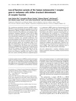
RE1 and REST-mediated gene repressionFigure 1
RE1 and REST-mediated gene repression. The expression levels in CD4+ T-cells are shown as boxplots for all human genes (All genes), RE1 genes without
REST (cRE1-REST and ncRE1-REST) and with REST (cRE1+REST and ncRE1+REST) in their promoters, and genes with RE1 motifs in the repetitive
sequences of their promoters (RpRE1-REST and RpRE1+REST). Conversely, the genes with REST in their promoters are also separated into two groups,
one with (REST+DJ-RE1) and the other without (REST-RE1) RE1s annotated in a previous study [19]. An asterisk indicates groups significantly (P < 0.001)
different from all human genes with respect to their expression scores.
0 500 1,000 1,500 2,000
Expression level
All genes
cRE1−REST
cRE1+REST *
ncRE1−REST
ncRE1+REST *
RpRE1−REST
RpRE1+REST
REST+DJ−RE1 *
REST−RE1 *
Genome Biology 2009, Volume 10, Issue 1, Article R9 Zheng et al. R9.6
Genome Biology 2009, 10:R9
which also had REST bound to their promoters. A compari-
son of these 115 cRE1/REST promoters and the remaining 33
cRE1 genes without REST showed clearly that binding of
REST to RE1s correlated with dramatic reduction in the
acetylation of H3K9 (Figure 3), a known target of HDACs
[10,46].
As gene repression is intimately correlated with histone
hypoacetylation [47], it is necessary to address to what extent
the observed histone deacetylation is merely a reflection of
gene repression rather than the direct target of REST com-
plexes. Therefore, we created two sets of genes as our con-
trols. Both control sets consisted of genes with neither an RE1
motif nor REST occupancy in their promoter regions, but one
set contained randomly chosen genes whose expression pro-
files matched that of cRE1/REST genes while the other set
exhibited expression as diverse as that of cRE1 genes without
REST binding. As such, the difference of a histone modifica-
tion between these two sets served as a reference for us to
determine the change contingent on gene expression but not
due specifically to REST occupancy on RE1 sites. As shown
here (Figure 3 and figures below), this strategy is highly
informative, and after taking into consideration the informa-
tion in our controls, we concluded that much of the reduction
in H3K9ac was in fact a direct consequence of REST binding
(Figure 3).
Further investigation of 17 additional lysine residues (Table 1)
in histones H2, H3, and H4 revealed significant REST-medi-
ated deacetylation in the following residues: H4K12, H4K5,
H4K8, H3K4, H3K18, H3K36, H2BK5, H3K27, and H3K9 (in
order of decreasing significance; Figure 4). As shown in Fig-
ure 3, the promoter profiles of H4K8ac and H3K9ac demon-
strated clearly that the binding of REST to cRE1 sites
Dynamics of nucleosomes near the promoter and non-promoter cRE1 modulated by REST bindingFigure 2
Dynamics of nucleosomes near the promoter and non-promoter cRE1 modulated by REST binding. The y-axis shows the normalized number of sequence
tags (in a 10 bp window) from the sense strand (red) and antisense strand (green). The x-axis shows the distance to the center of canonical RE1s (blue
box).
−1000 −750 −500 −250 −50
150
350 550 750 950
−2 −1 0 1 2
Promoter RE1 without REST
Distance to RE1
Nucleosome level
−1000 −750 −500 −250 −50 150 350 550 750 950
−1.0 −0.5 0.0 0.5 1.0
Non−promoter RE1 without REST
Distance to RE1
Nucleosome level
−1000
−750 −500 −250 −50 150 350 550 750 950
−1.0 −0.5 0.0 0.5 1.0
Promoter RE1 with REST
Distance to RE1
Nucleosome level
−1000 −750 −500 −250 −50
150
350 550
750
950
−1.0 −0.5 0.0 0.5 1.0
Non−promoter RE1 with REST
Distance to RE1
Nucleosome level
Genome Biology 2009, Volume 10, Issue 1, Article R9 Zheng et al. R9.7
Genome Biology 2009, 10:R9
correlated with reduced levels of histone acetylation. In both
cases, the magnitudes of deacetylation are significantly larger
than what were observed in their respective control groups
(Figure 3). For some other lysine residues the reduction of
their acetylations was prominent and significant, but the
change was not always greater than what was observed in
their corresponding controls (those not marked with an aster-
isk in Figure 4). Moreover, reductions of some specific
acetylations appeared more contingent on gene repression
than others (for example, H3K9ac versus H4K8ac; Figure 3).
While the systematic decline of histone acetylations likely
results from the actions of HDACs recruited by REST, the
decrease of H4K8ac appears to be inconsistent with a previ-
ous suggestion that an increase of H4K8ac would facilitate
and stabilize the binding of REST to RE1s through the associ-
ation of REST/CoREST and BRG1 [39], whose bromodomain
recognizes acetylated H4K8 (see Discussion).
As previously mentioned, REST binding to a promoter does
not always result in gene repression. However, our analyses
have revealed that even the upregulated cRE1 genes exhibited
REST-dependent deacetylations for most of the lysine resi-
dues interrogated (Figures 3 and 4). The REST-mediated his-
tone deacetylations were also analyzed for REST bound
ncRE1 genes. The magnitude of the reductions in histone
acetylations was largely comparable between REST-bound
H3K9ac and H4K8ac profiles in RE1 promotersFigure 3
H3K9ac and H4K8ac profiles in RE1 promoters. The profiles of these acetylations were generated and plotted for four groups of genes with different
colors (black, blue, red, and cyan), defined by the presences of cRE1, ncRE1, and REST in their promoters. The 'REST On & Exp Up' (red lines) refers to
the group of genes with cRE1 and REST but an expression score >300. The profiles of modifications for these RE1 genes are shown with solid lines. For
each of the four groups, a control was constructed by randomly selecting (5×) genes with the same expression levels but with neither RE1 nor REST in
their promoters (see Materials and methods). The profiles of these controls are shown with dashed lines and colors matching to their targeted group. For
the convenience of visual comparison, the zoom-in profiles for the four RE1 groups and their controls are re-drawn in the bottom panels. The color
scheme and line style in the bottom panels apply to Figures 5-7. The x-axis shows the distance to transcription start sites with a unit representing 200 bp,
and the y-axis shows the normalized counts of ChIP-Seq tags.
−20
−10
0
10 20
0246810
cRE1 & REST Off
−20
−10
0
10 20
01234
cRE1 & REST Off
−20 −10 0 10
20
0246810
cRE1 & REST On
−20 −10
0
10 20
01234
cRE1 & REST On
−20
−10 0
10
20
0246810
REST On & Exp Up
−20
−10 0 10 20
01234
REST On & Exp Up
−20 −10
0
10 20
0246810
ncRE1 & REST on
−20 −10 0 10 20
01234
ncRE1 & REST on
RE1 promoters
0246810
−10
−5 0
510
Non−RE1 genes as control
0246810
−10 −5
0
510
RE1 promoters
01234
−10 −5 0 5 10
Non−RE1 genes as control
01234
−10
−5 0 5 10
H4K8ac
H3K9ac
Genome Biology 2009, Volume 10, Issue 1, Article R9 Zheng et al. R9.8
Genome Biology 2009, 10:R9
ncRE1 genes and REST-bound cRE1 genes, in contrast to
cRE1 genes without REST (Figures 3 and 4). Therefore, our
results demonstrate convincingly that binding of REST to
RE1 promoters facilitates significant and broad histone
deacetylations.
REST binding correlates with reductions in histone
methylations implicated in gene activation
The extent of methylations on several lysine residues was also
found to be low in the group of cRE1/REST genes. In addition
to HDACs, LSD1 and SMCX are two other known histone
modifiers recruited by REST to remove H3K4 methylations.
Our data reveal that the cRE1/REST promoters had relatively
lower amounts of H3K4 methylations than the cRE1 promot-
ers without REST (Figure 5). The magnitude of the difference,
however, was smaller than what was seen for H3K4 acetyla-
tion, and appeared more prominent for H3K4me2 and
H3K4me3 than for H3K4me1 (Figure 5). However, these
reductions appeared inextricably linked to gene expression,
since the decline in H3K4 methylations was also very notice-
able in the genes of our controls, so that the changes in these
three methylations became statistically less significant by our
measurement, especially for the group of upregulated cRE1
genes (Figures 4 and 5). This observation is consistent with a
recent finding that the extent of H3K4me2/3 in several neu-
ronal genes was not affected by the introduction of a domi-
nant negative form of REST into the MPH36 neural stem cell
line [44].
In addition to H3K4 demethylations, REST also reduced the
levels of H3K27me1, H3K36me3 (Figure 6), H3K79me3,
H3K9me1, H2BK5me1, and H4K20me1; all of these methyla-
tion marks are enriched in the promoters of active genes
[47,48]. Whereas the enzymes for removing mono- (LSD1),
The P-values of paired t-test for comparing profiles between cRE1 promoters without REST and cRE1 with REST (or ncRE1 with REST, or cRE1 with REST and an expression value > 300)Figure 4
The P-values of paired t-test for comparing profiles between cRE1 promoters without REST and cRE1 with REST (or ncRE1 with REST, or cRE1 with REST
and an expression value > 300). The data for increased and decreased levels of modifications upon REST binding are shown in red and green, respectively.
Numbers are -log(10) transformation of P-values. An asterisk indicates histone modifications whose P-value from the comparison of RE1 genes is <0.0001
and at least ten times smaller than that from contrasting the corresponding control groups.
ncRE1 & REST on
15 10 5 0 5 10 15 20
cRE1 & REST on & exp up
15 10 5 0 5 10 15 20
cRE1 & REST on
15 10 5 0 5 10 15 20
H3K27me3*
H3K9me3*
H3K9me2*
H3K27me2
H4R3me2
H4K20me3
H3R2me2
H3K79me2*
H3K79me3*
H2AK5ac
H3K27me1*
H3K79me1
H4K12ac*
H4K5ac*
H4K8ac*
H2BK20ac
H3K36me3*
H3K4me1
H3K4ac*
H2BK12ac
PolII*
H3K9me1
H4K16ac
H3K18ac*
H3K36ac*
H2BK120ac
H4K91ac
H2BK5ac*
H3K27ac*
H2BK5me1
H3K4me2
H3K9ac*
H3K4me3
H4K20me1
H3K14ac
H2AZ
H3K23ac
H2AK9ac
H3R2me1
H3K36me1
Genome Biology 2009, Volume 10, Issue 1, Article R9 Zheng et al. R9.9
Genome Biology 2009, 10:R9
di- (LSD1 and SMCX) and tri-methylation of H3K4me3
(SMCX) are known to interact with REST/CoREST
[36,40,49,50] and LSD1 has also been suggested to act on
H3K9me [51], our data suggest that additional demethylases
could be recruited by REST because LSD1 and SMCX appear
unable to remove H3K36me3, H3K79me, and several other
methylation marks studied here [36,40,46], although some
recently identified JmjC domain-containing histone demeth-
ylases exhibit mixed activity profiles for H3K4 and H3K9
methylations [52,53].
REST binding correlates with enhancement of histone
methylations implicated in gene repression
While no histone residues in REST-bound cRE1 promoters
exhibited increased acetylation, several histone residues in
these regions displayed high amounts of methylations,
H3K4 profiles in RE1 promotersFigure 5
H3K4 profiles in RE1 promoters. The profiles are drawn in the same style as the bottom panels of Figure 3. The y-axis applies to a RE1 group and its
control (dashed lines).
H3K4me1
0246810
−20 −10 0 10 20 −20 −10 0 10 20
cRE1 & REST off
cRE1 & REST on
REST on & exp up
ncRE1 & REST on
H3K4me2
01234567
−20 −10 0 10 20 −20 −10 0 10 20
H3K4me3
0 10203040506070
−20 −10 0 10 20 −20 −10 0 10 20
H3K4ac
0123456
−20 −10 0 10 20 −20 −10 0 10 20
Genome Biology 2009, Volume 10, Issue 1, Article R9 Zheng et al. R9.10
Genome Biology 2009, 10:R9
including H3K27me2, H3K27me3, H3K9me2, H3K9me3,
and H4R3me2 (Figure 4 and 6). These modifications are
known to promote general gene repression [47,48], but the
surges in H3K27me3, H3K9me2, and H3K9me3 were higher
than what were observed in our control gene sets, indicating
that these changes are not simply a reflection of gene repres-
sion but are directly relevant to REST. In addition, it has been
reported that G9a, with a RING finger-like motif that inter-
acts with the carboxy-terminal domain of REST, could
increase the methylations in H3K9, predominantly di-meth-
ylation in nucleosomes within 2-kb regions of RE1s [41]. Our
analyses demonstrate that REST binding increased H3K9 di-
and tri-methylations but, surprisingly, reduced H3K9 mono-
methylations (Figures 4 and 7). We are not certain whether
the relatively uniform distribution of H3K9me2/me3 (that is,
no peak was detected) across TSSs could have contributed to
this intriguing observation, but we think the phenomenon
might be a consequence of competitive interaction between
G9a and LSD1, and a conversion of mono- to di- and tri-meth-
ylations. Since G9a is not known to methylate H3K27 in vivo
[41], our data suggest that REST likely interacts with addi-
tional histone methytransferase(s), such as polycomb repres-
sive complexes (PRCs).
REST binding has a similar influence on histone
modifications in promoter and non-promoter RE1 sites
We have also characterized the profiles of histone modifica-
tions near RE1 sites that are not in promoter regions. Here,
the profiles of histone modifications were anchored on the
centers of RE1 motifs (rather than TSSs for an obvious rea-
son). Such profiles were separately generated for non-pro-
moter RE1s with and without REST occupancy; for the
convenience of comparison, the profiles of histone modifica-
tions for promoter RE1s were also re-constructed using the
new anchoring system. Comparisons of the resulting profiles
demonstrate that, like binding to promoter RE1s discussed
above, the association of REST to non-promoter RE1s also
resulted in histone deacetylations and selective alterations of
H3K36me3 and H3K27me3 profiles in RE1 promoters, drawn in the same style as Figure 3Figure 6
H3K36me3 and H3K27me3 profiles in RE1 promoters, drawn in the same style as Figure 3. The y-axis applies to a RE1 group and its control (dashed lines).
−20
−10 0
10 20
0.0 0.5 1.0 1.5 2.0 2.5
cRE1 & REST off
−20 −10 0 10 20
0.0 0.5 1.0 1.5 2.0 2.5
cRE1 & REST off
−20
−10
01020
0.0 0.5 1.0 1.5 2.0 2.5
cRE1 & REST on
−20 −10 0 10 20
0.0 0.5 1.0 1.5 2.0 2.5
cRE1 & REST on
−20 −10 0
10
20
0.0 0.5 1.0 1.5 2.0 2.5
REST on & exp up
−20 −10 0 10 20
0.0 0.5 1.0 1.5 2.0 2.5
REST on & exp up
−20
−10 0
10 20
0.0 0.5 1.0 1.5 2.0 2.5
ncRE1 & REST on
−20
−10 0 10 20
0.0 0.5 1.0 1.5 2.0 2.5
ncRE1 & REST on
RE1 promoters
0.0 0.5 1.0 1.5 2.0 2.5
−10 −5
05
10
Non−RE1 genes as controls
0.0 0.5 1.0 1.5 2.0 2.5
−10
−5 0 5
10
RE1 promoters
0.0 0.5 1.0 1.5 2.0 2.5
−10
−5
0
510
Non−RE1 genes as controls
0.0 0.5 1.0 1.5 2.0 2.5
−10 −5 0 5
10
H3K36me3 H3K27me3
Genome Biology 2009, Volume 10, Issue 1, Article R9 Zheng et al. R9.11
Genome Biology 2009, 10:R9
a number of heterogeneous histone methylation profiles
(Table 1). The outcomes for canonical and ncRE1s were
almost identical, though minor and subtle variations existed.
It is interesting to note that H3R2me1/2 did not exhibit a
change in our analyses, as they are often associated with het-
erochromatin and generally do not affect gene expression
[46] (Table 1). Moreover, not all histone modifications impli-
cated in gene activation (for example, H3K36me1) or repres-
sion (H4K20me) displayed a detectable change. Taken
together, our data (Table 1) indicate that REST-mediated his-
tone modifications are more prominent at, but not restricted
to, RE1s in promoter regions.
H3K9 and Pol II profiles in RE1 promotersFigure 7
H3K9 and Pol II profiles in RE1 promoters. The profiles are drawn in the same style as the bottom panels of Figure 3. The y-axis applies to a RE1 group
and its control (dashed lines).
H3K9me1
0
123456
−20 −10 0 10 20
cRE1 & REST off
cRE1 & REST on
REST on & exp up
ncRE1 & REST on
−20 −10 0 10 20
H3K9me2
0.0 0.2 0.4 0.6 0.8 1.0 1.2
−20 −10 0 10 20 −20 −10 0 10 20
H3K9me3
0.0 0.2 0.4 0.6 0.8 1.0 1.2
−20 −10 0 10 20 −20 −10 0 10 20
Pol II
0
24
6810
12
−20 −10 0 10 20 −20 −10 0 10 20
Genome Biology 2009, Volume 10, Issue 1, Article R9 Zheng et al. R9.12
Genome Biology 2009, 10:R9
Correlation between RE1 motif strength, REST
binding, and histone modifications
As shown in Figures 3, 4, 5, 6, and 7, binding of REST to
ncRE1s caused dramatic loss of histone acetylations and sev-
eral key histone methylations, for example, H3K4me1 (Figure
5), H3K27me1 and H3K36me3 (Figure 6). For most histone
modifications, the patterns of change were very similar for
cRE1 and ncRE1 genes. However, to our initial surprise, the
magnitude of these changes appeared to be larger in ncRE1
than cRE1 sites when REST-bound ncRE1 and cRE1 genes
were compared in reference to cRE1 genes without REST.
Upon close inspection, we found that the average of our
ncRE1 PSFM scores was higher than that of cRE1s (data not
shown), suggesting that the degree of REST-mediated histone
modifications may be affected by the affinity of a RE1 motif
for REST. Such a correlation would also explain the signifi-
cant correlation of PSFM score with the strength of gene
repression regulated by REST [17].
In order to characterize this important observation in detail,
we examined all promoters bound by REST and utilized the
RE1 motifs and their normalized PSFM scores provided by
Johnson et al. [19]. Those RE1s are referred to as DJ-RE1
motifs here, which were generated with a lower threshold of
PSFM score than what was used in our own RE1 identification
process; they therefore represented an expansion of our lists
of RE1 sites (and consequently genes). We then computed the
correlations between the PSFM scores of the canonical DJ-
RE1s in REST-bound promoters and the extent of various his-
tone modifications (using the total number of ChIP-Seq reads
within ± 500 bp of DJ-RE1s as a metric). The results clearly
demonstrate that the amounts of all histone acetylations were
negatively correlated with the strength of RE1 motifs (Table
2). Many of these correlations were quite strong and highly
significant, such as those for H4K91ac, H2BK120ac, H3K4ac,
and H3K9ac (r < -0.2; Table 2). The PSFM scores of RE1
motifs also appear to be strongly but negatively correlated
with several histone methylations, including H3K36me1,
H3K4me3, H3K27me1, H3K4me2, H3K79me1, and
H3K9me1 (Table 2). For those methylations positively corre-
lated with the RE1 scores, the correlation coefficients were
relatively lower, but good correlations existed for H3K9me2,
H4K20me3, and H4R3me2. Interestingly, we found that the
correlation was positive for H3K9me2 but negative for
H3K9me1 (Table 2), suggesting a possible conversion of
mono- to di-/tri-methylations. The levels of H2A.Z (r = -
0.156) and Pol II (r = -0.132) present in promoters also
showed a negative correlation with the strength of RE1
motifs, consistent with RE1's general role in repressing tran-
scription.
A significant and positive correlation was found between the
RE1 PSFM scores and the amount of REST occupancy (r =
0.178), in agreement with previous finding that the fraction of
RE1 sites occupied by REST increases with RE1 motif scores
[19]. This correlation intriguingly did not lead to a highly par-
allel relationship between RE1 and REST with respect to their
separated correlations with individual histone modifications,
though the signs of these correlations were consistent; that is,
a negative correlation between a histone modification and
RE1s was usually accompanied by a negative correlation
Table 2
Pearson correlation coefficients between the levels of histone
modification and PSFM score, and REST occupancy
Factor PSFM scores of DJ-cRE1 REST ChIP-Seq reads
H2AK5ac -0.119 -0.141
H2AK9ac -0.074 0.138
H2BK120ac -0.226* -0.061
H2BK12ac -0.202* -0.101
H2BK20ac -0.220* -0.055
H2BK5ac -0.174* -0.072
H3K14ac -0.139 -0.054
H3K18ac -0.177* -0.102
H3K23ac -0.13 -0.024
H3K27ac -0.193* -0.070
H3K36ac -0.226* -0.120
H3K4ac -0.213* -0.119
H3K9ac -0.202* -0.084
H4K12ac -0.153 -0.050
H4K16ac -0.104 -0.053
H4K5ac -0.136 -0.071
H4K8ac -0.136 -0.007
H4K91ac -0.228* -0.103
H2BK5me1 0.036 0.413*
H3K27me1 -0.121 -0.018
H3K27me2 -0.075 -0.170*
H3K27me3 -0.027 -0.187*
H3K36me1 -0.203* 0.012
H3K36me3 -0.026 -0.108
H3K4me1 -0.059 -0.028
H3K4me2 -0.157 -0.041
H3K4me3 -0.200* -0.093
H3K79me1 -0.125 -0.135
H3K79me2 -0.096 -0.092
H3K79me3 -0.110 -0.044
H3K9me1 -0.147 0.052
H3K9me2 0.116 0.375*
H3K9me3 0.044 -0.091
H3R2me1 0.023 0.273*
H3R2me2 -0.041 0.355*
H4K20me1 0.081 0.418*
H4K20me3 0.13 0.039
H4R3me2 0.102 0.484*
H2AZ -0.156 -0.086
PolII -0.132 0.075
PSFM score - 0.178*
Significant correlations are marked by an asterisk.
Genome Biology 2009, Volume 10, Issue 1, Article R9 Zheng et al. R9.13
Genome Biology 2009, 10:R9
between REST and this particular modification (Table 2). But
the strengths of the correlations were often different. Moreo-
ver, no correlation was found between REST occupancy and
many histone modifications that exhibited a strong correla-
tion (r < -0.2) with RE1, such as H3K9ac, H2BK20ac,
H2BK120ac, and H3K36me1 (Table 2). Conversely, REST
abundance was correlated strongly (r > 0.2) with the levels of
H4K20me1, H2BK5me1, H3R2me1, H3R2me2, and
H4R3me2, but these histone modifications had no or weak
correlations with the RE1 PSFM scores (Table 2). While these
observations certainly need to be further characterized, with
cross-reactions of immunoprecipitation antibodies being
considered, they suggest that the relationship between RE1
motifs and REST occupancy is extremely complex and heter-
ogeneous, and perhaps inextricably linked to the modular
nature of REST complexes. For instance, as REST uses its
amino- and carboxy-terminal domains to recruit two distinct
groups of histone modifying enzymes and some REST protein
isoforms are truncated at the carboxyl terminus [10,24-
26,54,55], these patterns of correlations could be caused by
the presence of more than one REST isoform in T cells (see
Discussion for more details).
Discussion
Since the discovery of REST as a repressor for neuronal genes,
many studies have provided significant insights into the cel-
lular and molecular roles of REST in regulating diverse bio-
logical processes. What emerges from current literature is a
picture of dynamic REST complexes composed of multiple
proteins, many of which are involved in differentially estab-
lishing and regulating specific profiles of histone modifica-
tions or DNA methylation [8,10]. These REST-associated and
REST-dependent complexes cooperatively modulate the epi-
genetic properties in RE1 regions dynamically and help to
establish and maintain the cell- and tissue-specific expression
patterns of diverse classes of neuronal genes (and non-neuro-
nal genes as well). In this study, we have characterized 38 of
60 known histone modification sites [46,56], and provided a
broad overview of how REST macromolecular complexes
modulate histone modifications in human T cells. The results
of our study can be schematically summarized (Figure 8) in
spite of the complexity. Many of our observed changes have
been reported previously and, furthermore, the correspond-
ing enzymes have been identified (Figure 8) [10]; thus, strong
experimental evidence exists for some of our results, but a
significant subset of these histone modifications, particularly
those on H2 and H4, are now characterized for the first time
in our study. Moreover, our genome-wide analyses have iden-
tified some REST-mediated histone modifications (for exam-
ple, H4K8ac, H3K9me) that extend previous findings based
on studying a limited number of neuronal genes to novel
observations concerning their putative regulatory roles.
Promoter and non-promoter RE1s exhibit similar
affinity for REST and comparable profiles of REST-
mediated histone modifications
Most of our discussions have focused on the RE1/REST inter-
action in promoter regions of protein coding genes. This is
primarily due to the fact that the relationship between histone
modifications and gene expression is much better docu-
mented for promoters (or near TSSs) than for any other
regions [46,47]. The enrichments of many well-characterized
histone modifications in promoters [48,56,57] certainly war-
rant our choice of promoter RE1/REST as the focal point of
our report. Nevertheless, our comparisons of histone modifi-
cations in promoter and non-promoter RE1 regions produced
very similar results in terms of the influence of REST on local
histone modifications (Table 1), which strongly suggests that
REST coordinated histone modifications are not solely pro-
moter-dependent. This is an important general observation
considering that only about 10% of RE1s are located near
TSSs, whereas most RE1s are >50 kb away from protein cod-
ing genes. Overall, binding of REST to these non-promoter
RE1s (or remote RE1s) resulted in similar perturbations of
histone modifications as REST binding to promoter RE1s.
However, we cannot exclude the possibility that such changes
when examined in much greater detail may exhibit more sub-
tle and functional differences. For example, RE1/REST near
enhancers may be associated with a distinct pattern of histone
modifications. Our study represents the beginning of a much
more exhaustive inquiry regarding REST-mediated chroma-
tin remodeling, and in the future we plan to address many of
these seminal issues by designing experiments to separately
interrogate the differential profiles of chromatin remodeling
coordinated by REST but nevertheless occurring within dis-
tinct genomic, molecular and cellular contexts.
We did not characterize ncRE1s without REST occupancy in
this study. Compared to the high percentage of cRE1s with
bound REST, relatively few (11.5%) of our annotated ncRE1s
were occupied by REST. We believe this is largely due to the
current technical limitation of recognizing genuine ncRE1s in
the human genome. Nevertheless, it will be essential to deter-
mine whether macromolecular complexes recruited by RE1/
REST are substantially different from those recruited by
ncRE1/REST, though our data as well as previous studies
[19,20] have not sufficiently addressed the existence of such a
scenario.
REST-associated nucleosome reorganization and
histone modifications
With integrated high-resolution data, we have begun to illus-
trate the complex genome-wide landscape of chromatin
remodeling coordinated by a single transcription factor. Our
results indicate that the majority of RE1 sites are accessible
and bound to REST in differentiated T cells. Although we do
not know what triggers the association of REST with a partic-
ular RE1, our analyses have shown that REST binding induces
nucleosome repositioning, profound histone deacetylations,
Genome Biology 2009, Volume 10, Issue 1, Article R9 Zheng et al. R9.14
Genome Biology 2009, 10:R9
Figure 8 (see legend on next page)
RE1
REST
Nucleosome
BRG1
HDACs Methylases Demethylases
G9a
LSD-1
mSin3
coREST
REST
(a)
(b)
(c)
Acetyl group
Methyl group
HDAC1
HDAC2
??
??
??
PRC2
??
G9a
LSD-1
PRC2
SMCX
SMCX
H3K9ac, H4K8ac
H4K12ac, etc.
H3K9me,
H3K27me, etc.
H3K4me, H3K36me
H3K79me, etc.
Genome Biology 2009, Volume 10, Issue 1, Article R9 Zheng et al. R9.15
Genome Biology 2009, 10:R9
removal of histone methylation marks highly implicated in
gene activation, and the addition of selective methylations
involved in gene repression (Figures 4 and 8). It needs to be
mentioned that cRE1 genes without bound REST also exhib-
ited similar properties but to a much smaller extent compared
to genes without RE1s and evidence of REST binding (Figures
3, 4, 5, 6, and 7). This is likely due to the use of a threshold for
identifying regions enriched with REST; that is, some REST-
free regions might actually be occupied by a small number of
REST molecules.
It is important to emphasize that our data provide only a glo-
bal and static view of the consequence of REST binding, but
REST-mediated chromatin remodeling is a highly regulated,
cooperative, and sequential process orchestrated by a large
number of histone remodeling proteins, with some modifica-
tions occurring after and dependent upon earlier modifica-
tions [10]. For instance, removal of the acetyl group on H3K9
stimulates LSD1 activity, which subsequently removes methyl
groups from H3K4 [49]. Intriguingly, it was previously
reported that increased H4K8ac could potentially facilitate
the recruitment of REST to RE1 regions. The phase/position
of nucleosomes facilitated by REST in RE1 regions (Figure 2)
is consistent with this hypothesis as BRG1, one of the ATPases
of the SWI/SNF complex with a bromodomain that can rec-
ognize H4K8ac, can help reposition nucleosomes with
respect to DNA [39]. Our data nevertheless suggest that
H4K8ac might be relevant only in the initial recruitment of
REST, as it was found to decrease upon REST binding (Figure
3). After the interaction of REST and RE1s is established,
however, the subsequent recruitment of HDACs can presum-
ably lead to H4K8 deacetylation.
Our results suggest that histone modification enzymes other
than HDACs, LSD1, SMCX, and G9a may also interact with
REST and, thus, there are potentially additional and essential
components of REST co-repressors (Figure 8). Specifically,
our analyses found that REST binding changed the level of
several histone methylations that are not known targets of
currently identified REST co-repressors. The most noticeable
such histone marks are H3K27me2/me3, which were
increased upon REST binding, and H3K27me1 and
H3K36me3, which were conversely decreased by the pres-
ence of REST complexes (Figure 4). Of course, it is possible
that these histone methylations are in vivo targets of LSD1,
SMCX, or G9a, but such catalytic relationships have not yet
been elucidated. However, a more likely alternative scenario
is that previously unrecognized or never characterized his-
tone modification enzymes are present within distinct REST
macromolecular complexes, with PRC2 being a primary can-
didate as it catalyzes H3K27 tri-methylation (Figure 8) [46].
Moreover, our evolving comprehension of the multiple roles
played by individual histone modifications suggests that
these REST-associated chromatin remodeling events need to
be examined within the broader context of the fine-tuning of
local transcriptional control as well as more genome-wide
effects on heterochromatin dynamics, boundary elements,
and gene networks. Furthermore, it will be highly interesting
to study how the RE1/REST sites are enriched for LSD1,
SMCX, G9a, HDACs, or other histone modifying enzymes
when genome-wide ChIP-Seq data for these factors become
available in the future. This is also essential for further
unraveling the REST function in detail because it has been
shown that the composition of the REST complexes in differ-
ent RE1 genes could be different [44].
Histone deacetylases, methyltransferases, and
demethylases might have subtle and distinctive roles in
transacting REST functions
There is no doubt that binding to RE1s and the subsequent
recruitment of histone modifying enzymes are two activities
central to the REST regulatory network. These two primary
roles of REST are interdependent as reflected by the correla-
tions among RE1 motif score, REST occupancy, and levels of
various histone modifications (Table 2). However, the rela-
tionships appear much more complex and nuanced as multi-
ple proteins are involved in a sequential and interdependent
manner. As a result, we have found that most correlations
exist but are not particularly robust as judged by visual
inspection or statistical measurement. In particular, we did
not detect a significant functional interrelationship between
RE1 affinity and REST abundance with respect to their corre-
lations with the degree of individual histone modifications,
despite the observation that the correlations occurred in a
parallel mode (Table 2). The biochemical specificity and sen-
sitivity of some antibodies in ChIP-Seq might have contrib-
uted to this lack of parallel correlations. However, we do not
think that such a lack of a strong parallel relationship is
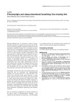
A schematic diagram illustrating the major components involved in REST-mediated local chromatin remodeling and their relationships to our findingsFigure 8 (see previous page)
A schematic diagram illustrating the major components involved in REST-mediated local chromatin remodeling and their relationships to our findings. (a)
RE1 is initially covered by a nucleosome. (b) A yet-to-identified cellular mechanism initiates nucleosome repositioning with the assistance of BRG1,
resulting in the exposure of the RE1 motif and the subsequent occupation of it by REST. The exact sequential order is not clear to date. (c) With the
assistance of mSin3 and coREST, the RE1-bound REST complexes then recruit histone deacetylases (HDACs) to promote histone deacetylations, histone
methylases (G9a, PRC2) to increase methylations on H3K9 and H3K27, and histone demethylases (LSD1, SMCX) to reduce methylations on H3K4. The
presence of PRC2 in REST complexes is unknown but suggested by our analysis, so we have drawn a dashed line around it. Our data also strongly suggest
that REST can recruit additional histone methylases and demethylases (represented by question marks) to target other lysine residues of histones, which
display RE1/REST-dependent changes in the current study. The enumeration of all the histone modifying enzymes in the REST complexes will enhance our
comprehension of how the complicated histone modifications are established; then, more investigations will be needed to decipher how these
modifications cross-talk and orchestrate the regulation of RE1 genes.
Genome Biology 2009, Volume 10, Issue 1, Article R9 Zheng et al. R9.16
Genome Biology 2009, 10:R9
caused by the fact that our REST binding data and histone
modification data came from different lines of T cells.
Instead, we believe that the weak interrelationship reflects
the complex amalgam of REST functional roles and dynamic
and more global molecular processes. Fundamentally, his-
tone modification is a highly regulated process not solely
dependent on REST interactions. It is also known that REST
utilizes its amino- and carboxy-terminal domains to recruit at
least two distinct groups of histone modifying enzymes.
Although it is still largely undefined how these two domains
and their associated co-repressors and complementary co-
modulatory complexes promote histone and higher-order
chromatin crosstalk, REST isoforms with altered or truncated
carboxy-terminal domains have been detected in neuronal
cells [26,54]. These different REST isoforms must possess a
spectrum of different activity profiles associated with their
binding of RE1s and the recruiting of selective histone modi-
fying enzymes. In fact, one truncated isoform, REST4, has
been found to have a lower affinity for DNA and to activate
the expression of neuronal genes by antagonizing the normal
function of full-length REST [24-26,55]. Although these
REST isoforms have not been reported in T cells, their pres-
ence would certainly help to explain at least a subset of our
observations regarding the intricate functional interrelation-
ship between RE1 binding affinity and REST occupancy, since
our current analyses and the underlying datasets cannot dis-
tinguish between different REST isoforms.
We suggest that the amino- and carboxy-terminal domains of
REST and their associated histone modifying enzymes might
have very distinct and subtle roles in the overall scheme of the
REST regulatory network. The HDACs recruited by REST are
less selective in their targeted residues, as manifested by the
significant reductions in broad histone acetylations observed
in our analyses; the histone demethylases and methytrans-
ferases recruited by REST are much more discriminative in
their molecular targets [10,46,56], as exemplified by the
diverse and complex changes in specific histone methylations
correlated with REST binding. As a result, HDACs appear to
induce a broad repression of RE1/REST genes, whereas the
histone demethylases and methyltransferases can cooperate
and dynamically alter the profiles of methylations on individ-
ual nucleosomes in a more selective, context-specific and
nuanced manner, and thus create an elaborate platform for
histone code readers [42]. As a result, the interaction of his-
tone methylases and methyltransferases has the potential to
fine-tune the expression levels and functions of individual
RE1 genes and to integrate gene networks in response to dis-
tinct developmental, environmental and interceptive cues
and imperatives. Furthermore, the distinct roles of REST-
mediated histone acetylation and methylation could be
important for multiple developmental processes, as histone
methylation has been considered to be more stable than
acetylation. In the future, we plan to address our hypothesis
with a double immunoprecipitation ChIP-Seq to define how
REST and a histone modification are correlated at the molec-
ular level.
Selection of a subset of histone marks for studying
REST-mediated chromatin remodeling
Our results (Figure 4, Table 1) indicate that it may be possible
and instructive to use a subset of histone marks to capture the
dynamic range of epigenetic modifications orchestrated by
REST. Although a genome-wide high-resolution map of his-
tone modifications can be readily obtained with the next gen-
eration high-throughput sequencing technology, it is unlikely
that we will be able to examine every possible post-transla-
tional modification of histones in every cell, particularly in a
dynamic fashion required to fully elucidate the functional sig-
nificance of the integrated higher-order chromatin code and
the associated spectrum of epigenetic modifications in the
foreseeable future. Therefore, we propose a subset of repre-
sentative and instructive histone marks that can be used to
investigate the overall patterns associated with REST-medi-
ated chromatin remodeling. Based on our results (Table 1,
Figure 4) and the observation that many modifications are
highly correlated with respect to their patterns of alterations
by REST in cRE1 promoters (data not shown), the primary
candidates for further study are H3K4ac, H3K9ac, H4K8ac,
H3K9me1, H3R2me1, H3K27me3, H3K36me3, and
H4R3me2. We believe that a survey of these histone marks
will provide considerable insight into the histone modifica-
tion platforms orchestrated by RE1/REST interaction in cells
(or tissues) and genes of interests.
Furthermore, we think that studying histone modifications in
a wider variety of cells will be essential for expanding our
knowledge of REST functions and will likely be more fruitful
than investigating a wider spectrum of histone modifications
in a more limited range of cell types over time or in response
to specific activation or stressor states. In particular, it will be
highly valuable to study whether the complicated and hetero-
geneous profiles of histone modifications, defined here for
RE1/REST in T cells, are specific in non-neuronal cells, and if
not, how the patterns evolve in neuronal cells. It is easy to
envisage that such a sophisticated and modulated epigenomic
remodeling program can play a significant role in neuron dif-
ferentiation and maturation. We also believe that studies of
additional classes of REST interacting factors, such as multi-
functional heterochromatin binding proteins (for example,
Heterochromatin protein 1, which interacts with G9a [41]),
DNA methylation effectors (for example, MeCP2, which rec-
ognizes methyl-DNA [58]) and specific subclasses of short
and longer non-coding RNAs that may promote sequence-
specific chromatin-modifications [59,60], will provide addi-
tional mechanistic insights required for an overall under-
standing of REST-mediated chromatin remodeling.
Genome Biology 2009, Volume 10, Issue 1, Article R9 Zheng et al. R9.17
Genome Biology 2009, 10:R9
REST-mediated histone modifications can be
associated with enhancement of gene expression in T
cells
The focus of this study has been to elucidate the local and
more global influences of REST on histone modifications. As
histone modifications, especially those on H3, are intrinsi-
cally linked to gene expression [46,47], we have constructed
and studied gene control groups to computationally 'uncou-
ple' this linkage in order to determine accurately the changes
of histone modifications that depend on REST binding to RE1
sites. Nonetheless, the predominant outcome of REST bind-
ing is overall gene repression in T cells (Figure 1), in accord-
ance with the originally proposed role of REST as a
transcriptional repressor to silence neuronal genes in non-
neuronal cell lineages. At the level of individual genes this is
surely more complex and dynamic as we have only examined
promoters and one particular aspect of the REST regulatory
network - histone modifications - whereas the expression of
most genes is regulated at multiple levels and by several inter-
related epigenetic mechanisms and both local and global
genomic modulatory processes.
We have examined a small group of genes with cRE1 and
REST in their promoters that were nonetheless highly
expressed in human T cells. These genes, such as CLK2,
DPH2 and RAB37, seemingly are not specifically related to
neuronal or T-cell development. The histone modification
data in the promoters of these genes are very valuable as they
have helped to demonstrate that REST binding is the cause of
reduced levels of histone acetylations and is not entirely con-
tingent on gene expression, and that the influence of REST on
methylations was much more complex than expected (Figure
4). For example, H3K4ac was lower in RE1 genes with REST
binding regardless of high or low levels of gene expression,
but the degree of H3K4 methylations (especially H3K4me1)
was noticeably higher only in the group of cRE1/REST genes
with up-regulated expression (red line in Figure 5). Several
other methylations whose magnitude of change was relatively
more contingent on expression level also exhibited such a pat-
tern (Figure 4), supporting our proposed role of methylations
in fine-tuning the expression of RE1/REST genes. In particu-
lar, compared to cRE1 genes without REST, H4K20me1 was
decreased upon REST binding but slightly increased in upreg-
ulated cRE1 genes with REST (Figure 4). The effects of the
H4K20me1 histone mark are known to be complex and con-
text-specific, including roles in active transcription, hetero-
chromatin formation, and DNA repair [46], as well as
potentially serving as a binding platform for the bifunctional
JMJD2A H3K9me2/3 and H3K36me2/3 histone demethyl-
ase [61]. The level of Pol II in the promoters of this group of
genes is also quite intricate as it is much lower than that of
REST-free cRE1 genes (Figure 7), suggesting that transcrip-
tion initiation might not be the key factor responsible for the
increased numbers of transcripts for these genes. Although
we cannot exclude the possibility that the REST co-repressors
associated with these genes might have a distinct molecular
configuration, we found that both the RE1 motif score and the
number of REST ChIP reads in these upregulated genes were
not statistically different from their corresponding values for
downregulated cRE1 genes with bound REST (p-value = 0.13
and 0.35, respectively). These observations suggest that
either additional pathways unrelated to REST are involved in
regulating the histone modifications (and consequently
expression) of these cRE1 genes, or other component(s) asso-
ciated with REST must exist to overcome the demethylation
activities of REST-associated LSD1 and SMCX. Non-coding
RNAs similar to the dsNRSE in rat neuronal stem cells [29]
certainly would be excellent and unique candidates for the
latter, since it has been shown that double-stranded RNA in
promoter regions can modulate histone modifications [62].
Conclusions
We have integrated multiple sets of genomic data obtained
from motif prediction, gene expression, and ChIP-Seq to
characterize in details the complex landscape of nucleosome
modifications mediated by RE1/REST interactions. Our
study reveals that the binding of REST to RE1 induces dra-
matic context-dependent chromatin remolding, including
nucleosome repositioning/phasing, systematic decline of
local histone acetylations and some key histone modifications
but increase of a different set of important histone modifica-
tions. Our findings show convincingly that REST-mediated
chromatin remodeling is extremely dynamic and complex
with novel histone modifying enzymes to be identified. Our
work provides valuable information for appreciating the com-
plexity of the REST regulatory network, and for further
decoding the roles of REST and its corepressors in stem cells,
and neuronal and non-neuronal lineage cells.
Materials and methods
Identification of RE1 sites in the human genome
The occurrences of the DNA motifs (RE1 sites) recognized by
REST were identified using the PSFM from the software
package Cistematic [17]. The PSFM was derived from a large
set of known instances of REST binding sequences and a set
of known negative cases. An efficient motif scanning algo-
rithm was implemented and a conserved threshold of 84% of
the best possible score [17] was used to select RE1 sites.
Whereas RE1 sites of 21 bp were called cRE1s (cRE1s), ncRE1s
(ncRE1s) refer to the RE1s with their left and right half sites
(10 bp each) separated by 0 or 3-9 nucleotides. The binding of
REST to ncRE1s was discovered recently by genome-wide
REST ChIP analyses; the ncRE1 motif has been found to be
highly similar to that of cRE1s, except for the non-conserved
distance between their two half sites. Therefore, we used the
same PSFM for cRE1s and ncRE1s but allowed various nucle-
otide insertions in ncRE1s. The program RepeatMasker was
used to identify repetitive regions in the human genome; then
RE1 sites fully embedded in repeats were designated as repeat
RE1s. We further segregated RE1s into promoter RE1s and
Genome Biology 2009, Volume 10, Issue 1, Article R9 Zheng et al. R9.18
Genome Biology 2009, 10:R9
non-promoter RE1s based on their locations with respect to
the promoters (-5 kb to +1 kb from the TSS) of known genes.
Accordingly, the genes with RE1 or REST in their promoters
were then termed RE1 genes or REST genes. The transcrip-
tion levels of known human genes in CD4+ T cells were
obtained from a previous microarray analysis [43]. The data
were processed using the Affymetrix software MAS 5.0
(MAS5) and low and not expressed genes had an expression
score <200.
Positioning of nucleosomes in relation to RE1 sites
The positioning of nucleosomes near RE1s was characterized
with the genome-wide map of nucleosome positions in rest-
ing CD4+ T cells constructed by direct high-throughput
sequencing of nucleosome ends [45]. The density of nucleo-
somes was profiled by totaling the reads mapped to a 10 bp
window sliding from -1 kb to +1 kb from the center of cRE1s.
The reads aligned to sense and antisense strands were treated
separately.
ChIP-Seq data for REST-bound regions and histone
modifications
The human genomic regions bound with REST were obtained
from a ChIP-Seq assay using a monoclonal antibody against
REST in Jurkat T cells [19]. The REST data included a list of
genomic regions with numbers of mapped ChIP-Seq reads.
The authors also provided the locations of RE1s (with their
PSFM scores) within or adjacent to each of these REST bound
regions. These RE1s were called DJ-RE1s as they were gener-
ated with a motif score threshold lower than what was used in
the current study. This is feasible because the identification of
DJ-RE1s was applied only to sequences near genomic regions
with REST binding; otherwise, this threshold would result in
a great number of false positive RE1s.
The genome-wide data for histone modifications have been
described in two previous studies, one targeted at histone
acetylations [57] and the other focused on histone methyla-
tions (plus H2A.Z and RNA polymerases II) [48]. The specif-
icities of individual antibodies have been described [48,57].
The data are lists of genomic coordinates for individual ChIP-
Seq reads that could be mapped to the human genome unam-
biguously.
Generation and comparison of aggregated profiles of
histone modifications
RE1 sites binding REST were inferred computationally by
intersecting the predicted RE1 sites with the REST-bound
regions. To construct an aggregated profile of a histone mod-
ification for RE1 promoters (or RE1 sites), we summed the
ChIP-Seq reads in a window of 200 bp moving from -5 kb to
+5 kb of TSSs (or the center of RE1s where applicable). This
profile was then normalized by sample size (for example,
number of TSSs) to generate average histone modifications
spanning TSSs (or RE1s) for subsequent direct comparisons.
Moreover, in the comparisons of profiles with and without
REST in their promoters, a control group was a set of genes
whose expression scores in CD4+ T cells matched to those of
genes under investigation. For example, to compare the pro-
files of cRE1 genes with REST (group A) and without REST
(group B), five genes without a RE1 and REST were selected
randomly from the pool of all human genes for every gene in
group A on the condition that these six genes would have the
same expression score. Application of this approach to group
A thus yielded a control group A', likewise B' for group B.
Paired t-test was then used to quantify statistically the differ-
ence between groups A and B, and the corresponding P-value
is shown in Figure 4. In this study, the difference between
groups A and B would not be considered significant unless the
P-value was <0.0001 and at least ten times smaller than the
corresponding P-value from the comparison of groups A' and
B'. The goal was to computationally uncouple the change (of
histone modifications) directly modulated by REST from that
intimately correlated with gene repression. As shown in Fig-
ures 3, 4, 5, 6, and 7, this strategy was both effective and
highly informative. The profiles anchored on the center of
RE1s were visually compared for determining the outcome of
REST binding to promoter and non-promoter RE1s.
Correlation of RE1 PSFM score, REST occupancy and
histone modifications
We used the DJ-RE1s for studying the correlation between
the strength of RE1s and the degree of histone modifications,
because these RE1 had a bigger range of PSFM scores than
those RE1s identified in current work. Moreover, these RE1s
were derived from genomic regions known to bind REST in
vivo [19] and thus should have very low false positive sites.
The DJ-RE1s were also used to characterize the correlation
between RE1 motif score and REST occupancy, and that
between REST binding and histone modifications. The metric
for REST occupancy was the number of ChIP-Seq reads
obtained from the previous study [19], and the metric for a
histone modification (or H2A.Z and PolII) is defined here as
the number of ChIP-Seq reads within 500 bp of RE1s. An
alternative window size of ± 2 kb yielded similar results.
Abbreviations
ChIP: chromatin immunoprecipitation; ChIP-Seq: cChIP and
high-throughput sequencing; cRE1: canonical RE1; HDAC:
histone deacetylase; L2: type 2 long interspersed nuclear ele-
ment; LSD: lysine specific demethylase; ncRE1: non-canoni-
cal RE1; NRSE: neuron-restrictive silencer element; PRC:
polycomb repressive complex; PSFM: position specific fre-
quency matrix; RE1: repressor element 1; REST: RE1 silenc-
ing transcription factor; TSS: transcription start site.
Authors' contributions
DZ and MM conceived of the study. KZ's group produced all
the ChIP-Seq data for histone modifications and nucleosome
positioning. DZ designed the experiments and carried out the
Genome Biology 2009, Volume 10, Issue 1, Article R9 Zheng et al. R9.19
Genome Biology 2009, 10:R9
analyses. DZ interpreted the results with help from KJ. DZ,
KJ and MM wrote the paper together. All authors read and
approved the final manuscript.
Acknowledgements
This work was supported by startup funds from Albert Einstein College of
Medicine of Yeshiva University (to DZ), by research funds from the Intra-
mural Research Programs of National Heart, Lung, and Blood Institute,
National Institutes of Health (NIH) (to KZ), and by grants from the NINDS
and NIMH, NIH and from the Roslyn and Leslie Goldstein, the Mildred and
Bernard H Kayden, the FM Kirby, the Alpern Family and the Rosanne H Sil-
berman Foundations (to MFM).
References
1. Chong JA, Tapia-Ramirez J, Kim S, Toledo-Aral JJ, Zheng Y, Boutros
MC, Altshuller YM, Frohman MA, Kraner SD, Mandel G: REST: a
mammalian silencer protein that restricts sodium channel
gene expression to neurons. Cell 1995, 80:949-957.
2. Mori N, Schoenherr C, Vandenbergh DJ, Anderson DJ: A common
silencer element in the SCG10 and type II Na+ channel genes
binds a factor present in nonneuronal cells but not in neuro-
nal cells. Neuron 1992, 9:45-54.
3. Schoenherr CJ, Anderson DJ: The neuron-restrictive silencer
factor (NRSF): a coordinate repressor of multiple neuron-
specific genes. Science 1995, 267:1360-1363.
4. Scholl T, Stevens MB, Mahanta S, Strominger JL: A zinc finger pro-
tein that represses transcription of the human MHC class II
gene, DPA. J Immunol 1996, 156:1448-1457.
5. Kraner SD, Chong JA, Tsay HJ, Mandel G: Silencing the type II
sodium channel gene: a model for neural-specific gene regu-
lation. Neuron 1992, 9:37-44.
6. Li L, Suzuki T, Mori N, Greengard P: Identification of a functional
silencer element involved in neuron-specific expression of
the synapsin I gene. Proc Natl Acad Sci USA 1993, 90:1460-1464.
7. Schoenherr CJ, Paquette AJ, Anderson DJ: Identification of poten-
tial target genes for the neuron-restrictive silencer factor.
Proc Natl Acad Sci USA 1996, 93:9881-9886.
8. Ballas N, Mandel G: The many faces of REST oversee epige-
netic programming of neuronal genes. Curr Opin Neurobiol
2005, 15:500-506.
9. Zuccato C, Belyaev N, Conforti P, Ooi L, Tartari M, Papadimou E,
MacDonald M, Fossale E, Zeitlin S, Buckley N, Cattaneo E: Wide-
spread disruption of repressor element-1 silencing transcrip-
tion factor/neuron-restrictive silencer factor occupancy at
its target genes in Huntington's disease. J Neurosci 2007,
27:6972-6983.
10. Ooi L, Wood IC: Chromatin crosstalk in development and dis-
ease: lessons from REST. Nat Rev Genet 2007, 8:544-554.
11. Guardavaccaro D, Frescas D, Dorrello NV, Peschiaroli A, Multani AS,
Cardozo T, Lasorella A, Iavarone A, Chang S, Hernando E, Pagano M:
Control of chromosome stability by the beta-TrCP-REST-
Mad2 axis.
Nature 2008, 452:365-369.
12. Westbrook TF, Hu G, Ang XL, Mulligan P, Pavlova NN, Liang A, Leng
Y, Maehr R, Shi Y, Harper JW, Elledge SJ: SCFbeta-TRCP controls
oncogenic transformation and neural differentiation
through REST degradation. Nature 2008, 452:370-374.
13. Majumder S: REST in good times and bad: roles in tumor sup-
pressor and oncogenic activities. Cell Cycle 2006, 5:1929-1935.
14. Singh SK, Kagalwala MN, Parker-Thornburg J, Adams H, Majumder S:
REST maintains self-renewal and pluripotency of embryonic
stem cells. Nature 2008, 453:223-227.
15. Bruce AW, Donaldson IJ, Wood IC, Yerbury SA, Sadowski MI, Chap-
man M, Gottgens B, Buckley NJ: Genome-wide analysis of repres-
sor element 1 silencing transcription factor/neuron-
restrictive silencing factor (REST/NRSF) target genes. Proc
Natl Acad Sci USA 2004, 101:10458-10463.
16. Johnson R, Gamblin RJ, Ooi L, Bruce AW, Donaldson IJ, Westhead
DR, Wood IC, Jackson RM, Buckley NJ: Identification of the REST
regulon reveals extensive transposable element-mediated
binding site duplication. Nucleic Acids Res 2006, 34:3862-3877.
17. Mortazavi A, Leeper Thompson EC, Garcia ST, Myers RM, Wold B:
Comparative genomics modeling of the NRSF/REST repres-
sor network: from single conserved sites to genome-wide
repertoire. Genome Res 2006, 16:1208-1221.
18. Wu J, Xie X: Comparative sequence analysis reveals an intri-
cate network among REST, CREB and miRNA in mediating
neuronal gene expression. Genome Biol 2006, 7:R85.
19. Johnson DS, Mortazavi A, Myers RM, Wold B: Genome-wide map-
ping of in vivo protein-DNA interactions. Science 2007,
316:1497-1502.
20. Otto SJ, McCorkle SR, Hover J, Conaco C, Han JJ, Impey S, Yochum
GS, Dunn JJ, Goodman RH, Mandel G: A new binding motif for
the transcriptional repressor REST uncovers large gene net-
works devoted to neuronal functions. J Neurosci 2007,
27:6729-6739.
21. Johnson R, Teh CH, Kunarso G, Wong KY, Srinivasan G, Cooper ML,
Volta M, Chan SS, Lipovich L, Pollard SM, Karuturi RK, Wei CL, Buck-
ley NJ, Stanton LW: REST regulates distinct transcriptional
networks in embryonic and neural stem cells. PLoS Biol 2008,
6:e256.
22. Palm K, Belluardo N, Metsis M, Timmusk T: Neuronal expression
of zinc finger transcription factor REST/NRSF/XBR gene. J
Neurosci 1998, 18:1280-1296.
23. Ballas N, Grunseich C, Lu DD, Speh JC, Mandel G: REST and its
corepressors mediate plasticity of neuronal gene chromatin
throughout neurogenesis. Cell 2005, 121:645-657.
24. Spencer EM, Chandler KE, Haddley K, Howard MR, Hughes D, Bely-
aev ND, Coulson JM, Stewart JP, Buckley NJ, Kipar A, Walker MC,
Quinn JP: Regulation and role of REST and REST4 variants in
modulation of gene expression in in vivo and in vitro in epi-
lepsy models. Neurobiol Dis 2006, 24:41-52.
25. Abramovitz L, Shapira T, Ben-Dror I, Dror V, Granot L, Rousso T,
Landoy E, Blau L, Thiel G, Vardimon L: Dual role of NRSF/REST
in activation and repression of the glucocorticoid response.
J Biol Chem 2008, 283:110-119.
26. Lee JH, Shimojo M, Chai YG, Hersh LB: Studies on the interaction
of REST4 with the cholinergic repressor element-1/neuron
restrictive silencer element. Brain Res Mol Brain Res 2000,
80:88-98.
27. Shimojo M, Hersh LB: REST/NRSF-interacting LIM domain pro-
tein, a putative nuclear translocation receptor. Mol Cell Biol
2003, 23:9025-9031.
28. Conaco C, Otto S, Han JJ, Mandel G: Reciprocal actions of REST
and a microRNA promote neuronal identity. Proc Natl Acad Sci
USA 2006, 103:2422-2427.
29. Kuwabara T, Hsieh J, Nakashima K, Taira K, Gage FH: A small mod-
ulatory dsRNA specifies the fate of adult neural stem cells.
Cell 2004, 116:779-793.
30. Hsieh J, Gage FH: Epigenetic control of neural stem cell fate.
Curr Opin Genet Dev 2004,
14:461-469.
31. Andres ME, Burger C, Peral-Rubio MJ, Battaglioli E, Anderson ME,
Grimes J, Dallman J, Ballas N, Mandel G: CoREST: a functional
corepressor required for regulation of neural-specific gene
expression. Proc Natl Acad Sci USA 1999, 96:9873-9878.
32. Grimes JA, Nielsen SJ, Battaglioli E, Miska EA, Speh JC, Berry DL,
Atouf F, Holdener BC, Mandel G, Kouzarides T: The co-repressor
mSin3A is a functional component of the REST-CoREST
repressor complex. J Biol Chem 2000, 275:9461-9467.
33. Huang Y, Myers SJ, Dingledine R: Transcriptional repression by
REST: recruitment of Sin3A and histone deacetylase to neu-
ronal genes. Nat Neurosci 1999, 2:867-872.
34. Naruse Y, Aoki T, Kojima T, Mori N: Neural restrictive silencer
factor recruits mSin3 and histone deacetylase complex to
repress neuron-specific target genes. Proc Natl Acad Sci USA
1999, 96:13691-13696.
35. Roopra A, Sharling L, Wood IC, Briggs T, Bachfischer U, Paquette AJ,
Buckley NJ: Transcriptional repression by neuron-restrictive
silencer factor is mediated via the Sin3-histone deacetylase
complex. Mol Cell Biol 2000, 20:2147-2157.
36. Shi Y, Lan F, Matson C, Mulligan P, Whetstine JR, Cole PA, Casero RA,
Shi Y: Histone demethylation mediated by the nuclear amine
oxidase homolog LSD1. Cell 2004, 119:941-953.
37. Tachibana M, Sugimoto K, Fukushima T, Shinkai Y: Set domain-con-
taining protein, G9a, is a novel lysine-preferring mammalian
histone methyltransferase with hyperactivity and specific
selectivity to lysines 9 and 27 of histone H3. J Biol Chem 2001,
276:25309-25317.
38. Battaglioli E, Andres ME, Rose DW, Chenoweth JG, Rosenfeld MG,
Anderson ME, Mandel G: REST repression of neuronal genes
requires components of the hSWI.SNF complex. J Biol Chem
Genome Biology 2009, Volume 10, Issue 1, Article R9 Zheng et al. R9.20
Genome Biology 2009, 10:R9
2002, 277:41038-41045.
39. Ooi L, Belyaev ND, Miyake K, Wood IC, Buckley NJ: BRG1 chro-
matin remodeling activity is required for efficient chromatin
binding by repressor element 1-silencing transcription factor
(REST) and facilitates REST-mediated repression. J Biol Chem
2006, 281:38974-38980.
40. Tahiliani M, Mei P, Fang R, Leonor T, Rutenberg M, Shimizu F, Li J, Rao
A, Shi Y: The histone H3K4 demethylase SMCX links REST
target genes to X-linked mental retardation. Nature 2007,
447:601-605.
41. Roopra A, Qazi R, Schoenike B, Daley TJ, Morrison JF: Localized
domains of G9a-mediated histone methylation are required
for silencing of neuronal genes. Mol Cell 2004, 14:727-738.
42. Goldberg AD, Allis CD, Bernstein E: Epigenetics: a landscape
takes shape. Cell 2007, 128:635-638.
43. Su AI, Wiltshire T, Batalov S, Lapp H, Ching KA, Block D, Zhang J,
Soden R, Hayakawa M, Kreiman G, Cooke MP, Walker JR, Hogenesch
JB: A gene atlas of the mouse and human protein-encoding
transcriptomes. Proc Natl Acad Sci USA 2004, 101:6062-6067.
44. Greenway DJ, Street M, Jeffries A, Buckley NJ: RE1 Silencing tran-
scription factor maintains a repressive chromatin environ-
ment in embryonic hippocampal neural stem cells. Stem Cells
2007, 25:354-363.
45. Schones DE, Cui K, Cuddapah S, Roh TY, Barski A, Wang Z, Wei G,
Zhao K: Dynamic regulation of nucleosome positioning in the
human genome. Cell 2008, 132:887-898.
46. Kouzarides T: Chromatin modifications and their function.
Cell 2007, 128:693-705.
47. Li B, Carey M, Workman JL: The role of chromatin during tran-
scription. Cell 2007, 128:707-719.
48. Barski A, Cuddapah S, Cui K, Roh TY, Schones DE, Wang Z, Wei G,
Chepelev I, Zhao K:
High-resolution profiling of histone meth-
ylations in the human genome. Cell 2007, 129:823-837.
49. Lee MG, Wynder C, Cooch N, Shiekhattar R: An essential role for
CoREST in nucleosomal histone 3 lysine 4 demethylation.
Nature 2005, 437:432-435.
50. Yang M, Gocke CB, Luo X, Borek D, Tomchick DR, Machius M,
Otwinowski Z, Yu H: Structural basis for CoREST-dependent
demethylation of nucleosomes by the human LSD1 histone
demethylase. Mol Cell 2006, 23:377-387.
51. Metzger E, Wissmann M, Yin N, Muller JM, Schneider R, Peters AH,
Gunther T, Buettner R, Schule R: LSD1 demethylates repressive
histone marks to promote androgen-receptor-dependent
transcription. Nature 2005, 437:436-439.
52. Cloos PA, Christensen J, Agger K, Helin K: Erasing the methyl
mark: histone demethylases at the center of cellular differ-
entiation and disease. Genes Dev 2008, 22:1115-1140.
53. Lan F, Nottke AC, Shi Y: Mechanisms involved in the regulation
of histone lysine demethylases. Curr Opin Cell Biol 2008,
20:316-325.
54. Lee JH, Chai YG, Hersh LB: Expression patterns of mouse
repressor element-1 silencing transcription factor 4 (REST4)
and its possible function in neuroblastoma. J Mol Neurosci 2000,
15:205-214.
55. Shimojo M, Paquette AJ, Anderson DJ, Hersh LB: Protein kinase A
regulates cholinergic gene expression in PC12 cells: REST4
silences the silencing activity of neuron-restrictive silencer
factor/REST. Mol Cell Biol 1999, 19:6788-6795.
56. Schones DE, Zhao K: Genome-wide approaches to studying
chromatin modifications. Nat Rev Genet 2008, 9:179-191.
57. Wang Z, Zang C, Rosenfeld JA, Schones DE, Barski A, Cuddapah S,
Cui K, Roh TY, Peng W, Zhang MQ, Zhao K: Combinatorial pat-
terns of histone acetylations and methylations in the human
genome. Nat Genet 2008, 40:897-903.
58. Lunyak VV, Burgess R, Prefontaine GG, Nelson C, Sze SH, Che-
noweth J, Schwartz P, Pevzner PA, Glass C, Mandel G, Rosenfeld MG:
Corepressor-dependent silencing of chromosomal regions
encoding neuronal genes. Science
2002, 298:1747-1752.
59. Brower-Toland B, Findley SD, Jiang L, Liu L, Yin H, Dus M, Zhou P,
Elgin SC, Lin H: Drosophila PIWI associates with chromatin and
interacts directly with HP1a. Genes Dev 2007, 21:2300-2311.
60. Sanchez-Elsner T, Gou D, Kremmer E, Sauer F: Noncoding RNAs
of trithorax response elements recruit Drosophila Ash1 to
Ultrabithorax. Science 2006, 311:1118-1123.
61. Lee J, Thompson JR, Botuyan MV, Mer G: Distinct binding modes
specify the recognition of methylated histones H3K4 and
H4K20 by JMJD2A-tudor. Nat Struct Mol Biol 2008, 15:109-111.
62. Janowski BA, Younger ST, Hardy DB, Ram R, Huffman KE, Corey DR:
Activating gene expression in mammalian cells with pro-
moter-targeted duplex RNAs. Nat Chem Biol 2007, 3:166-173.