Báo cáo y học: "Reality check for malaria proteomics" pdf
Bạn đang xem bản rút gọn của tài liệu. Xem và tải ngay bản đầy đủ của tài liệu tại đây (982.28 KB, 4 trang )
Genome
BBiioollooggyy
2009,
1100::
211
Minireview
RReeaalliittyy cchheecckk ffoorr mmaallaarriiaa pprrootteeoommiiccss
Robert E Sinden
Address: The Malaria Centre, Department of Life Sciences, Imperial College London, SW7 2AZ, UK. Email:
AAbbssttrraacctt
New studies highlight the wide diversity of post-translational protein modifications in the intra-
erythrocytic stages of the malaria parasite, raising new avenues for inquiry.
Published: 26 February 2009
Genome
BBiioollooggyy
2009,
1100::
211 (doi:10.1186/gb-2009-10-2-211)
The electronic version of this article is the complete one and can be
found online at />© 2009 BioMed Central Ltd
Now is an exciting time to be in malaria research. The
science is moving at an ever faster pace, and the malaria
research community has been challenged by Bill and
Melinda Gates to re-engage with the ambitious concept of
global eradication of malaria. Fundamental to any new
efforts to attack the parasite (Plasmodium) or its mosquito
vectors (Anopheles species) is the need to understand the
regulation and molecular organization of parasite develop-
ment throughout its complex life cycle (Figure 1). A new
study by Foth et al. [1] published in Genome Biology adds a
significant new dimension to this understanding by using
methods that detect and delineate a diversity of post-
translational modifications to proteins in the asexual stages
of the parasite infecting the red blood cells of its human
host, the stage that causes the debilitating clinical symptoms
of malaria.
‘‘JJuusstt iinn ttiimmee’’ rreegguullaattiioonn aanndd iittss eexxcceeppttiioonnss
The sequencing of the genome of Plasmodium falciparum in
2002 made possible high-throughput global analysis of the
transcriptome [2-5]. Interpreted in the light of the limited
previous work on the expression of individual proteins, these
transcriptome analyses suggested that a significant fraction
of the genome is regulated in a ‘just-in-time’ manner; that is,
immediate translation (implicitly of bioactive proteins) of
newly synthesized transcripts [3]. The first proteomic studies
emerged soon after, looking at large datasets from individual
or multiple parasite life stages [6-12].
While proteomic studies confirmed the expression of many
proteins as consistent with the ‘just-in-time’ hypothesis, they
also found that a previously described disjunction of trans-
cription and translation [13] was not the rarity suspected,
but might represent a ‘master strategy’ by which quiescent
stages of the parasite life cycle are pre-programmed for rapid
developmental transitions - for example, when the cell-cycle-
arrested gametocytes are transferred from the human blood-
stream into the stomach of the mosquito vector. Here,
induction of gametogenesis (see Figure 1) by mosquito-
derived xanthurenic acid, and a fall in temperature of the
bloodmeal, activates calcium- and protein-kinase-mediated
pathways that control gamete formation [14]. Transcripts for
as many as 370 proteins expressed in the gamete or in the
zygote (for example, the candidate vaccine targets P25 and
P28), were found to be stabilized by a DDX6-class RNA
helicase, DOZI [15]. These mRNAs are translated within
minutes following ingestion of infected blood into the
mosquito’s stomach.
There is a second (and reciprocal) life-stage transition when
another cell-cycle-arrested form (the sporozoite) leaves the
mosquito salivary gland and enters the liver of the human
host to initiate infection (see Figure 1) but, interestingly, here
there is less compelling evidence for translational control
[16]. It is somewhat surprising, therefore, that a growing
body of evidence, exemplified by the study of Foth et al. [1],
indicates that translational control can regulate differen-
tiation of the rapidly replicating asexual stage of the parasite
during its pathogenic development inside red blood cells.
PPoosstt ttrraannssllaattiioonnaall rreegguullaattiioonn iinn
PP ffaallcciippaarruumm
Exciting though high-throughput global transcriptome and
proteome comparisons are, they do not grapple with the fact
that development of eukaryotic organisms is significantly
regulated by post-translational modifications of protein
structure and function, for example, protease cleavage [17],
phosphorylation, glycosylation, covalent addition of lipid
groups and formation of molecular complexes (Figure 2). Foth
et al. [1] now make the first substantive effort to understand
how changes in both protein structure and protein amount
modulate Plasmodium development in its asexual blood
stages. There have been previous attempts to produce
quantitative data on protein expression levels, but the elegant
and logistically demanding methodology of that work, using
radiolabeling methods [18], lacked the higher-throughput
potential of the methods deployed by Foth et al. These authors
[1] used experimentally standardized two-dimensional
difference gel electrophoresis (2D-DIGE) with fluorescent
labeling to compare protein expression in four samples (each
of a 6-hour ‘bandwidth’) taken from cultures from infected red
blood cells 34, 38, 42 and 46 hours post-invasion.
Analysis of some 9,000 spots in the gels showed that the
abundance of 278 proteins changed more than 1.4-fold
between samples, the most extreme being the translation
initiation factor eIF5a, which exhibited a 15-fold change.
Detailed analysis including identification by mass spectro-
metry (MS) was achieved for 54 proteins, a small but
/>Genome
BBiioollooggyy
2009, Volume 10, Issue 2, Article 211 Sinden 211.2
Genome
BBiioollooggyy
2009,
1100::
211
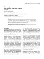
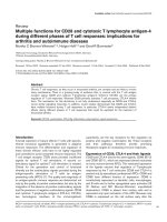
FFiigguurree 11
A generic life cycle of
Plasmodium
species. Sporozoites delivered from the salivary glands of a biting mosquito (8) enter the human bloodstream and are
carried to the liver, where they infect hepatocytes (1) and produce liver-stage schizonts. These burst open to release merozoites, which enter red blood
cells and undergo multiple rounds of replication as the erythrocytic schizont (3). The stages shown at (3) are those analyzed by Foth
et al
. [1]. A minority
of merozoites at each cycle form the sexual stage gametocytes (4), which persist in the blood until ingested by another mosquito. Within minutes,
gametes differentiate in the mosquito gut and fertilization follows (5). The zygote then develops into an ookinete (6), which penetrates the gut wall to
form another ‘schizogonic’ stage, the oocyst (7). Daughter sporozoites are released from the oocysts and invade the salivary glands (8). Gametocytes (4)
are terminally arrested cells while within the bloodstream. The expression of many proteins required for gamete function just minutes after the parasite
is ingested by the mosquito is under translational control. Sporozoites (8), which are similarly responsible for transmission between hosts, have not yet
exhibited similar regulation of gene expression: note that their development in the new host is less urgent. Figure modified with permission from [20].
2
1
3
8
5
6
7
4
significant return for the massive investment made when
compared with previous less discriminatory approaches
using multidimensional protein identification technology
(MudPIT) or one-dimensional gel/liquid chromatography/
MS technologies [6-11], methods that have identified many
hundreds of proteins at individual life stages.
What the new data lack in quantity is, however, more than
compensated for by the new information on protein abun-
dance and isoform changes. Foth et al. [1] detected multiple
isoforms for 50% of all the proteins identified. Different
isoforms of equivalent mobility (Mr) were considered to be
due to changes in phosphorylation. An increase in mobility
between two isoforms was interpreted to be due to post-
translational protein cleavage (or proteasomal degradation).
One protein, enolase, was described in no less than seven
different isoforms, of which two appeared to be truncated.
By comparing the proteomic data from these samples with
previous transcriptomic data from comparable samples [5],
Foth et al. [1] found that expression of some proteins or
isoforms - for example, the chaperone protein HSP40 and
four actin isoforms - were concordant with the ‘just-in-time’
synthesis model. Interestingly, peak protein abundance of
another actin isoform was delayed following transcription,
indicating regulated post-translational modification. The
expression of yet other proteins, for example HSP60, was
negatively correlated with their mRNA levels.
LLooookkiinngg ttoo tthhee ffuuttuurree
Where does this leave us? Reductionists can argue strongly
that this paper [1] reinforces the concept that it is essential
to treat each molecule and pathway separately and investi-
gate each and every one in depth, whereas ‘synthesizers’ can
emphasize that such global approaches have the potential,
perhaps not fully realized in this work, to understand
‘master regulatory mechanisms’, which require
consideration before examining individual pathways, each of
which will be, by definition, unique. It will be interesting to
see whether the application of systems approaches to data of
this type will permit resolution of these questions at the
global level.
Above all, Foth et al. [1] provide a healthy reality check as to
the complexity of the molecular mechanisms regulating the
development of this important parasite, which should
caution the researcher against making assumptions as to the
time and place of protein activity from transcriptome, or
indeed proteome, analyses. Even the phenotypic analysis of
genetic mutations may not provide unequivocal solutions to
these questions [19]. For those enjoying the ‘thrill of the
academic chase’ there is clearly ample room for more
exciting research. For those seeking to control this global
scourge, an understanding of the fundamental yet multi-
faceted mechanisms regulating parasite development may
bring ways of interrupting the parasite’s life cycle, or
perhaps of generating new attenuated strains for therapy or
transmission blockade.
RReeffeerreenncceess
1. Foth BJ, Zhang N, Mok S, Preiser PR, Bozdech Z:
QQuuaannttiittaattiivvee
pprrootteeiinn eexxpprreessssiioonn pprrooffiilliinngg rreevveeaallss eexxtteennssiivvee ppoosstt ttrraannssccrriippttiioonnaall
rreegguullaattiioonn aanndd ppoosstt ttrraannssllaattiioonnaall mmooddiiffiiccaattiioonn iinn sscchhiizzoonntt ssttaaggee
mmaallaarriiaa ppaarraassiitteess
Genome Biol
2008,
99::
R177.
2. Hayward RE, Derisi JL, Alfadhli S, Kaslow DC, Brown PO, Rathod
PK:
SShhoottgguunn DDNNAA mmiiccrrooaarrrraayyss aanndd ssttaaggee ssppeecciiffiicc ggeennee eexxpprreessssiioonn iinn
PPllaassmmooddiiuumm ffaallcciippaarruumm
mmaallaarriiaa
Mol Microbiol
2000,
3355::
6-14.
3. Le Roch KG, Zhou Y, Blair PL, Grainger M, Moch JK, Haynes JD, De
La Vega P, Holder AA, Batalov S, Carucci DJ, Winzeler EA:
DDiissccoovv
eerryy ooff ggeennee ffuunnccttiioonn bbyy eexxpprreessssiioonn pprrooffiilliinngg ooff tthhee mmaallaarriiaa ppaarraassiittee
lliiffee ccyyccllee
Science
2003,
330011::
1503-1508.
4. Bozdech Z, Mok S, Hu G, Imwong M, Jaidee A, Russell B, Ginsburg
H, Nosten F, Day NP, White NJ, Carlton JM, Preiser PR:
TThhee ttrraann
ssccrriippttoommee ooff
PPllaassmmooddiiuumm vviivvaaxx
rreevveeaallss ddiivveerrggeennccee aanndd ddiivveerrssiittyy ooff
ttrraannssccrriippttiioonnaall rreegguullaattiioonn iinn mmaallaarriiaa ppaarraassiitteess
Proc Natl Acad Sci
USA
2008,
110055::
16290-16295.
5. Bozdech Z, Zhu J, Joachimiak MP, Cohen FE, Pulliam B, DeRisi JL:
EExxpprreessssiioonn pprrooffiilliinngg ooff tthhee sscchhiizzoonntt aanndd ttrroopphhoozzooiittee ssttaaggeess ooff
PPllaass
mmooddiiuumm ffaallcciippaarruumm
wwiitthh aa lloonngg oolliiggoonnuucclleeoottiiddee mmiiccrrooaarrrraayy
Genome
Biol
2003,
44::
R9.
6. Florens L, Washburn MP, Raine JD, Anthony RM, Grainger M,
Haynes JD, Moch JK, Muster N, Sacci JB, Tabb DL, Witney AA,
Wolters D, Wu Y, Gardner MJ, Holder AA, Sinden RE, Yates JR,
/>Genome
BBiioollooggyy
2009, Volume 10, Issue 2, Article 211 Sinden 211.3
Genome
BBiioollooggyy
2009,
1100::
211
FFiigguurree 22
Application of ‘omics’ technologies to understanding the regulation of
expression of functional proteins. The area in which 2D-DIGE approaches
(as applied by Foth
et al.
[1]) are particularly valuable is indicated.
Transcription
Spatial localization in cytoplasm
inactivation
Activation
Translation
mRNA
degraded
Protein
mRNA
Folding
Covalent modification = activation or deactivation
Phosphorylation
Glycosylation
Lipid addition
Multimer formation
Homopolymer
Heteropolymer
Proteolytic activation?
Protein degradation
Transcriptomics
Proteomics
2D-DIGE
Interactomics
Carucci DJ:
AA pprrootteeoommiicc vviieeww ooff tthhee
PPllaassmmooddiiuumm ffaallcciippaarruumm
lliiffee
ccyyccllee
Nature
2002,
441199::
520-526.
7. Lasonder E, Ishihama Y, Andersen JS, Vermunt AM, Pain A, Sauer-
wein RW, Eling WM, Hall N, Waters AP, Stunnenberg HG, Mann M:
AAnnaallyyssiiss ooff tthhee
PPllaassmmooddiiuumm ffaallcciippaarruumm
pprrootteeoommee bbyy hhiigghh aaccccuurraaccyy
mmaassss ssppeeccttrroommeettrryy
Nature
2002,
441199::
537-542.
8. Lasonder E, Janse CJ, van Gemert GJ, Mair GR, Vermunt AM,
Douradinha BG, van Noort V, Huynen MA, Luty AJ, Kroeze H, Khan
SM, Sauerwein RW, Waters AP, Mann M, Stunnenberg HG:
PPrroo
tteeoommiicc pprrooffiilliinngg ooff
PPllaassmmooddiiuumm
ssppoorroozzooiittee mmaattuurraattiioonn iiddeennttiiffiieess nneeww
pprrootteeiinnss eesssseennttiiaall ffoorr ppaarraassiittee ddeevveellooppmmeenntt aanndd iinnffeeccttiivviittyy
PLoS
Pathog
2008,
44::
e1000195.
9. Hall N, Karras M, Raine JD, Carlton JM, Kooij TW, Berriman M,
Florens L, Janssen CS, Pain A, Christophides GK, James K, Ruther-
ford K, Harris B, Harris D, Churcher C, Quail MA, Ormond D,
Doggett J, Trueman HE, Mendoza J, Bidwell SL, Rajandream MA,
Carucci DJ, Yates JR 3rd, Kafatos FC, Janse CJ, Barrell B, Turner CM,
Waters AP, Sinden RE:
AA ccoommpprreehheennssiivvee ssuurrvveeyy ooff tthhee
PPllaassmmooddiiuumm
lliiffee ccyyccllee bbyy ggeennoommiicc,, ttrraannssccrriippttoommiicc,, aanndd pprrootteeoommiicc aannaallyysseess
Science
2005,
330077::
82-86.
10. Khan SM, Franke-Fayard B, Mair GR, Lasonder E, Janse CJ, Mann M,
Waters AP:
PPrrootteeoommee aannaallyyssiiss ooff sseeppaarraatteedd mmaallee aanndd ffeemmaallee ggaammeettoo
ccyytteess rreevveeaallss nnoovveell sseexx ssppeecciiffiicc
PPllaassmmooddiiuumm
bbiioollooggyy
Cell
2005,
112211::
675-687.
11. Patra KP, Johnson JR, Cantin GT, Yates JR 3rd, Vinetz JM:
PPrrootteeoommiicc
aannaallyyssiiss ooff zzyyggoottee aanndd ooookkiinneettee ssttaaggeess ooff tthhee aavviiaann mmaallaarriiaa ppaarraassiittee
PPllaassmmooddiiuumm ggaalllliinnaacceeuumm
ddeelliinneeaatteess tthhee hhoommoollooggoouuss pprrootteeoommeess ooff
tthhee lleetthhaall hhuummaann mmaallaarriiaa ppaarraassiittee
PPllaassmmooddiiuumm ffaallcciippaarruumm
Proteomics
2008,
88::
2492-2499.
12. Tarun AS, Peng X, Dumpit RF, Ogata Y, Silva-Rivera H, Camargo N,
Daly TM, Bergman LW, Kappe SH:
AA ccoommbbiinneedd ttrraannssccrriippttoommee aanndd
pprrootteeoommee ssuurrvveeyy ooff mmaallaarriiaa ppaarraassiittee lliivveerr ssttaaggeess
Proc Natl Acad Sci
USA
2008,
110055::
305-310.
13. Paton MG, Barker GC, Matsuoka H, Ramesar J, Janse CJ, Waters AP,
Sinden RE:
SSttrruuccttuurree aanndd eexxpprreessssiioonn ooff aa ppoosstt ttrraannssccrriippttiioonnaallllyy rreegguu
llaatteedd mmaallaarriiaa ggeennee eennccooddiinngg aa ssuurrffaaccee pprrootteeiinn ffrroomm tthhee sseexxuuaall ssttaaggeess
ooff
PPllaassmmooddiiuumm bbeerrgghheeii
Mol Biochem Parasitol
1993,
5599::
263-275.
14. Billker O, Dechamps S, Tewari R, Wenig G, Franke-Fayard B,
Brinkmann V:
CCaallcciiuumm aanndd aa ccaallcciiuumm ddeeppeennddeenntt pprrootteeiinn kkiinnaassee rreegguu
llaattee ggaammeettee ffoorrmmaattiioonn aanndd mmoossqquuiittoo ttrraannssmmiissssiioonn iinn aa mmaallaarriiaa ppaarraa
ssiittee
Cell
2004,
111177::
503-514.
15. Mair GR, Braks JA, Garver LS, Wiegant JC, Hall N, Dirks RW, Khan
SM, Dimopoulos G, Janse CJ, Waters AP:
RReegguullaattiioonn ooff sseexxuuaall ddeevveell
ooppmmeenntt ooff
PPllaassmmooddiiuumm
bbyy ttrraannssllaattiioonnaall rreepprreessssiioonn
Science
2006,
331133::
667-669.
16. Srinivasan P, Abraham EG, Ghosh AK, Valenzuela J, Ribeiro JM,
Dimopoulos G, Kafatos FC, Adams JH, Fujioka H, Jacobs-Lorena M:
AAnnaallyyssiiss ooff tthhee
PPllaassmmooddiiuumm
aanndd
AAnnoopphheelleess
ttrraannssccrriippttoommeess dduurriinngg
ooooccyysstt ddiiffffeerreennttiiaattiioonn
J Biol Chem
2004,
227799::
5581-5587.
17. Pachebat JA, Kadekoppala M, Grainger M, Dluzewski AR, Gunaratne
RS, Scott-Finnigan TJ, Ogun SA, Ling IT, Bannister LH, Taylor HM,
Mitchell GH, Holder AA:
EExxtteennssiivvee pprrootteeoollyyttiicc pprroocceessssiinngg ooff tthhee
mmaallaarriiaa ppaarraassiittee mmeerroozzooiittee ssuurrffaaccee pprrootteeiinn 77 dduurriinngg bbiioossyynntthheessiiss aanndd
ppaarraassiittee rreelleeaassee ffrroomm eerryytthhrrooccyytteess
Mol Biochem Parasitol
2007,
115511::
59-69.
18. Nirmalan N, Sims PF, Hyde JE:
QQuuaannttiittaattiivvee pprrootteeoommiiccss ooff tthhee
hhuummaann mmaallaarriiaa ppaarraassiittee
PPllaassmmooddiiuumm ffaallcciippaarruumm
aanndd iittss aapppplliiccaattiioonn ttoo
ssttuuddiieess ooff ddeevveellooppmmeenntt aanndd iinnhhiibbiittiioonn
Mol Microbiol
2004,
5522::
1187-
1199.
19. Ecker A, Bushell ES, Tewari R, Sinden RE:
RReevveerrssee ggeenneettiiccss ssccrreeeenn
iiddeennttiiffiieess ssiixx pprrootteeiinnss iimmppoorrttaanntt ffoorr mmaallaarriiaa ddeevveellooppmmeenntt iinn tthhee mmooss
qquuiittoo
Mol Microbiol
2008,
7700::
209-220.
20. Peters W:
A Colour Atlas of Arthropods in Medicine.
Barcelona,
Spain: Wolfe Publishing; 1992.
/>Genome
BBiioollooggyy
2009, Volume 10, Issue 2, Article 211 Sinden 211.4
Genome
BBiioollooggyy
2009,
1100::
211