Báo cáo y học: "How to build a paraspeckle" docx
Bạn đang xem bản rút gọn của tài liệu. Xem và tải ngay bản đầy đủ của tài liệu tại đây (576.93 KB, 5 trang )
Sasaki and Hirose: Genome Biology 2009, 10:227
Abstract
Noncoding RNAs have recently been identified as essential
components of the nuclear suborganelles called paraspeckles.
This finding will facilitate our understanding of the molecular
dynamics and physiological role of these enigmatic macro-
molecular structures.
Discovery of paraspeckles
Paraspeckles are large ribonucleoprotein structures around
0.5 μm in diameter that can be detected in nuclei with a
light microscope and appropriate antibody staining, and
are currently of unknown function. They were discovered
quiteunexpectedlyasrecentlyas2002[1,2].Lamondand
colleagues conducted a large-scale mass-spectrometric
analysisofnucleoliisolatedfromHeLacells,whichidenti-
fied 271 nucleolar proteins.Of these proteins, morethan
30%werenoveloruncharacterized[1].Thelocalizationof
asubsetofthenovelproteinsfusedwithyellowfluorescent
protein(YFP)forvisualdetectionwasthendetermined[2].
Surprisingly, one of those fusion proteins was found to
co-localizenottothenucleolusitself,buttoanovelnuclear
compartmentorsuborganelle.
Theproteinwasfoundtobeubiquitouslyexpressedinall
humancelllinesexamined[2],andislocalizedingranular
foci often adjacent to ‘splicing-speckles’, which are impli-
cated as the reservoir of various splicing factors. Hence,
thenewlydiscoveredfociweredubbed‘paraspeckles’and
the newly characterized protein was named paraspeckle
protein 1 (PSP1) [2]. Mass spectrometric analysis of
nucleolar proteins demonstrated that a small fraction of
this protein, undetectable by fluorescence microscopy,
transientlyassociatedwiththenucleolus,whichexplained
itsoriginaldetectionasanucleolarprotein[1].
Thenumberofparaspecklesperinterphasenucleiinhuman
celllinesvaries between10and 20, andtheirtypical sizeis
0.5μmindiameter.InadditiontoPSP1,threeproteins,p54
nrb
(alsoknownasNONO,non-POUdomaincontainingoctamer-
binding protein), polypyrimidine tract-binding protein-
associated splicing factor (PSF), and paraspeckle protein 2
(PSP2), exhibit a punctate nucleoplasmic distribution,
co-localizing to paraspeckles as seen by immunnostaining
usingantibodiesagainstcorrespondingproteins[2,3].
These paraspeckle proteins each contain two RNA-
recognitionmotifs(RRMs).Thepropertiesandinteraction
behavior of PSF, p54
nrb
, and their homologs in species
ranging from Drosophila to mouse have been extensively
characterized. PSF and p54
nrb
interact with a nuclear
receptor and with RNA, and also with both single- and
double-stranded DNA [4-9]. Both p54
nrb
and PSF are
multifunctional proteins that are implicated in nuclear
processes such as transcriptional control, splicing regu-
lation, mRNA 3’-end formation, DNA repair and recom-
bination, and nuclear retention of hyperedited RNAs in
various human and mousecell lines [4-9]. Chromosomal
translocationsinvolvingthegenesencodingPSForp54
nrb
can produce chimeric proteins that cause tumorigenesis
(see [4] and references therein). Furthermore, if trans-
criptionisinhibitedbyactinomycinD,alltheparaspeckle
proteins relocate to a perinucleolar cap [10]. There are
severalmoreproteinsthatmeetsomeoftheabovecriteria,
andthelistofparaspeckleproteinsisthereforeexpectedto
expand in the near future. Indeed, Cardinale et al. [11]
recently reported that a pre-mRNA 3’-end processing
factor,mammaliancleavagefactorI(CFI
m
68),localizesto
paraspeckles. The protein contains one RRM instead of
twoandmovestotheperinucleolarcapwhentranscription
isinhibited[11].
The identification of paraspeckle proteins immediately
prompted investigations of the molecular mechanism by
which this membranelesssuborganelle is assembled.Fox
et al. [3] reportedthat PSP1 heterodimerizeswith p54
nrb
both in vivo and in vitro, andthat the functioning RRM
domainsarecriticalfortargetingPSP1totheparaspeckle.
Furthermore, the paraspeckle structure is sensitive to
RNase,indicatingthatRNAisalsoan essentialstructural
component[3].
Noncoding RNAs as ‘architectural RNAs’
Given that the paraspeckle was predicted to be a large
ribonucleoproteincomplex[3],thepresumedRNA-protein
interactions have become a focus of research into the
molecularmechanismsunderlyingparaspeckleformation.
Threegroupshavenowindependentlyidentifiedthelong-
sought architectural RNAs [12-14]. These groups began
working from different research perspectives but
eventually found the same noncoding RNAs (ncRNAs) -
Review
How to build a paraspeckle
Yasnory TF Sasaki and Tetsuro Hirose
Address: Functional RNomics Team, Biomedicinal Information Research Center, National Institute of Advanced Industrial Science and
Technology (AIST), 2-42 Aomi, Koutou, Tokyo 135-0064, Japan. Email: ;
227.2
Sasaki and Hirose: Genome Biology 2009, 10:227
two isoforms, MENε and MENβ, which are transcribed
fromthe same RNA polymeraseII promoter but differin
thelocationoftheir3’ends,andthefunctionsofwhichare
largelyuncharacterized[15].Ourlaboratory[12]identified
MENε and MENβfromtheLeLacellnucleiasacomponent
of the paraspeckle-enriched fraction by biochemical puri-
fication. Sunwoo et al. [13] identified some200 ncRNAs
thatareeitherup-ordownregulatedduringdifferentiation
oftheC2C12mousemyoblastcelllineintomyotubes[13].
They narrowed down their target to Menε/β by manual
examinationandsubcellularlocalizationanalyses.Looking
for nuclear-retained abundant ncRNAs in both humans
andmousecells,Clemsonandcolleagues[14,16]identified
three:theinactivatedX-chromosometranscriptXIST,and
two ncRNAs they called nuclear-enriched abundant
transcripts1and2,NEAT1 and NEAT2. NEAT1 is identical
to MENε and NEAT2 to the noncoding ncRNA MENα,
whichresidesdownstreamofMenε/βintheMENlocus.
Inhumans,twoMENisoforms,MENε(3.7kb)andMENβ
(approximately23kb),aretranscribed from a single pro-
moterattheMENε/βlocusatchromosome11q13.1;simi-
larly, the mouse counterparts, Menε (3.2 kb) and Menβ
(approximately 20 kb), share the same promoter at
chromosome19qA[12-14].Inbothhumanandmouse,the
shortertranscript,MENε/Menε,ispolyadenylatedatits3’
end; however, the 3’ end of the longer isoform, MENβ/
Menβ, is formed by RNase P cleavage [13]. The physio-
logicalsignificanceofthisnoncanonical3’-endprocessing
is not yet clear. In all cases, the exclusive paraspeckle
localization of MENε/β was confirmed by RNA fluores-
cence in situ hybridization analysis combined with
immunofluorescent detection of paraspeckle marker
proteins[12-14](Figure1).
The MENε/β depletion phenotype was also examined in
both human and mouse cells, using knockdown with
chimericantisenseoligonucleotides[12,13]orsmallinter-
feringRNA(siRNA)[14].MENε/βknockdownresultedin
disruptionoftheparaspecklesbutnotofotherintranuclear
bodies [12-14] (Figure 1). Importantly, there is no
degradation of paraspeckle proteins in these knockdowns
and no paraspeckles remained intact without MENε/β.
Furthermore,thereassemblyofparaspecklesdisassembled
by treatment with an RNA polymerase II inhibitor,
5,6-dichloro-1-β-d-ribofuranosylbenzimidazole (DRB), was
suppressed in MENε/β-depletedcells[12,13].Theseresults
stronglysupportthehypothesisthatMENε and MENβ are
essentialfortheintegrityoftheparaspecklestructure.
The physical associations of MENε/β RNAs with para-
speckle proteins have been investigated using immuno-
precipitation and the following RNA-protein interactions
havebeenreported:MENβandp54
nrb
and MENβ and PSF
[12], Menε/β andp54
nrb
[13], andMENε and p54
nrb
and
MENε and PSP1 [14]. Clemson et al. [14] demonstrated
that deletion of the RRM domains of PSP1 abrogates its
association with MENε in paraspeckles. Our group [12]
examined the effect of paraspeckle protein depletion on
MENε/βRNAlevelsandparaspecklestructure.Wefound
that depletion of either p54
nrb
or PSF preferentially
decreases MENβ but not MENε,anddisruptsparaspeckle
structure. Notably, PSP1 depletion did not affect either
MENε/β levels or paraspeckle structure. These results
suggestthatPSP1playsaroleinparaspeckleorganization
distinctfromp54
nrb
andPSF.Despitesomediscrepancies
among the reports of the three research groups, the
consensus that the ncRNAs MENε/β are essential to
paraspeckle formation via interactions with the RRM
domainsofeachparaspeckleproteinisclear.
Prasanthet al.[17]haveproposed aroleforparaspeckles
in the posttranscriptional regulation of expression of
cationicaminoacidtransporter2(CAT2)genemRNAs.An
RNA called CTN-RNA is transcribed from the protein-
coding mouse cationic amino acid transporter 2 gene
throughalternativepromoterandpoly(A)siteusageandis
retainedinthenucleus[17].Understress,thisRNAcanbe
cleavedtoproducetheprotein-codingCAT2mRNA.How-
ever,CTN-RNAisthoughttoberetainedinthenucleusas
aresultofA-to-IRNAeditinginthe3’untranslatedregion
[17], whereas MENε/β RNAs do not appear to be edited
[12-14].
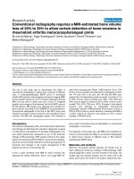
Figure 1
Knockdown of MENε/β ncRNAs leads to disintegration of the
paraspeckles. Confocal images of HeLa cells treated either with a
control scrambled antisense oligonucleotide (upper panels) or with
a MENε/β knockdown antisense oligonucleotide (lower panels).
Upper panel: MENε/β ncRNAs (magenta) co-localize to
paraspeckles defined by PSF immunofluorescence (green). Lower
panel: the paraspeckle-associated PSF signal disappeared when
the MENε/β ncRNAs were successfully depleted, indicating that the
paraspeckles have disintegrated. Note that the nucleoplasmic PSF
signal remains intact. The HeLa cell nuclei were counterstained with
DAPI (blue). Scale bar, 10 μm.
ControlKnockdown
MEN
ε
/
β
PSF Merge
227.3
Sasaki and Hirose: Genome Biology 2009, 10:227
Withthecurrently available knowledge, what elsecanwe
determine regarding the physiological function of para-
speckles? The ubiquity of paraspeckles across different
tissuesmustbetakenintoconsideration.Giventhatmost
paraspecklecomponentshavepreviouslybeenidentifiedas
involvedintranscriptionalregulationandRNAprocessing,
it is tempting to speculate that paraspeckles control gene
expression.However,themechanismofparaspeckleaction
is open to question, as the ‘paraspeckle proteins’ in fact
seemtofunctionprimarilyinnuclearcompartmentsother
than MENε/β-containing paraspeckles [4-10]. One
plausible assumption, as has been hypothesized for other
intranuclear compartments such as the nucleolus and
splicingspeckles,isthatparaspecklesserveasawarehouse
foranumberofregulatoryproteinsthataresequesteredin
theparaspeckleuntilrequiredinresponsetophysiological
conditions [18-21]. Thus, the availability of regulatory
proteinsatatargetgenelocuscanbestrictlycontrolledby
theparaspeckle.
Paraspeckle dynamics
The remarkable dynamics of paraspeckle proteins have
been noted since the discovery of paraspeckles, as
proteomicanalysesalsoidentifiedalltheseproteinsinthe
perinucleolar compartment [1,2]. When paraspeckle
proteins relocate to the perinucleolar compartment, the
MENε/βRNAshavedissociated,andaredegraded[12]or
relocate to either splicing speckles [13] or the nucleolus
[14]. Paraspeckle proteins diffuse across the nucleoplasm
intheabsenceoftheMENε/βRNAs[6,12,13].Itispossible
that posttranslational modifications such as phosphory-
lationandmethylationcouldaltertheinteractionbetween
the MENε/β RNAs and paraspeckle proteins, and could
increase the affinity of paraspeckle proteins for the
perinucleolarcompartment.
The number of paraspeckles varies with the cell cycle:
paraspeckles increase during interphase, disappear at
telophase, when paraspeckle proteins translocate to the
perinucleolar compartment, andreappear early inG1 [3]
(Figure2).Thisvariationinparaspecklenumbercoincides
with the transcriptional activity of RNA polymerase II,
and, hence, perhaps with the expression level of the
MENε/βRNAs.Intriguingly,Clemsonet al.[14]reported
paraspeckleformationattranscriptionallyactiveMENε/β
loci.NewlygeneratedMENε/βfociseemtobelargerthan
those found later in the cell cycle, and are constrained
withinanuclearsubvolume,mostprobablyinthevicinity
of the MENε/β locus [14]. These data imply that nascent
MENε/βtranscriptsareconcentratedinthevicinityofthe
MENε/β loci and serve as a platform for paraspeckle
proteinrecruitment(Figure2).Consistentwiththeabove
observation, stable expressionof ectopicMenε causes an
increase in paraspeckle number [14], whereas transient
expressiondoesnot[12].
There is an apparent difference in the number and
distributionpatternofparaspecklesinthenucleusbetween
theG1phaseandtherestofinterphase.Inaddition,each
cell line that has been observed displays a unique
paraspeckledistributionpattern,whichmayrepresentthe
physiological status of the cells. These observations
inevitablyraisequestionsastotheprecisemechanismsof
paraspeckle formation and translocation. Is an individual
paraspeckle formed on the MEN locus, or is a large
paraspeckle precursor formed and then subsequently
divided into several daughter paraspeckles? How do
paraspeckles depart from the MENε/β loci? Do para-
specklesroamthroughthenucleusoraretheydestinedfor
specific target locations? These questions are inextricably
intertwined if both the formation and movement of
Figure 2
Paraspeckle dynamics. A model illustrating paraspeckle dynamics
in the cell cycle. Three representative stages are shown: early G1;
interphase; and telophase. The localization and behavior of
paraspeckles throughout the cell cycle are highly dynamic. Early G1
(top): the nucleus of a human cell (large oval) contains two MENε/β
loci (green circle), one on each chromosome 11q13 (blue
territories). Paraspeckles (red circles or ovals) are generated at the
transcriptionally active MENε/β loci, where paraspeckle proteins
(smaller white, grey and black ovals in inset) associate with nascent
MENε/β RNAs (black helices) to generate the paraspeckle.
Interphase (lower right): the number of paraspeckles increases,
typically to between 10 and 20 per nucleus. Newly generated
paraspeckles are first localized to the MENε/β loci and then become
distributed throughout the nucleus (indicated by arrows) by an
unknown mechanism. Intact paraspeckles appear to be in a
dynamic equilibrium, in which the flux of constituents between
paraspeckles and nucleoplasm is balanced. The trajectories of
redistribution of paraspeckles throughout the nucleus may be
random as paraspeckles roam the interchromatin space by
scanning specific target sites. Telophase (lower left): RNA
polymerase II transcriptional activity is undetectable at this stage
and, therefore, the levels of MENε/β decrease, which in turn causes
paraspeckle disassembly. Paraspeckles are reassembled once
MENε/β transcription restarts in the daughter cells.
Cell cycle
Early G1
Assembly
Key
MENε/β
MENε/β loci
Paraspeckles
Chr11 territories
Paraspeckle proteins
Disassembly
Telophase
Interphase
Dynamic
equilibrium
227.4
Sasaki and Hirose: Genome Biology 2009, 10:227
paraspeckles are dependent on the nuclear domains with
whichparaspecklesassociate,thatis,the MENε/β loci and
putative target gene loci. In addressing these questions,
comparisons with the formation of other nuclear bodies
may be useful. The nucleolus is formed at the nucleolar
organizerregion(NOR)containingtherRNAgenes,andits
formationisdependentonrRNAtranscription.Additional
nucleoli can be formed by introducing extrachromosomal
NORs [22]. Cajal bodies, involved in small nuclear ribo-
nucleoprotein(snRNP) and smallnucleor RNP (snoRNP)
biogenesis, also closely interact with particular gene loci
such as those for spliceosomal small nuclear RNAs
(snRNAs) and histones, and are recruited or formed de
novo in a microenvironment in which the local concen-
trationoftheirsubstrates,snRNAs,iselevated[23].Thus,
genelociprovidenucleationsitesfornuclearbodyforma-
tionand may be a targetfor transcriptional regulationor
modulation by nuclear bodies [18-21]. Interestingly, the
RRMproteinNonA,theDrosophilacounterpartofp54
nrb
,
forms a complex with other RNA-binding proteins in
developmentally regulated ‘puffs’ on polytene chromo-
somes[7].Itwillbeofgreatinteresttodeterminewhether
paraspeckles also target particular gene loci in specific
physiologicalconditions(Figure2).
Having ncRNAs as part of their structure gives para-
speckles unique properties; for example, unlike other
intranuclear bodies, paraspeckle structure persists during
most of mitosis, with the exception of telophase, in the
absenceofassociationwithcondensedchromatin[3].This
observation implies that long ncRNAs can themselves
functionas a scaffold fornucleation. In contrast, nucleoli
and Cajal bodies disassemble when cells enter mitosis
becauseassociationwiththeirtargetlociisa prerequisite
for nucleation [24,25]. It should be noted that RNAs
associated with these nuclear bodies (for example, pre-
rRNA and snRNA) are relatively small compared to
MENε/β).ThebiogenesisofCajalbodiesexhibitsthehall-
marks of stochastic self-organization [26]. An important
focusoffutureinvestigationswillbetodeterminetowhat
extent paraspeckle formation is consistent with the self-
organizationmodel.
The identification of MENε/β as a component of para-
speckles has raised many more questions, rather than
simply answering the question of what a paraspeckle is.
The depletion of MENε/β RNA profoundly affects the
structural integrity of paraspeckles, which does not
necessarilyexcludethepossibilityofthepresenceofother
structural/functionalRNAsinparaspeckles.Transcriptome
analysisofisolatedparaspeckles,forexample,mayleadto
the identification ofancillary RNA components.Through
mechanical and functional characterization of para-
speckles,withemphasisontheRNAcomponents,we will
gainsubstantialinsightsintothedynamicnatureofthese
nuclearbodies-inparticular,howtheyareassembledinto
largeribonucleoproteincomplexesandhowtheyfindtheir
targetsonchromatinand/orinparticularnucleardomains.
Theseinsightsshouldberelevanttoourunderstandingof
thedynamicsofothernuclearbodiesaswell.
Acknowledgements
We thank members of the Hirose laboratory, in particular T
Naganuma, K Aoki and T Kawaguchi for helpful discussions. We
also thank K Watanabe and T Misteli for their continuous support
and encouragement.
References
1. Andersen JS, Lyon CE, Fox AH, Leung AKL, Lam YW, Steen
H, Mann M, Lamond AI: Directed proteomic analysis of the
human nucleolus. Curr Biol 2002, 12:1-11.
2. Fox AH, Lam YW, Leung AKL, Lyon CE, Andersen J, Mann M,
Lamond AI: Paraspeckles: A novel nuclear domain. Curr Biol
2002, 12:13-25.
3. Fox AH, Bond CS, Lamond AI: P54nrb forms a heterodimer
with PSP1 that localizes to paraspeckles in an RNA-
dependent manner. Mol Biol Cell 2005, 16:5304-5315.
4. Shav-Tal Y, Zipori D: PSF and p54(nrb)/NonO - multi-func-
tional nuclear proteins. FEBS Lett 2002, 531:109-114.
5. Auboeuf D, Dowhan DH, Li X, Larkin K, Ko L, Berget SM,
O’Malley BW: CoAA, a nuclear receptor coactivator protein
at the interface of transcriptional coactivation and RNA
splicing. Mol Cell Biol 2004, 24:442-453.
6. Dong X, Sweet J, Challis JRG, Brown T, Lye SJ:
Transcriptional activity of androgen receptor is modulated
by two RNA splicing factors, PSF and p54nrb. Mol Cell Biol
2007, 27:4863-4875.
7. Reim I, Stanewsky R, Saumweber H: The puff-specific RRM
protein NonA is a single-stranded nucleic acid binding
protein. Chromosoma 1999, 108:162-172.
8. Zhang Z, Carmichael GG: The fate of dsRNA in the nucleus:
A p54
nrb
-containing complex mediates the nuclear reten-
tion of promiscuously A-to-I edited RNAs. Cell 2001,
106:465-475.
9. Bladen CL, Udayakumar D, Takeda Y, Dynan WS:
Identification of the polypyrimidine tract binding protein-
associated splicing factor p54(nrb) complex as a candidate
RNA double-strand break rejoining factor. J Biol Chem
2005, 280:5205-5210.
10. Shav-Tal Y, Blechman J, Darzacq X, Montagna C, Dye BT,
Patton JG, Singer RH, Zipori D: Dynamic sorting of nuclear
components into distinct nucleolar caps during transcrip-
tional inhibition. Mol Biol Cell 2005, 16:2395-2413.
11. Cardinale S, Cisterna B, Bonetti P, Aringhieri C, Biggiogera M,
Barabino SML: Subnuclear localization and dynamics of the
pre-mRNA 3’ end processing factor mammalian cleavage
factor I 68-kDa subunit. Mol Biol Cell 2007, 18:1282-1292.
12. Sasaki YT, Ideue T, Sano M, Mituyama T, Hirose T: MENε/β
noncoding RNAs are essential for structural integrity of
nuclear paraspeckles. Proc Natl Acad Sci USA 2009, 106:
2525-2530.
13. Sunwoo H, Dinger ME, Wilusz JE, Amaral PP, Mattick JS,
Spector DL: MENε/β nuclear-retained non-coding RNAs are
up-regulated upon muscle differentiation and are essential
components of paraspeckles. Genome Res 2009, 19:347-
359.
14. Clemson CM, Hutchinson JN, Sara SA, Ensminger AW, Fox
AH, Chess A, Lawrence JB: An architectural role for a
nuclear noncoding RNA: NEAT1 RNA is essential for the
structure of paraspeckles. Mol Cell 2009, 33:717-726.
15. Guru SC, Agarwal SK, Manickam P, Olufemi SE, Crabtree JS,
Weisemann JM, Kester MB, Kim YS, Wang Y, Emmert-Buck
MR, Liotta LA, Spiegel AM, Boguski MS, Roe BA, Collins FS,
Marx SJ, Burns L, Chandrasekharappa SC: A transcript map
for the 2.8-Mb region containing the multiple endocrine
neoplasia type 1 locus. Genome Res 1997, 7:725-735.
227.5
Sasaki and Hirose: Genome Biology 2009, 10:227
16. Hutchinson JN, Ensminger AW, Clemson CM, Lynch CR,
Lawrence JB, Chess A: A screen for nuclear transcripts
identifies two linked noncoding RNAs associated with
SC35 splicing domains. BMC Genomics 2007, 8:39.
17. Prasanth KV, Prasanth SG, Xuan Z, Hearn S, Freier SM,
Bennett CF, Zhang MQ, Spector DL: Regulating gene expres-
sion through RNA nuclear retention. Cell 2005, 123:249-
263.
18. Misteli T: Protein dynamics: Implications for nuclear archi-
tecture and gene expression. Science 2001, 291:843-84719.
Misteli T: Concepts in nuclear architecture. BioEssays 2005,
27:477-487.
20. Shav-Tal Y, Darzacq X, Singer RH: Gene expression within a
dynamic nuclear landscape. EMBO J 2006, 25:3469-3479.
21. Misteli T: Physiological importance of RNA and protein
mobility in the cell nucleus. Histochem Cell Biol 2008, 129:5-
11.
22. Oakes M, Aris JP, Brockenbrough JS, Wai H, Vu L, Nomura M:
Mutational analysis of the structure and localization of the
nucleolus in the yeast Saccharomyces cerevisiae. J Cell
Biol 1998, 143:23-34.
23. Dundr M, Ospina JK, Sung M-H, John S, Upender M, Ried T,
Hager GL, Matera SG: Actin-dependent intranuclear reposi-
tioning of an active gene locus in vivo. J Cell Biol 2007, 179:
1095-1103.
24. Leung AK, Gerlich D, Miller G, Lyon C, Lam YW, Lleres D,
Daigle N, Zomerdijk J, Ellenberg J, Lamond AI: Quantitative
kinetic analysis of nucleolar breakdown and reassembly
during mitosis in live human cells. J Cell Biol 2004, 166:787-
800.
25. Carmo-Fonseca M, Ferreira J and Lamond AI: Assembly of
snRNP-containing coiled bodies is regulated in interphase
and mitosis - evidence that the coiled body is a kinetic
nuclear structure. J Cell Biol 1993, 120:841-852.
26. Kaiser TE, Intine RV, Dundr M: De novo formation of a sub-
nuclear body. Science 2008, 322:1713-1717.
Published: 16 July 2009
doi:10.1186/gb-2009-10-7-227
© 2009 BioMed Central Ltd