EFFECTS OF CHOLINE KINASE ACTIVITY ON PHOSPHOLIPID METABOLISM AND MALIGNANT PHENOTYPE OF PROSTATE CANCER CELLS
Bạn đang xem bản rút gọn của tài liệu. Xem và tải ngay bản đầy đủ của tài liệu tại đây (2.46 MB, 121 trang )
EFFECTS OF CHOLINE KINASE ACTIVITY ON PHOSPHOLIPID
METABOLISM AND MALIGNANT PHENOTYPE OF PROSTATE CANCER
CELLS
Aditya Bansal
Submitted to the faculty of the University Graduate School
in partial fulfillment of the requirements
for the degree
Doctor of Philosophy
in the Department of Biochemistry and Molecular Biology,
Indiana University
October 2010
ii
Accepted by the Faculty of Indiana University, in partial
fulfillment of the requirements for the degree of Doctor of Philosophy.
____
Timothy R. DeGrado, Ph.D., Chair
Department of Biochemistry and Molecular Biology
____
Robert A. Harris, Ph.D.
Department of Biochemistry and Molecular Biology
Doctoral Committee
____
William F. Bosron, Ph.D.
Department of Biochemistry and Molecular Biology
June 15, 2010
____
James E. Klaunig, Ph.D.
Department of Toxicology
iii
DEDICATION
This study is dedicated to my family and friends for their unconditional love
and support; to my wife Pragya Sharma for her love, encouragement, and
patience; to my child, Sabhya Bansal, for bringing joy to my life; to my sister
Megha Bansal for her love, support and encouragement.
iv
ACKNOWLEDGEMENTS
I am indebted to many people who have helped me throughout my
graduate study. My sincere appreciation goes to all of them.
Dr. Timothy R. DeGrado for his wonderful mentoring, enormous
support, and being my role model.
Dr. Robert A. Harris for his great discussion, helpful criticism and
suggestions.
Dr. William F. Bosron and Dr. James E. Klaunig for their valuable
advices and reference.
All current and previous members in laboratory of Dr. Timothy R.
DeGrado
All members in laboratory of Dr. Robert A. Harris.
All members in laboratory of Dr. William F. Bosron.
v
ABSTRACT
Aditya Bansal
EFFECTS OF CHOLINE KINASE ACTIVITY ON PHOSPHOLIPID
METABOLISM AND MALIGNANT PHENOTYPE OF PROSTATE CANCER
CELLS
High choline uptake and increased choline kinase activity have been
reported in many cancers. This has motivated the use of choline as a biomarker
for tumor imaging. Tumors in general are heterogeneous in nature with respect to
oxygen tension. There are regions of hypoxia and normoxia that are expected to
have different metabolism but regulation of choline metabolism under hypoxia is
poorly understood. It is important to clarify the status of choline metabolism in
hypoxic microenvironment as it will have an impact on potential of choline as a
cancer biomarker. The primary goal was to determine the status of choline
phosphorylation in hypoxic cancer cells and its effect on uptake of choline. This
was examined by tracer studies in cancer cells exposed to hypoxia. It was
observed that hypoxia universally inhibits choline uptake /phosphorylation in
cancer cells. Decreased choline phosphorylation resulted in transient uptake of
choline radiotracers in cultured cancer cells and 9L tumors suggesting potential
problem in using choline as a biomarker for cancers in hypoxic
microenvironment. To investigate the mechanism behind decrease in choline
phosphorylation, steady state levels of choline metabolites were measured and
choline kinase catalyzed choline phosphorylation step was found to be rate-
vi
limiting in PC-3 cells. This suggested that modulation in choline kinase levels can
alter choline metabolism in hypoxic cancer cells. Expression and activity assays
for choline kinase revealed that choline kinase expression is down-regulated in
hypoxia. This regulation involved transcriptional level mediation by HIF1 at the
conserved HRE7 site in choline kinase promoter. To further understand the
importance of down-regulation of choline kinase in hypoxia, stable prostate
cancer cell lines over-expressing choline kinase were generated. Effect of over-
expression of choline kinase in hypoxia was evaluated in terms of malignant
phenotypes like proliferation rate, anchorage independent growth and invasion
potential. Both over-expression of choline kinase and hypoxia had a pronounced
effect on malignant phenotypes of prostate cancer cells. Further study showed
that increased choline kinase activity and hypoxic tumor microenvironment are
important for progression of early-stage, androgen-dependent LNCaP prostate
cancer cells but confer little survival advantage in undifferentiated, androgen-
independent PC-3 prostate cancer cells.
Timothy R. DeGrado, Ph.D., Chair
vii
TABLE OF CONTENTS
LIST OF TABLES xiii
LIST OF FIGURES xiv
GENERAL INTRODUCTION 1
1. Phosphatidylcholine metabolism 1
1.1. Overview of phosphatidylcholine metabolism 1
1.2. Reactions of CDP-choline pathway 1
1.3. Regulation of CDP-choline pathway 4
1.3.1. Regulatory reactions in CDP-choline pathway 4
1.4. Choline kinase (ChK) 4
1.4.1. Overview of choline kinase 4
1.4.1.2. Structure of choline kinase 5
1.4.1.3 Mechanism of reaction catalyzed by choline kinase 10
1.4.1.4 Kinetic parameters of choline kinase 11
1.4.1.5 Choline kinase knockouts 12
1.4.2. Regulation of choline kinases 13
1.4.2.1. Regulation of choline kinase activity by allosteric effectors 13
1.4.2.1. Regulation of choline kinase activity by phosphorylation 14
2. Transcriptional regulation of choline kinase 15
2.1. Transcriptional factors that are involved in
choline kinase expression 15
2.1.1. Activator protein 1 (AP-1) 15
viii
2.1.2. Hypoxia Inducible Factor (HIF-1) 16
2.1.3. Sp/KLF transcription factor (SP-1) 17
2.1.4. Cyclic AMP response element (CRE) -binding protein (CREB) 17
2.1.5. Xenobiotic response element (XRE) binding Aryl hydrocarbon
receptor (AhR)/Hypoxia Inducible Factor 18
2.2. Regulatory mechanism responsible for ChK gene expression 19
2.2.1. Regulation of rodent ChK gene expression 19
2.2.2. Promoter analysis of human ChK gene 19
3. Choline transport and metabolism in normal and cancer cells 21
3.1. Choline transport in normal cells 21
3.2. Choline transport in cancer cell lines and cancerous tissues 21
3.3 Choline metabolism 23
4. Choline kinase as an oncogene 24
4. 1. Choline kinase and cell signaling 24
4.2. Choline as a cancer biomarker 25
4.2.1. Choline based Positron Emission Tomography (PET) imaging
of malignant cancers 25
4.2.2. Choline based Magnetic Resonance Spectrometry Imaging (MRSI)
of malignant cancers 25
5. Specific aims and hypotheses 26
CHAPTER I 28
1. Abstract 28
2. Introduction 29
ix
3. Materials and Methods 32
3.1. Material chart 32
3.2. Tumor xenograft model 33
3.3. Establishment of hypoxic environment 33
3.4. Uptake of radiolabeled choline in cancer cells 33
3.5. Pulse chase experiment 34
3.6. Measurement of choline metabolite levels in cells 35
3.7. Uptake of radiolabeled choline in tumor xenograft 35
3.8. Analysis of radiolabeled choline metabolites in cancer cells
and tumor tissue 36
3.9. Measurement of choline kinase activity in cancer cells
and tumor tissue 37
3.10. Tumor perfusion assay and spatial localization of
radiolabeled choline in tumor xenograft 37
3.11. Statistical analysis 38
4. Results 38
4.1. Hypoxia decreased choline uptake and phosphorylation in
cancer cells 38
4.2. Hypoxia increased choline/phosphocholine ratio in cancer cells 39
4.3. Uptake of radiolabeled choline in tumor xenograft is transient 43
4.4. Choline uptake pattern coincides with perfusion pattern of the
tumor xenograft 44
5. Discussion 45
x
CHAPTER II 49
1. Abstract 49
2. Introduction 50
3. Materials and Methods 52
3.1. Material chart 52
3.2. Isolation of total RNA from cancer cells 53
3.3. Quantification of mRNA signal 53
3.4. Western blot analysis 54
3.5. Over-expression of hypoxia inducible factor 1 (HIF1) 54
3.6. Isolation of promoter region upstream of ChK55
3.7. Promoter alignment 55
3.8. Site-directed mutagenesis 56
3.9. DNA sequencing 57
3.10. Transient expression assay 57
3.11. Electrophoretic Mobility Shift Assay (EMSA) 58
3.12. Chromatin Immunoprecipitation (ChIP) assay 59
3.13. Statistical analysis 61
4. Results 61
4.1. Hypoxia decreases the expression of ChK in
prostate cancer cells 61
4.2. Over-expression of HIF1 decreases uptake and phosphorylation
of choline in prostate cancer cells 62
4.3. HIF1 binding sites and promoter alignment 62
xi
4.4. Mutation of a putative HRE site reduced the inhibitory effect of
HIF1 on ChK promoter activity in PC-3 prostate cancer cells 66
4.5. HIF1 is able to bind to the putative HRE site in ChK promoter 67
5. Discussion 70
CHAPTER III 75
1. Abstract 75
2. Introduction 76
3. Materials and Methods 79
3.1. Material 79
3.2. Over-expression of choline kinase 79
3.3. Measurement of population doubling time 80
3.4. Colony formation assay 80
3.5. Cell invasion assay 81
3.6. Statistical analysis 82
4. Results 82
4.1. Effect of over-expression of choline kinase on choline uptake 82
4.2. Effect of over-expression of choline kinase and hypoxia on
population doubling time 83
4.3. Effect of over-expression of choline kinase on
cancer cell morphology 83
4.4. Effect of over-expression of choline kinase and hypoxia on
anchorage independent growth 84
xii
4.5. Effect of over-expression of choline kinase and hypoxia on
invasion potential of prostate cancer cells 86
4.6. Effect of over-expression of choline kinase and hypoxia on
expression of pro-invasion factor, urokinase plasminogen
activator (uPa) 87
5. Discussion 88
CONCLUSIONS 93
REFERENCES 95
CURRICULUM VITAE
xiii
LIST OF TABLES
Table 1 Kinetic parameters of choline kinase reaction obtained with
highly purified or recombinant enzyme preparations 12
Table 2 Kinetic characteristic of choline kinase isoforms, ChK and
ChK12
Table 3 Metabolite levels in PC-3 cell extracts in serum-supplemented
medium after 24 h normoxia (1% O
2
) and hypoxia (21% O
2
)
(n =3, each condition). Choline kinase activity is expressed
as nmol choline phosphorylated/min/mg protein 40
Table 4 Tumor- to-background ratio of [
14
C]choline and [
18
F]FCH in
9L-glioma bearing Fisher rat 43
Table 5 Blood flow and FCH retention estimates of 9L glioma tumors
at 5 min and 20 min post-injection 45
Table 6 Sequences of primers used for ChKα promoter isolation
and nested PCRs 56
Table 7 Sequence of double stranded probe used for
electrophoretic mobility shift assays 59
Table 8 Sequences of primers used for PCR for amplification of
promoter regions in ChIP assay 60
Table 9 Effect of 24h of hypoxic (1% O
2
) exposure on expression of
ChKα and VEGF 62
xiv
LIST OF FIGURES
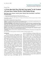
Figure 1 Biosynthesis of phosphatidylcholine in eukaryotes 3
Figure 2 Expression profile of choline kinase isoforms in various
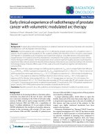
breast cancer cell lines 6
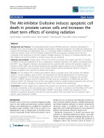
Figure 3 A stereo ribbon drawing of human choline kinase α dimer 7
Figure 4 Amino acid alignment of choline kinase isoforms 8
Figure 5 Comparison of structure of choline kinase, aminoglycoside
3’phospho transferase (APH(3’)-Illa) and catalytic domain of
cAMP-dependent protein kinase (PKA) 9
Figure 6 Schematic diagram showing reaction mechanism
of choline kinase 10
Figure 7 Domain structure of yeast choline kinase 16
Figure 8 Effect of CCl4 treatment on expression of choline kinase 20
Figure 9 Schematic diagram of promoter region upstream of
human ChK gene 20
Figure 10 Hypoxia inhibits phosphorylation of choline and formation of
CDP-choline in atrial cardiomyocytes 30
Figure 11 Uptake of [
3
H]choline, [
14
C]acetate and [
18
F]FDG in prostate
cancer cells 31
Figure 12 Uptake of radiolabeled choline in PC-3 prostate cancer cells 41
Figure 13 Uptake of radiolabeled choline in LNCaP prostate cancer cells 41
xv
Figure 14 Efflux of choline radioactivity from normoxic and
hypoxic PC-3 cells 42
Figure 15 Efflux of choline radioactivity from normoxic and
hypoxic 9L glioma cells 42
Figure 16 Representative autoradiograph of sections of
9L glioma tumors 44
Figure 17 Schematic diagram of upstream promoter of human ChK51
Figure 18 Schematic diagram of HIF1 inserts in the expression vector 55
Figure 19 Effect of hypoxia on expression of HIF1, ChK and VEGF 61
Figure 20 Effect of HIF1 on choline metabolism 64
Figure 21 Nucleotide alignments of segments of promoter region
upstream of ChK gene 65
Figure 22 Schematic diagram of human ChK promoter showing the site
and description of mutation 66
Figure 23 Promoter – reporter construct assay 67
Figure 24 In vitro binding of HIF1 to HRE 7 of human ChK promoter 69
Figure 25 In vivo binding of HIF1 to HRE 7 of ChK promoter region in
PC-3 cells by ChIP assay 70
Figure 26 Effect of over-expression of hChK on choline uptake in
prostate cancer cells 82
Figure 27 Effect of hypoxia (1% O
2
, 24h) and over-expression
of hChK on cell population doubling time of
prostate cancer cells 83
xvi
Figure 28 Effect of over-expression of hChK on cell morphology of
prostate cancer cells 84
Figure 29 Effect of over-expression of hChK on anchorage
independent growth of LNCaP prostate cancer cells 85
Figure 30 Effect of hypoxia and over-expression of hChK on
invasion potential of PC-3 and LNCaP prostate cancer cells 87
Figure 31 Effect of hypoxia and over-expression of hChK on expression
of promigratory factor, uPA in LNCaP prostate cancer cells 88
1
GENERAL INTRODUCTION
1. Phosphatidylcholine metabolism
1.1. Overview of phosphatidylcholine metabolism
Phosphatidylcholine is quantitatively the most important membrane lipid in
eukaryotic cells (Pelech and Vance, 1984). The majority (40-60%) of the
eukaryotic membrane phospholipids are phosphatidylcholines (Kent, 2005). The
biosynthesis of phosphatidylcholine in eukaryotes is done by two distinct
pathways - CDP-choline pathway (also known as Kennedy pathway) (Figure 1A)
and successive methylation of phosphatidylethanolamine to phosphatidylcholine
(Figure 1B). Among the two, the CDP-choline pathways represents the major
phosphatidylcholine synthesis pathway in eukaryotes (Kent, 2005).
1.2. Reactions of CDP-choline pathway
The CDP-choline pathway consists of three steps: 1. phosphorylation of
choline to form phosphocholine, catalyzed by choline kinase; 2. transfer of CMP
from CTP to phosphocholine to form CDP-choline, catalyzed by CTP-
phosphocholine cytidylyltransferase (CCT); and 3. transfer of phosphocholine
from CDP-choline to diacylglycerol to form phosphatidylcholine , catalyzed by
CDP-choline: sn-1,2-diacylglycerol choline phospho-transferase (Figure 1A)
(Kent, 2005).
2
3
Figure 1. Biosynthesis of phosphatidylcholine in eukaryotes. (A) CDP-
choline pathway and (B) successive methylation of phosphatidylethanolamine.
ATP = Adenosine triphosphate, CTP = Cytosine triphosphate, CCT = CTP-
phosphocholine cytidylyltransferase, CPT = CDP-choline: sn-1,2-diacylglycerol
choline phospho- transferase, SAM = S-Adenosyl Methionine, PEMT -
Phosphatidylethanolamine Methyltransferase. Modified from Kent, 2005.
4
1.3. Regulation of CDP-choline pathway
1.3.1. Regulatory reactions in CDP-choline pathway
The two regulatory enzymes in the CDP-choline pathway are choline
kinase and CCT. Traditionally, CCT was believed to be the rate-limiting enzyme
in phosphatidylcholine biosynthesis with choline kinase having low control
strength in the flux in CDP-choline pathway (Ishidate, 1997). Presence of inactive
cytosolic and active membrane bound form of CCT (Wilgram and Kennedy,
1963), and activation of CCT by certain phospholipids (Fiscus and Schneider,
1966) established CCT as an important regulatory enzyme for
phosphatidylcholine synthesis. Later evidence grew to support choline kinase as
also being rate-limiting (Infante, 1977; Infante and Kinsella, 1978) and regulatory
in some circumstances for instance in phosphatidylcholine synthesis in mitogen
stimulated NIH3T3 cells (Warden and Friedkin, 1985) and ras oncogene
expressing mouse fibroblasts (Ratnam and Kent, 1995).
1.4. Choline kinase (ChK)
1.4.1. Overview of choline kinase
Located in the cytoplasm, choline kinase catalyzes phosphorylation of
choline in the presence of ATP and Mg
2+
(Wittenberg and Kornberg, 1953).
Mammalian choline kinase has 3 isoforms: ChK isoforms α1 (ChKα1); ChK
isoforms α2 (ChKα2); and ChK isoformβ (ChKβ). ChKα1 and ChKα2 are splice
variants with addition of 18-residue segment (RSCNKEGSEQAQNENEFQ) in the
amino-half of the ChK α2 protein. ChK α1 is shorter than ChK α2 and is 60%
5
homology to ChKβ (Aoyama et al., 1998). The 2.7-kb gene encoding human ChK
α is located on chromosome 11 and has 12 exons (NCBI). The 1.6-kb gene
encoding human ChKβ is located on chromosome 22 and has 11 exons (NCBI).
Choline kinase exists in homo- or hetero-dimeric forms (Aoyama et al., 2002).
The proportion of the different homo- (αα or ββ) or hetero- (αβ) dimer population
has been proposed to be tissue-specific (Aoyama et al., 2002). Furthermore, the
combination between choline kinase isoforms results in a different level of
choline kinase activity in vitro under cell-free systems conditions. The α/α
homodimer is the most active choline kinase form, the β/β homodimer the less
active, and the α/β heterodimer has an intermediate phenotype (Aoyama et al.,
2002). Distribution of isoform’s dimer population is still not known but ChKα is the
major form of choline kinase which positively correlates with malignant
phenotype of cancer cells (Figure 2).
1.4.1.2. Structure of choline kinase
Choline kinase has an N-terminal domain and a C-terminal domain, with an
active site situated between the two domains (Figure 3) (Peisach et al., 2003).
The N-terminal domain is composed of a five-stranded antiparallel β-sheet (β-
strands A-E) and a single α-helix (helix 1). Inserted between the third (C) and the
fourth (D) strands of this β-sheet is an α-helix that forms the interface stabilizing
the protein as a dimer (helix 2). Amino acids belonging to the region connecting
the third β-strand with the interface helix are not visible in electron density maps
of all three structures. Interestingly, this sequence segment represents the 18
amino acids missing in α1. This insertion in hChKα2 lowers the K
m
for choline to
6
0.10 mM, in comparison to the much higher value of 1.69 mM for hChKα1. The
C-terminal domain is primarily helical, and contains many of the conserved and
functionally important residues. The loop comprising residues 302-311 is the
Brenner’s motif, whereas the region including residues 326-354 is the choline
kinase motif. Recently the apo structure of the nematode C. elegans choline
kinase (CKA2), which exhibits 42% sequence identity with hChKα2, has been
reported (Malito et al., 2006).
Figure 2. Expression profile of choline kinase isoforms mRNA in various
breast cancer cell lines. Choline kinase α isoform is upregulated in majority of
breast cancer cell lines. Modified from Gallego-Ortega et al., 2009. The data in Y-
axis were normalized with the endogenous 18S ribosomal RNA. For the
comparison between tumoral and non-tumoral cell line (HMEC), the 2
-ΔΔCt
method was applied and log10 RQ (relative quantity) represents Log
10
of (mRNA
signal in tumoral cells/mRNA signal in HMEC). ChKα represents both choline
kinase alpha1 and alpha2 while ChKβ represents choline kinase beta isoform.
7
Comparison of the isoforms of choline kinase showed 5 conserved motifs,
ATP-binding loop, dimer interface, link, Brenner’s motif and choline kinase motif
(Figure 4). Out of these , Brenner’s phosphotransferase motif and putative
choline kinase motif are reported to be part of catalytic domains (Aoyama et al.,
2004). The functions of other conserved motifs are currently unknown. Recently,
X-ray diffraction analysis of crystallized isoform of Caenorhabditis elegans
choline kinase (CKA2) has shown involvement of these domains in the formation
of active dimer complex (Peisach et al., 2003).
Figure 3. A stereo ribbon drawing of human choline kinase α dimer. α
helices are numbered and drawn as coils, β strands are lettered and drawn as
arrows, and other elements are drawn as tubes. ball-and-stick representation of
ADP and PCho molecules are shown with their carbon atoms colored in orange
and green, respectively. Oxygen, nitrogen and phosphate atoms are shown in
red, blue and magenta, respectively. Modified from Malito et al., 2006.
8
Figure 4. Amino acid alignment of choline kinase isoforms. Alignment of
human choline kinase isoforms (hCKα2, α1, β2), C. elegans choline kinase
isoform alpha-2 (cCKA2), mouse choline kinase isoform alpha-1 and beta
(mCKα1 and mCKβ), atypical enzymes - aminoglycoside 3’phospho transferase
(APH) and cAMP-dependent protein kinase (cAPK) showing conserved ATP-
binding loop, dimer interface, link, Brenner’s motif and choline kinase motif. The
alignment shows that atypical enzyme that show resemblance in structure do not
share any sequence homology. Modified from Malito et al., 2006.
9
Choline kinase is known to be an enzyme primarily responsible for
catalyzing phosphorylation of choline. No other substrate of choline kinase is
known. Although protein phosphorylation ability of choline kinase has not yet
confirmed but based on structure of choline it is possible that it can potentially
phosphorylate proteins too. This is because structure of choline kinase reveals a
typical protein kinase fold found in “atypical kinases” (AKs) family of enzymes
(Malito et al., 2006) (Figure 5). These “atypical kinases” clearly share homology
with the eukaryotic protein kinases (ePKs) catalytic core but do not conserve all
of the usual kinase motifs (Figure 5).
Figure 5. Comparison of structure of choline kinase, aminoglycoside
3’phospho transferase (APH(3’)-Illa) and the catalytic domain of cAMP-
dependent protein kinase (PKA). Modified from Kent, 2005.