IDENTIFICATION OF PUTATIVE TARGETS OF NKX2-5 IN XENOPUS LAEVIS USING CROSS-SPECIES ANNOTATION AND MICROARRAY GENE EXPRESSION ANALYSIS
Bạn đang xem bản rút gọn của tài liệu. Xem và tải ngay bản đầy đủ của tài liệu tại đây (25.82 MB, 200 trang )
IDENTIFICATION OF PUTATIVE TARGETS OF NKX2-5 IN
XENOPUS LAEVIS USING CROSS-SPECIES ANNOTATION
AND MICROARRAY GENE EXPRESSION ANALYSIS
Marcus R. Breese
Submitted to the Faculty of the University Graduate School
in partial fulfillment of the requirements
for the degree
Doctor of Philosophy
in the Department of Biochemistry and Molecular Biology,
Indiana University
October 2011
Accepted by the Faculty of Indiana University, in partial
fulfillment of the requirements for the degree of Doctor of Philosophy.
_____________________________
Howard J. Edenberg, Ph.D., Chair
_____________________________
Thomas D. Hurley, Ph.D.
Doctoral Committee
_____________________________
Simon J. Rhodes, Ph.D.
June 10, 2011
_____________________________
David G. Skalnik, Ph.D.
ii
DEDICATION
This work is dedicated to my mom.
iii
ACKNOWLEDGEMENTS
This work would not have been possible without the help and guidance of my thesis
advisor, Howard Edenberg. He was gracious to take me on as a student when my
previous advisor, Matt Grow, left the university. Even though he may have asked tough
questions or wanted things done in a very specific way, he was usually right. I don’t think
that anyone thought that this process would take nearly as long as it did, but throughout it
all, he was a great mentor, and I will forever be grateful to him.
I would also like to acknowledge my original advisor, Matt Grow, for getting me started
on this crazy journey with frogs. This project took many strange turns, starting with
spotted microarrays, pivoting to GeneChips, and finally ending with a lot of
computational analysis. Throughout each of those steps, Matt gave me a great deal of
leeway and help when I needed it. He also let me explore the bioinformatics side of
science before jumping back into benchwork. Even though he left the university before
the end of my work, he set me up with a solid foundation with which to continue. His
enthusiasm for science was infectious, and I learned a great deal from him.
For the past year and a half, Yunlong Liu has kindly let me work in his lab while I
finished this work. He let me play in the world of next-generation sequencing by day
while I worked on my thesis by night (and quite often vice-versa). He has been very
supportive of me, and I am quite appreciative.
I also want to thank my thesis committee members: Tom Hurley, David Skalnik, and
Simon Rhodes. I am especially thankful to Dr. Rhodes for stepping in when Matt left. My
iv
committee was always very patient with my work, allowing me the opportunity to
explore the computational aspects of this research, while kindly reminding me that I was
in the Department of Biochemistry and Molecular Biology and needed to finish my
benchwork. Together, they helped me to get everything possible from my data.
Finally, I’d like to thank my family for putting up with me and my schedule. This work is
the result of many late nights (and quite a few late mornings). My wife, Erin, has dealt
with it all in stride, putting up with me in the process. Throughout the duration of this
project, we got married and went from daily walks with the dog to less-frequent walks
with the kids (and the dog). None of this would have been possible without her.
v
ABSTRACT
Marcus R. Breese
Identification of putative targets of Nkx2-5 in Xenopus laevis using cross-species
annotation and microarray gene expression analysis
The heart is the first organ to form during development in vertebrates and Nkx2-5 is the
first marker of cardiac specification. In Xenopus laevis, Nkx2-5 is essential for heart
formation, but early targets of this homeodomain transcription factor have not been fully
characterized. In order to discover potential early targets of Nkx2-5, synthetic Nkx2-5
mRNA was injected into eight-cell Xenopus laevis embryos and changes in gene
expression measured using microarray analysis. While Xenopus laevis is a commonly
used model organism for developmental studies, its genome remains poorly annotated.
To compensate for this, a cross-species annotation database called CrossGene was
constructed. CrossGene was created by exhaustively comparing UniGene transcripts from
Homo sapiens, Mus musculus, Rattus norvegicus, Gallus gallus, Xenopus laevis, Danio
rerio, Drosophila melanogaster, and Caenorhabditis elegans using the BLAST family of
algorithms. Networks were then assembled by recursively combining reciprocal best
matches into groups of orthologous genes. Gene ontology annotation from all organisms
could then be applied to all members of the reciprocal group. In this way, the CrossGene
database was used to augment the existing genomic annotation of Xenopus laevis.
vi
Combining cross-species annotation with differential gene expression analysis of Nkx2-5
overexpression led to the discovery of 99 potential targets of Nkx2-5.
Howard J. Edenberg, Ph.D., Chair
vii
TABLE OF CONTENTS
List of Tables .................................................................................................................... xii!
List of Figures...................................................................................................................xiv!
Abbreviations ................................................................................................................. xvii!
Chapter 1: Introduction........................................................................................................ 1!
Cardiogenesis .................................................................................................................. 1!
Nkx2-5 ............................................................................................................................. 2!
Other cardiogenic factors ................................................................................................ 6!
Induction of stem cells to cardiomyocytes ...................................................................... 8!
Use of Xenopus laevis in research ................................................................................... 9!
Microarray analysis of gene expression ........................................................................ 13!
Gene Ontology............................................................................................................... 15!
Scope of this work ......................................................................................................... 16!
Chapter 2: Identification of putative targets of Nkx2-5 in Xenopus laevis ....................... 19!
Introduction ................................................................................................................... 19!
Methods ......................................................................................................................... 21!
Plasmid constructs ..................................................................................................... 21!
Generation of synthetic mRNA for microinjection ................................................... 23!
Culturing of Xenopus laevis embryos........................................................................ 23!
Microinjection of synthetic mRNA into Xenopus laevis embryos ............................ 24!
Harvesting RNA from Xenopus laevis embryos ....................................................... 27!
Reverse transcription PCR confirmation ................................................................... 27!
viii
Head versus tail dissection ........................................................................................ 28!
Microarray analysis ................................................................................................... 31!
Statistical data analysis .............................................................................................. 32!
Gene ontology enrichment and annotation ................................................................ 33!
Network / pathway analysis....................................................................................... 33!
Nkx2-5 binding site search ........................................................................................ 34!
Results ........................................................................................................................... 34!
Nkx2-5 overexpression .............................................................................................. 34!
Development and transcription related genes enriched ............................................. 35!
Developmental pathways activated ........................................................................... 40!
Prioritization of potential Nkx2-5 targets .................................................................. 45!
Classification by head/tail expression ................................................................... 45!
Heart and transcription-related classification ........................................................ 51!
Presence of possible Nkx2-5 binding sites ............................................................ 51!
Discussion...................................................................................................................... 64!
Chapter 3: Expression profiling of selected targets ........................................................... 67!
Introduction ................................................................................................................... 67!
Semi-quantitative RT-PCR profiling ......................................................................... 67!
Quantitative real-time PCR ....................................................................................... 68!
Methods ......................................................................................................................... 71!
Candidate gene selection ........................................................................................... 71!
Semi-quantitative RT-PCR profiling ......................................................................... 74!
Quantitative real-time PCR profiling ........................................................................ 74!
ix
Primer design ......................................................................................................... 74!
Cloning control PCR fragments ............................................................................ 75!
RNA extraction from fixed embryos ..................................................................... 76!
Real-time qPCR profiling ...................................................................................... 77!
Measuring RNA abundance .................................................................................. 77!
Results ........................................................................................................................... 85!
Discussion.................................................................................................................... 104!
Chapter 4: Construction and use of the CrossGene annotation database ........................ 106!
Introduction ................................................................................................................. 106!
Methods ....................................................................................................................... 108!
Sequence retrieval and processing........................................................................... 108!
Best-match calculations ........................................................................................... 110!
Reciprocal group assembly...................................................................................... 110!
GO annotation ......................................................................................................... 111!
GO rescue and HomoloGene comparisons.............................................................. 112!
Results ......................................................................................................................... 112!
Interface and searching ............................................................................................ 112!
Reciprocal group assembly...................................................................................... 113!
GO annotation ......................................................................................................... 121!
Robustness of GO annotations ................................................................................ 121!
HomoloGene ortholog comparison ......................................................................... 128!
Discussion.................................................................................................................... 133!
Identification and annotation ................................................................................... 133!
x
Sequence and algorithm choice ............................................................................... 133!
Reciprocal group composition ................................................................................. 134!
Reciprocal group GO annotation ............................................................................. 141!
Conclusions ............................................................................................................. 141!
Chapter 5: Conclusions.................................................................................................... 143!
Appendix 1: PCR primers ............................................................................................... 151!
Appendix 2: GO enrichment in Nkx2-5 overexpression microarrays ............................. 156!
References ....................................................................................................................... 171!
Curriculum Vitae
xi
LIST OF TABLES
Table 1.1 – Summary of PubMed records and GEO datasets by organism ...................... 11!
Table 2.1 – Microarray filtering for Nkx2-5 overexpression and head vs. tail ................. 39!
Table 2.2 – Molecular function enrichment in up-regulated genes ................................... 41!
Table 2.3 – Biological process enrichment in up-regulated genes .................................... 42!
Table 2.4 – Molecular function enrichment in down-regulated genes .............................. 43!
Table 2.5 – Biological process enrichment in down-regulated genes ............................... 44!
Table 2.6 – Differentially represented physiological pathways ........................................ 48!
Table 2.7 – Prioritized list of potential targets of Nkx2-5 ................................................. 54!
Table 3.1 – Selection criteria for candidate genes ............................................................. 72!
Table 3.2 – GO terms used for candidate gene selection .................................................. 73!
Table 3.3 – Genes selected for RT-PCR profiling ............................................................ 86!
Table 3.4 – The number of copies present in the control standard curves ........................ 91!
Table 3.5 – Copy number for selected genes .................................................................... 96!
Table 3.6 – Correlation of expression profiles to Nkx2-5 ............................................... 103!
Table 4.1 – Sources of data included in CrossGene ........................................................ 109!
Table 4.2 – HTTP API URLs .......................................................................................... 119!
Table 4.3 – Size of best-match and high-quality reciprocal groups ................................ 120!
Table 4.4 – Transcripts with at least one reciprocal best or high-quality match ............. 124!
Table 4.5 – Transcript annotation levels before and after CrossGene best-match
reciprocal group annotation ..................................................................... 125!
Table 4.6 – GO annotation rescue (best-match) .............................................................. 126!
Table 4.7 – GO annotation rescue (high-quality) ............................................................ 127!
xii
Table 4.8 – HomoloGene confirmation percentage ........................................................ 130!
Table 4.9 – Percentage of organism-to-organism pairs confirmed (best-match) ............ 131!
Table 4.10 – Percentage of organism-to-organism pairs confirmed (high-quality) ........ 132!
Table A1.1 – Primer3 design parameters ........................................................................ 151!
Table A1.2 – Primer sequences used in this study .......................................................... 152!
Table A2.1 – Biological Process – up-regulated genes ................................................... 156!
Table A2.2 – Biological Process – down-regulated genes .............................................. 165!
Table A2.3 – Molecular Function – up-regulated genes ................................................. 167!
Table A2.4 – Molecular Function – down-regulated genes ............................................ 168!
Table A2.5 – Cellular Component – up-regulated genes ................................................ 169!
Table A2.6 – Cellular Component – down-regulated genes ........................................... 170!
xiii
LIST OF FIGURES
Figure 1.1 – Location of amino-acid change in the homeodomain of Nkx2-5LP
dominant negative ....................................................................................... 4!
Figure 1.2 – Simplified model of known signaling in early cardiogenesis ......................... 7!
Figure 1.3 – Hybridization of Xenopus tropicalis heart RNA to a Xenopus laevis
spotted cDNA microarray.......................................................................... 14!
Figure 2.1 – Plasmid map of Nkx2-5HA........................................................................... 22!
Figure 2.2 – Location of synthetic mRNA injection ......................................................... 25!
Figure 2.3 – Sorted embryos showing GFP expression in the cardiac crescent ................ 26!
Figure 2.4 – Nkx2-5HA primers do not amplify endogenous Nkx2-5.............................. 29!
Figure 2.5 – Head versus tail bisection ............................................................................. 30!
Figure 2.6 – RT-PCR confirmation of the presence of injected Nkx2-5HA RNA ........... 36!
Figure 2.7 – Microarray results for Nkx2-5 over-expression samples .............................. 37!
Figure 2.8 – Fold change and FDR filtering...................................................................... 38!
Figure 2.9 – Selected IPA Network: Embryonic Development, Tissue
Development, Organismal Development .................................................. 46!
Figure 2.10 – Selected IPA Network: Cellular Development, Nervous System
Development and Function, Embryonic Development ............................. 47!
Figure 2.11 – IPA Canonical pathway: Factors Promoting Cardiogenesis in
Vertebrates ................................................................................................. 49!
Figure 2.12 – IPA Canonical pathway: Cardiomyocyte Differentiation via BMP
Receptors ................................................................................................... 50!
Figure 2.13 – Known Nkx2-5 interacting partners ............................................................ 52!
xiv
Figure 2.14 – Microarray results head versus tail samples ............................................... 53!
Figure 2.15 – Venn diagram showing the number of genes matching each
classification type ...................................................................................... 63!
Figure 3.1 – Model of gene expression for an auto-regulatory gene in a
developing organism ................................................................................. 69!
Figure 3.2 – Equations for calculating copy number from concentration and size
of a DNA fragment .................................................................................... 78!
Figure 3.3 – Slope finding in a qPCR sample ................................................................... 80!
Figure 3.4 – Ct finding for a qPCR plate ........................................................................... 81!
Figure 3.5 – Equations describing PCR amplification ...................................................... 82!
Figure 3.6 – Standard curve plot ....................................................................................... 83!
Figure 3.7 – Pearson sample correlation coefficient ......................................................... 84!
Figure 3.8 – Semi-quantitative RT-PCR profile of selected genes ................................... 89!
Figure 3.9 – Standard curves for qPCR profiled genes ..................................................... 92!
Figure 3.10 – qPCR expression profiles of selected genes (normalized to ODC) ............ 98!
Figure 3.11 – Expression profile correlation with Nkx2-5.............................................. 102!
Figure 4.1 – Screenshot showing the best-matches and transcript overview .................. 114!
Figure 4.2 – Screenshot showing the BLAST results...................................................... 115!
Figure 4.3 – Reciprocal group overview screen .............................................................. 116!
Figure 4.4 – Reciprocal group matches screen ................................................................ 117!
Figure 4.5 – GO annotations screen ................................................................................ 118!
Figure 4.6 – Best-match reciprocal group for Nkx2-5 .................................................... 122!
Figure 4.7 – High-quality reciprocal group for Nkx2-5 .................................................. 123!
xv
Figure 4.8 – Reciprocal best-match group for CHN1/CHN2 .......................................... 137!
Figure 4.9 – High-quality reciprocal group for CHN1/CHN2 ........................................ 138!
Figure 4.10 – Reciprocal group 654 ................................................................................ 139!
Figure 4.11 – Reciprocal group 654, trimmed ................................................................ 140!
xvi
ABBREVIATIONS
BMP
Bone morphogenic protein
BLAST
Basic Local Alignment Search Tool
Bp
Base pairs
C0
Cycle zero, prior to amplification
Cp
Crossing point
Ct
Threshold cycle
cDNA
Complementary DNA
ChIP
Chromatin-immunoprecipitation
cRNA
Complementary RNA
DNA
Deoxyribonucleic acid
dATP
Deoxyadenosine triphosphate
dCTP
Deoxycytidine triphosphate
dGTP
Deoxyguanosine triphosphate
DMSO
dimethyl sulfoxide
dNTP
Mixture of dATP, dCTP, dGTP, dTTP
DTT
Dithiothreitol
dTTP
Deoxythymidine triphosphate
EDTA
ethylenedinitrilotetraacetic acid
EST
Expressed sequence tag
EtBr
Ethidium bromide
FDR
False discovery rate
GAPDH
Glyceraldehyde 3-phosphate dehydrogenase
GFP
Green fluorescent protein
GO
Gene Ontology
xvii
GOA
Gene Ontology Annotation database from EBI
HA
Human influenza hemagglutinin epitope
HCG
Human chorionic gonadotropin
HCl
Hydrochloric acid
HBOX
Homeobox
IEA
GO annotation that was inferred using electronic annotation
IPA
Ingenuity Pathway Analysis software
LB
Luria broth
MMR
Mark’s modified Ringer’s solution
mRNA
Messenger RNA
NaCl
Sodium chloride
NCBI
National Center for Biotechnology Information
NKE
Nkx2-5 enhanced binding site
ODC
Ornithine decarboxylase
ORF
Open reading frame
PCR
Polymerase chain reaction
qPCR
Quantitative PCR
RNA
Ribonucleic acid
RT-PCR
Reverse transcription PCR
SDS
Sodium docecyl-sulfate
Taq
Thermus aquaticus
TGF-!
Transforming growth factor beta
Tris-HCl
Tris base, pH balanced with HCl
UTR
Untranslated region
UV/Vis
Ultraviolet / visible light
xviii
CHAPTER 1: INTRODUCTION
Cardiogenesis
The heart is the first major organ to develop and it does so via a well-coordinated series
of events including timed changes in gene expression and cellular migration. The
mechanisms of heart development are similar for all vertebrates, indicating that the
developmental mechanisms are highly conserved evolutionarily. Indeed, the mechanisms
for heart development are so well conserved that much of the early work in the field was
performed by studying the formation of the Drosophila equivalent of the vertebrate heart,
the dorsal vessel (Zaffran et al. 2002). Cells first become specified to the cardiac lineage
soon after gastrulation (Srivastava et al. 2000) when mesoderm cells migrate laterally to
form a cardiac field (or crescent) (Harvey et al. 2002).
Subtle mutations in cardiogenic genes can have a profound effect on the formation of the
heart. Some of these mutations result in congenital heart disease. Congenital heart defects
are the most common cause of death for infants, amounting to almost one third of all
deaths due to a congenital condition (Lloyd-Jones et al. 2010). It is estimated that heart
defects are present in nearly 1% of live births, of which approximately 2.3 out of 1000
will require some form of invasive treatment (Lloyd-Jones et al. 2010). Defects can range
from asymptomatic ventricular septal defects that resolve themselves spontaneously to
more major anatomical defect that require surgical intervention, including tetralogy of
Fallot, transposition of the great arteries, atrioventricular defects, and severe ventricular
septal defects. Mutations in several genes have been directly implicated in congenital
heart disease in humans, these genes include Nkx2-5 (Schott et al. 1998; Benson et al.
1
1999), TBX5 (Basson et al. 1999), and Jagged1 (Krantz et al. 1999). Studies in Mus
musculus, Xenopus laevis, Danio rerio, and other organisms, have uncovered mutations
in additional genes that have been linked to cardiac malformations; these include TGF-!
(Brown et al.), GATA4 (Kuo et al. 1997; Molkentin et al. 1997), GATA5 (Reiter et al.
1999), dHand (Srivastava et al. 1997), Nkx2-5 (Schott et al. 1998), Smad6 (Galvin et al.
2000), and Pax3 (Conway et al. 1997a; Conway et al. 1997b). Nkx2-5 is particularly
interesting as it is the most commonly mutated gene in congenital heart disease (Schott et
al. 1998). Recently, it has been shown that the expression of Nkx2-5 is significantly
increased in patients with hypertrophic cardiomyopathy. (Kontaraki et al. 2007).
Nkx2-5
The earliest known marker of cardiogenesis in vertebrates is Nkx2-5, also known as CSX
(Tonissen et al. 1994; Harvey 1996). Nkx2-5 is a homeodomain transcription factor that
starts being expressed during gastrulation and continues to be expressed throughout
adulthood. In vertebrates, the expression of Nkx2-5 starts in presumptive cardiac cells
and continues to be restricted to the adult heart. Nkx2-5 is a member of the NK2 family
of homeodomain transcription factors. It is a DNA binding protein that acts as a dimer
with itself or another family member (Kasahara et al. 2001). Nkx2-5 has two DNA
binding domains: a homeodomain that binds the sequence TYAAGTG and an Nk2
domain that binds the sequence CWTAATTG (Chen et al. 1995). In some known targets,
such as the gene atrial natriuretic factor (ANF), the two binding sites are in close
proximity in what is known as an Nk2 enhanced element (NKE) (Small et al. 2003).
2
A common name for Nkx2-5 is tinman, due to its orthology to the Drosophila
melanogaster gene tinman (Tonissen et al. 1994; Evans et al. 1995). In Drosophila
melanogaster, tinman is required for the formation of the insect equivalent of the heart –
the dorsal vessel (Bodmer 1993). Tinman is named after the character in Baum’s The
Wonderful Wizard of Oz, because when the gene is knocked out, the organism lacks a
heart, like the Tin Man in the story (Bodmer 1993). Drosophila tinman directly regulates
other cardiac related factors, such as myocyte enhancer factor-2 (Mef2) (Gajewski et al.
1998).
In vertebrates, the role of Nkx2-5 isn’t so clear. At least ten Nkx2 family members have
been identified across many vertebrate species. In Xenopus laevis, Nkx2-3, Nkx2-5, and
Nkx2-10 are all expressed in the heart field (Sparrow et al. 2000). Overexpression of
Nkx2-5 in Xenopus laevis causes a large-heart phenotype (Cleaver et al. 1996; Harvey
1996). However, knocking down Nkx2-5 in Xenopus laevis or Danio rerio using a genespecific morpholino oligonucleotide causes cardia bifida, but no loss of the heart organ,
in contrast to Drosophila (Nagao et al. 2008; Tu et al. 2009). This is thought to be due to
functional redundancy between the various family members (Fu et al. 1998; Grow et al.
1998). In order to test this, a dominant negative mutant was developed: Nkx2-5LP (Grow
et al. 1998) (Figure 1.1). Nkx2-5LP does not effectively bind DNA, rendering it
incapable of directing cardiogenic transcription. Furthermore, because Nkx2-5 operates
as a heterodimer, the functional redundancy afforded by other family members was also
blocked.
3
A
B
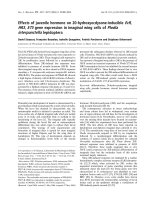
Figure 1.1 – Location of amino-acid change in the homeodomain of Nkx2-5LP
dominant negative
A) Drosophila melanogaster vnd/NK-2 homeodomain protein bound to DNA (PDB:
1NK3) (Gruschus et al. 1997) (rendered using pymol) (DeLano Scientific 2009). B)
Location of leucine to proline substitution in Nkx2-5LP is shown in red. In Nkx2-5, this
substitution results in the total loss of cardiac tissue (Grow et al. 1998).
4
In Mus musculus, Nkx2-5 isn’t required for cardiac specification, as it is in Xenopus
laevis. Nkx2-5 knockouts are embryonic lethal at day 9.5-11.5 in the mouse – not
because of the lack of cardiac tissue, but rather due to improper looping of the heart tube
(Lyons et al. 1995). However, in murine P19 carcinoma stem cells, over-expression of
Nkx2-5 is enough to drive the cells to differentiate into the cardiac lineage (Jamali et al.
2001).
Nkx2-5 is auto-regulatory, meaning that it can regulate its own expression though a
positive feedback loop (Oka et al. 1997). Nkx2-5 is also known to directly interact with a
number of other gene products, including GATA4 (Durocher et al. 1997; Riazi et al.
2009) and Tbx5 (Bruneau et al. 2001; Hiroi et al. 2001) to regulate the transcription of
genes specific to cardiomyocytes (Figure 1.2). Examples of these targets are "-cardiac
actin, ANF and myosin light chain 2 (MLC2) (Sepulveda et al. 1998; Tanaka et al. 1999).
Many of these targets are expressed only in terminally differentiated, adult,
cardiomyocytes. One of the known targets of Nkx2-5 in earlier development is
myocardin (Myocd), which is required for cardiomyogenesis (Ueyama et al. 2003). In
Xenopus laevis, myocardin doesn’t start to be expressed until stage 24, well after the start
of Nkx2-5 expression (Small et al. 2005). The lack of knowledge about early stage targets
means that the role(s) of Nkx2-5 in early development have still not been fully explored.
5
Other cardiogenic factors
Initial cardiogenesis patterning seems to occur in response to positive and negative
morphogen gradients such as the members of the bone morphogenic protein (BMP)
family, Wnt, and Wnt antagonists (Harvey et al. 2002). In addition to Nkx2-5, there are
many other genes that have a role in early cardiomyocyte determination (Figure 1.2).
The TGF-! signaling pathway is one such contributor. The TGF-! signaling cascade
starts with BMP4 and ultimately results in activation of SMAD1 and SMAD4 (Brown et
al. 2004). SMAD4 can then interact with GATA4 to regulate Nkx2-5 expression and
drive cardiogenesis (Brown et al. 2004). The role of TGF-! is further supported by
experiments demonstrating that a constitutively active TGF-! receptor can result in the
upregulation of cardiogenic factors (Brown et al. 2004). Like BMP4, treatment with
activin can also initiate cardiomyocyte differentiation via TGF-! signaling (Ariizumi et
al. 2003).
The GATA family members are also important in early cardiogenic determination.
GATA family members are zinc-finger transcription factors that bind to the rough
consensus sequence [AT]GATA[AG] (Molkentin et al. 2000) and all are expressed in the
presumptive heart field, and exhibit an overlapping expression pattern, suggesting
functional redundancy (Peterkin et al. 2005). The idea of functional redundancy is
reinforced by experiments involving GATA4 deficient mice where heart development
continued, apparently compensated for by an increase in GATA6 expression (Pikkarainen
et al. 2004). GATA6 has also been shown to activate BMP4 in adjacent endoderm, which
might be required for maintenance of Nkx2-5 expression (Peterkin et al. 2003). Nkx2-5 is
6
7
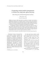
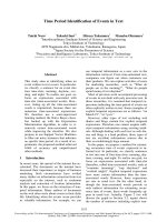
These are some of the signaling known to be involved in early cardiogenesis. TGF-! signaling in a Cardiomyocyte precursor is
activated by BMP4 signaling from adjacent endoderm. This results in SMAD4 activation which causes transcription of Nkx2-5. In
addition to BMP4, exposure to activin can have the same effect. Nkx2-5, with other co-factors, regulates the transcription of terminal
cardiogenic factors, such as "-cardiac actin, cardiac troponin, ANF, and others. Canonical Wnt signaling has been shown to block the
transcription of cardiogenic factors through !-catenin, but non-canonical Wnt signaling has been shown to have a positive effect by
either promoting the transcription of cardiogenic factors or by inhibiting the action of !-catenin. Exposure to DMSO can also cause
differentiation of mouse P19 cells to cardiomyocytes, but it does so via an unknown mechanism.
Figure 1.2 – Simplified model of known signaling in early cardiogenesis