CONTRIBUTION OF K+ CHANNELS TO CORONARY DYSFUNCTION IN METABOLIC SYNDROME
Bạn đang xem bản rút gọn của tài liệu. Xem và tải ngay bản đầy đủ của tài liệu tại đây (1.09 MB, 118 trang )
CONTRIBUTION OF K+ CHANNELS TO CORONARY
DYSFUNCTION IN METABOLIC SYNDROME
Reina Watanabe
Submitted to the faculty of the University Graduate School
in partial fulfillment of the requirements
for the degree
Doctor of Philosophy
in the Department of Cellular & Integrative Physiology,
Indiana University
May 2009
Accepted by the Faculty of Indiana University, in partial
fulfillment of the requirements for the degree of Doctor of Philosophy.
Johnathan D. Tune, Ph.D., Chair
H. Glenn Bohlen, Ph.D.
Doctoral Committee
Kieren J. Mather, M.D.
January 13, 2009
______________
Alexander G. Obukhov, Ph.D.
______________
Michael Sturek, Ph.D.
ii
ACKNOWLEDGEMENTS
I would like to thank my graduate advisor, Dr. Johnathan D. Tune, as well as the
members of my research committee, Drs. H. Glenn Bohlen, Kieren J. Mather, Alexander
G. Obukhov, and Michael Sturek, for their invaluable guidance. This work was supported
by American Heart Association grant 0810048Z (RW), National Health Institute grants
HL67804 (JDT), RR13223 (MS), HL62552 (MS), HL52490 (MHL) AR048523 (MHL), and
the Fortune-Fry Ultrasound Research Fund of the Department of Cellular and Integrative
Physiology, Indiana University School of Medicine.
iii
ABSTRACT
Reina Watanabe
CONTRIBUTION OF K+ CHANNELS TO CORONARY DYSFUNCTION
IN METABOLIC SYNDROME
Coronary microvascular function is markedly impaired by the onset of the
metabolic syndrome and may be an important contributor to the increased
cardiovascular events associated with this mutlifactorial disorder. Despite increasing
appreciation for the role of coronary K+ channels in regulation of coronary microvascular
function, the contribution of K+ channels to the deleterious influence of metabolic
syndrome has not been determined. Accordingly, the overall goal of this investigation
was to delineate the mechanistic contribution of K+ channels to coronary microvascular
dysfunction in metabolic syndrome. Experiments were performed on Ossabaw miniature
swine fed a normal maintenance diet or an excess calorie atherogenic diet that induces
the classical clinical features of metabolic syndrome including obesity, insulin resistance,
impaired glucose tolerance, dyslipidemia, hyperleptinemia, and atherosclerosis.
Experiments involved in vivo studies of coronary blood flow in open-chest anesthetized
swine as well as conscious, chronically instrumented swine and in vitro studies in
isolated coronary arteries, arterioles, and vascular smooth muscle cells. We found that
coronary microvascular dysfunction in the metabolic syndrome significantly impairs
coronary vasodilation in response to metabolic as well as ischemic stimuli. This
impairment was directly related to decreased membrane trafficking and functional
expression of BKCa channels in vascular smooth muscle cells that was accompanied by
augmented L-type Ca2+ channel activity and increased intracellular Ca2+ concentration.
In addition, we discovered that impairment of coronary vasodilation in the metabolic
iv
syndrome is mediated by reductions in the functional contribution of voltage-dependent
K+ channels to the dilator response. Taken together, findings from this investigation
demonstrate that the metabolic syndrome markedly attenuates coronary microvascular
function via the diminished contribution of K+ channels to the overall control of coronary
blood flow. Our data implicate impaired functional expression of coronary K+ channels as
a critical mechanism underlying the increased incidence of cardiac arrhythmias,
infarction and sudden cardiac death in obese patients with the metabolic syndrome.
Johnathan D. Tune, Ph.D., Chair
v
TABLE OF CONTENTS
Chapter 1: Introduction
The Epidemic of Obesity and Metabolic Syndrome................................................. 1
Metabolic Syndrome and the Coronary Circulation ................................................. 2
Coronary K+ Channels and Metabolic Syndrome .................................................... 6
KCa Channels ........................................................................................................... 8
KV Channels ............................................................................................................ 13
KATP Channels ......................................................................................................... 14
Hypothesis and Aims of the Investigation ............................................................... 15
Chapter 2: Impaired Functional Expression of Coronary BKCa Channels
in Metabolic Syndrome
Abstract ................................................................................................................... 20
Introduction.............................................................................................................. 21
Methods................................................................................................................... 23
Results .................................................................................................................... 28
Discussion ............................................................................................................... 33
Chapter 3: Role of BKCa Channels in Local Metabolic Coronary Vasodilation
in Ossabaw Swine with Metabolic Syndrome
Abstract ................................................................................................................... 39
Introduction.............................................................................................................. 40
Methods................................................................................................................... 42
Results .................................................................................................................... 46
Discussion ............................................................................................................... 52
Chapter 4: Contribution of K+ Channels to Ischemic Coronary Vasodilation
in Metabolic Syndrome
Abstract ................................................................................................................... 56
vi
Introduction.............................................................................................................. 57
Methods................................................................................................................... 59
Results .................................................................................................................... 61
Discussion ............................................................................................................... 66
Chapter 5: Discussion
Major Findings of this Investigation ......................................................................... 71
Future Directions ..................................................................................................... 76
Closing Remarks ..................................................................................................... 81
References.......................................................................................................................... 83
Curriculum Vitae
vii
CHAPTER 1: INTRODUCTION
The epidemic of obesity and metabolic syndrome
Obesity in Western society has reached epidemic proportions, as an estimated 100
million Americans are overweight or obese (66). In addition, recent estimates indicate
that there are approximately 1 billion persons worldwide who are overweight (body mass
index 25 – 30 kg/m2) (161). Many of these individuals are affected for years by the so
called “metabolic syndrome,” the combined disorder of obesity, insulin resistance,
hypertension and dyslipidemia before therapeutic measures are initiated or the
development of overt type II diabetes mellitus occurs. Presently, an estimated 30% of
the U.S. adult population exhibits characteristics of the pre-diabetic metabolic syndrome
(3; 57; 116). According to the commonly used diagnostic definition of the National
Cholesterol Education Program’s Adult Treatment Panel-III, a patient is diagnosed with
metabolic syndrome when three or more of the following clinical criteria are present in
one individual: elevated waist circumference (≥ 40 in for men, 35 in for women), elevated
triglycerides (≥ 150 mg/dL), reduced HDL cholesterol (< 40 mg/dL for men, 50 mg/dl for
women), elevated blood pressure (≥ 130/85 mmHg), and elevated fasting glucose (≥110
mg/dL) (101). These contribute to the cluster of metabolic risk factors including
abdominal obesity, atherogenic dyslipidemia, elevated blood pressure, insulin resistance
and/or glucose intolerance, prothrombotic state, and proinflammatory state that comprise
the syndrome (66).
Earlier studies have established that each component of metabolic syndrome is
an independent risk factor for cardiovascular disease (66). Recent estimates suggest
that individuals with metabolic syndrome have a 61% increased risk of cardiovascular
disease compared to those without metabolic syndrome (59). Follow-up data of the 1948
Framingham Heart Study, which sought to identify common factors that contribute to
1
cardiovascular disease, demonstrated significantly increased incidence of coronary
atherosclerotic disease, cardiomyopathies, myocardial infarction, sudden death,
congestive heart failure, and atherothrombotic stroke in obese subjects relative to lean
(59; 66; 70; 75; 76; 96; 129). Obese subjects were also found to be at twice the risk of
coronary disease (75). Despite the known link between obesity and cardiovascular
disease the pathophysiologic mechanisms underlying obesity- and metabolic syndromeinduced cardiovascular diseases remain poorly understood. Accordingly, the long-term
goal our research is to delineate mechanisms of obesity-related coronary vascular
disease and thereby elucidate pathways and novel therapeutic targets to reduce the
incidence of cardiovascular complications in this patient population. The central premise
of our studies is that impaired coronary microvascular function is an important
contributor to increased cardiovascular morbidity and mortality in obese patients with the
metabolic syndrome.
Metabolic syndrome and the coronary circulation
Due to the limited anaerobic capacity of the myocardium, the heart depends on a
continuous supply of oxygen from the coronary circulation to meet its metabolic
requirements (43; 151; 154). To ensure adequate balance between coronary blood flow
and myocardial metabolism, powerful regulatory mechanisms exist to maintain nutritive
blood flow to the heart to protect the myocardium from ischemia. If this need for oxygen
is not met, the resulting ischemia substantially diminishes cardiac function within
seconds (25; 68; 69; 132). Thus, under normal physiological conditions myocardial
oxygen delivery is closely matched with the rate of myocardial oxidative metabolism.
2
There is mounting evidence that this ability to match oxygen delivery to
myocardial demand is diminished in metabolic syndrome. Data from human patients
demonstrate diminished coronary flow reserve (the difference between maximal and
baseline coronary blood flow) with obesity and metabolic syndrome (27; 33; 87; 92; 137),
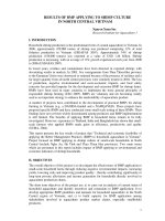
indicating that the capacity to vasodilate is greatly reduced in obesity. For instance,
Kiviniemi et al. reported a negative correlation between coronary flow reserve and waist
to hip ratio (Figure 1.1) such that maximal flow capacity diminished in proportion to the
degree of obesity (87). In addition to diminished flow reserve, recent investigations
provide evidence for impaired insulin-mediated capillary recruitment, altered obesityrelated endocrine signaling, and increased arterial stiffening in metabolic syndrome (125;
139). Remodeling of the microcirculation is considered a hallmark of established
vascular disease, and reduced perfusion has been linked to reduced wall compliance
and increased wall thickness in peripheral arterioles of obese Zucker rat (144).
Therefore, microvascular defects play an important role in the end-organ damage
associated with this combined disorder.
Figure 1.1 Correlation of waist-to-hip ratio
with coronary flow velocity reserve (CFVR;
n = 36). Taken from Kiviniemi et al. (73)
To examine the effects of the metabolic syndrome on myocardial oxygen supply
demand balance, our laboratory explored the control of coronary blood flow in
conscious, instrumented control and chronically high-fat-fed dogs at rest and during
3
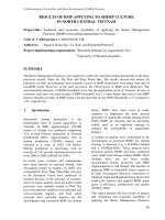
graded treadmill exercise (141). We found that the metabolic syndrome significantly
attenuated exercise-induced coronary hyperemia. Specifically, slopes of coronary blood
flow vs. MVO2 and coronary conductance (coronary blood flow normalized to mean aortic
pressure) vs. MVO2 relationships were significantly reduced in metabolic syndrome dogs
relative to lean controls (Figure 1.2A), indicating that metabolic syndrome impairs the
ability of the coronary circulation to match myocardial oxygen delivery to metabolism. In
addition, there was a significant parallel downward shift in the relationship between
coronary venous oxygen tension and MVO2 (a sensitive measure of tissue oxygenation
that reflects whether changes in coronary blood flow adequately match changes in
MVO2) in the high-fat-fed dogs (Figure 1.2B), indicating that in these animals, oxygen
delivery was not sufficient to meet myocardial metabolic demand such that these
animals had to increase extraction in order to meet their requirements for oxygen.
Further, this finding suggests a loss of a tonic vasodilator mechanism and/or activation
of a tonic vasoconstrictor mechanism (151; 154) that was present at rest as well as
during exercise. These data are consistent with attenuated coronary flow responses to
Figure 1.2 Metabolic syndrome impairs the ability of the coronary circulation to
match myocardial oxygen delivery to myocardial metabolism. Taken from Setty
et al. (118)
4
cardiac pacing in obese patients (27) and indicate that metabolic syndrome significantly
impairs regulation of coronary microvascular function to the extent that the balance
between coronary blood flow and myocardial metabolism is compromised.
The mechanisms responsible for coronary microvascular dysfunction in
metabolic syndrome have not been fully elucidated. Recent data from our laboratory
indicate that coronary vasomotor dysfunction in the metabolic syndrome is related to
sensitization of key coronary vasoconstrictor pathways (39; 88; 89; 141; 173). In
particular, metabolic syndrome is associated with elevated basal plasma epinephrine
and norepinephrine levels and sensitization of α1- and α2-adrenoceptor signaling in
metabolic syndrome dogs that limits control of coronary blood flow in response to
sympathetic activation (39). In addition, metabolic syndrome was associated with
elevated plasma renin activity and angiotensin II levels that act via increased angiotensin
II type 1 receptors (173). These data indicate that coronary vasomotor dysfunction in
metabolic syndrome is related to chronic activation of the renin-angiotensin and
sympathetic nervous system that leads to augmented AT1 and α1-adrenoceptor
mediated coronary vasoconstriction. Further, coronary vasoconstriction in canine
isolated arterioles to endothelin-1 was similar in control and metabolic syndrome despite
significantly decreased ETA-receptor transcript levels and protein expression, indicating
that ETA-receptor signaling is also sensitized by induction of metabolic syndrome (90).
These pathways converge on smooth muscle L-type Ca2+ channels to increase
intracellular Ca2+ concentrations and depolarize smooth muscle cells, thereby inducing
vasoconstriction (88). In addition, the sensitization of the angiotensin signaling pathway
directly contributes to microvascular dysfunction as inhibition of angiotensin II type 1
(AT1) receptors significantly improved the balance between coronary blood flow and
myocardial metabolism in dogs with the metabolic syndrome (173).
5
Several recent studies suggest that alterations in these mechanisms, especially
angiotensin II, could directly inhibit K+ channels, in particular large conductance Ca2+activated (BKCa) K+ channels. Agonist-induced vasoconstriction by angiotensin II has
been found to involve inhibition of BKCa channels by c-Src tyrosine kinase via direct
phosphorylation of the channel protein (4). Further, administration of angiotensin IIactivated calcineurin/NFATc3 signaling in murine arterial smooth muscle significantly
diminished BKCa channel function via down-regulation of the BKCa channel β1 subunit
expression (114). Consistent with these findings, patch-clamp studies of coronary
vascular smooth muscle cells have demonstrated that angiotensin II inhibits BKCa
channel function by altering the open and closed states of the channel thereby
prolonging the closed confirmation (150). These findings suggest that sustained
angiotensin II signaling, such as seen in hypertension or metabolic syndrome, impair K+
channel-mediated dilatory mechanisms. This can in turn be linked to depolarization of
vascular smooth muscle membrane potential (Em) and induction of arterial dysfunction
(4; 8; 21; 23; 88; 114; 150). These previous findings indicate that in addition to enhanced
vasoconstrictor pathways, depressed vasodilator mechanisms could also contribute to
impaired microvascular function in metabolic syndrome.
Coronary K+ channels and metabolic syndrome
Vascular smooth muscle cells express a variety of ion channels involved in a
wide number of physiological and pathophysiological mechanisms. K+ channel activity is
an important factor in the determination and regulation of membrane potential and
vascular tone (81; 113). The opening of K+ channels in smooth muscle cells leads to
diffusion of K+ ions out of the cell, causing membrane hyperpolarization and closure of
voltage-gated Ca2+ channels (Figure 1.3). This in turn results in a decreased
concentration of intracellular Ca2+ and induces vasorelaxation. The closure of K+
6
channels has the opposite effect, leading to the opening of L-type Ca2+ channels and
membrane depolarization, inducing an increase in intracellular Ca2+ levels, and
ultimately resulting in vasoconstriction. Many cellular metabolites, such as endothelialdependent factors (e.g. nitric oxide (NO), prostacyclin, endothelial-derived
hyperpolarizing factors (EDHF)) as well as endogenous cardiomyocyte-derived
metabolites (e.g. adenosine, hydrogen peroxide) are released in response to increased
myocardial metabolism and/or decreased tissue oxygenation to induce vasodilation
through smooth muscle K+-channel mediated pathways (82).
Figure 1.3 Schematic diagram of electromechanical coupling in
vascular smooth muscle. Taken from Jackson (68).
Importantly, recent studies have linked metabolic syndrome with diminished
functional expression of vascular smooth muscle K+ channels (26; 38; 73; 108). BKCa
channels of insulin resistant rat mesenteric arterial myocytes were found to have
reduced current density relative to lean while single channel activity and channel protein
expression remained similar (38). In arterial myocytes of diabetic fatty rat models of type
II diabetes mellitus, BKCa channel current was diminished despite absence of changes in
7
channel expression (26). Alternatively, diminished BKCa channel function could be
related to reduced activation of the channels by factors such as prostacyclin (101).
Further, decreased coupling of sarcoplasmic reticulum-mediated Ca2+ sparks to
spontaneous transient outward K+ currents were demonstrated in coronary microvessels
of alloxan-diabetic dyslipidemic swine (108). These changes are not limited to BKCa
channels, as studies also report impaired functional dilation of KATP channels in obese
Zucker rat models without changes in protein expression, linking impaired dilation to KATP
channel sensitivity (73). Therefore, decreases in K+ channel function could represent an
important component of coronary vascular dysfunction in disease states such as
metabolic syndrome. However, despite decades of research, the contribution of K+
channels to the regulation of coronary blood flow has not been fully elucidated,
especially in the setting of the metabolic syndrome.
KCa channels
There are three major classifications of K+ channels regulated by intracellular
Ca2+ levels based according to the biophysical property of conductance (i.e. by the slope
of their single channel current-voltage relationships): small (SKCa), intermediate (IKCa)
and large/big (BKCa) conductance K+ channels. Very little is known about the role of SKCa
and IKCa channels in the regulation of coronary blood flow. No patch-clamp data are
available regarding SKCa channels in coronary vascular smooth muscle. IKCa channels
are expressed in cultured coronary smooth muscle cells and may contribute to
phenotypic modulation (148), but their role in coronary vascular reactivity remains
unknown. The few studies conducted of SKCa and IKCa channel involvement in coronary
vasodilation seem to suggest an involvement of these channels in mediating EDHFinduced dilation (31; 58). Smooth muscle cell function of SKCa and IKCa channels and its
contribution to coronary blood flow will be an interesting area of future research.
8
More is known about the molecular and biophysical properties of coronary
vascular smooth muscle BKCa channels (32; 146). BKCa channels are highly expressed in
the coronary vascular smooth muscle (20; 108; 143) and have been implicated in
coronary endothelial-dependent dilation under normal-lean conditions (71; 105; 106). In
particular, bradykinin-induced endothelial-dependent relaxation in coronary arteries is
mediated, in part, by the activation of BKCa channels (71; 105; 106). Studies in swine
implicate endothelial NO release, hydroxyl radicals, or cytochrome-P450-independent
endothelial hyperpolarizing factors (71), whereas studies in human coronary artery do
not support a role for NO (105). Flow-induced vasodilation of human coronary arteries
has also been reported to involve BKCa channels but via an NO-mediated pathway that is
lost in patients with coronary artery disease (106). Despite the evidence for BKCa
channel contribution to endothelial-dependent dilation, earlier studies failed to show a
significant effect of BKCa channel blockade on resting coronary blood flow, though
evidence suggests a role in exercise-induced and ischemic vasodilation (103; 115).
Merkus et al. found that administration of the BKCa channel inhibitor tetraethylammonium
resulted in a significant decrease in the relationship between coronary venous PO2 and
MVO2 (Figure 1.4A) in normal-lean swine both at rest and during exercise (103). Animal
and human studies indicate that this relationship under control conditions is similar in
pigs, dogs, and in humans patients (42; 50; 103; 155). In addition, Node et al. reported
that the BKCa channel antagonist iberiotoxin significantly decreased coronary blood flow
during ischemia (Figure 1.4B), suggesting that ischemic vasodilators may activate BKCachannel mediated dilation (115). Therefore, previous studies implicate BKCa channels in
the regulation of endothelial-dependent dilation, metabolic control of coronary blood flow
during increases in MVO2 and also during episodes of cardiac ischemia.
9
Control
TEA
Figure 1.4 Role of BKCa channels in coronary vasodilation during
exercise (A; Merkus et al. (84)) and myocardial ischemia (B; Node et al.
(94)).
Even fewer studies have examined a role for BKCa channels in obesity and
metabolic syndrome. Interpretation of the role for BKCa channels in vascular disease is
confounded by the presence of conflicting results among the different components of
metabolic syndrome, as summarized in Table 1.1. Evidence for diminished BKCa channel
function has been observed in models of insulin resistance (38) and high-fat feeding
(168). Numerous studies investigating BKCa channel role in hypertension alone observed
evidence for increased BKCa channel function (98-100; 174) whereas others found
diminished function (7; 21; 23). Findings in models of overt type I and type II diabetes
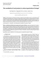
Figure 1.5 Spontaneous
transient outward currents
(STOCS) are attenuated
in smooth muscle cells
from
microvessels
obtained from diabetic
dyslipidemic animals but
not in cells from conduit
arteries.
Taken from
Mokelke et al. (88)
10
mellitus are less clear, as there is evidence for decreased BKCa channel function (102),
no change (169), as well as increased function (109). These may be due to species,
vascular bed examined, or artery size (108; 109). Recent data from the Sturek laboratory
indicates that diabetic dyslipidemia increases the functional coupling of BKCa channels to
sarcoplasmic reticulum Ca2+ release in vascular smooth muscle cells from large-conduit
coronary arteries (108). This increase was attributed to a compensatory change in
response to the increase in Ca2+ influx (or a vasoconstrictor influence) as previously
noted in conduit arteries of aldosterone-salt hypertensive rats (99). However, additional
studies in coronary microvessels revealed a significant decrease in the coupling of
sarcoplasmic reticulum-mediated Ca2+ spark events (which activate BKCa channels) with
spontaneous transient outward K+ current (STOC) frequency (Figure 1.5) (108). These
findings indicate that diabetic dyslipidemia impairs microvascular BKCa channel function,
however, the contribution of BKCa channel defects to the control of coronary blood flow
and vascular dysfunction in metabolic syndrome has not been directly examined.
Table 1.1. Summary of the role of BKCa channels in vascular disease.
Investigators
Dimitropoulou
et al. 2002
(38)
Species
Rats
Model
Insulin
resistance
Vascular bed
Mesenteric
microvessels
Yang et al.
2007 (168)
Du et al. 2006
(41)
Pig
High fat diet
Rabbit
Godlewski et
al. 2009 (62)
JeremyMcCarron
2000 (83)
Ye et al. 2004
(169)
HEK
cells
Rabbit
High
cholesterol
diet
High
cholesterol
High
cholesterol
Coronary
artery
Sphincter of
Oddi
McGahon et
Rat
Mouse
Mesenteric
artery
Femoral
artery
Streptozotocin Aorta
type I
diabetes
mellitus
Streptozotocin Retinal
11
Role of BKCa channels
↓ current
protein expression
unitary conductance
Ca2+/voltage sensitivity
↓ current
↓ protein expression
↓ current
↓ dilation
↓ unitary conductance
↑ open probability
↓ current
al. 2007 (102)
type I
diabetes
mellitus
arterioles
Mokelke et al.
2005 (108)
Pig
Alloxaninduced
diabetic
dyslipidemia
Coronary
conduit
arteries &
microvessels
Mokelke et al.
2003 (109)
Pig
Coronary
artery
Burnham et
al. 2006 (26)
Rat
Lu T et al.
2005 (101)
Rat
Liu et al. 1995
(99)
Rat
Liu et al.1997
(100)
Rat
Alloxaninduced
diabetic
dyslipidemia;
High
cholesterol
ZDF type II
diabetes
mellitus
ZDF type II
diabetes
mellitus
Aldosteronesalt
hypertension
Spontaneous
hypertension
Liu et al. 1998
(98)
Rat
Spontaneous
hypertension
Cerebral
arterioles
Zhang Y et al.
2005 (174)
Rat
Spontaneous
hypertension
Mesenteric
artery
Amberg et al.
2003 (7)
Rat
Cerebral
artery
Bratz et al.
2005 (23)
Rat
Bratz et al.
2005 (21)
Rat
Ang IIinduced
hypertension
l-NNAinduced
hypertension
l-NNAinduced
hypertension
Mesenteric
artery
Coronary
artery
Aorta
Aorta
Mesenteric
artery
Mesenteric
artery
12
↓ STOCs
↑ activation (Ca2+ sparks)
↓ protein expression
↓ mRNA expression
↓ activation (Ca2+ sparks)
↓ STOCs, microvessels
STOCs, conduit
↓ current, microvessels
current, conduit
↑ current
protein expression
↓ current
protein expression
mRNA expression
current
↓ activation (PGI)
protein expression
↑ STOCs
unitary conductance
↑ current
↑ protein expression
mRNA expression
↑ current
↑ protein expression
unitary conductance
Ca2+/voltage sensitivity
↑ STOCs
↑ current
↑ protein expression
↓ current
↑ activation (Ca2+ sparks)
↓ protein expression
↓ current
unitary conductance
Ca2+/voltage sensitivity
↓ protein expression
KV channels
Voltage-gated, delayed rectifier K+ (KV) channels are prominently expressed in
coronary artery smooth muscle cells (63). These channels conduct outward,
hyperpolarizing K+ current that is important for the regulation of cell membrane potential
(80; 126) and determining coronary vascular resistance and blood flow (131). Recent
studies implicate KV channels as important end-effectors in the regulation of local
metabolic control of coronary blood flow as well as in ischemic coronary vasodilation.
More specifically, Saitoh et al. demonstrated that coronary infusion of the KV channel
antagonist 4-aminopyridine in dogs significantly impaired the vasodilatory response to
pacing and norepinephrine-induced increases in MVO2 (135). Importantly, they observed
a significant difference in the slope of the relationship between coronary venous oxygen
tension and MVO2 (Figure 1.6A), indicating a mismatch in the balance between
myocardial oxygen delivery and myocardial metabolism with KV channel inhibition. In
addition, Dick et al. recently found that KV channels play an important role in the
regulation of baseline coronary blood flow and the reactive hyperemic response. This
effect was evidenced by marked reductions in baseline coronary blood flow following
administration of 4-aminopyridine; to the extent that myocardial ischemia was evident
Figure 1.6 Role for KV channels in coronary vasodilation in response to
increases in cardiac metabolism (A) and ischemia (B). Taken from Saito et
al. (112) and Dick et al. (33)
13
(ST segment depression). In addition, 4-aminopyridine significantly decreased the
coronary debt to repayment ratio (Figure 1.6B) thus implicating a role for KV channels in
mediating vasodilation in response to cardiac ischemia (36).
Despite mounting evidence for KV channel contribution in mediating local
metabolic and ischemic vasodilation under normal physiological conditions, the effect of
obesity and metabolic syndrome on KV channel-mediated vasodilation remains
unknown. Recent studies have linked components of metabolic syndrome, in particular
hypertension, to diminished KV channel current and expression in other vascular beds
(21; 23). However, the effect of obesity and metabolic syndrome on KV channelmediated vasodilation warrants further investigation.
KATP channels
KATP channels are comprised of pore-forming KIR6 family subunits combined with
sulfonylurea receptor subunits. They are regulated by the ATP:ADP ratio and the
channels close as ATP concentrations increase. The majority of evidence indicates that
KATP channels are not obligatory for exercise-induced dilation, but rather contribute to the
tonic regulation of coronary blood flow. In the presence of the selective KATP inhibitor
glibenclamide, Merkus et al. found that KATP channel inhibition in a swine diminished
coronary venous oxygen tension at any given rate of MVO2 (103). This was especially
prominent under resting conditions and its effect diminished with increasing MVO2, which
suggests that these channels are important for the regulation of coronary blood flow
under baseline-resting conditions. These findings are consistent with previous data from
our laboratory which found that KATP channel inhibition with glibenclamide in dogs
resulted in a parallel downward shift of the relationship between coronary venous
oxygen tension and MVO2, again indicating that KATP channels are not required for local
metabolic coronary dilation (45; 127; 128; 156). Although one study in humans found
that glibenclamide attenuated metabolic dilation in response to cardiac pacing (53),
14
experiments in dogs and pigs, as well as a recent study in human forearm (138),
demonstrate only a role for tonic regulation of coronary blood flow and no role for KATP
channels in mediating local metabolic dilation to exercise under physiological conditions.
Alternatively, in alloxan-diabetic canines glibenclamide made the slope of the
relationship between coronary venous oxygen tension and MVO2 more negative,
indicating a more prominent role for KATP channels in local metabolic dilation in type I
diabetes mellitus (156). These data suggest that KATP channel function and activity is
possibly altered under pathophysiological conditions. However, this effect has not been
shown in other animal models, and the contribution of KATP channels to the regulation of
coronary blood flow and vascular tone in obesity and metabolic syndrome remains
unexplored.
Based on these earlier studies, we suggest that BKCa, KV, and KATP channels
merit investigation as potential end-effectors of metabolic vasodilators. Elucidating the
role of these important channels in the regulation of coronary blood flow, further attention
can be focused to uncover the relative contribution of coronary K+ channels to the
coronary vascular complications of obesity and metabolic syndrome.
Hypothesis and aims of the investigation
Earlier studies have demonstrated that coronary microvascular function is
markedly impaired by the onset of the metabolic syndrome (27; 39; 90; 137; 141; 173)
and indicate that this may be an important contributor to increased cardiovascular events
in obese patients with this combined disorder (66; 70; 96). Although recent investigations
have made important strides in elucidation of key mechanisms underlying this
dysfunction, much remains unclear. Although there is mounting evidence of coronary K+
channel dysfunction in the metabolic syndrome (26; 38; 73; 108), there are no
conclusive data to directly address the contribution of K+ channels to impaired coronary
15
microvascular responsiveness in the setting of the metabolic syndrome. Accordingly, the
overall goal of this investigation was to delineate the mechanistic contribution of K+
channels to coronary microvascular dysfunction in metabolic syndrome (Figure 1.7).
Figure
1.7
Schematic
diagram of proposed studies
to examine the contribution of
K+ channels to coronary
microvascular dysfunction in
metabolic syndrome.
Aim 1 was designed to examine the molecular and functional expression of
BKCa channels that regulate coronary vascular function and to elucidate
mechanisms underlying the deleterious influence of metabolic syndrome. The
rationale for this study came from recent investigations which found that BKCa channel
activity was significantly impaired in coronary microvascular smooth muscle of alloxandiabetic dyslipidemic swine (108), mesenteric microvessels of insulin resistant rats (38),
and coronary arterial myocytes of diabetic fatty rats (26; 101). In addition, BKCa channels
are highly expressed in the coronary vascular smooth muscle cells (20; 108; 143) and
have been implicated as end-effectors in the regulation of coronary vasodilation in
response to key endothelial and metabolic metabolites (71; 105; 106). However, the
16
contribution of BKCa channel defects to the control of coronary blood flow and vascular
dysfunction in metabolic syndrome has not been examined.
Aim 2 was designed to examine whether impaired functional expression of
coronary microvascular BKCa channels in metabolic syndrome significantly
attenuates the balance between myocardial oxygen delivery and metabolism at
rest and during exercise-induced increases in MVO2. The rationale for this aim stems
from studies which have shown that BKCa channels contribute to coronary endothelialdependent and exercise-induced dilation under normal-lean conditions (16; 103; 105;
106). To date, no study has examined the contribution of BKCa channels to metabolic
coronary vasodilation in the setting of the metabolic syndrome. We propose that
decreases in BKCa channel function could be an underlying mechanism of impaired
metabolic control of coronary blood flow in the metabolic
Aim 3 was designed to elucidate the relative contribution of specific K+
channels to coronary reactive hyperemia and test the hypothesis that metabolic
syndrome impairs coronary vasodilation to cardiac ischemia via decreases in the
relative contribution of BKCa, KV, and KATP channels to the reactive hyperemic
response. The rationale for this set of experiments is supported by previous
investigations which documented a role for BKCa channels (115), KV channels (36), as
well as KATP channels (10; 28; 172) in coronary vasodilation in response to a brief
episode of myocardial ischemia, i.e. reactive hyperemia. However, the relative
contribution of these specific K+ channels to ischemic coronary vasodilation has not
been delineated in lean or obese subjects with the metabolic syndrome.
This study integrated molecular, cellular, and systems approaches to directly
examine these three aims. Experiments were performed on Ossabaw miniature swine
fed a normal maintenance diet or an excess calorie atherogenic diet that induces many
common features of metabolic syndrome, including: obesity, insulin resistance, impaired
17
glucose tolerance, and dyslipidemia (22; 49; 145). Experiments involved in vivo studies
of coronary blood flow in open-chest anesthetized swine as well as conscious,
chronically instrumented swine and in vitro studies in isolated coronary arteries,
arterioles, and vascular smooth muscle cells. Results from this investigation stand to
offer novel mechanistic insight into the role and contribution of K+ channels in metabolic
syndrome-induced coronary vascular disease.
18