- Trang chủ >>
- Y - Dược >>
- Ngoại khoa
oxford case histories in neurosurgery
Bạn đang xem bản rút gọn của tài liệu. Xem và tải ngay bản đầy đủ của tài liệu tại đây (8.15 MB, 464 trang )
Oxford Case Histories
00-Hasegawa-FM.indd i 2/28/2013 4:11:45 PM
Oxford Case Histories
Series Editors
Sarah Pendlebury and Peter Rothwell
Published:
Neurological Case Histories (Sarah Pendlebury, Philip Anslow, and
Peter Rothwell)
Oxford Case Histories in Cardiology (Rajkumar Rajendram,
Javed Ehtisham, and Colin Forfar)
Oxford Case Histories in Gastroenterology and Hepatology (Alissa Walsh,
Otto Buchel, Jane Collier, and Simon Travis)
Oxford Case Histories in Respiratory Medicine (John Stradling,
Andrew Stanton, Najib Rahman, Annabel Nickol, and Helen Davies)
Oxford Case Histories in Rheumatology (Joel David, Anne Miller,
Anushka Soni, and Lyn Williamson)
Oxford Case Histories in TIA and Stroke (Sarah Pendlebury, Ursula Schulz,
Aneil Malhotra, and Peter Rothwell)
Oxford Case Histories in Neurosurgery (Harutomo Hasegawa,
Matthew Crocker, and Pawan Singh Minhas)
00-Hasegawa-FM.indd ii 2/28/2013 4:11:45 PM
1
Oxford Case
Histories in
Neurosurgery
Harutomo Hasegawa
Specialty Registrar in Neurosurgery, London Deanery, UK
Matthew Crocker
Consultant Neurosurgeon, Atkinson Morley Wing,
St. George’s Hospital, London, UK
Pawan Singh Minhas
Consultant Neurosurgeon, Atkinson Morley Wing,
St. George’s Hospital, London, UK
00-Hasegawa-FM.indd iii 2/28/2013 4:11:45 PM
1
Great Clarendon Street, Oxford OX2 6DP
United Kingdom
Oxford University Press is a department of the University of Oxford.
It furthers the University’s objective of excellence in research, scholarship,
and education by publishing worldwide. Oxford is a registered trade mark of
Oxford University Press in the UK and in certain other countries
© Oxford University Press, 2013
The moral rights of the authors have been asserted
First published 2013
Impression: 1
All rights reserved. No part of this publication may be reproduced, stored in
a retrieval system, or transmitted, in any form or by any means, without the
rior permission in writing of Oxford University Press, or as expressly permitted
by law, by licence or under terms agreed with the appropriate reprographics
rights organization. Enquiries concerning reproduction outside the scope of the
above should be sent to the Rights Department, Oxford University Press, at the
address above
You must not circulate this work in any other form
and you must impose this same condition on any acquirer
British Library Cataloguing in Publication Data
Data available
ISBN 978–0–19–959983–7
Printed and bound by
CPI Group (UK) Ltd, Croydon, CR0 4YY
Oxford University Press makes no representation, express or implied, that the drug dosages in this
book are correct. Readers must therefore always check the product information and clinical
procedures with the most up-to-date published product information and data sheets provided by the
manufacturers and the most recent codes of conduct and safety regulations. The authors and the
publishers do not accept responsibility or legal liability for any errors in the text or for the misuse or
misapplication of material in this work. Except where otherwise stated, drug dosages and
recommendations are for the non-pregnant adult who is not breast-feeding.
Links to third party websites are provided by Oxford in good faith and
for information only. Oxford disclaims any responsibility for the materials
contained in any third party website referenced in this work.
00-Hasegawa-FM.indd iv 2/28/2013 4:11:45 PM
Acknowledgements
We would like to thank Anthony Pereira and Phil Rich for their helpful comments in
reviewing the manuscript and Oxford University Press for their care and attention
throughout the publishing process. We would also like to thank Steve Connor for
Fig. 62.1, Mihai Danciut for Figs. 21.5 and 47.3, James Laban for Fig. 55.1, and Donal
Walsh for his help in preparing cases 18 and 23. We are grateful to our teachers in
neurosurgery, and to our patients whom we were privileged to treat.
A note from the series editors
Case histories have always had an important role in medical education, but most
published material has been directed at undergraduates or residents. The Oxford Case
Histories series aims to provide more complex case-based learning for clinicians in
specialist training and consultants, with a view to aiding preparation for entry- and
exit-level specialty examinations or revalidation.
Each case book follows the same format with approximately 50 cases, each compris-
ing a brief clinical history and investigations, followed by questions on differential
diagnosis and management, and detailed answers with discussion.
At the end of each book, cases are listed by mode of presentation, aetiology, and
diagnosis. We are grateful to our colleagues in the various medical specialties for their
enthusiasm and hard work in making the series possible.
Sarah Pendlebury and Peter Rothwell
Foreword
Safe, successful care of patients requires both a sound knowledge base and the skill to
apply it effectively. In neurosurgery there is no shortage of didactic, factual accounts to
support the systematic study of disciplines such as neuroanatomy, neurophysiology, neu-
ropathology, neuroimaging and how abnormalities are expressed and managed in vari-
ous clinical conditions. Unfortunately these subjects have a reputation for being difficult,
complicated, even mysterious, leaving doctors within, or those liaising with neurosur-
gery, experiencing hesitancy and insecurity in the face of the complexities of the care of a
patient. An antidote to this situation is now available through this compendium of pres-
entations which convey how the key information relevant to a range of clinical problems
can be selected and used to achieve timely, effective decision-making and treatment.
The emphasis is on learning from vividly described case histories portraying the
presentation, investigation and management of individual patients suffering from a
wide breadth of clinical problems. The flow of information mirrors clinical experi-
ence. The successive sets of questions that are posed and then answered throughout
each case engage, stimulate and inform the reader and convey how knowledge and
understanding are applied to the clinical situation of real-world cases. This problem-
based learning approach is familiar to modern students and graduates but until now
there has been little written material to support case-based learning as part of private
study. This is increasingly relevant to the emphasis on scenario and patient-based
questions in speciality training exit examinations.
The principle of placing the patient at the centre of learning fits well with the philosophy
of key figures in the original emergence of neurosurgery as a separate discipline. While a
resident in general surgery, Harvey Cushing was stimulated and encouraged to specialise
in neurosurgery by Sir William Osler, then professor of medicine in Baltimore, later Regius
Professor in Oxford. In his Pulitzer prize-winning biography of Osler, Cushing paid trib-
ute to how his mentor had made clinical teaching the foundation of modern medical
education, as expressed in his dictum ‘
He who studies medicine without books sails an
uncharted sea, but he who studies medicine without patients does not go to sea at all’.
Standard texts retain a place in neurosurgical education but it is through the study
of individual patients that the skills necessary for confident and competent clinical
diagnosis and management are gained. The wealth of information conveyed so mem-
orably by the patient stories assembled by
Messrs Hasegawa, Crocker and Minhas will
powerfully promote these abilities in u
ndergraduates, trainees and qualified special-
ists, whether in neurosurgery or in specialties interfacing with it,
and hence the quality
of care they give to their patients.
Sir Graham Teasdale
FRCS, FRCP, F Med Sci, FRSE
Emeritus Professor of Neurosurgery, University of Glasgow
Past President of the Society of British Neurological Surgeons and of the Royal
College of Physicians and Surgeons of Glasgow
Contents
Abbreviations viii
Section 1. Cranial trauma 1
Cases 1–8 3
Section 2. Spinal trauma 67
Cases 9–16 69
Section 3. Vascular neurosurgery 117
Cases 17–28 119
Section 4. Neuro-oncology 213
Cases 29–45 215
Section 5. Spinal neurosurgery 319
Cases 46–52 321
Section 6. Paediatric neurosurgery and hydrocephalus 355
Cases 53–61 357
Section 7. Miscellaneous 405
Cases 62–67 407
List of cases by diagnosis 439
List of cases by principal clinical features at presentation 441
List of cases by aetiological mechanism 442
Index 443
00-Hasegawa-FM.indd vii 2/28/2013 4:11:46 PM
ACA anterior cerebral artery
ACD anterior cervical discectomy
ACom anterior communicating artery
ADC apparent diffusion coefficient
ADH antidiuretic hormone
AF atrial fibrillation
AAGBI Association of Anaesthetists of
Great Britain and Ireland
AICA anterior inferior cerebellar artery
AP anteroposterior
ASIA American Spinal Injury
Association
ATLS Advanced Trauma Life Support
ATP adenosine triphosphate
AVM arteriovenous malformation
bd twice daily
BIH benign intracranial hypertension
bpm beats per minute
CBF cerebral blood flow
CPP cerebral perfusion pressure
CRP C-reactive protein
CSF cerebrospinal fluid
CSW cerebral salt wasting
CT computed tomography
CTA CT angiography/angiogram
CTS carpal tunnel syndrome
CVP central venous pressure
CVR cerebral vascular resistance
DAI diffuse axonal injury
DBS deep brain stimulation
DCI delayed cerebral ischaemia
DDAVP 1-deamino-8 d -arginine
vasopressin
DI diabetes insipidus
DIND delayed ischaemic neurological
deficit
DNET dysembryoplastic neuroepithelial
tumour
DVLA Driver and Vehicle Licensing
Agency
DVT deep vein thrombosis
DWI diffusion-weighted imaging
E eye-opening (GCS)
EC extracranial
ECF extracellular fluid
ENT ear, nose, and throat
ETV endoscopic third
ventriculostomy
EVD external ventricular drain
FLAIR fluid attenuated inversion
recovery
GCS Glasgow Coma Scale/Score
GP general practitioner
GPi globus pallidus internus
HIV human immunodeficiency virus
IC intracranial
ICA internal carotid artery
ICH intracranial haemorrhage
ICP intracranial pressure
ICU intensive care unit
IGF-1 insulin-like growth factor 1
IIH idiopathic intracranial
hypertension
INR international normalized ratio
ISAT International Subarachnoid
Aneurysm Trial
L litre
LP lumbar puncture
M motor response (GCS)
MAP mean arterial pressure
MCA middle cerebral artery
MEP motor evoked potential
mg milligram
MIP maximum intensity projection
mL millilitre
MRA magnetic resonance angiography
Abbreviations
00-Hasegawa-FM.indd viii 2/28/2013 4:11:46 PM
ix
MRC Medical Research Council
MRI magnetic resonance
imaging/image
MRS magnetic resonance
spectroscopy
ng nasogastric
NICE National Institute for Health
and Clinical Excellence
NPH normal pressure hydrocephalus
PCA posterior cerebral artery
PCom posterior communicating
artery
PCV procarbazine–lomustine
(CCNU)–vincristine
PE pulmonary embolism
PET positron emission tomography
PICA posterior inferior cerebellar
artery
PNET primitive neuroectodermal
tumour
po by mouth
RCT randomized controlled trial
RTA road traffic accident
SAH subarachnoid haemorrhage
SCA superior cerebellar artery
SIADH syndrome of inappropriate
ADH secretion
SSEP somatosensory evoked potential
STN subthalamic nucleus
TB tuberculosis
TIA transient ischaemic attack
V verbal response (GCS)
VP ventriculoperitoneal
VTE venous thromboembolism
WHO World Health Organization
00-Hasegawa-FM.indd ix 2/28/2013 4:11:46 PM
This page intentionally left blank
Section 1
Cranial trauma
01-Case01-Hasegawa.indd 1 1/22/2013 2:17:58 PM
This page intentionally left blank
CASE 1
3
Case 1
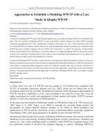
A 78-year-old man was admitted to hospital with a 2 week history of progressive con-
fusion and unsteadiness. His medical history included parkinsonism and a metallic
mitral valve replacement. On examination his GCS was 14/15 (E4, V4, M6) (see
‘Glasgow Coma Scale and Score’, p. 196), and he had left-sided weakness. He was
taking warfarin, and the INR was 3.8.
Questions
1. What is the differential diagnosis?
2. A CT scan of the brain is performed (Fig. 1.1 ). Describe the appearances.
Fig. 1.1
01-Case01-Hasegawa.indd 3 1/22/2013 2:17:58 PM
OXFORD CASE HISTORIES IN NEUROSURGERY
4
Answers
1. What is the differential diagnosis?
Progressive confusion and gait disturbance with a left hemiparesis point to a right
hemisphere lesion. The differential diagnosis includes cerebral infarction or haem-
orrhage, subdural haematoma, and a neoplastic lesion. The time course of the
symptoms is central to distinguishing them: intracerebral haemorrhage or stroke
typically presents with sudden-onset symptoms; progressive symptoms suggest a
slowly enlarging mass such as a tumour or chronic subdural haematoma.
2. A CT scan of the brain is performed (Fig. 1.1 ). Describe the
appearances (Fig. 1.2 ).
There is an extra-axial crescent shaped fluid collection over the right cerebral con-
vexity (A, B) indicating a chronic subdural haematoma (Fig. 1.2 ). The patient is
scanned supine. There is layering according to density, with a hypodense fluid
supernatant (A) above hyperdense thrombus or cellular precipitant (B). This
appearance could be due to a single episode of haemorrhage or rebleeding into a
chronic collection. There is midline shift (C) with obliteration of cerebral sulci and
the trigone (not seen, D) on the right.
Fig. 1.2
01-Case01-Hasegawa.indd 4 1/22/2013 2:17:59 PM
CASE 1
5
Questions
3. What is the pathophysiology of chronic subdural haematomas?
4. What are the initial considerations in the management of this case?
5. What is the urgency of surgery? When should surgery be performed if the patient
presents in the middle of the night?
6. What are the surgical options?
7. What are the complications of surgery?
8. The wife of the patient expresses her concern about plans for surgery. She tells
you that her husband was never keen on surgery and that he would not have
liked to survive with neurological impairment. She does not want you to
perform the operation.
(a) How would you approach this conversation and what points would you
cover in the discussion?
(b) What is the legal position of the family’s views on a patient’s treatment?
9. The subdural haematoma is evacuated with burrholes, and the patient makes a
good recovery. How should his anticoagulation be managed postoperatively?
01-Case01-Hasegawa.indd 5 1/22/2013 2:17:59 PM
OXFORD CASE HISTORIES IN NEUROSURGERY
6
Answers
3. What is the pathophysiology of chronic
subdural haematomas?
Chronic subdural haematomas are typically caused by tearing of dural bridging
veins. Cerebral atrophy (e.g. in the elderly or in alcoholic patients) causes increased
tension on these veins, predisposing them to tearing. The trauma causing the initial
bleed can be sufficiently mild to be absent from the history, even in retrospect, in
over 50 % of patients. A local inflammatory reaction follows the haemorrhage and
results in the formation of a haematoma cavity with membranes within it. The clot
liquefies over time and this collection may expand. The processes that mediate this
are poorly understood, but may include recurrent microbleeds from dural capillar-
ies and haematoma membranes, secretion of fluid from haematoma membranes,
and osmotic fluid shifts into the haematoma cavity.
4. What are the initial considerations in the
management of this case?
The initial consideration is whether the patient should be managed operatively or
conservatively. Operative management is appropriate in the presence of a neurologi-
cal deficit or severe and persistent headache. In either case the INR requires normali-
zation and blood tests, including serum sodium and clotting, should be performed.
5. What is the urgency of surgery? When should surgery be
performed if the patient presents in the middle of the night?
Surgery should be performed as soon as possible, but the practicalities of operating
overnight require consideration if the patient presents in the middle of the night.
Surgery should be considered overnight if symptoms have progressed rapidly or if
the haematoma is large (e.g. with significant midline shift and contralateral
ventricular enlargement from encystment). However, if deterioration has occurred
over several days or weeks, it would be reasonable to wait until the morning.
6. What are the surgical options?
There are several options for chronic subdural haematomas. Burrhole drainage is
the most common. There are specific indications for performing a craniotomy,
such as the presence of subdural membranes and recurrent episodes (see ‘Surgery
for chronic subdural haematomas ’, p. 8 and ‘Varieties of chronic subdural
haematomas’ , p. 9).
7. What are the complications of surgery?
Seizures, intracranial haematoma, pneumocephalus, and infection (including sub-
dural empyema). Patients should also be advised of the risk of recurrence (up to
30 % ) and risk to life with a general anaesthetic, especially in a condition affecting
an almost exclusively elderly population.
01-Case01-Hasegawa.indd 6 1/22/2013 2:17:59 PM
CASE 1
7
8. The wife of the patient expresses her concern about plans
for surgery. She tells you that her husband was never
keen on surgery and that he would not have liked to survive
with neurological impairment. She does not
want you to perform the operation.
a) How would you approach this conversation and
what points would you cover in the discussion?
The patient’s present condition and his prognosis with and without surgery should be
carefully communicated to the family. If this is done effectively and there is a clear case
for intervention, it is unusual for the family to disagree with the proposed treatment.
The existence of advance directives or a legal guardian (an individual who is legally
authorized to make decisions on behalf of the patient) should also be determined.
b) What is the legal position of the family’s
views on a patient’s treatment?
If a patient lacks capacity to consent for treatment, in the UK the doctor is required to
make a decision in the patient’s best interests. The views of the family will inform this
decision but they (or any other individual) cannot consent on behalf of the patient.
Therefore a discussion with the family is essential before proceeding to surgery
(although this should not delay life-threatening surgery). If there is any doubt about
advance directives or legal guardians, the doctor should make a decision in the
patient’s best interests based on available information (for further guidance on patient
autonomy and consent see Good Medical Practice , General Medical Council, UK).
9. The subdural haematoma is evacuated with burrholes, and the
patient makes a good recovery. How should his anticoagulation
be managed postoperatively?
The risk of further intracranial bleeding must be balanced against the risk from
systemic embolization from a metallic heart valve. In general, the latter risk is
greater and anticoagulation should be recommenced early, although observational
studies have shown that stopping anticoagulation perioperatively for up to 2 weeks
in patients with mechanical heart valves is safe. In this patient a CT scan was per-
formed 48 hours after surgery to exclude ongoing haemorrhage. This was negative
and he was restarted on warfarin ( see ‘Anticoagulation in neurosurgery’, p. 11 ).
Further reading
General Medical Council (UK) ( 2011 ). Good Medical Practice . Available online at: http://www.
gmc-uk.org/static/documents/content/GMP_0910.pdf (accessed 27 February 2011) .
Haines DE , Harkey HL , Al-Mefty O ( 1993 ). The ‘subdural’ space: a new look at an outdated
concept . Neurosurgery ; 32 : 111 – 20 .
Wilberger JE ( 2000 ). Pathophysiology of evolution and recurrence of chronic subdural
hematoma . Neurosurg Clin N Am ; 11 : 435 – 8 .
Yamashima T , Yamamoto S ( 1985 ). The origin of inner membranes in chronic subdural
hematomas . Acta Neuropathol ; 67 : 219 – 25 .
01-Case01-Hasegawa.indd 7 1/22/2013 2:17:59 PM
OXFORD CASE HISTORIES IN NEUROSURGERY
8
Surgery for chronic subdural haematomas
Chronic subdural haematomas are a very common neurosurgical condition but
remain challenging to treat for various reasons.
◆ They are frequently due to multiple bleeds and hence have membranes causing
compartmentalization or ‘loculation’ of the haematoma, making it harder to
drain via a single hole.
◆ They usually occur in elderly people with multiple comorbidities.
◆ The brains of elderly people are slower to re-expand and fill the subdural space
after the haematoma is evacuated. Therefore there is a large space between the
brain and the skull which continues to stretch the bridging veins and has a tendency
to fill with venous blood, causing re-accumulation of the haematoma .
◆ They are more common in patients on anticoagulation. If there is a compelling
reason for anticoagulation (e.g. mechanical heart valve), there is justifiable anxiety
about temporary withdrawal of anticoagulation.
Various surgical options are available and a balance is required between minimiz-
ing discomfort (performing the operation under local anaesthesia) and minimiz-
ing risk of recurrence (which may require a larger operation). The options (in
increasing order of magnitude) are as follows.
1. Twist drill craniostomy: this can be done under local anaesthetic, even on the ward. A
small-diameter drill bit is used, similar to that used to place an ICP monitor, and the
burrhole drilled without direct vision. The skin is closed over the burrhole without
formal irrigation in the hope that a completely liquefied haematoma will be absorbed
into the galea. This is less invasive than all the other options and probably less effective.
2. Burrhole drainage: this can also be performed under local anaesthetic with or without
sedation in a suitable patient, but an anaesthetist should be available in case the need
for urgent general anaesthesia arises. It must be performed in the operating theatre.
The burrholes allow formal irrigation of the clot either in and out of a single burrhole
or through two burrholes. The burrholes are left open and the haematoma cavity again
communicates with the subgaleal space. High-quality evidence supports a period of
postoperative drainage using a soft subdural catheter for 2 days (Santarius et al . 2009 ).
3. Craniotomy: this is usually reserved for re-collected subdural haematomas or
those with loculations that cannot be managed using burrholes alone. A modest
craniotomy will allow direct visualization of the subdural space and the opportu-
nity to divide or excise the membranes that form compartments within the
haematoma cavity. This typically requires general anaesthesia.
Decisions to be made in the postoperative period include the following.
◆ When to allow the patient to sit up and mobilize: theoretically maintaining the
patient supine will reduce venous return and encourage the brain to re-expand
and obliterate the subdural space . This is probably associated with a lower risk
of recurrence (Abouzari et al. 2007 ).
◆ When to restart anticoagulation (see ‘Anticoagulation in neurosurgery’, p. 11).
01-Case01-Hasegawa.indd 8 1/22/2013 2:17:59 PM
CASE 1
9
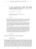
Varieties of chronic subdural haematomas
This 86-year-old man (Fig. 1.3 ) has bilateral chronic
subdural haematomas. Bilateral subdural haematomas
may exert considerable pressure on the brain. There is
midline shift to the right as the larger haematoma on
the left exerts more pressure than the smaller collection
on the right. There is greater sulcal effacement on the
left under the larger collection. As a consequence of the
mass effect there is often also vertical shift of the brain
which is harder to appreciate on axial images. Bilateral
burrholes are required to manage this condition. If
only one side is evacuated, more midline shift will result
from the unopposed haematoma on the other side.
The patient returns to hospital one week after drain-
age of the subdural haematomas due to increasing
drowsiness. The scan (Fig. 1.4 ) shows bilateral sub-
dural collections and some air over the right frontal
lobe (A). There is less mass effect and the midline shift
has resolved. The question is whether the residual col-
lections are responsible for the symptoms. In this case
the patient is clinically worse but the scan looks better.
Therefore other causes for the drowsiness should be
considered before surgery to re-evacuate the residual
collections is contemplated. This patient had hyponat-
raemia and he improved when this was corrected. The
term ‘recurrent chronic subdural haematoma’ is often used when a patient who
has had a chronic subdural haematoma drained returns with a scan showing per-
sisting subdural collections. This could represent a new episode of subdural haem-
orrhage, re-accumulation of fluid secreted by membranes, or simply saline wash
used to irrigate the subdural cavity in the previous operation. A postoperative sub-
dural collection could also be infected, presenting with sepsis with worsening
headache or neurological deficit.
The chronic subdural haematoma in this 87-year-old
man contains septations within the collection (Fig. 1.5 :
arrows) representing membranes. There is mass effect
on the right hemisphere causing effacement of sulci.
The right lateral ventricle is displaced downwards out
of the imaging plane of this slice, indicating downward
brain herniation. Little midline shift is evident as this
image is at the level of the falx (the bright line in the
mid-sagittal plane) which restrains brain herniation
apart from adjacent to the left lateral ventricle where
subfalcine herniation of the medial right frontal lobe
Fig. 1.3
Fig. 1.5
Fig. 1.4
(continued)
01-Case01-Hasegawa.indd 9 1/22/2013 2:17:59 PM
OXFORD CASE HISTORIES IN NEUROSURGERY
10
Varieties of chronic subdural haematomas (continued)
is apparent. Burrholes are unlikely to be successful because it will not be possible to
access all the subdural compartments formed by the membranes. A larger ( > 2.5cm
diameter) burrhole or a craniotomy enables the membranes to be accessed and
divided, and will offer the best chance of improvement.
This 74-year-old woman
(Fig. 1.6 ) presented with
headaches but without
any neurological deficits.
She has a left-sided
chronic subdural hae-
matoma with mass effect
(note the effacement of
sulci on the left) but no
midline shift (Fig. 1.6 (A)).
Surgery in such a situation
is unlikely to make her
better. However, it could
be argued that she may deteriorate if untreated because of expansion of the hae-
matoma. Some surgeons may operate but it would also be reasonable to manage her
conservatively. A small dose of dexamethasone (2mg bd for 10 days) will tend to settle
the headache and even a mild neurological deficit somewhat faster. Its mechanism of
action is unknown, but it is thought to stabilize the chronic subdural membrane and
have a protective effect on the cerebral cortex. She was managed conservatively and her
CT scan one week later (Fig. 1.6 (B)) shows reduction in the size of the haematoma and
less mass effect (the sulci are now visible in the left hemisphere).
AB
Fig. 1.6
01-Case01-Hasegawa.indd 10 1/22/2013 2:17:59 PM
CASE 1
11
Anticoagulation in neurosurgery
An increasing number of patients are anticoagulated. Common indications are
prevention of cardiovascular disease, prevention of stroke in atrial fibrillation
and prosthetic heart valves, and treatment of venous thromboembolism (DVT
and PE). Here we discuss the perioperative management of anticoagulation in
neurosurgical patients.
Reversal of anticoagulation
Elective patients
Antiplatelet therapy and warfarin should be stopped a few days before surgery.
Warfarin bridging can be performed if the thromboembolic risk is particularly high:
patients are admitted to hospital a few days before surgery and commenced on
heparin while warfarin is stopped. Full anticoagulation can continue until several
hours before surgery (typically 6 hours for unfractionated heparin and 12 hours
for low molecular weight heparin). Elective surgery should be postponed if the
acute event necessitating anticoagulation is recent, as the thromboembolic risk is
particularly high and surgery will increase the risk further.
Emergency patients
Patients requiring emergency surgery and those with intracranial haemorrhage
(ICH) require rapid and complete reversal of anticoagulation.
Warfarin
Intravenous vitamin K and prothrombin complex should be given.
Antiplatelets
Aspirin irreversibly blocks platelet function for the life of the platelet (approxi-
mately 10 days). Restoration of platelet function depends on the synthesis of new
platelets. The number of new functional platelets can be estimated (10 % of platelets
are replenished per day; hence if the platelet count is 250 × 10
9
/L, 25 × 10
9
new
platelets will be produced per day). A platelet transfusion can be given if a patient is
deemed to have insufficient functional platelets. One pool of platelets will raise the
platelet count by approximately 50 × 10
9
platelets. Clopidogrel has stronger
antiplatelet activity and two pools of platelets may be given (Beshay et al. 2010 ).
The role of platelet transfusions in conservatively managed intracerebral haemor-
rhage is unclear (Morgenstern et al. 2010 ).
Postoperative issues
Venous thromboembolism (VTE) prophylaxis
The incidence of VTE in neurosurgical patients is high and many are asympto-
matic (Iorio and Agnelli 2000 ). A recent meta-analysis showed that low-dose
(continued)
01-Case01-Hasegawa.indd 11 1/22/2013 2:18:00 PM
OXFORD CASE HISTORIES IN NEUROSURGERY
12
Anticoagulation in neurosurgery (continued)
heparin reduced the risk of VTE but with a slight increase in haemorrhagic events
(9.1 % absolute risk reduction in VTE; 0.7 % absolute risk increase in ICH)
(Hamilton et al. 2011 ). NICE ( 2010 ) advises mechanical prophylaxis for neurosur-
gical patients at increased risk of VTE with postoperative heparin (usually com-
menced 12–24 hours postoperatively) if the risk of major bleeding is low. If the
presentation is with cranial or spinal haemorrhage, heparin prophylaxis is not rec-
ommended until the lesion is secured or the condition is stable (Morgenstern et al.
2010 ; NICE 2010 ).
Recommencement of anticoagulation
Anticoagulation should be restarted as soon as the risk of haemorrhage from
a particular condition has passed. Retrospective studies show that withholding
warfarin for up to 2 weeks is safe in patients with prosthetic heart valves (Romualdi
et al. 2009 ).
Intracranial haemorrhage (ICH)
All anticoagulants (including antiplatelet agents) increase the risk of ICH.
The majority are intracerebral and subdural haematomas. Population estimates
for the absolute risk of ICH on anticoagulants are 0.2–0.3 % /year for aspirin,
0.3–0.4 % /year for aspirin plus clopidogrel, and 0.3–1 % /year for warfarin (vs.
0.15 % /year in the general population aged 70) (Hart et al. 2005 ). The individual
risk varies considerably depending on age, comorbidities, intensity of anticoagula-
tion, and lifestyle.
When an anticoagulated patient survives an ICH, a decision is required on
whether it should be continued. This decision is based on the risk of recurrent
ICH, the risk of thromboembolism (Table 1.1 ) and the overall neurological status
of the patient. One systematic review found an aggregate recurrence rate for
ICH without anticoagulation of 2.3 % /year (Bailey et al. 2001 ). In one study, anti-
coagulation increased the risk of recurrent ICH three-fold (Vermeer et al. 2002 ).
The individual risk of recurrent ICH (influenced by age, comorbidities, mobility,
lifestyle, and anticoagulant use) requires careful consideration and needs to be
balanced against the thromboembolic risk derived from cardiovascular risk strati-
fication. Antiplatelet agents are safer than warfarin and have been recommended
for patients at a relatively low risk of thromboembolism and a higher risk of ICH,
or in those with very poor neurological function (Furie et al. 2011 ). If warfarin is
to be continued, a CT scan may be helpful to exclude a persistent or postoperative
haematoma. Some guidelines (e.g. Furie et al. 2011 ) suggest that all anticoagulants,
including antiplatelet drugs, should be withheld for at least 1–2 weeks following
ICH (including intracerebral, subdural, and subarachnoid haemorrhage) although
individual practices vary according to experience and the perceived balance of risks
and benefits.
01-Case01-Hasegawa.indd 12 1/22/2013 2:18:00 PM
CASE 1
13
Table 1.1 Thromboembolic risk without anticoagulation
Condition Risk of thromboembolic
complications off
warfarin ( % /year)
Notes
Metallic heart valve
(Mok et al. 1985 ;
Cannegieter et al.
1994 )
4–12 Increased risk in mitral valves, ball-cage
valves, increasing age, comorbidities (e.g.
atrial fibrillation, left ventricular dysfunction)
Atrial fibrillation
(Gage et al. 2001 )
1.9–18.2 Increased risk with additional comorbidities
(congestive heart failure, hypertension, age
≥ 75, diabetes, previous stroke)
DVT/PE (Kearon
and Hirsh 1997 )
15 40 % in first month, 10 % in next 2 months
after initial event
Risk increased 100-fold in postoperative
period
References
Abouzari M , Rashidi A , Rezaii J , et al . ( 2007 ). The role of postoperative patient posture in the
recurrence of traumatic chronic subdural hematoma after burr-hole surgery . Neurosurgery
2007 ; 61 : 794 – 7 .
Bailey RD , Hart RG , Benavente O , Pearce LA ( 2001 ). Recurrent brain hemorrhage is more
frequent than ischemic stroke after intracranial hemorrhage . Neurology ; 56 : 773 – 7 .
Beshay JE , Morgan HM , Madden C , Yu W , Sarode R ( 2010 ). Emergency reversal of
anticoagulation and antiplatelet therapies in neurosurgical patients . J Neurosurg ; 112 :
307 – 18 .
Cannegieter SC , Rosendaal FR , Briet E ( 1994 ). Thromboembolic and bleeding complications
in patients with mechanical heart valve prosthesis . Circulation ; 89 : 635 – 41 .
Furie KL , Kasner SE , Adams RJ , et al . ( 2011 ). Guidelines for the prevention of stroke in
patients with stroke or transient ischemic attack: a guideline for healthcare professionals
from the American Heart Association/American Stroke Association . Stroke ; 42 : 227 – 76 .
Gage BF , Waterman AD , Shannon W , et al . ( 2001 ). Validation of clinical classification schemes
for predicting stroke: results from the National Registry of Atrial Fibrillation . JAMA ; 285 :
2864 – 70 .
Hamilton MG , Yee WH , Hull RD , Ghali WA ( 2011 ). Venous thromboembolism prophylaxis
in patients undergoing cranial neurosurgery: a systematic review and meta-analysis .
Neurosurgery ; 68 : 571 – 81 .
Hart RG , Boop BS , Anderson DC ( 1995 ). Oral anticoagulants and intracranial hemorrhage:
Facts and hypotheses . Stroke ; 26 : 1471 – 7 .
Hart RG , Tonarelli SB , Pearce LA ( 2005 ). Avoiding central nervous system bleeding during
antithrombotic therapy: recent data and ideas . Stroke ; 36 : 1588 – 93 .
Iorio A , Agnelli G ( 2000 ). Low molecular weight and unfractionated heparin for prevention
of venous thromboembolism in neurosurgery . Ann Int Med ; 160 : 2327 – 32 .
Kearon C , Hirsh J ( 1997 ). Management of anticoagulation before and after surgery . N Engl
J Med ; 336 : 1506 – 11 .
01-Case01-Hasegawa.indd 13 1/22/2013 2:18:00 PM
OXFORD CASE HISTORIES IN NEUROSURGERY
14
Mok CK , Boey J , Wang R , et al . ( 1985 ). Warfarin versus dipyridamole-aspirin and
pentoxifyllineaspirin for the prevention of prosthetic heart valve thromboembolism: a
prospective clinical trial . Circulation ; 72 : 1059 – 63 .
Morgenstern LB , Hemphill C , Anderson C , et al . ( 2010 ). Guidelines for the management of
spontaneous intracerebral hemorrhage. A guideline for healthcare professionals from the
American Heart Association/American Stroke Association . Stroke ; 41 : 2108 – 29 .
NICE ( 2010 ). Venous thromboembolism — reducing the risk. NICE Guideline CG92 . Available
online at: (accessed
24 April 2011).
Romualdi E , Micieli E , Ageno W , Squizzato A ( 2009 ). Oral anticoagulant therapy in patients
with mechanical heart valve and intracranial haemorrhage . Thromb Haemost ; 101 : 290 – 7 .
Santarius T , Kirkpatrick PJ , Ganesan D , et al . ( 2009 ). Use of drains versus no drains after
burr-hole evacuation of chronic subdural haematoma: a randomised controlled trial .
Lancet ; 374 : 1067 – 73 .
Vermeer SE , Algra A , Franke CL , Koudstaal PJ , Rinkel GJE ( 2002 ). Long-term prognosis after
recovery from primary intracerebral hemorrhage . Neurology ; 59 : 205 – 9 .
01-Case01-Hasegawa.indd 14 1/22/2013 2:18:00 PM