- Trang chủ >>
- Y - Dược >>
- Ngoại khoa
oxford case histories in respiratory medicine
Bạn đang xem bản rút gọn của tài liệu. Xem và tải ngay bản đầy đủ của tài liệu tại đây (6.09 MB, 406 trang )
Oxford Case Histories in
Respiratory Medicine
OXFORD CASE HISTORIES
Series Editors
Peter Rothwell and Sarah Pendlebury
Published:
Neurological Case Histories (Sarah Pendlebury and Peter Rothwell)
Oxford Case Histories in Gastroenterology and Hepatology (Alissa
Walsh, Otto Buchel, Jane Collier, and Simon Travis)
Oxford Case Histories in Respiratory Medicine (John Stradling, Andrew
Stanton, Najib Rahman, Annabel Nickol, and Helen Davies)
Forthcoming:
Oxford Case Histories in Cardiology (Colin Forfar, Javed Ehtisham and
Rajkumar Rajendram)
Oxford Case Histories in Nephrology (Chris Pugh, Chris O’Callaghan,
Aron Chakera, Richard Cornall and David Mole)
Oxford Case Histories in Rheumatology (Joel David, Anne Miller,
Anushka Soni and Lyn Williamson)
Oxford Case Histories in Stroke and TIA (Sarah Pendlebury and Peter
Rothwell)
Oxford Case
Histories in
Respiratory
Medicine
John Stradling
Professor of Respiratory Medicine, Oxford University,
Consultant Physician, Oxford Centre for Respiratory
Medicine, Churchill Hospital, Oxford
Andrew Stanton
Specialist Registrar in Respiratory Medicine
Oxford Centre for Respiratory Medicine
Churchill Hospital, Oxford
Najib Rahman
Specialist Registrar and MRC Training Fellow
Oxford Centre for Respiratory Medicine
Churchill Hospital, Oxford
Annabel Nickol
Clinical Lecturer in Respiratory Medicine
Oxford Centre for Respiratory Medicine
Churchill Hospital, Oxford
Helen Davies
Specialist Registrar in Respiratory Medicine
Oxford Centre for Respiratory Medicine
Churchill Hospital, Oxford
1
1
Great Clarendon Street, Oxford OX2 6DP
Oxford University Press is a department of the University of Oxford.
It furthers the University’s objective of excellence in research, scholarship,
and education by publishing worldwide in
Oxford New York
Auckland Cape Town Dar es Salaam Hong Kong Karachi
Kuala Lumpur Madrid Melbourne Mexico City Nairobi
New Delhi Shanghai Taipei Toronto
With offices in
Argentina Austria Brazil Chile Czech Republic France Greece
Guatemala Hungary Italy Japan Poland Portugal Singapore
South Korea Switzerland Thailand Turkey Ukraine Vietnam
Oxford is a registered trade mark of Oxford University Press
in the UK and in certain other countries
Published in the United States
by Oxford University Press Inc., New York
© Oxford University Press, 2010
The moral rights of the author have been asserted
Database right Oxford University Press (maker)
First published 2010
All rights reserved. No part of this publication may be reproduced,
stored in a retrieval system, or transmitted, in any form or by any means,
without the prior permission in writing of Oxford University Press,
or as expressly permitted by law, or under terms agreed with the appropriate
reprographics rights organization. Enquiries concerning reproduction
outside the scope of the above should be sent to the Rights Department,
Oxford University Press, at the address above
You must not circulate this book in any other binding or cover
and you must impose the same condition on any acquirer
British Library Cataloguing in Publication Data
Data available
Library of Congress Cataloging in Publication Data
Data available
Typeset in Minion by Cepha Imaging Private Ltd., Bangalore, India
Printed in Great Britain
on acid-free paper through
The MPG Group
ISBN 978-0-19-955637-3 (Pbk.)
10 9 8 7 6 5 4 3 2 1
Oxford University Press makes no representation, express or implied, that the drug dosages in this book
are correct. Readers must therefore always check the product information and clinical procedures with
the most up-to-date published product information and data sheets provided by the manufacturers and
the most recent codes of conduct and safety regulations. The authors and the publishers do not accept
responsibility or legal liability for any errors in the text or for the misuse or misapplication of material
in this work. Except where otherwise stated, drug dosages and recommendations are for the non-preg-
nant adult who is not breast-feeding.
A note from the series editors
Case histories have always had an important role in medical education, but
most published material has been directed at undergraduates or residents. The
Oxford Case Histories series aims to provide more complex case-based learn-
ing for clinicians in specialist training and consultants, with a view to aiding
preparation for entry and exit-level specialty examinations or revalidation.
Each case book follows the same format with approximately 50 cases, each
comprising a brief clinical history and investigations, followed by questions on
differential diagnosis and management, and detailed answers with discussion.
All cases are peer-reviewed by Oxford consultants in the relevant specialty. At
the end of each book, cases are listed by mode of presentation, aetiology and
diagnosis.
We are grateful to our colleagues in the various medical specialties for their
enthusiasm and hard work in making the series possible.
Sarah Pendlebury and Peter Rothwell
Quotes on the first book in the series – “Neurological Case Histories”
“I recommend this excellent volume highly this book will enlighten and
entertain consultants, and all readers will learn something.”
Lancet Neurology 2007; 6: 951
“This short and well-written text is …. designed to enhance the reader’s diag-
nostic ability and clinical understanding …. A well documented and practical
book”
European Journal of Neurology 2007; 14: e19
This page intentionally left blank
Introduction
Postgraduate medical education has changed considerably over the last
30 years. There is greater emphasis on structured learning, but apprenticeship
time has decreased. Thus specialist registrars may reach the end of their training
without having seen cases of either rare diseases, rare presentations of common
diseases or unusual problems in association with common diseases. Most phy-
sicians learn from cases they have seen. This collection of cases is a second-best
alternative, providing vignettes that hopefully will come to mind when a
similar case is encountered in the future.
The cases are not meant to comprehensively cover the ‘syllabus’ of a specialist
registrar in respiratory medicine, but are selected for their interest, or to eluci-
date points that the authors feel are important but may be under-appreciated.
The style of presentation thus inevitably varies depending on the type of message
and some of the problems discussed have no right answer, ours may well be
disputed!
We hope the question-and-answer format will keep the reader on their toes
and make reading through the cases more fun.
This page intentionally left blank
Acknowledgements
Many people have given their time to read through these cases and correct
errors or improve clarity. We are very grateful for their input; in particular
Rachel Benamore has provided considerable help with the radiology, and Rolf
Smith read through all the cases to provide us with invaluable help. These are
the individuals who reviewed one or more cases for us: Lesley Bennett,
Malcolm Benson, Di Bilton, Steve Chapman, Sonya Craig, Ling-Pei Ho, Rob
Davies, Colin Forfar, Maxine Hardinge, Robin Howard, Gary Lee, Raashid
Luqmani, Lorna McWilliam, Grace Robinson, Rana Sayeed, Claire Shovlin,
Catherine Swales, Catherine Thomas, Chris Winearls, and John Wrightson.
Needless to say any errors remain our responsibility.
This page intentionally left blank
Contents
Abbreviations xiii
Normal ranges xvi
Cases 1–44 1
List of cases by aetiology 387
List of cases by diagnosis 388
This page intentionally left blank
AAFB Acid- and alcohol-fast bacilli
ABG Arterial blood gases
ABPA Allergic bronchopulmonary
aspergillosis
ACE Angiotensin converting
enzyme
ANA Anti-nuclear antibody
ANCA Anti-nuclear cytoplasmic
antibody
ARDS Adult respiratory distress
syndrome
ASD Atrial septal defect
AVM Arteriovenous malformation
BAL Bronchoalveolar lavage
BAPE Benign asbestos-related pleural
effusion
BE Base excess
BMI Body mass index (kgs/metre
2
)
BMT Bone marrow transplant
BNP Brain natriuretic peptide
BO bronchiolitis obliterans
BPM Beats per minute
C a
++
Calcium
CBG Capillary blood gases
CCAM Congenital cystic adenomatoid
malformation
CETTE Contrast-enhanced
transthoracic echo
CF Cystic fibrosis
CFTR Cystic fibrosis transmembrane
conductance regulator
CLL Chronic lymphatic leukaemia
COP Cryptogenic organizing
pneumonia
CPAP Continuous positive airway
pressure
CRP C-reactive protein
CT Computerized tomography
CTEPH Chronic thromboembolic
pulmonary hypertension
CTPA Computerized tomographic
pulmonary angiogram
CVID Common variable
immunodeficiency
CXR Chest radiograph
DBP Diastolic blood pressure
DCT Direct Coombs test
DNA Deoxyribonucleic acid
DOT Directly observed therapy
DPB Diffuse panbronchiolitis
DVLA Driver vehicle licensing
authority
DVT Deep vein thrombosis
EIA Enzyme immunoassay
ELCs Emphysema-like changes
ELS extralobar sequestration
EPP extrapleural pneumonectomy
ESS Epworth sleepiness score
FEV
1
Forced expiratory volume in
one second
FRC Functional residual volume
FVC Forced expiratory volume
GVHD Graft-versus-host-disease
H&E Haematoxylin and Eosin
Hb Haemoglobin
[HCO
3
]¯ Bicarbonate
HES Hypereosinophilic syndrome
HGV Heavy goods vehicle
HHT Hereditary haemorrhagic
telangiectasia
HIV Human immunodeficiency
virus
HLA Human leukocyte antigen
HP Hypersensitivity pneumonitis
HPS Hepatopulmonary syndrome
Abbreviations
xiv
ABBREVIATIONS
HRCT High resolution computerized
tomography
HR Heart rate
ICS Inhaled corticosteroid
IL1 Interleukin 1
ILS intralobar sequestration
INR International normalized ratio
IPF Interstitial pulmonary fibrosis
IVC Inferior vena cava
JVP Jugular venous pressure
K
+
Potassium
K
CO
Carbon-monoxide transfer
coefficient
LAM Lymphangioleiomyomatosis
LCH Langerhans cell histiocytosis
LDH Lactate dehydrogenase
LFTs Liver function tests
LIP Lymphoid interstitial
pneumonia
LTOT Long-term oxygen therapy
LV Left ventricle
LVSF Left ventricular systolic
function
MAC Mycobacteria avium complex
MCS Microscopy, culture and
sensitivity
MCT Medium-chain triglycerides
MCV Mean corpuscular volume
MDR-TB Multi-drug resistant TB
MGUS Monoclonal gammopathy of
unknown significance
MI Myocardial infarction
MPO Myeloperoxidase
MSLT Multiple sleep latency test
MWT Maintenance of wakefulness
test
N a
+
Sodium
NSAID Non-steroidal anti-
inflammatory agent
NICE National Institute for Health
and Clinical Excellence
NSIP Non-specific interstitial
pneumonia
NTM Non-tuberculous mycobacteria
OSA Obstructive sleep apnoea
OSAS Obstructive sleep apnoea
syndrome
OSLER Oxford sleep resistance test
PA Pulmonary artery
P
a
CO
2
Partial pressure of arterial
carbon-dioxide
P
a
O
2
Partial pressure of arterial
oxygen
PAP Pulmonary artery pressure
PAVM Pulmonary arteriovenous
malformations
PCD Primary ciliary dyskinesia
PCR Protein creatinine ratio
PEFR Peak expiratory flow rate
PH Pulmonary hypertension
PFO Patent foramen ovale
PFTs Pulmonary function tests
PND Post-nasal drip or paroxysmal
nocturnal dyspnoea
PSP Primary spontaneous
pneumothorax
RA-ILD Rheumatoid associated
interstitial lung disease
RAW Airway resistance (from body
box)
RBILD Respiratory bronchiolitis–
interstitial lung disease
RPO Re-expansion pulmonary
oedema
RV Residual volume/Right
ventricle
S
a
O
2
Arterial oxygen saturations
SBP Systolic blood pressure
SOB Shortness of breath
SVC Superior vena cava
T4 Thyroxine
TB Tuberculosis
TBB Transbronchial biopsy
TLC Total lung capacity
T L
CO
Carbon-monoxide transfer
factor
TNM Tumour/nodes/metastases
classification
xv
SYMBOLS AND ABBREVIATIONS
TPN Total parenteral nutrition
U&Es Urea and electrolytes
UACS Upper airway cough
syndrome
UIP Usual interstitial pneumonia
USS Ultrasound scan
VATS Video-assisted thoracoscopy
VCD Vocal cord dysfunction
VTE Venous thrombo-embolism
V/Q Ventilation/perfusion
Normal ranges
Lower limit Upper limit units
Hb (men) 13 18 g/dL
Hb (women) 11.5 15 g/dL
MCV 83 105 fL
WCC 4 11 ×10
9
/L
Neutrophils 2 7 ×10
9
/L
Lymphocytes 1 4 ×10
9
/L
Eosinophils 0.02 0.5 ×10
9
/L
Platelets 150 400 ×10
9
/L
PTT 10 14 s
APTT 22 34 s
ESR 0 about half the age mm/hr
Na 135 145 mmol/L
K 3.5 5 mmol/L
Urea 2.5 6.7 mmol/L
Creatinine 70 150 umol/L
Bilirubin 3 17 umol/L
AST 3 35 IU/L
ALT 10 45 IU/L
ALP 75 250 IU/L
Albumin 35 50 g/L
GGT (men) 11 51 IU/L
GGT (women) 7 33 IU/L
Ca (corr) 2.12 2.62 mmol/L
PO
4
0.8 1.45 mmol/L
Glucose (fasting) 3.5 5.5 mmol/L
CRP 0 8 mg/L
ACE 18 55 IU/L
α1 anti trypsin
107 209 mg/dL
PSA 0 4 ng/mL
PaO
2
12 14 kPa
PaCO
2
4.7 5.9 kPa
pH 7.36 7.44
Base excess −2 2 meq/L
Bicarbonate 23 27 meq/L
IgG 6 13 g/L
IgA 0.8 3 g/L
IgM 0.4 2.5 g/L
IgE 5 120 kU/L
Case 1
A 42-year-old lady was referred for respiratory review with a history of asthma,
which had become difficult to control over the last 3 years, with increased
nocturnal cough and peak flow variability. She had received multiple courses
of oral antibiotics and steroids to which she would briefly respond, and was on
a long-term combined inhaled steroid and long-acting beta agonist. She used
a nasal steroid for nasal polyps. She had not moved house or changed jobs, she
worked as a gardener and had no pets.
Questions
1a) What reasons could explain this deterioration after many years of good
control?
CASE HISTORIES IN RESPIRATORY MEDICINE
2
Answers
1a) What reasons could explain this deterioration after many years of good
control?
There are multiple reasons to fail to respond to asthma therapy, including
a poor inhaler technique or adherence to therapy. Reasons for deteriora-
tion in symptoms after good control include:
◆
Development of oesophageal reflux
◆
New exposure to asthma triggers, e.g. house-dust-mite, cat fur, pollen
or occupational exposure
◆
New psychological or social pressure
◆
Alternative diagnoses, such as the development of allergic bronchopul-
monary aspergillosis (ABPA) or Churg–Strauss syndrome
◆
Gain in weight.
Investigations showed
◆
Full blood count: Hb, 13.5g/dL
◆
WCC, 7.29 × 10
9
/L
◆
Eosinophils, 3.21 × 10
9
/L
◆
Platelets, 362 × 10
9
/L
◆
Total IgE, 620 ng/ml (normal range 5–120)
◆
Aspergillus RAST (IgE), strongly positive
◆
Aspergillus precipitins (IgG), 2 lines (where 1 line = weakly positive and
6 = strongly positive)
◆
Sputum culture, mucoid Pseudomonas aeruginosa
◆
CF genetic screening, negative for common CF mutations.
Questions
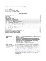
1b) What do the CXR and CT scan in Fig. 1.1 show?
1c) What diagnosis do investigations support?
1d) What are the typical clinical features of this condition?
1e) Discuss treatments options for this lady.
CASE 1
3
Fig. 1.1 (a) CXR and (b) CT chest.
(a)
(b)
CASE HISTORIES IN RESPIRATORY MEDICINE
4
Answers
1b) What do the CXR and CT scan in Fig. 1.1 show?
The CXR shows hyper-expanded lung fields, with widespread bronchiec-
tatic changes. The CT slice shows dilated airways, much larger than the
adjacent blood vessel, in keeping with bronchiectasis. There is also ‘tree
in bud nodularity’, which may be suggestive of small airway chronic or
atypical infection.
Fig. 1.2 Portion of CT-chest illustrating features in keeping with allergic
bronchopulmonary aspergillosis.
1c) What diagnosis do investigations support?
Investigations support a diagnosis of allergic bronchopulmonary aspergil-
losis, ABPA. Atopic patients with asthma and cystic fibrosis with IgE-
mediated allergy to inhaled Aspergillus spores are vulnerable to this
condition. They may develop IgE and IgG reactions to Aspergillus in the
airways, provoking mucous plugging with distal consolidation, and then
ABPA, with inflammatory damage to the airways and resultant bron-
chiectasis. Damp conditions, composting organic material and thunder-
storms are associated with high Aspergillus spore counts, and so may lead
to exacerbations. Since simple atopic asthma is at one end of a continuum,
with ABPA at the other, there is no single diagnostic test that defines the
transition. The presence of the features in Box 1.1 would support the diag-
nosis, with the first four being the most important. Many asthmatics and
patients with cystic fibrosis have one or more findings suggestive of ABPA,
but do not fulfil all criteria listed.
ABPA is a complex hypersensitivity reaction, often in patients with asthma
or cystic fibrosis that occurs when bronchi become colonized by Aspergillus .
Tree-in-bud nodularity
Dilated airways
CASE 1
5
Repeated episodes of bronchial obstruction, inflammation, and mucoid
impaction can lead to bronchiectasis, fibrosis and respiratory compro-
mise. It is thought healthy, unaffected individuals are able to effectively
eliminate fungal spores. They have low levels of IgG against fungal anti-
gens in the circulation, and low anti-fungal secretory IgA in bronchoal-
veolar fluid. In contrast, exposure of atopic individuals to fungal spores or
mycelial fragments results in the formation of IgE and IgG antibodies.
Aspergillus responsive T-cells generate the cytokines interleukin (IL)-4,
IL-5 and IL-13, which account for the eosinophilia and raised IgE in
ABPA. Aspergillus colonization of the asthmatic airway leads to vigorous
IgE- and IgG-mediated immune responses superimposed on the asth-
matic milieu. In spite of these vigorous responses in ABPA, the fungus is
able to colonize the airway and cause recurrent symptoms. Proteolytic
enzymes are released by Aspergillus as part of its exophytic feeding strat-
egy, and these enzymes may in theory damage airway walls. However,
exuberant host defence mechanisms are thought to be the dominant
method of damage, hence there is a good response to steroids. Spores and
hyphae (indicating germination of the spores in the airway) are some-
times seen on direct microscopy, and Aspergillus is cultured from sputum
in up to two-thirds of patients with ABPA. As in most cases of ABPA, the
patient in this case had a background history of atopic asthma.
1d) What are the typical clinical features of this condition?
Typical clinical features of ABPA are long-standing asthma with a more
recent deterioration, complicated by recurrent episodes of bronchial
obstruction and expectoration of brownish mucous plugs, fever, malaise,
peripheral blood eosinophilia and sometimes episodic haemoptysis.
Box 1.1 Diagnostic features of ABPA
◆
Longstanding history of asthma
◆
Immediate positive IgE reaction to Aspergillus on skin testing, or on
serum testing using RAST (radioallergosorbent test)
◆
Precipitating serum IgG antibodies to Aspergillus fumigatus
◆
Central bronchiectasis on chest CT
◆
Peripheral blood eosinophilia
◆
Serum total IgE concentration elevated > 1000ng/mL
◆
Flitting lung infiltrates on CXR or chest HRCT.
CASE HISTORIES IN RESPIRATORY MEDICINE
6
Wheezing is not always present, and some patients present with asympto-
matic ‘flitting’ pulmonary consolidation.
1e) Discuss treatment options for this lady.
Treatment of ABPA involves optimal care of bronchiectasis and asthma,
plus early use of oral steroids and consideration of itraconazole with drug
level monitoring where this is available. This needs to be prescribed for 3
to 6 months (regular liver function tests are needed as the drug may be
hepatotoxic) and the IgG levels to Aspergillus should fall with fungal load
reduction. Inhaled steroids may help control symptoms of asthma, but do
not have documented efficacy in preventing acute episodes of ABPA.
Further reading
Denning D , O’Driscoll B , Hogaboam C , Bowyer P and Niven RM ( 2006 ). The link between
fungi and severe asthma: a summary of the evidence . Eur Respir J ; 27 : 615 – 626 .
Stevens DA , Schwartz HJ , Lee JY , Moskovitz BL , Jerome DC , Catanzaro A et al. ( 2000 ).
A randomized trial of itraconazole in allergic bronchopulmonary aspergillosis .
New Eng J Med ; 342 : 756 – 762 .
Case 2
A 77-year-old lady was referred with progressive breathlessness over 3 years.
She was breathless walking 100 yards on the flat and could not manage stairs.
There were no other respiratory symptoms. Past history was of myocardial
infarction (MI) in 1984, and duodenal ulcer 1988. She had stopped smoking
after her MI, with a prior 40 pack year smoking history. Her medication con-
sisted of simvastatin, lisinopril, furosemide, aspirin, amiodarone, salbutamol
and omeprazole. All of her cardiac medications were commenced post-MI.
She kept no pets. On examination there was central cyanosis, finger clubbing
and resting oxygen saturations of 83% on room air. JVP was not elevated and
there was no peripheral oedema. Cardiac examination revealed an aortic scle-
rotic murmur and respiratory examination revealed bibasal fine inspiratory
crackles in the lower zones. Abdominal and musculoskeletal examination was
unremarkable.
Investigations
◆
Hb 14.3g/dL, WCC 5.94 × 10
9
/L (eosinophils 0.18 × 10
9
/L), platelets
145 × 10
9
/L
◆
ESR, 48mm/h
◆
U&Es, normal
◆
Bilirubin 39 μmol/L, ALT 18 IU/L, ALP 308 IU/L
◆
Albumin, 27g/L
◆
Rheumatoid factor, 69.4U (<10, negative; 10–30, borderline; >30, posi-
tive)
◆
ANA, anti-smooth muscle antibody, anti-mitochondrial antibody, and
anti-gastric parietal cell antibody: negative
◆
Alpha 1 antitrypsin, 185mg/dL (normal 107–209 mg/dL)
◆
ABG (on air), PaO
2
6.7 kPa, PaCO
2
4.17 kPa, [HCO
3
]¯ 23.3 mol/L,
pH 7.45
◆
ECG, normal
◆
Abdominal USS, liver appeared slightly enlarged with an irregular
outline. Spleen was also slightly irregular. Pancreas and kidneys were
normal.
CASE HISTORIES IN RESPIRATORY MEDICINE
8
Table 2.1 Pulmonary function tests
Measured % Predicted
FEV
1
(L) 2.0 131
FVC(L) 2.8 144
FEV
1
/FVC(%) 71
FRC(L) 2.8 110
RV(L) 1.9 97
TLC(L) 4.7 108
VA(L) 3.6 82
TL
CO
(mmol/min/kPa) 2.05 34
K
CO
(mmol/min/kPa/L) 0.57 41
Fig. 2.1 CXR.