nghiên cứu ứng dụng phương pháp phẫu thuật nội soi qua lỗ liên hợp lấy đĩa đệm trong thoát vị đĩa đệm cột sống thắt lưng bản tóm tắt tiếng anh
Bạn đang xem bản rút gọn của tài liệu. Xem và tải ngay bản đầy đủ của tài liệu tại đây (609.57 KB, 30 trang )
MINISTRY
OF EDUCATION AND TRANING
MINISTRY
OF HEALTH
HANOI MEDICAL UNIVERSITY
Specialism: Orthopaedic and Trauma
Code: 62720129
MEDICAL DOCTORAL THESIS
HA NOI - 2013
The thesis has been completed at:
HANOI MEDICAL UNIVERSITY
Supervisor:
1. Ass prof. NGUYEN CONG TO
2. Ass prof. NGUYEN VAN THACH
Reviewer 1: Ass prof. VU VAN HOE
Reviewer 2: Prof. LE DUC HINH
Reviewer 3: Ass prof. CAO MINH CHAU
The Thesis will be present in front of board of university examiner and reviewer level hold at Hanoi Medical
University.
At …………, on …… , 2013.
The thesis can be found at:
- National Library
- National Medical Informatic Library
- Library of Hanoi Medical University
THE LIST OF WORKS RELATED TO THE THESIS
THAT HAS BEEN PUBLISHED
1. Dinh Ngoc Son, Nguyen Van Thach (2010),"A result of transforaminal endoscopic discectomy for lumbar
disc herniation ", Vietnam Medical Journal, October, (2), pp. 5-10.
2. Dinh Ngoc Son, Nguyen Van Thach, Nguyen Thi Ngoc Lan, Nguyen Van Chuong, Pham Xuan Phong,
Nguyen Le Bao Tien, Hoang Gia Du, Nguyen Hoang Long, Do Manh Hung (2011), "Summarize results of
some methods in treatment of lumbar disc herniation ”, Vietnam Medical Journal 7/2011, pp. 64-69.
3. Nguyen Van Thach, Dinh Ngoc Son, Nguyen Le Bao Tien, Nguyen Hoang Long, Do Manh Hung, Dinh
Manh Hai, Tran Quoc Khanh (2012), "Researching on applying device interspinous assisted motion into
laminotomie to treat lumbar stenosis disease", Journal of surgery, 61(1, 2, 3), pp. 351-356.
4. Nguyen Van Thach, Dinh Ngoc Son, Nguyen Le Bao Tien, Hoang Gia Du, Nguyen Hoang Long, Tran
Dinh Toan, Dinh Manh Hai, Do Manh Hung (2012),"Minimal invasive lumbar spine surgery: Treatment
and outcomes", Journal of the Vietnam Orthopeadic Association, (1), pp. 1-5.
5. Dinh Ngoc Son, Nguyen Van Thach (2013), "Research on morphologic of the triangular working zone for
the current practice of endoscopic lumbar discectomy through cadaver workshop”, Journal of the Vietnam
Orthopeadic Association, (3), pp. 31-35.
1
INTRODUCTION
The importance of the study
Disc herniation refers to the displacement of disc material
beyond the limits of the intervertebral disc space. A herniated disc
may engage the nucleus, the cartilage, the gristle, or annulous
fibrosus.
A spinal specialist provides the definitive determination of the
herniated disc position. With the application of MRI today, it has
become easier to find and classify a herniated disc.
Two methods of treating lumbar disc heniation include
conservative treatment and surgical treatment. Surgical treatment is
laid out with a herniated disc causing nerve root compression or when
conservative treatment fails after 3 months.
Currently, in the world, open surgical procedure only applies to
the case of large herniated disc and migrated disc herniation
accompanied by other conditions of the spine such as spinal instability,
spinal stenosis and alternative methods are minimally invasive
discectomy. Endoscopic surgery with access via a natural opening
outside the spinal canal - the intervertebral foramen - to remove the
herniated disc is a minimally invasive discectomy overcoming the
disadvantages of posterior elements – the yellow ligament is not
removed; it does not affect the rear part of the spine including the
lamina, the spinal process, and the posterior longitudinal ligament;
virtually no organized nerve fibers stick. Endoscopic surgery is more
favourable with distinctive features such as local anesthesia, small
incision of 0.7 cm, 1-2 days in hospital after surgery, lower costs, and
fewer complications. According to some authors, the success rate is
85-95%. However, this method can be only applied for lateral,
foraminal and extra-foraminal herniation. Endoscopy is commonly
used in the U.S, Korea, Japan, Europe and has some applied
anatomical research. In Vietnam ,transforaminal endoscopic surgery is
2
performed in the center of Saigon EXSon from 10/2007 and the
Vietnam-Germany Hospital from 9/2008. In order to put a new method
in practice, it is important to carry out the basic research, which is the
applied anatomical research and using its results in clinical practice.
Therefore, “The applied research of endoscopic discectomy
transforamen to treat lumbar herniated disc” is carried out with two
goals:
1. Identify the anatomical measurements through surgery on
cadavers to determine the safety approach of transforaminal
lumbar endoscopic discectomy.
2. Evaluate the results of transforaminal endoscopic surgery in
lumbar herniated disc.
Practical values:
1. The study shows the practical value of applied anatomical
research to identify the safe access path in endoscopic surgery.
2. The study also shows the effectiveness of transforaminal lumbar
endoscopic discectomy.
Contributions:
- The first study of applied anatomy through the foramen in
Vietnam, proposing some new points compared with previous studies
in the world; it is the scientific basis for the implementation of the
surgical method through the foramen.
- The first study in Vietnam on a new surgical method:
transforaminal endoscopic discectomy.
Layout:
The thesis consists of 149 pages, including: introduction (2
pages), overview (page 46), subjects and research methods (33
pages), results (35 pages), discussion (30 pages), conclusion (2
pages), proposal (1 page). Thesis has 45 tables, 82 images. Reference
materials in both Vietnamese and English.
3
Chapter 1
OVERVIEW
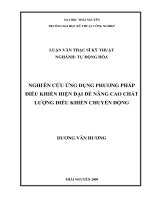
1.1. APPLIED ANATOMY THROUGH THE FORAMEN
* The foramen: the access path of endoscopic surgery.
+ The intervertebral foramen is the aperture that gives exit to the
segmental spinal nerves and entrance to the vessels and nerve
branches that supply the bone and soft tissues of the vertebral canal.
It is superiorly and inferiorly bounded by the respective pedicles of
the adjacent vertebrae. Its ventral and dorsal components involve the
two major intervertebral articulations. The dorsum of the
intervertebral disc, covered by the lateral expansion of the posterior
longitudinal ligament, provides a large part of its ventral boundary,
whereas the joint capsule of the articular facets and the ligamentum
flavum contribute the major parts of its dorsal limitation. Along with
the root, the remaining space is filled with loose areolar tissue and fat
+ The internal structure of the foramen: nerve root, stem branch of
the artery, the connection between the venous plexus TM inside and
outside, the organization surrounding tissue structures.
Image 1.1. Foramen
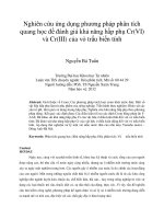
* The safety triangle:
Dr. Parviz Kambin defined this area as “a right triangle over the
dorsolateral disc. The hypotenuse is the exiting nerve root, the base
(width) is the superior border of the caudal vertebra and the height is
the dura/traversing nerve root.’ (Kambin P, Sampson S Clin Orthop
4
Relat Res. 1986 Jun; (207):37-43) In practice, approximately around
the pedicle and disc are selected milestones identified in the surgical
process because it is expressed on a fluorescent screen sang.
Understanding of the safety zone is important in entering the
endoscope into a surgical field.
Image 1.2 and 1.3. The surgical triangle ,.
(the safety triangle)
A. Hypotenuse B. Inside edge C. Dura/traversing nerve root
D. inferior edge E. Pedicle slide F. Safety triangle
1.2. CLINICAL
1.2.1. Clinical symptoms:
- Spinal syndrome: back pain, scoliosis
- Nerve root syndrome: pain, disorders of sensation along the
dominant roots, reduced tendon reflexes, muscle atrophy.
- Irritation nerve root sign: good diagnosis
* LassÌgue Test: Positive >60°.
* Pushing bell sign: pain along the leg
* Valleix pain point: Using the thumb to push deeply on the
points through the way of nerve root. Patient can feel pain at the
pushing points.
5
- Lesions of nerve root sign: disorders of sensation, moving,
reduction of reflex, bladder disorder.
1.2.2. Diagnostic imaging
Conventional X-ray: Take lumbosacral spine straight, tilt left or
right with three positions: lowered maximum, intermediate and
maximum hyperextension; angling three quarters to the left or aim to
eliminate damage loss strong, physically waist.
Computerized tomography (CT scan): to assess bone lesions
suspected on X-ray, calcified discs.
Magnetic resonance imaging (MRI): the best diagnostic
method available
Classification by MRI:
-By Fardon 2001:
+Stage 1: Protrusion occurs when the outermost layers of the
annulus fibrosus of the intervertebral discs are intact, but bulge when
one or more of the discs are under pressure. MRI vertical slices show
that the distance between the edges of the disc herniation is less than
the distance between the edges of the base.
+Stage 2: Extrusion occurs when the outer part of the spinal disc
ruptures, allowing the inner, gelatinous part of the disc to squeeze out.
MRI vertical slices show the distance between the edges of the disc
material is greater the distance at the base.
Image 1.4:
Stage 1 and Stage 2
A : Protrusion B,C : Extrusion
6
+Stage 3: Migration indicates displacement of disc material
away from the site of extrusion
+Stage 4: Sequestration indicates that the displaced disc
material has lost completely any continuity with the parent disc
-Axial localisation of herniated discs.
+Central or medial: occurs in the midportion of the disc.
+Paramedian or lateral recess: the zone occupied by the base of
the lateral expansions of the posterior longitudinal ligament.
+Foraminal or subarticular: between the base of the lateral
expansions of the posterior longitudinal ligament and the entrance of
the intervertebral foramen.
+Extraforaminal or lateral: outside the neuroforamen.
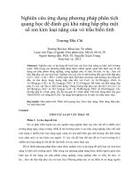
-Classification of disc migration by Lee SH:
+Zone 1: From the
inferior margin of upper
pedicle to 3 mm below of the
inferior margin of upper
pedicle.
+Zone 2: From 3 mm
below of the inferior margin of
upper pedicle to the inferior
margin of upper vertebral
body.
+Zone 3: From the superior margin of lower vertebral body to the
center of lower pedicle.
+Zone 4: From the center to the inferior margin of lower pedicle.
Zones 1 and 4 are far-migrated disc herniation. Zones 2 and 3 are
near-migrated disc herniation.
Image 1.21. Disc migration classified
by Lee SH
7
1.3. SURGICAL TREATMENT FOR DISC HERNIATIONS
Absolutely indication: Caudal equina syndrome or nerve root
compression due to unilateral or bilateral paralysis, severe pain.
Fairly indication: conservative treatment fails after 3 months
1.3.1. Open surgery: This is still the most popular treatment in
Vietnam. In developed countries, it is only performed when required.
Advantages: easy and inexpensive. Disadvantages: large incision,
the risk of bleeding, soft tissue injury, instability.
1.3.2.Minimally invasive percutaneous techniques:
-General principals: Using chemonucleolysis to dissolve the
nucleus pulposus (papaya enzyme injections), using radiofrequency
nucleotomy, using laser disc decompression to vaporize a small amount
of nucleus with laser energy, or using nucleotomy to physically remove
part of the nucleus pulpous, reducing pressure on nerve roots.
-Indications: cases where the anulus fibrosus is inact.
-Advantages and disadvantages: Advantages are simple, are
the method between conservative treatment and surgery. High
efficiency of 70 – 80% success rate with proper indications.
However, it is an expensive technique. Papaya enzyme injections is
not performed any more due to complications.
1.3.3. Minimally invasive surgical techniques.
- Microscopic discectomy and microscopic discectomy
transendoscopic tube:
+ Indication: likes open surgery. Technique: Using microscopic
discectomy through open incision, microinstruments, and cannula
system. The basic stages are as same as open surgery. The
instruments are burr, nerve root retractor.
+ Advantages: Less nerve root injury, scar. Disadvantages: due to
injury of posterior elements. Microscope is a little bit expensive. Using
microscope and cannula need to be have training and practice regularly.
8
- Endoscopic surgery to remove lumbar disc herniation :
+ Endoscopic discectomy through interlamina.
*Indications: Migrated herniations and lateral herniations based
on Fardon’s classification.
*Technique: Making an incision from 7-8mm at the interlamina.
Using canule to make a bigger incision and checking by C-arm.
Evaluation and remove the yellow ligament, retract the nerve root
due to remove the herniation block.
*Advantages: this is a minimally invasive technique, short time in
hospital, quick recovery, clearly operation field, few complications.
Disadvantages: long-term training, high costs, just only indication for
extrusion and migration herniation.
- Endoscopic discectomy transforamen.
*Indication : foramen herniation, extraforamen herniation,
subarticular and lateral herniation.
* Advantages: clear images, less recurrent herniation because of
less anulus fibrosus injury. Less the risk of nerve root injury, scar and
adhesion fibrosus. No spinal muscle retraction and reduce the risk of
spinal instability, reduce the height of disc.
* Disadvantages: endoscopic instruments are expensive.
* Complications: injured exiting nerve root, inadequate removal
of disc materials, and postoperative disc infections are recorded in
Vietnam and other places.
1.4. Endorscopic surgery to remove the nucleus pulposus in
Vietnam
+ In 2010, Nguyen Van Thach conducted a study of 70
patients with mean folow-up period of 12 months. According to the
modified Macnab criteria, excellent and good outcomes comprise
92.9%. 6.7% of patients had fair outcomes. One patient experienced
recurrent hernia requiring additional operation (1.4% ).
9
+ In 2010, Nguyen Trong Thien also reported the outcome of a
study on 32 patients. Good and fair outcomes were 85,7% at 6-month
follow-up. 3 participants needed additional operation - one due to
dicitis; one due to poor preoperative assessment of the migrated
herniation; and one due to missing diagnosis. The author emphasized
the need of better skills and indications.
+ In 2010, Vo Xuan Son reported a study of 26 patients. 2
patients (7.7%) experienced postoperative tingling and weakness in
foot flexsion. These syndromes improved and patients were discharged
after three days. This method is effective in treating lumbar disc
herniation.
Chapter 2
SUBJECTS AND RESEARCH METHODS
2.1. RESEARCH SUBJECTS
2.1.1. Anatomical research subjects
120 foramen was studied on adult cadavers preserved with
formalin in Anatomy faculty, Hanoi Medical University. Average
height of the cadavers is 162cm ±9.18, maximum 179 cm, minimum
151cm. None had history of previous spinal surgery.
2.1.2. Clinical research population:
80 patients diagnosed with lumbar disc herniations were treated
with endorscopic surgery through the foramen in Viet-Germany
Hospital from 09/2008 to 10/2009. Among those, 52 were male and 28
were female.
Exclusion criteria:
- Syndroms as: spinal instability, spinal stenosis, spinal tumors
- Disc herniation accompanied with nerve root compression
10
- Diseases as: diabetes, liver conditions, kidney conditions…
2.2. RESEARCH METHODS:
2.2.1. Anatomical research
- Research location: Anatomy School, Hanoi Medical
University performed dissection on on formalin preserved cadavers
to study 120 foramens.
- Procedure: Cadavers in a prone position, make a midline
incision of the lumbar spine. Disclosing the edge of life and this
detachment from the horizontal spikes. Dissection, removing weeds
curret use pots. Reveal the complex holes. Nerve roots exit from the
hole conjugation. Disclosure nerve roots exit the index measured.
Tien surgery and meticulously recorded. As a graduate student
measuring just measured.
Image 2.1. Cadaver dissection
1. Exiting nerve roots 2. Spinous processes
- Anatomical measurements:
+ The index relates the way to put the needle of endoscopic
surgery:
* Measure the distance from strating point to the midline.
* Measure the angle between the needle and the surface of
11
lumbar using the surface rule and the Insize angle rule.
+ The index relates exiting nerve root and superior facet join:
The length indicates the suitability of endoscopic instruments.
* The distance from the inferior margin of the exiting nerve root
to the superior margin of the spinous process at the coronal layer
through superior face of vertebrae.
* The distance between anterisuperior facet join, the conection of
superior facet join and pedicle to the inside edge of nerve root.
+ Kambin triangle measurements: The hypotenuse, the base, the
angle of the exiting nerve and the superior border of the caudal
vertebra, and the height of the triangle.
+ The index relates triangle through superior pedicle: inside edge,
inferior edge and root-superior vertebral.
+ Determine the area of two angles and compare to the circle slide
through endoscopic instruments (Canule , tube).
+ The distance between two transverse nerve roots and the
parallel line through the inferior vertebra.
2.2.2. Clinical Study: a longitunal study to assess the outcomes of
endoscopic surgery transforamen.
2.2.2.1. Evaluation the clinical and MRI image of lumbar
herniation.
- Evaluation the clinical: age, gender, onset symptoms…
+ Examination to find the lumbar-sacrum nerve root symtom.
+ Caudal equinal syndrome: sphincter disorder.
- Diagnostic imaging:
+Conventional X-ray:
* Anterior-posterior and lateral X-ray: all patients.
* Moving X-ray: To exclude spinal instability, spondylolisthesis.
+ CT scan: when bone damage is suspected.
12
+ MRI: applied to all of the patients.
*Determine herniation level: for example L34, L45…
*Classify herniations: lateral, paramedian, and foraminal
herniations.
*Classify herniations based on Lee SH’s zones: Near-migrated
herniations: zones 2 and 3; Far-migrated
herniations: zones 1 and 4
*Herniation staging: Stage 1:
protrusion, Stage 2: extrusion, Stage 3:
migration, Stage 4: sequestration
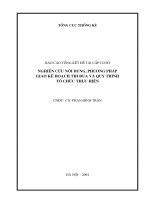
2.2.2.2. Transforaminal endoscopic
surgery
-Instruments: Joimax endoscopic
system, guide line system, canule,
remove dics instrument, bleeding
control tools.
-Patients: lying on the side with
pillows supporting lumbar region.
-Surgical Procedures:
Step 1: Determine the access
Step 2: Put the needle inside the disc, check the disc by anti-
radiolucent.
Step 3: Open the incision
Step 4: Widen the foramen
Step 5: Put the canule
Step 6: Enter the endoscope into the surgical field
Step 7: Remove the herniation
Step 8: Check the exiting nerve root and its mobility as well as
patients’ clinical syndrom
Image 2.2. Endoscopic system
13
Image 2.2.ABC. The image of canule and instruments
intraoperation.
2A. A-P film
1. Midline of pedicle. Anti-radiolucent inside the herniation
2C.Endoscopic image:
1. Herniation 2. Exiting nerve root
2.2.2.3. Outcome assessment
-Pain level measured by VAS
-Spinal dysfunction evaluation ODI
-By modified Macnab criteria:
+Excellent: No pain and no limitation of normal life.
+Good: Occasional pain or paresthesia, but no need of
medication, and no limitation of normal life.
+Fair: Pain is somewhat improved but needs medication, and
some limitation of normal life.
+Poor: No improvement or worsening, additional operation is
needed due to incomplete decompression, development of instability.
-Postoperative MRI:
-Complications: recorded
-Recurrent hernia: not recorded as a complication
14
2.3. DATA PROCESSING: SPSS 13.5
2.4. ETHICS IN RESEARCH: The study information is confidential
and serves the research purposes only.
Chapter 3
RESEARCH RESULTS
3.1.RESULTS ON CADAVERS
Table 3.1.The index relates the guide line of needle intra-
operation:
Anatomy index Mean SD
Distance of the needle put inside edge of pedicle (mm) 64.24 21.542
Distance of the needle put middle edge of pedicle
(mm)
48.38 14.257
Distance of the needle put outside edge of pedicle
(mm)
35.42 10.560
The angle of inside edge pedicle needle (degree) 44.97 11.698
The angle of middle edge pedicle needle (degree) 57.08 10.531
The angle of outside edge pedicle needle (degree) 70.08 12.747
Distance between needle and exiting nerve root (mm) 1.85 0.964
Table 3.2. The index between exiting nerve root and superior
facet joint
Anatomy index Mean SD
Distance between nerve root and outside edge
of facet join (mm)
14.03 2.652
15
Distance between nerve root and anterior edge
of facet join (mm)
6.77 1.599
Table 3.3. Kambin triangle measurements
Kambin index Mean SD
Inside edge (mm) 16.55 4.819
Inferior edge (mm) 12.59 2.466
Height (mm) 9.66 1.753
Nevre root - superior facet angle (degree) 52.48 8.837
Triangle in area (mm square) 107.08 44.604
Table 3.4. The index relates the angle through superior pedicle
Triangle of superior pedicle Mean SD
Inside edge (mm) 19.20 5.041
Inferior edge (mm) 14.44 2.600
Height (mm) 11.66 1.812
Nevre root - superior facet angle (degree) 52.48 8.837
Triangle in area (mm square) 141.14 50.589
Table 3.5. Classify the triangle area due to the slide of endoscopic
instruments.
Triangle
Slide through canule (mm
square)
Slide through tube (mm
square)
S≤38.46 S>38.46 S≤44.15 S>44.15
N Rate N Rate N Rate N Rate
Kambin
1 0.8% 119 99.2% 4 3.3% 116 96.7%
Superior
pedicle
0 0 120 100% 0 0 120 100%
3.2. CLINICAL OUTCOMES:
3.2.1.General characteristics:
16
-Gender: 52 males and 28 females. Males/females is 1.85.
-Age: 36.84 ± 12,02 on average. The youngest is 19, and the
oldest 71.
-Past operative history: 5 patients had past disc herniation
operation
3.2.2.Clinical syndromes:
-Spinal syndromes: 100% had mechanical back pain. 26.5%
spinal muscle spasm, 4.9% scoliosis
- Nerve root syndrom
Table 3.6. Lumbar sacrum nerve root syndrom
Symptoms N Rate
Lasseque sign positive 74 92.5%
Oblique Lasseque sign 32 40%
Touching bell sign 8 10%
Valeix points system 10 12.8%
Reduce or disappear the sensation base on
nerve root
72 90%
Reflex disorder: Patella tendon, heel. 31 38.75%
Moving disorder depend on nerve root 12 15%
Nutrition disorders, amyotrophy 29 36.25%
Radicular pain 79 98.75%
Mean VAS leg pain 6.26 ± 1.202
3.2.3.MRI imaging:
-Herniation level: L5S1: 43 patients (53.8%). L4L5: 36 patients
(45%). L3L4: 01 patient
-Location: Lateral : 9 patients (11.3%), sub-articular : 61
17
patients (76.2%), foraminal: 8 patients (10%), extra-foraminal: 02
patients (2.5%).
-Herniation stage: Extrusion: 75 patients. Migration: 05 patients.
3.2.4.Surgical Treatment:
-Surgical time: 99.7 minutes ±34.37. The longest is 250 minutes,
and shortest 40 minutes
-Difficulties: put the needle again:06 BN, put the canule
again:02BN, difficult in drilling:02 BN, bleeding intra-operation:
01BN.
-In hospital: minimum 01 day, maximum 03 days. Average: 02
days.
3.2.5.Surgical outcomes:
Table 3.7. VAS score before and after operation
VAS (score)
Preoperatio
n
1-month
follow-up
6-month
follow-up
24-month
follow-up
N 80 80 79 65
Mean 6.3 2.96 2.04 1.23
SD 1.195 1.427 0.953 1.057
Average
improvemen
t rate
53% 67.61% 80.47%
Table 3.8. ODI score before and after operation
Time
Preoperatio
n
1-month
follow-up
6-month
follow-up
24-month
follow-up
N 80 80 79 65
Mean (%) 60.58 33.20 19.35 10.49
SD 12.444 13.887 12.000 8.958
18
Average
improvement
rate
45.2% 68.1% 82.68%
Table 3.9. ODI rate at different time pre and post operation
ODI
Preoperation
(N)
1-month
follow-up
(N)
6-month
follow-up
(N)
24-month
follow-up
(N)
Level 1 0 15 50 56
Level 2 3 46 26 9
Level 3 47 15 3 0
Level 4 26 4 0 0
Level 5 5 0 0 0
Total 80 80 79 65
Table 3.10. Outcome assessment based on Macnab criteria
Macnab
6 month follow-up >24 month follow-up
N % N %
Excellent 19 23.75 40 60.6
Good 58 72.5 21 31.8
Fair 2 2.5 4 6.1
Poor 1 1.25 1 1.5
Total 80 100 66 100
-Postoperative MRI: 29 patients taken. One patient needed
additional surgery due to recurrent disc herniation. Two patients
19
needed follow-up on disc protrusion.
-Postoperative complications: Sensation disorders: 01/80
patients (1.25%); Pseudocysts: 01/29 patients (3.5%).
-Recurrence: one patient needed additional surgery after 5
months.
Chapter 4
DISCUSSION
4.1. Result of cadaver research
4.1.1.Index relates approach of endoscopic surgery:
- When the needle position is inside edge pedicle, the distance
form starting point to midline: 64.24mm ±21.542 (Table 3.1). Depend on
Kambin, The final position of needle is inside edge pedicle, The distance
from starting point to midline is about 110-120 mm. Yeung TA publish
the general technique for all kind of hernation with the distance is about
120 ±20mm. Our results are lower due to the smaller measeares of
Vietnamese.
- When the needle is in middle of pedicle: The mean distance is
about 48.38mm ±14.257. Theo Kim HD, fluctuate from 50-80mm.
- When the needle is in outside edge of pedicle: We think that the
distance from exiting nerve root to annulus fibrous is mean about
1.85mm ±0.964. So with the 7mm canule we have 95% cases let to
nerve root injury.
For that reason, we think that the distance safety for needle
entry point is the mean distance between medial edge of pedicle
and middle pedicle.
4.1.2. The index relates exiting nerve root and superior
facet joint
-The distance from medial edge of nerve root and lateral facet
20
joint: Depend on table 3.2, The mean distance is 4.03mm± 2.652. So,
it is easy for the procedure when the canule is 7mm and the tube is
5mm.
- The distance from anterior of superior facet join to exiting nerve
root is about 6.77mm±1.599. This is the anatomy diameter when
endoscopic instrument is used with migration herniation. So with 8mm
canule, we need to drill the foramen. That is the most important point
of endosopic transforamen technique.
4.1.3.Kambin triangle: When the needle is parallel with the disc relate
to the inferior triangle, and when the needle goes down, it relates to the
height of triangle. Depend on table 3.3, the length mean of inside edge
is 16.55mm±4.819 and the height is 9.66mm±1.753, and also easy for
7.5mm instruments.
4.1.4. Safety Kambin area and superior triangle compare with the
slide of tube and canule: When compare with the slide of canule
(table 3.5), we had a case with smaller area (0.8%). With 7.5mm tube,
we had 4 cases with smaller area (3.3%). But, all of triangle areas
through superior pedicle are bigger than the slide of endoscopic
instruments. For that reason, we think that drilling facet join will help
to easier touching to superior pedicle and easier for safety endocsopis
systems.
4.2.Clinical outcome:
4.2.1. Pain level measured by VAS:
Table 3.7 shows the mean preoperative VAS score is 6.3 ± 1.19
with 95% falling between 5.05-6.69, the lowest score is 5 and the
highest 9. The mean postoperative score is 2.96 ± 1.427; 2.04 ± 0.953
and 1.23 ± 1.057 at 1-month, 6-month, and 24-month follow-up
respectively. 95% confidence intervals are: 2.73-3.41; 1.8-2.32 and
1.12-1.63. There is a significant improvement in the VAS preoperation
and postoperation (p<0.001). 1-month follow-up shows improvement