biodiversity and antibiotic activity of actinomycetes isolated from cat ba island, vietnam = đa dạng sinh học và hoạt tính kháng sinh của các chủng xạ khuẩn phân lập ở đảo cát bà, việt nam
Bạn đang xem bản rút gọn của tài liệu. Xem và tải ngay bản đầy đủ của tài liệu tại đây (2.34 MB, 52 trang )
CONTENTS
Acknowledgements i
Abbreviations ii
List of figures iii
List of tables iv
Abstract 1
Tóm tắt 3
Foreword 5
Chapter 1. Introduction 6
1.1 Antibiotic 6
1.1.1 General introduction 6
1.1.2 History of the development of antibiotics 7
1.1.3 Classification of antibiotics 9
1.1.4 Anti-tumor antibiotics 13
1.1.5 The need of developing new antibiotics 14
1.2 Actinomycetes 14
1.2.1 General characteristics 14
1.2.2 Actinomycetes and secondary metabolites 16
1.3 Objectives of the study 16
Chapter 2. Materials and methods 18
2.1 Work flow 18
2.2 Methods 19
2.2.1 Isolation of actinomycetes 19
2.2.2 Targeted microorganisms 21
2.2.3 Screening antibiotic producing actinomycetes 21
2.2.4 Ethyl-acetate extraction 22
2.2.5 Chromatography analyses of antibiotics 22
2.2.6 Screening for cytotoxicity 24
2.2.7 Taxonomical identification of actinomycete isolates 25
Chapter 3. Results and discussion 28
3.1 Biodiversity of the actinomycete isolated from Catba island 28
3.2 Screening for antibiotic producing actinomycetes 29
3.3 Chromatography analyses of crude extracts of the selected strains 31
3.3.1 Thin Layer Chromatography (TLC) 31
3.3.2 High – Performance Liquid Chromatography (HPLC) 32
3.4 Primary study on cytotoxicity activity 33
3.4.1 pH-dependent color change by the selected strains 33
3.4.2 Cytotoxicity assay against human cell lines 33
3.5 Identification of the actinomycete isolates 34
3.5.1 Morphology-based identification for the Streptomyces isolates 35
3.5.2 16S rDNA sequence-based identification for the
non-Streptomyces isolates 37
Conclusion and Suggestion 38
Bibliography 40
Appendix 1 Culture media 46
Appendix 2 The 16S rDNA sequence………………………………………… …….46
iii
LIST OF FIGURES
Figure Title Page
Figure 1 Discovery of important antibiotics and other natural products over the
years .7
Figure 2 Evolution of penicillin G 8
Figure 3 Chemical structures of daunorumycin and idarumycin .13
Figure 4 Catba national park 19
Figure 5 Evaluation of antimicrobial activity of the actinomycete isolates 30
Figure 6 Analyses of crude extracts of the selected actinomycete strains via
TLC 31
Figure 7 HPLC analysis of crude extract from actinomycetes 32
Figure 8 Color change by strains A1018 and A1073 depending on pH in the
medium 33
Figure 9 Colony morphology of the representative Streptomycete strains… 35
Figure 10 Spore-bearing aerial hyphae of the representative Streptomyces
strains… 36
Figure 11 Neighbor-joining tree of 16S rDNA partial sequences showing
phylogenetic positions of the 7 actinomycete strains in the relationship
to type strains of the genus Nonomuraea 37
iv
LIST OF TABLES
Chapter 1
Table 1.1 Grouping of antibiotics bases on their chemical structures 10
Chapter 2
Table 2.1 Condition for HPLC analyses of the standards 23
Chapter 3
Table 3.1 Taxonomical grouping of the actinomycete isolates 28
Table 3.2 Antimicrobial activity of the 17 selected actinomycete strains 30
Table 3.3 Toxicity assay against three human cell lines 34
1
ABSTRACT
In this study, a total of 424 actinomycete strains isolated from soil and
litter samples on Catba island (Haiphong, Vietnam) were subjected to a
screening for the inhibitory activities against microorganisms, including
bacteria (Micrococcus luteus, and Escherichia coli) and eukaria (Candida
albicans and Fusarium oxysporium). Through two screening steps, 17 strains
were selected for their inhibitory activity against one or more target
microorganisms. Crude extracts in ethyl acetate from culturing media of the
selected strains were analyzed via thin-layer chromatography (TLC) and high
performance liquid chromatography (HPLC), in which chloramphenicol,
kitasamycin, erythromycin and raw extract of anthracycline were used as
standards. The obtained results showed that antibiotic substances produced by
the selected strains could not be able to classify in any group of the analyzed
standards, except the strain A396 which appeared to produce
chloramphenicol-like antibiotic.
Besides that, several human tumor cell lines have also been used for
testing the inhibitory effects. The results showed that among the 17 selected
strains, 3 strains (A1018, A1022 and A1073) exhibited cytotoxicity against all
three cell lines including hepatocellular carcinoma, lung cancer, and
rhabdosarcoma.
Taxonomical studies based on the morphology and 16S rDNA gene
sequencing indicated that the collection of actinomycetes isolated from Catba
island contained mainly Streptomyces species (about 70%) and the group of
rare actinomycetes (non-Streptomyces) which made of 30% of the collection
was dominated by Micromonospora, Nonomureae and Nocardia genera. Of
the 17 selected strains with highest antimicrobial activity, ten strains affiliated
to the genus Streptomyces (as based on morphology) and seven strains to the
genus Nonomuraea (as based on 16S rDNA sequence analyses). The strains
2
selected in this study could serve as valuable sources for discovering new
antibiotic substances in Vietnam.
3
TÓM TẮT
Tên luận văn: Đa dạng sinh học và hoạt tính kháng sinh của các chủng xạ
khuẩn phân lập ở ñảo Cát Bà, Việt nam
Người hướng dẫn: TS. Nguyễn Huỳnh Minh Quyên
TS. Nguyễn Quỳnh Uyển
Viện Vi sinh vật và Công nghệ Sinh học,
Đại học Quốc gia Hà nội
Ngành: Công nghệ Sinh học Chuyên ngành: Công nghệ Sinh học
Mã số: 60 42 80
Tổng số 424 chủng xạ khuẩn phân lập từ mẫu ñất và lá mục thu thập ở
ñảo Cát bà (Hải Phòng, Việt Nam) ñược sử dụng ñể sàng lọc hoạt tính kháng
ñối với cả vi khuẩn (Micrococcus luteus và Escherichia coli) và vi sinh vật
nhân thực (Candida albicans và Fusarium oxysporium). Qua hai bước sàng
lọc, 17 chủng ñã ñược chọn lọc dựa trên hoạt tính ức chế ñối với một hoặc
nhiều vi sinh vật kiểm ñịnh. Chiết xuất thô trong ethyl acetate của các chủng
lựa chọn ñược phân tích bằng sắc ký bản mỏng và sắc ký lỏng hiệu năng cao,
trong ñó chloramphenicol, kitasamycin, erythromycin và chiết xuất thô của
chủng vi sinh vật sinh anthracyclin ñược sử dụng làm chất chuẩn. Kết quả thu
ñược cho thấy các chất kháng sinh do các chủng lựa chọn tạo ra không nằm
trong nhóm của các chất kháng sinh sử dụng làm chất chuẩn. Trường hợp
ngoại lệ duy nhất là chủng A396 tạo chất kháng sinh cùng nhóm với
chloramphenicol.
Bên cạnh ñó, việc thử nghiệm khả năng ức chế một số dòng tế bào ung
thư cũng ñược thực hiện. Kết quả cho thấy rằng, 3 trong số 17 chủng ñược
chọn (A1018, A1022 và A1073) thể hiện tính ñộc ñối với cả 3 dòng tế bào
gồm ung thư gan, ung thư phổi và ung thư cơ vân tim.
4
Nghiên cứu phân loại dựa trên các ñặc ñiểm hình thái và trình tự gen 16S
rDNA cho thấy xạ khuẩn phân lập từ Cát Bà tương ñối ña dạng, trong ñó
nhóm Streptomyces chiếm ña số (70%) và nhóm xạ khuẩn hiếm (non-
Streptomyces) chiếm 30%, gồm các chi chính là Micromonospora,
Nonomureae và Nocardia. 10 trong số 17 chủng lựa chọn có hoạt tính kháng
khuẩn cao ñược xếp vào chi Streptomyces (dựa vào các ñặc ñiểm hình thái), 7
chủng còn lại ñược xếp vào chi Nonomuraea (dựa trên so sánh trình tự gen
16S rDNA). Các chủng lựa chọn từ nghiên cứu này có thể sử dụng làm ñối
tượng ñể nghiên cứu phát hiện các chất kháng sinh mới ở Việt Nam.
5
FOREWORD
Vietnam is located in the tropical to subtropical region, has 1,700 km
coastline from the north to the south with many islands that are known for
highly rich biodiversity. Some islands have been recognized as national parks
for conservation and sustainable use of bioresources. Since microorganisms
play a crucial role in the development of biotechnology, a great interest has
been given to microbial sources in these conserved areas of the country.
Whereas diversity of plants and animals in these conserved areas has been
intensively studied, there is still not much known about the diversity and
applicable capability of microorganisms there.
Cat Ba Island presents the biggest island in Halong Bay, a World Natural
Heritage in the North of Vietnam. The National park on the island has been
reported to contain 620 species of higher plants, 32 species of mammals, 69
species of birds and 20 species of reptiles. Similar to other conserved areas in
Vietnam, the National Park on Cat Ba Island has not yet been investigated on
microbial diversity and their potential use. The study “Biodiversity and
antibiotic activity of actinomycetes isolated from Catba island, Vietnam”
presented here was conducted for providing first information about the
actinomycete community on the island, one of the most abundant group of
microbes with high applicable potential. Thus, a high number of actinomycete
isolates were obtained from diverse soil and litter samples collected at Cat Ba
island by using different isolation methods. Taxonomical diversity of the
isolates was assessed via morphological classification as well as comparative
study of the 16S rDNA sequences. On the other hand, potential use of the
isolates was preliminarily investigated based on their activity in production of
useful secondary metabolites such as antibiotic and antitumor compounds.
Main part of the research was carried out at the Institute of Microbiology and
Biotechnology, Vietnam National University, Hanoi.
6
CHAPTER 1. INTRODUCTION
1. 1 Antibiotic
1.1.1 General introduction
Microbes produce an extraordinary array of defense systems. These
include broad-spectrum of classical antibiotics, metabolic by products, such
as lactic acid produced by lactobacilli, lytic agents like lysozymes, numerous
types of protein exotoxins, bacteriocins, etc, which are loosely defined as
bioactive compounds with a bacteriocidal mode of action. This biological
arsenal is striking not only in its diversity, but also in its natural abundance
[26].
According to current definition, antibiotics are chemical compounds
produced by microorganisms and in low concentrations they are capable of
inhibiting the growth of, or killing, other microorganisms [30]. Antibiotics are
also produced by higher plants and animals. Such substances are however
excluded by this definition. Although produced by microorganisms,
bacteriocins are also not included in this definition because they are not only
larger in molecular size than the usual antibiotics, furthermore they affect
mainly organisms related to the producing organism. In comparison to
bacteriocins, conventional antibiotics however are more diverse in their
chemical nature and attack organisms distantly related to themselves. Most
importantly, while the information specifying the formation of ‘regular’
antibiotics is carried on several genes, bacteriocins need single genes [30].
Currently about 16,500 antibiotics have been discovered from
microorganisms, and every year dozens of new antibiotic are discovered [17].
7
1.1.2 History of the development of antibiotics
Antibiotics have been major weapon of human against infectious
diseases. Looking back at the history of human diseases, infectious diseases
have accounted for a very large proportion of diseases as a whole. Until the
latter half of the 19th century, microorganisms were found to be responsible
for a variety of infectious diseases. Accordingly, chemotherapy aimed at the
causative organisms was developed as the main therapeutic strategy [42].
In 1928, Fleming discovered penicillin. He found that the growth of
Staphylococcus aureus was inhibited in a zone surrounding a contaminated
blue mold (a fungus from the Penicillium genus) in culture dishes, leading to
the finding that a microorganism would produce substances that could inhibit
the growth of other microorganisms. The antibiotic was named penicillin, and
it came into clinical use in the 1940s. Penicillin, which is an outstanding agent
in terms of safety and efficacy, led in the era of antimicrobial chemotherapy
by saving lives of many wounded soldiers during World War II [33].
Figure 1: Discovery of important antibiotics and other natural products
over the years [17]
8
During the subsequent two decades, from late 1940s to the late 1960s,
many new antibiotics were identified, mostly from the actinomycetes, leading
to a golden age of antimicrobial chemotherapy (Fig. 1).
In 1944, streptomycin was obtained from the soil bacterium
Streptomyces griseus. Thereafter, chloramphenicol, tetracycline, macrolide,
and glycopeptide (e.g., vancomycin) were discovered from soil bacteria. The
synthesized antibiotic such as nalidixic acid, a quinolone antimicrobial drug,
was obtained in 1962. Improvements in each class of antibiotics continued to
achieve a broader antimicrobial spectrum and higher antimicrobial activity,
for example β-lactam antibiotics. β-lactam class includes penicillins,
cephems, carbapenems, and monobactams. Penicillins were originally
effective against Gram-positive organisms such as S. aureus. Later, S. aureus
produces the penicillin-hydrolysing enzyme penicillinase, methicillin was
developed. On the other hand, attempts to expand the antimicrobial spectrum
yielded ampicillin, which is also effective against Gram-negative
Enterobacteriaceae, and piperacillin, which is effective even against
Pseudomonas aeruginosa (Fig. 2).
Figure 2: Evolution of penicillin G [42]
9
The discovery, development, and clinical use of highly effective
antibiotics during the 20
th
century have decreased substantially the mortality
from bacterial infections. However, since 1980 the introduction of new
antibiotics for clinical use has declines, in part because of enormous expense
of developing and testing new drugs. Parallel to this, there has been an
alarming increase in bacterial resistance to the existing antibiotics. Currently,
bacterial resistance is combated by the discovery of new drugs. However,
microorganisms are becoming resistant more quickly than new drugs are
made available. Thus, future research in antimicrobial therapy may focus on
finding how to overcome the antibiotic resistance and new antibiotics with
different mechanisms of action are also needed [10].
1.1.3 Classification of antibiotics
There are several methods of antibiotic classification that have been
adopted by various authors. One of the methods, which has been used, is
based on mode of action, e.g. whether antibiotics act on the cell wall, or
inhibit proteins, etc. However, several mechanisms of action may operate
simultaneously making such method of classification difficult to sustain. In
some cases antibiotics have been classified on the basis of the producing
organisms. But the same organism may produce several antibiotics, e.g. the
production of penicillin N and cephalosporin by a Streptomyces sp. The same
antibiotics may also be produced by different organisms. Antibiotics have
been classified by routes of biosynthesis, yet several different biosynthetic
routes often have large areas of similarity. The spectra of organisms attacked
have also been used, e.g. those affecting bacteria, fungi, protozoa, etc.
However, antibiotics belonging to one group, e.g. aminoglycosides, may have
different spectra [30], [43].
The classification presented here is based on the chemical structure of
the antibiotics and according to that antibiotics are classified into 13 groups
(Table 1.1) [30].
10
Table 1.1 Groups of antibiotics based on their chemical structures
Chemical group Example
Aminoglycosides Streptomycin
Ansamacrolides Rifamycin
Beta-lactams Penicillin
Chloramphenicol and analogues Chloramphenicol
Linocosaminides Linocomycin
Macrolides Erythromycin
Nucleosides Puromycin
Puromycin Curamycin
Peptides Neomycin
Phenazines Myxin
Polyenes Amphothericin B
Polyethers Nigericin
Tetracyclines Tetracycline
As mentioned above, there are different ways to classify antibiotics, non
of which could perfectly cover the diversity of antibiotics. Not being an
exception, the classification based on chemical structure has relative
character, and generally a well-known example is given to facilitate
recognition of the group it belongs to. Here characteristics of few common
groups of antibiotics are discussed in more details.
a. Aminoglycosides is a group of antibiotics in which amino sugars are
linked by glycoside bonds. This class of antibiotic binds to the bacterial 30S
ribosomal subunit, interfering with protein synthesis. By binding to the
ribosome, aminoglycosides inhibit the translocation of tRNA during
translation and leaving the bacterium unable to synthesize proteins necessary
for growth. Representative aminoglycosides include kanamycin,
streptomycin, gentamicin and a whole slew of other "-mycins". Streptomycin
and gentamicin are well-known examples of the group. Streptomycin is still
11
used as alternative drug in the treatment of tuberculosis, but rapid
development of resistance and serious toxic effects have diminished its
usefulness. The aminoglycosides inhibit protein synthesis in many Gram-
negative and some Gram-positive bacteria. They are sometimes used in
combination with penicillin. Members of this group tend to be more toxic
than other antibiotics [43].
b. Beta-lactam antibiotics include the well-established and clinically
important penicillins and cephalosphorins as well as some relatively newer
members such as cephamycins, nocardicins, thienamycins, and clavulanic
acid. Penicillins and cephalosphorins work by interfering with interpeptide
linking of peptidoglycan, a strong, structural molecule found specifically
bacterial cell walls. Cell walls without intact peptidoglycan cross-links are
structurally weak, prone to collapse and disintegrate when the bacteria
attempts to divide [30].
Penicillins are bactericidal, inhibiting formation of the cell wall. There
are four types of penicillins: the narrow-spectrum penicillin-G types,
ampicillin and its relatives, the penicillinase-resistant penicillins, and the
extended spectrum penicillins that are active against pseudomonads.
Penicillin-G types are effective against gram-positive strains of streptococci,
staphylococci, and some gram-negative bacteria such as meningococcus.
Penicillin-G is used to treat such diseases as syphilis, gonorrhea, meningitis,
anthrax, and yaws. The related penicillin V has a similar range of action but is
less effective. Ampicillin and amoxicillin have a range of effectiveness
similar to that of penicillin-G, with a slightly broader spectrum, including
some Gram-negative bacteria [43].
c. Chloramphenicol inhibits growth of many Gram-positive and
Gram-negative bacteria; it was the first broad-spectrum antibiotic to be used
clinically. Chloramphenicol was originally obtained from cultures of the soil
bacterium Streptomyces venezuelae. Chloramphenicol inhibits bacterial
12
protein biosynthesis by interfering with the intrinsic catalytic activity of the
peptidyl-transferase of the ribosome during the elongation phase of
translation. Because of its small molecular size, it promotes diffusion into
areas of the body that are normally inaccessible to many other drugs.
However, chloramphenicol has serious adverse effects, the most important is
the suppression of bone marrow activity, therefore affecting the formation of
blood cells [6], [30].
d. Macrolides are a group of antibiotics named for the presence of a
macrocyclic lactone ring in their molecules. Macrolides exert their
bateriostatic effect by binding irreversibly to the 50S subunit of bacterial
ribosomes. By binding to the ribosome, macrolides inhibit translocation of
tRNA during translation (the production of proteins under the direction of
DNA). Although the cells of humans also have ribosomes, the eukaryotic
cellular protein factories differ in size and structure from the ribosomes of
prokaryotes. Erythromycin, azithromycin and clarithromycin are a few
examples of macrolide antibiotics. Erythromycin is effective against Gram-
positive cocci and is often used as a substitute for penicillin against
streptococcal and pneumococcal infections. Other uses for macrolides include
diphtheria and bacteremia. Side effects may include nausea, vomiting, and
diarrhea; infrequently, there may be temporary auditory impairment [30].
e. Tetracyclines are a group of closely related broad-spectrum
antibiotics produced by Streptomyces spp. The tetracyclines interfere with the
attachment of the tRNA carrying the amino acids to the ribosome at the 30S
subunit of the 70S ribosome, preventing the addition of amino acids to the
growing polypeptide chain. Tetracyclines are broad-spectrum antibiotics
effective against strains of streptococci, gram-negative bacilli, rickettsia
(causing typhoid fever), and spirochetes (causing syphilis). They are also used
to treat urinary-tract infections and bronchitis. Because of their wide range of
effectiveness, tetracyclines can sometimes upset the balance of resident
13
bacteria that are normally held in check by the body's immune system, leading
to secondary infections in the gastrointestinal tract and vagina, for example.
Tetracycline use is now limited because of the increase of resistant bacterial
strains [30].
1.1.4 Anti-tumor antibiotics
Each cell in higher organisms has a definite function which is carried out
in cooperation with other cells. Sometimes, a cell lost the cooperation with
other cells surrounding it, starting to divide indiscriminately and
independently to form a structure called a tumor or neoplasm. Neoplasms are
treated by one or more of three methods i.e. by surgery to remove the tumor,
by radiation to selectively destroy the cancer cells or by chemotherapy. Many
of chemotherapeutic agents used in cancer treatment are secondary
metabolites produced by microorganisms (so called anti-tumor antibiotics),
especially strains of the genus Streptomyces [4], [30]. Some of the best known
groups used in clinical practice include anthracyclines, actinomycins and
bleomycins.
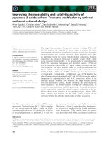
Anthracyclines have been widely
used in the treatment of hematological
and solid neoplasms such as leukemias,
lymphomas, breast cancer, prostate
cancer and bladder cancer [12]. The
anthracycline antibiotics are produced by
Streptomyces coeruleorubidus or
Streptomyces peucetius and include
daunorumycin (DNR), doxorubicin,
epirubicin and idarumycin (IDA) (Fig. 3).
Daunorumycin and adriamycin link up
base pairs and thus inhibit RNA and
DNA synthesis. Mithramycin and
Figure
3
:
Chemical structures of
daunorumycin and idarumycin
14
chromomycin A3 which are actinomycins, inhibit DNA-dependent RNA
synthesis. Bleomycins react with DNA and cause it to break [30], [4].
1.1.5 The need of developing new antibiotics
One of the triumphs of modern medicine is the development of
antibiotics and other antimicrobial substances. However, it is clear that
microbes have developed and will continue to develop resistance to currently
available antibiotics by either new mutations or exchange of genetic
information [2], [8]. New antibiotics that are active against resistant bacteria
are required, especially those are anti-tumor and anti-parasitic compounds.
The search for anti-tumor antibiotics however is more difficult than that for
antibacterial or antifungal in terms of methodology and interpretation [30].
Moreover, new antibiotics are required for the use in agriculture as drugs
for plant or animal diseases, since they could have effect on human through
the food chain.
1.2 Actinomycetes
1.2.1 General characteristics
Actinomycetes make a big group of diverse bacteria, most of which
grow aerobically and form branching mycelia similar to those of fungi. The
name actinomycete derives from the Greek “actys” (ray) and “mykes”
(fungus) and actinomycetes were initially regarded as minute fungi because of
their mycelium-like growth. The branching network of hyphae usually grows
critically both on the surface of the solid substrata (forming aerial mycelia) as
well as into it, leading to formation of substrate mycelia [3]. Most of
actinomycetes produce spores varying widely in shape and size, which can
serve as taxonomical characteristics.
Actinomycetes are Gram positive bacteria having high G+C content
(>55%) in their DNA. The majority of actinomycetes has free living,
saprophytic life form and is widely distributed in soil, water and plant litter.
15
Actinomycetes play an ecologically important role in recycling substances in
nature. They decompose and utilize difficult-to-degrade organic matters such
as humic acid in the soil. Many strains have the ability to solubilize lignin and
degrade lignin-related compounds by producing cellulose- and hemicellulose-
degrading enzymes and extracellular peroxidases [24], [32]. Some
actinomycete strains also occur in other environments rich in organic matter
such as composts, in both the mesophilic and thermophilic phases [38], and
sewage sludge, where, notably the mycolic acid-containing actinomycetes are
associated with the extensive and undesirable formation of stable foams and
scum [14], [36].
In general, mesophilic temperatures of 25-30°C and neutral pH are
optimal conditions for the growth of actinomycetes. Nevertheless, many
species have been isolated from extreme environments, for example the
psychrophilic Arthrobacter ardleyensis isolated from sediment from an
Antarctic lake was able to grow at temperatures as low as 0°C [5] and
Nocardiopsis alkaliphila isolated from desert soil in Egypt could grow at pH
9.5-10 [18].
At the presence actinomycetes are defined as the order Actinomycetales
which consists of 13 suborders, 42 families and about 200 genera.
Actinomycetes are often divided into two categories, the Streptomyces and
non-Streptomyces [7], [28].
The best known actinomycete genus is Streptomyces, which contains
approximately 500 species, all with a characteristically high GC content (69–
73 %) in the DNA. Streptomyces are extremely prevalent in soil, where they
saprophically degrade a wide range of complex organic substrates by means
of extracellular enzymes. Indeed, the characteristic musty smell of many soils
is due to the production of a volatile organic compound called geosmin. A
high proportion of therapeutically useful antibiotics derive from Streptomyces
16
species, well-known examples are streptomycin, erythromycin and
tetracycline [39].
1.2.2 Actinomycetes and secondary metabolites
Special interest in actinomycetes lies in their ability to produce
secondary metabolites of highly applicable values. Of all the reported natural
products from microbes 45% are produced by actinomycetes, 38% by fungi
and 17% by unicellular bacteria [2]. Two-thirds of known antibiotics are
produced by actinomycetes, mainly by Streptomyces species [28]. Various
antibiotic substances from actinomycetes have been characterized, including
aminoglycosides, anthracyclins, glycopeptides, β-lactams, macrolides,
nucleosides, peptides, polyenes, polyester, polyketides, actinomycins and
tetracyclines [13]. These substances have been succesfully used as herbicides,
anticancer agents, drugs, immunoregulators and antiparasitic agents [44].
1.3 Objectives of the study
This study aimed to investigating biodiversity and to screening
actinomycete strains with high antimicrobial activity among a collection of
actinomycetes isolated from Catba island, a national park with rich
biodiversity in Vietnam. The selected strains were then subjected to studies on
the antibiotic substances they produced as well as the phylogenetic affiliation
of selected strains. The particular objectives were as follows:
- Isolate actinomycetes from soil and litter samples on Catba island
(Haiphong, Vietnam).
- Screen actinomycetes having high antimicrobial activity and activity
against human cell lines.
- Study property of antibiotics produced by the selected actinomycetes
via thin-layer chromatography (TLC) and high performance liquid
chromatography (HPLC).
17
- Study taxonomy of actinomycetes based on morphological
characteristics and 16S rDNA gene sequences.
18
CHAPTER 2. MATERIALS AND METHODS
2.1 Work flow
Collection and isolation of actinomycetes
Collection of samples from Catba island
Isolation by dry – heating and rehydration – centrifugation
Screening of antibiotic producing actinomycetes
Primary screening by agar disc method
Secondary screening by culture broth method
Chromatography analyses of antibiotics
Ethyl-acetate extraction
Analysis via thin-layer chromatography (TLC)
Analysis via high performance liquid chromatography (HPLC)
Primary study on cytotoxicity activity
Color test method
Cytotoxicity assay
Taxonomical identification of actinomycetes
Morphological characterization
DNA extraction
PCR amplification of the 16S rDNA sequencing, and
phylogenetic analysis
19
2.2 Methods
2.2.1 Isolation of actinomycetes
Catba is an island with the territory of approximately 140 km
2
in Halong
Bay, Northern Vietnam. It is the largest island in the Bay and the preserved
National Park occupies about half of its area. The park contains both land and
marine areas with high biodiversity. Catba national park was recognized
by UNESCO in December 2004 as a Biosphere reserve in the world.
Figure 4: Catba national park
Soil and leaf litter samples were collected from Catba island and used as
sources for actinomycete isolation. Two isolation methods were used in this
study, the dry-heating method [31] and the rehydration-centrifugation method
[15].
In the dry-heating method, soil and littler samples were first dried at
room temperature for 5 – 7 days, then possessed a heat treatment at 90 – 110
0
C for 10 – 30 minutes for killing non-spore forming bacteria (most
actinomycete spores did not die at this condition). Afterward, the samples
were spreaded onto HV medium (Humic acid - vitamin agar medium; humid
acid 1 g, CaCO
3
0.02 g, FeSO
4
.7H
2
O 0.01 g, KCl 1.71 g, MgSO
4
.7H
2
O 0.05
g, Na
2
HPO
4
0.5 g, cycloheximine 50 mg, nalidixic acid 20 mg, kabicidine 14
20
mg, agar 18 g, H
2
O 1 L, pH 7.2) and incubated at 28 – 30
0
C for 7 to 14 days.
Actinomycete colonies of different morphology were picked up and
transferred to new HV agar medium for the second purification. The isolates
were then transferred to YS medium (yeast extract – starch agar medium;
glucose 10 g, yeast extract 2 g, agar 17 g, distilled water 1 L, pH 7.0) for
producing higher cell mass.
The rehydration–centrifugation method is an isolation method specific
for motile actinomycetes. The sequential steps are as following:
- Take 0.5 g of air–dried soil or litter samples in 100 ml beakers, gently
added 50 ml of 10 mM phosphate buffer (pH = 7) containing 10% soil extract
solution (mix 500 g soil and 500 ml distilled water in a flask, autoclave at 121
°C for 30 minutes, afterward filtrate twice, autoclave again and kept the
solution at 4 °C).
- Cover the beakers with aluminium foil and incubate statically at 30 °C
for 90 minutes to allow liberation of motile zoospores.
- Transfer 8 ml of the supernatant into 15 ml falcon tubes, centrifuge at
room temperature for 20 minutes at 1,500 g to eliminate streptomycetes and
other non-motile actinomycetes from the supernatant. The tubes were then
allowed to settle for 30 minutes.
- Take 1 ml of the supernatant for serial dilution in sterile tap water,
aliquots 0.2 ml of serial dilution of 10
-2
, 10
-3
and 10
-4
were plated onto HV
agar and incubated at 28 – 30 °C for 2 – 3 weeks to produce colonies.
- Actinomycete colonies were then picked up, transferred to new HV agar
medium for second purification and then to YS agar medium for biomass
production.
All actinomycete strains isolated by two methods described above were
maintained as stock cultures frozen at -80 °C in 20% glycerol solution at
Vietnam Type Culture Collection (VTCC).
21
2.2.2 Targeted microorganisms
Four microorganism strains, including Micrococcus luteus (a Gram
positive bacterium), Escherichia coli (a Gram negative bacterium), Candida
albicans (a yeast) and Fusarium oxysporium (a filamentous fungus) were used
as targets in screening for antimicrobial activity. These strains were cultivated
in proper nutrient media, i.e. Mueller-Hinton medium (MHA; meat extract
0.3%, hydrolysis casein 1.75%, starch 0.15%, pH 7.4) for E. coli and M.
luteus, yeast/malt extract medium (YM; glucose 1%, peptone 0.5%, yeast
extract 0.3%, malt extract 0.3%) for C. albicans and F. oxysporum. The
cultures were incubated under shaking condition at 37 °C for E. coli and M.
luteus or 30 °C for C. albicans and F. oxysporum. These strains were
provided by the VTCC.
2.2.3 Screening antibiotic producing actinomycetes
Before using in experiments, all actinomycete strains were refreshed on
YS medium plates at 30 °C for 3–4 days. After reactivation, the strains were
cultivated in liquid soybean meal medium (starch 25 g, soybean meal 15 g,
yeast extract 1 g, CaCO
3
4 g, distilled water 1 L, pH 6.2) at 30 °C for 3 days
on shaker at 100 rpm. Culture broths were then centrifuged at 8,000 rpm for
15 minutes, the supernatants were used to evaluate antimicrobial activity.
Agar disc method
Agar discs (5 mm in diameter) picked from YS agar plates of well grown
actinomycete cultures were placed onto surface of agar plates previously
seeded with a target microorganism and incubated at proper conditions for 2
days. The inhibitory effect was assessed based on the formation of clear
inhibition zones around the agar discs and the activity was measured as the
diameter of these zones [19].