Vai trò của điện tâm đồ
Bạn đang xem bản rút gọn của tài liệu. Xem và tải ngay bản đầy đủ của tài liệu tại đây (4.09 MB, 105 trang )
Vai trò của điện tâm đồ
Điện tâm đồ bình thường
ThS. Lê Hoài Nam
Bộ môn Nội – ĐHYD TPHCM
History of the ECG/EKG
• During the late 1800’s and early 1900’s, Dutch physiologist Willem
Einthoven developed the early electrocardiogram He won the Nobel
prize.
!
• Hubert Mann first uses the electrocardiogram to describe
electrographic changes associated with a heart attack in 1920
!
• Electrocardiograms must be viewed in the context of demographics,
health history, and other clinical test correlates. They are especially
useful when compared across time to see how electrical activity of
the heart has changed (perhaps as the result of some pathology).
1887 British physiologist Augustus D. Waller of St Mary's Medical School, London publishes
the first human electrocardiogram. It is recorded with a capilliary electrometer from Thomas
Goswell, a technician in the laboratory. Waller AD. A demonstration on man of electromotive
changes accompanying the heart's beat. J Physiol (London) 1887;8:229-234 1889 Dutch
physiologist Willem Einthoven sees Waller demonstrate his technique at the First International
Congress of Physiologists in Bale. Waller often demonstrated by using his dog "Jimmy" who
would patiently stand with paws in glass jars of saline. 1889
!
R.E.Mason.,
I.Likar
( 1966)
Augustus waller first to His electrocardiograph machine consisted of a Lippmann capillary electrometer fixed to a projector. The
trace from the heartbeat was projected onto a photographic plate which was itself fixed to a toy train. This allowed a heartbeat
to be recorded in real time. In 1911 he still saw little clinical application for his work.
An initial breakthrough came when Willem Einthoven, working in Leiden, Netherlands, used the string galvanometer that he
invented in 1903.
[8]
This device was much more sensitive than both the capillary electrometer that Waller used and the string
galvanometer that had been invented separately in 1897 by the French engineer Clément Ader.
[9]
. Rather than using today's
self-adhesive electrodes Einthoven's subjects would immerse each of their limbs into containers of salt solutions from which the
ECG was recorded.
Einthoven assigned the letters P, Q, R, S and T to the various deflections, and described the electrocardiographic features of a
number of cardiovascular disorders. In 1924, he was awarded the Nobel Prize in Medicine for his discovery limb leads
Many advancements such as Goldbergers chest leads
Electrocardiography
• A recording of the electrical activity of the heart over time
• Gold standard for diagnosis of cardiac arrhythmias
• Helps detect electrolyte disturbances (hyper- &
hypokalemia), arrhythmias, myocardial ischemia and
infarction, pericarditis, chamber hypertrophy, drug toxicity
(i.e. digoxin and drugs which prolong the QT interval)
• Allows for detection of conduction abnormalities
• Screening tool for ischemic heart disease during stress
tests
• Helpful with non-cardiac diseases (e.g. pulmonary
embolism or hypothermia)
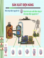
Cardiac Electrical Activity
• SA Node - Dominant
pacemaker with an
intrinsic rate of 60 - 100
beats/minute.
!
• AV Node - Back-up
pacemaker with an
intrinsic rate of 40 - 60
beats/minute.
!
• Ventricular cells - Back-
up pacemaker with an
intrinsic rate of 20 - 40
bpm.
Lead “Views”
EKG Leads
Leads are electrodes which measure the
difference in electrical potential between
either:
1. Two different points on the body (bipolar leads)
!
2. One point on the body and a virtual reference point with
zero electrical potential, located in the center of the
heart (unipolar leads)
EKG Leads
The standard EKG has 12 leads: 3 Standard Limb Leads
3 Augmented Limb Leads
6 Precordial Leads
The axis of a particular lead represents the viewpoint from
which it looks at the heart.
Standard Limb Leads
Standard Limb Leads
Augmented Limb Leads
All Limb Leads
Precordial Leads
Adapted from: www.numed.co.uk/electrodepl.html
Precordial Leads
Summary of Leads
Limb Leads
Precordial Leads
Bipolar
I, II, III
(standard limb leads)
-
Unipolar
aVR, aVL, aVF
(augmented limb leads)
V
Limb Leads Chest Leads
I aVR V1 V4
!
II aVL V2 V5
!
III aVF V3 V6
Lead Groups
Lead “Views”
Inferior Wall
• II, III, aVF
– Left Leg
I
II
III
aVR
aVL
aVF
V1
V2
V3
V4
V5
V6
Inferior Wall
Inferior Wall
I
II
III
aVR
aVL
aVF
V1
V2
V3
V4
V5
V6
Lateral Wall
• I and aVL
– Left Arm
I
II
III
aVR
aVL
aVF
V1
V2
V3
V4
V5
V6
Lateral Wall
• V5 and V6
– Left lateral chest
I
II
III
aVR
aVL
aVF
V1
V2
V3
V4
V5
V6
Lateral
• I, aVL, V5, V6
I
II
III
aVR
aVL
aVF
V1
V2
V3
V4
V5
V6
Lateral Wall
Anterior Wall
• V3, V4
– Left anterior chest
I
II
III
aVR
aVL
aVF
V1
V2
V3
V4
V5
V6
Anterior Wall
• V3, V4
I
II
III
aVR
aVL
aVF
V1
V2
V3
V4
V5
V6