Varicella-Zoster Virus Epithelial Keratitis in Herpes Zoster Ophthalmicus In Vivo Morphology in the Human Cornea
Bạn đang xem bản rút gọn của tài liệu. Xem và tải ngay bản đầy đủ của tài liệu tại đây (14.35 MB, 96 trang )
Varicella-Zoster Virus Epithelial Keratitis
in Herpes Zoster Ophthalmicus
Helena M. Tabery
Varicella-Zoster Virus
Epithelial Keratitis
in Herpes Zoster
Ophthalmicus
In Vivo Morphology
in the Human Cornea
ISBN 978-3-642-14486-8 e-ISBN 978-3-642-14487-5
DOI 10.1007/978-3-642-14487-5
Springer Heidelberg Dordrecht London New York
© Springer-Verlag Berlin Heidelberg 2011
This work is subject to copyright. All rights are reserved, whether the whole or part of the material is
concerned, specifically the rights of translation, reprinting, reuse of illustrations, recitation, broadcasting,
reproduction on microfilm or in any other way, and storage in data banks. Duplication of this publication
or parts thereof is permitted only under the provisions of the German Copyright Law of September 9, 1965,
in its current version, and permission for use must always be obtained from Springer. Violations are liable
to prosecution under the German Copyright Law.
The use of general descriptive names, registered names, trademarks, etc. in this publication does not imply,
even in the absence of a specific statement, that such names are exempt from the relevant protective laws
and regulations and therefore free for general use.
Product liability: The publishers cannot guarantee the accuracy of any information about dosage and appli-
cation contained in this book. In every individual case the user must check such information by consulting
the relevant literature.
Cover design: eStudioCalamar, Figueres/Berlin
Printed on acid-free paper
Springer is part of Springer Science+Business Media (www.springer.com)
Helena M. Tabery
Ögonkliniken UMAS
20502 Malmö
Sweden
v
Preface
This book treats varicella-zoster virus (VZV) caused corneal epithelial changes cap-
tured in high-magnification photographs in herpes zoster ophthalmicus (HZO). The
images highlight the typical substructure of VZV lesions clinically presenting in a
large variety of shapes and sizes, both in conjunction with and in the absence of typi-
cal HZO rash; the accompanying case reports illustrate the varying clinical features
of the disease, ranging between typical and rare ones.
In addition, the book shows serial photographs capturing the dynamic features of
VZV impact on the corneal epithelial architecture. The opportunity was unique, not
only because the corneal epithelium is the only one in the human body in which mor-
phological changes can be directly observed and followed without intervention, and
highlighted by in vivo staining, but also because the follow-up was not terminated by
treatment. Contrary to expectations, the at that time recommended antiviral drug (acy-
clovir or valacyclovir) showed no detectable effect, neither on the morphology nor on
the dynamics of the epithelial disease.
In the interpretation of the disturbances of the epithelial architecture, this book
partly relates to the morphology of herpes simplex virus (HSV) caused changes, for
reasons extending beyond differential diagnostics. The point is that it is not only the
impact of the infection that has to be taken in account, but also epithelial healing
responses. When the similarities between the two viruses are sorted out, very different
reparative patterns emerge; these patterns indicate that after having reached the cor-
neal epithelium via the same route, the two viruses strongly diverge in their behaviour.
Because all this is reflected in the individual lesions, the comparison between them
can explain at least some mechanisms behind their appearance.
With this book I intended to fill a void in the literature by adding high-magnification
in vivo images that capture several aspects of an intriguing disease so far defying
attempts to be reproduced in laboratory animals. I hope I have done that.
Malmö, Sweden Helena M. Tabery
January 2010
vii
Contents
1 The Morphology of Varicella-Zoster Virus Epithelial Keratitis
in Herpes Zoster Ophthalmicus 1
VZV Cytopathic Effect in Cell Cultures 2
VZV Cytopathic Effect in the Living Human Corneal Epithelium 3
VZV Epithelial Keratitis: Surface Elevations and Disruptions 4
VZV Epithelial Keratitis: Dynamics of Fluorescein Sodium Staining 5
VZV Epithelial Keratitis: Surface Plaques 6
VZV Epithelial Keratitis and Epithelial Edema 8
Epithelial Erosion: A Sequela of VZV Epithelial Keratitis 11
Subepithelial Opacity: A Sequela of VZV Epithelial Keratitis (1) 12
Subepithelial Opacity: A Sequela of VZV Epithelial Keratitis (2) 13
Inflammatory Cells on the Endothelium in VZV Epithelial Keratitis (1) 14
Inflammatory Cells on the Endothelium in VZV Epithelial Keratitis (2) 15
2 The Dynamics of Varicella-Zoster Virus Epithelial
Keratitis in Herpes Zoster Ophthalmicus 17
Case 1: Changing Shapes of a Large VZV Lesion 18
Case 2: Changing Shapes of a Smaller VZV Lesion 22
Case 3: Appearance and Disappearance of VZV
Corneal Epithelial Lesions . . . . . . . . . . . . . . . . . . . . . . . . . . . . . . . . 30
Development of VZV Corneal Epithelial Lesions
in the Same Location 39
3 Recurrent VZV Epithelial Keratitis in HZO; HZO Sine Herpete 43
Case 1: Recurrent VZV Epithelial Keratitis in HZO 44
Case 2: Recurrent VZV Epithelial Keratitis in HZO 46
Case 3: Recurrent VZV Epithelial Keratitis in HZO 52
Case 1: VZV Epithelial Keratitis in HZO Sine Herpete 53
Case 2: VZV Epithelial Keratitis in HZO Sine Herpete 54
Case 3: VZV Epithelial Keratitis in HZO Sine Herpete 62
viii Contents
4 Three Rare Cases of Ocular Surface Involvement in Acute HZO 65
Case 1: HZO, Epithelial Edema, and (Presumed) VZV
Epithelial Keratitis 66
Case 2: HZO and Corneal Epithelial Cysts 70
Case 3: HZO, VZV Epithelial Keratitis, and VZV
Conjunctival Lesions 72
5 Comparison of HSV and VZV Epithelial Keratitis 75
Swollen Epithelial Cells; Surface Ulceration (HSV) 76
Subsurface Changes, Surface Elevations 77
Light-Reflecting Properties 78
Light-Reflecting Properties and Staining Features 79
Various Aspects of an HSV Lesion 80
Various Aspects of a VZV Lesion 81
The Origin of HSV Dendrites and VZV Pseudodendrites 82
Fluorescein Staining of HSV Dendrites and VZV Pseudodendrites 83
Rose Bengal Staining of HSV Dendrites and VZV Pseudodendrites 84
Addendum. Interplay of Destructive and Healing Forces
in HSV Epithelial Keratitis 86
Final Remark 88
Bibliography 89
Index 91
ix
About Herpes Zoster Ophthalmicus
Infection with varicella-zoster virus (VZV) causes varicella (chickenpox), a disease
that manifests as a disseminated vesicular body rash. After that, the virus remains
latent in the sensory ganglia; it reactivates later on and causes new symptoms – herpes
zoster (HZ).
In herpes zoster ophthalmicus (HZO), the reactivated virus descends from the
trigeminal ganglion through the first division of the fifth nerve, the nervus ophthal-
micus, which via its different branches supplies the skin of the forehead, the lids, the
nose, and the eye. HZO is a very common disease affecting the elderly; it is rare in chil-
dren and young adults. At all ages, immunosuppression is a predisposing factor. HZO
might severely damage any eye structure and even result in a destruction of the eye.
The HZO diagnosis is clinical. It is easy in patients presenting with a typical vesic-
ular rash, challenging when mimicked by vesicles caused by herpes simplex virus
(HSV), and may be missed when skin eruptions are lacking (zoster sine herpete). The
problem is that almost all HZO ocular manifestations are per se unspecific and often
indistinguishable from those occurring for other causes in general and those caused
by HSV infections in particular. Yet, there is one exception – VZV epithelial keratitis.
Clinically, it is the least troublesome of VZV ocular manifestations, but it occupies an
outstanding position because of its typical features. VZV epithelial keratitis may pre-
cede the rash, accompany it, develop later on, and recur; in some patients, it may be
the only clue revealing the true cause of their disease.
xi
The photographs presented in this book have been chosen to show
The • in vivo morphology of VZV corneal epithelial lesions in patients with HZO,
accompanying signs and sequelae (Chap. 1)
The • dynamic features of VZV corneal epithelial lesions in patients with HZO
(Chap. 2)
The morphological and dynamic features of VZV epithelial lesions in • HZO sine
herpete and of recurrent VZV epithelial lesions (Chap. 3)
Three rare cases• of ocular surface involvement in HZO (Chap. 4)
A • comparison of (HZO) VZV and (recurrent) HSV corneal epithelial lesions
(Chap. 5)
The photographs were taken by non-contact in vivo photomicrography, a method that
requires neither contact with the epithelium nor the use of anesthetics. By this method
structures that optically differ from their regularly organized surroundings are visual-
ized; a normal corneal epithelium or stromal cells cannot be discerned. As there is no
contact with the ocular surface, the architecture of epithelial changes is not disturbed
by the examination, and there is no risk of spreading infections. The technique allows
the use of various illumination modes to complement each other and a free applica-
tion of diagnostic dyes to expand the information, e.g., 1% fluorescein sodium and
1% rose bengal (preservative-free solutions). These dyes are commonly used in clini-
cal practice.
The diagnosis was clinical; in some cases, it was verified by PCR.
The photographs of cell cultures were taken by the same method.
The bars indicate 200 mm throughout the book.
About This Book
xiii
CPE Cytopathic effect
Fluorescein Fluorescein sodium
IOP Intraocular pressure
HIV Human immunodeficiency virus
HSV Herpes simplex virus
HZO Herpes zoster ophthalmicus
KCS Keratoconjunctivitis sicca
PCR Polymerase chain reaction
VZV Varicella-zoster virus
Abbreviations
H. M. Tabery, Varicella-Zoster Virus Epithelial Keratitis in Herpes Zoster Ophthalmicus
DOI: 10.1007/978-3-642-14487-5_1, © Springer-Verlag Berlin Heidelberg 2011
Before the introduction of newer methods, the gold
standard of detection and identication of viruses was
virus isolation test in cell culture. In living cells, virus
replication causes cell swelling and rounding (a phe-
nomenon termed the virus cytopathic eect, CPE), fol-
lowed by cell bursting and disappearance.
When the multilayered living human corneal epithe-
lium in situ becomes infected with varicella-zoster
virus (VZV), the virus CPE generates secondary phe-
nomena: Subsurface cell swelling causes volume
increase resulting in surface elevations and disruptions;
later on, degenerating and dead cells appear on the sur-
face from which they are shed. e surface debris has
propensity to conuence resulting, probably with
mucus contribution, in plaque-like formations. Surface
ulcerations (in the sense of missing substance) are not
a morphologic feature of VZV lesions but might occur
as a sequela (see below). With the exception of the rare
patient seen very early aer onset, VZV lesions usually
show both incipient and more advanced changes in
adjacent areas.
e shapes of VZV lesions vary greatly. ose appear-
ing as branching gures have been termed pseudoden-
drites to dierentiate them from branching gures
caused by herpes simplex virus (HSV) infections. e
resemblance between the two is only supercial; their
substructures dier from each other (Chap. 5). It is
only during the very early stage, i.e. the stage showing
subsurface cell swelling and surface elevations, in
which the impact of the two viruses appears similar. In
clinical practice, such situation in the absence of other
clues seems rare. (I happened to see it only once. e
following day, the diagnosis was clear – HZO sine her-
pete, Chap. 3.)
As accompanying signs, anterior uveitis with keratic
precipitates on the endothelium is frequently seen con-
currently with epithelial keratitis; a concurrent epithe-
lial edema (oen associated with elevated intraocular
pressure) is occasionally encountered.
A sequela, or complication, of VZV epithelial kerati-
tis might be epithelial erosions resulting from sloughing-
o of whole involved areas. is occurs infrequently,
in corneae probably predisposed by a poor quality of
the epithelium. Another sequelae of epithelial keratitis,
developing in some but not all corneae, are subepithelial
opacities showing abnormal cells located about the
level of the epithelial basement membrane. e exact
nature of these cells is not clear, but their persistence,
in some patients for several months, implies invading
inammatory cells possibly attracted by the virus anti-
gen. In the photographs, such cells are per se indistin-
guishable from virus-damaged ones; it is their presence,
and persistence, under a restored surface that implies
their dierent nature (cf. also Chap. 2).
Chapter 1
The Morphology of Varicella-Zoster Virus Epithelial
Keratitis in Herpes Zoster Ophthalmicus
2 Chapter 1 The Morphology of Varicella-Zoster Virus Epithelial Keratitis in Herpes Zoster Ophthalmicus
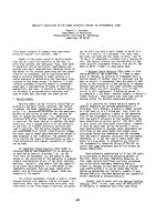
VZV Cytopathic Effect in Cell Cultures
a
b
Fig. 1.1 VZV cytopathic eect in cultured cells. (a) is culture shows swollen/rounded cells, individual (arrowhead) or
aggregated (straight arrow). Cell death and detachment from the underlying surface has resulted in cell-devoid areas (bowed
arrow) (GMK, green monkey kidney). (b) Also in this cell culture are visible swollen/rounded cells (arrowhead) and cell-
devoid areas (bowed arrow). Additionally, there is a propensity to cell conuence (straight arrows) (A549, human lung cell
carcinoma). (Adapted from [7])
VZV Cytopathic Effect in the Living Human Corneal Epithelium 3
VZV Cytopathic Effect in the Living Human Corneal Epithelium
b
a
d
c
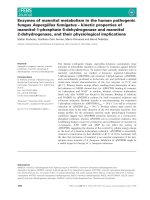
Fig. 1.2 a–d VZV cytopathic eect in the living human corneal epithelium. In all photographs are visible swollen/rounded
cells (arrowheads) distributed at random; in (b) is additionally visible a corneal nerve (arrows), and in (d) a more advanced
light-reecting lesion (arrow)
b
a
Fig. 1.3 VZV cytopathic eect in the living human cornea epithelium. (a) is lesion contains many swollen/rounded cells
(arrowheads). (b) shows a lesion in which swollen/rounded cells (arrowheads) are visible at the edges but dicult to see in the
area indicated by arrow; whether the cells are conuent or obscured by overlying debris cannot be discerned
4 Chapter 1 The Morphology of Varicella-Zoster Virus Epithelial Keratitis in Herpes Zoster Ophthalmicus
VZV Epithelial Keratitis: Surface Elevations and Disruptions
a
b
Fig. 1.5 (a) In the tear lm stained green with uorescein sodium, incipient VZV lesions located below an intact surface
(arrows) appear dark. In the right lesion are visible grouped swollen/rounded cells (arrowhead). (b) In this part of a larger lesion,
protruding swollen/rounded cells appear as dark dots (arrowheads) in the tear lm stained green with uorescein sodium
Fig. 1.4 Incipient foci of VZV corneal epithelial infection (arrows) visualized with uorescein sodium and blue lter. Elevated
foci with intact surfaces appear dark; bright uorescein staining indicates surface disruptions
VZV Epithelial Keratitis: Dynamics of Fluorescein Sodium Staining 5
VZV Epithelial Keratitis: Dynamics of Fluorescein Sodium Staining
a
b
c
Fig. 1.6 e same part of a larger VZV
corneal epithelial lesion. (e arrows
are placed in corresponding locations)
(a) Shortly aer the application of
uorescein sodium, the staining shows
a broken pattern; some areas appear
intensively green, others only weakly.
e white arrow indicates an area of a
small incipient lesion, the black arrows
point to small cysts
(b) Aer a short while, with ongoing
diusion, the staining is more pro-
nounced; the individual parts oat
together, which gives rise to an impres-
sion of a continuous, branching gure
(c) A few minutes later is visible a
smooth, green stained branching g-
ure with no discernible details except
for some cysts (brightly green dots).
Rose bengal reveals diseased surface
cells and cell debris; in places, the red
staining is conuent
6 Chapter 1 The Morphology of Varicella-Zoster Virus Epithelial Keratitis in Herpes Zoster Ophthalmicus
VZV Epithelial Keratitis: Surface Plaques
b
a
d
c
VZV Epithelial Keratitis: Surface Plaques 7
Rose Bengal Staining of Surface Plaques
Fig. 1.7 (Opposite page) Shows the same area (a) before staining, (b and c) aer staining with uorescein and (d) with addi-
tion of rose bengal. Surface plaques (arrows) are strongly light reecting, stain yellow with (adherent) uorescein and red with
rose bengal. In addition, this series shows an enlargement with uorescein diusion of the visible area of damage (cf. Fig. 1.6).
(e arrows are placed in corresponding locations)
Fig. 1.8 Low-magnication photograph of VZV epithelial
keratitis visualized with rose bengal. e area indicated by
circular frame is shown in Fig. 1.9 (right), and that in rectan-
gular frame in Fig. 1.10 (below)
Fig. 1.10 e plaque-like rose bengal staining of these VZV epithelial lesions ranges between a dense (black arrows) and a
weak or a barely perceptible one (white arrows). Some diseased areas do not stain (arrowhead). (Composed photograph)
Fig. 1.9 Bizarre appearance of a VZV epithelial lesion stained
with rose bengal
8 Chapter 1 The Morphology of Varicella-Zoster Virus Epithelial Keratitis in Herpes Zoster Ophthalmicus
VZV Epithelial Keratitis and Epithelial Edema
Fig. 1.11 (Right) Composed low-magnication photograph showing a
part of a large VZV pseudodendrite. e branching conguration mim-
ics an HSV dendrite. e epithelium was edematous over the whole
cornea and two days later suered a large erosion. Details and staining
features are shown in Fig. 1.12 (below), Figs. 1.13 and 1.14 (opposite
page), and Fig. 1.15 (overleaf); the erosion is shown in Fig. 1.16
b
a
Fig. 1.12 Central part of the VZV epithelial lesion shown in Fig. 1.11. (a) Before staining, the epithelium shows light-reect-
ing (plaque-like) structures (long arrows) that (b) stain red with rose bengal. Brightly green staining with uorescein sodium
reveals surface disruptions in an additional area of damage (short arrow). (e arrows are placed in corresponding locations).
e area indicated in (b) by circular frame is shown in Fig. 1.13 and that within rectangular frame in Fig. 1.14, opposite page
VZV Epithelial Keratitis and Epithelial Edema (cont.) 9
VZV Epithelial Keratitis and Epithelial Edema (cont.)
b
a
Fig. 1.13 e part of the lesion indicated by circular frame in Fig. 1.12b. (a) e light-reecting areas (arrow) (b) stain red
with rose bengal. In (b), in the in-between areas, are additionally visible supercial damaged/diseased surface cells staining
red with rose bengal (arrowhead). Cf. also Fig. 1.14 (below) and Fig. 1.15 (overleaf). (e arrows are placed in corresponding
locations)
b
a
Fig. 1.14 e part of the lesion indicated by rectangular frame in Fig. 1.12b. (a) Yellow staining of the lesion’s surface (arrows)
with (adherent) uorescein corresponds to (b) red staining with rose bengal. e arrowhead in (b) indicates red-stained diseased
surface cells in the surrounding epithelium. Cf. also Fig. 1.15 (overleaf). (e arrows are placed in corresponding locations)
10 Chapter 1 The Morphology of Varicella-Zoster Virus Epithelial Keratitis in Herpes Zoster Ophthalmicus
VZV Epithelial Keratitis and Epithelial Edema (cont.)
Fig. 1.15 Upper part of the large VZV epithelial lesion shown in Fig. 1.11. e mottled appearance of the surrounding epi-
thelium is caused by large numbers of diseased/damaged surface cells staining red with rose bengal and seen against a back-
ground of a diuse green staining with uorescein sodium of edematous epithelium. VZV lesions (long arrows) stain heavily
with rose bengal. An additional area of damage appears as a brightly green channel (short arrows) that seems to be connecting
the rose bengal–stained patches
Epithelial Erosion: A Sequela of VZV Epithelial Keratitis 11
Epithelial Erosion: A Sequela of VZV Epithelial Keratitis
Fig. 1.16 Sequela of VZV epithelial keratitis. A part of a large epithelial erosion (bowed arrow) surrounded by edematous
epithelium staining green (long arrow). e detached epithelium is partly folded at the edge (short arrow). e adjacent sur-
face shows diseased/damaged surface cells staining red with rose bengal (arrowhead). (e same cornea as shown in Figs. 1.11–
1.15, two days later)
Addendum
e patient suered from diabetes; aer the keratitis episode, KCS was diagnosed in both eyes.
Fig. 1.17 For comparison with Fig. 1.15, two VZV lesions seen against a background of a normal epithelium. Fluorescein has
disappeared from the tear lm. e lesions show patches of cell debris staining red with rose bengal (arrows); the green stain-
ing with uorescein is limited to the lesions (cf. inset)
12 Chapter 1 The Morphology of Varicella-Zoster Virus Epithelial Keratitis in Herpes Zoster Ophthalmicus
Subepithelial Opacity: A Sequela of VZV Epithelial Keratitis (1)
b
a
c
Fig. 1.18 is subepithelial opacity captured 8 weeks aer the onset of VZV epithelial keratitis is (a) light reecting, has a
granular structure, and (b) contains abnormal cells (arrowheads). (c) shows a close view of the area in frame in (b).
(e arrowheads in (b) and (c) are placed in corresponding locations)
Subepithelial Opacity: A Sequela of VZV Epithelial Keratitis (2) 13
Subepithelial Opacity: A Sequela of VZV Epithelial Keratitis (2)
a
b
Fig. 1.19 a-b A subepithelial opacity 12 months aer VZV epithelial keratitis. In (b) are visible abnormal cells (arrowheads)
Fig. 1.20 For comparison with Fig. 1.18 (opposite page), inammatory cells (arrowheads) attached to the endothelium
captured in anterior uveitis occurring concurrently with VZV epithelial keratitis
14 Chapter 1 The Morphology of Varicella-Zoster Virus Epithelial Keratitis in Herpes Zoster Ophthalmicus
Inflammatory Cells on the Endothelium in VZV Epithelial Keratitis (1)
a
b
Fig. 1.21 a–b ese patients had anterior uveitis concurrently with VZV epithelial keratitis. e majority of inammatory
cells adhering to the endothelium (white arrowheads) are round; some appear fusiform (black arrowheads)
Inflammatory Cells on the Endothelium in VZV Epithelial Keratitis (2) 15
Inflammatory Cells on the Endothelium in VZV Epithelial Keratitis (2)
a
b
Fig. 1.22 a–b Two additional examples of rounded (white arrowheads) and fusiform (black arrowheads) inammatory cells
adhering to the endothelium in patients with VZV epithelial keratitis accompanied by anterior uveitis
H. M. Tabery, Varicella-Zoster Virus Epithelial Keratitis in Herpes Zoster Ophthalmicus
DOI: 10.1007/978-3-642-14487-5_2, © Springer-Verlag Berlin Heidelberg 2011
The morphology of an individual VZV lesion reflects
a sequence of events triggered by the virus impact
on corneal epithelial cells. When seen in time
perspective, it becomes evident that the morphology
of the lesions at each moment is a result of an
ongoing, highly dynamic process that involves not
only the destructive action of the virus but also an
action of natural healing forces. Serial observations
reveal that the shapes of the lesions change rapidly
(as fast as within 24 hours). This feature is relatable
to two phenomena: partly a disappearance of dis-
eased/damaged cells in some locations and, as judged
by the absence of ulcerations, their substitution by
fresh ones, and partly an appearance of new sites of
damage in adjacent areas. The first phenomenon
implies that in some locations the virus noxious
action has ceased and the second one that new infec-
tions in new locations have occurred. In larger
lesions, this process results in their changing shapes;
in smaller lesions located at some distance from each
other the same is demonstrated by their coming and
going.
e VZV lesions presented in this chapter have been
captured in patients treated with an antiviral drug in vitro
arresting the virus replication (acyclovir, 800mg ve times
a day) administered for a week. It is notable that: (a) new
lesions continued to develop aer the treatment was
started; these lesions were morphologically indistinguish-
able both from those which had developed before that
and from those developing aer the treatment was
stopped; and (b) that the features of VZV epithelial kera-
titis in treated patients were indistinguishable from the
natural course of the disease observed in patients not
treated with antiviral drugs (cf. Chap. 3). And the same
applied to lesions treated with topical acyclovir (Chap. 3).
In the absence of a detectable eect of treatment (so
clearly visible in HSV infections) in conjunction with
the absence of knowledge on how rapidly invading
abnormal (inammatory?) cells appear, it is possible
that in patients subsequently developing subepithelial
opacities in the same areas some images captured both
virus-damaged and invading cells (Case 2, Figs. 2.12–
2.13 and Case 3, Figs. 2.21–2.22). Before the surface is
restored, a distinction between the two is not possible.
Chapter 2
The Dynamics of Varicella-Zoster Virus Epithelial
Keratitis in Herpes Zoster Ophthalmicus